MORPHOLOGICAL TRANSFORMING FACTOR:
INFLUENCE ON HISTOTYPIC ORGANIZATION OF EPITHELIAL BRAIN CELLS
RAMON LIM DAVID E. TURRIFF
SHUANG S. TROY KATSUSUKE MITSUNOBU*
Departments of Surgery (Neurosurgery) and Biochemistry and the Brain Research Institute
University of Chicago Chicago, Illinois
I. INTRODUCTION
Tissue culture is the only artificial system where cells in the living state can be brought under direct observation for a relatively long period of time. Since brain cells during matura- tion undergo striking changes in morphology and histotypic pat- tern, they serve as a useful model for the study of morphogenesis at the cellular level and its relationship with chemical differ- entiation. In this article we summarize our experience with fetal brain cells in a monolayer culture as well as the effects of a tissue factor on the cells.
*Present address: Department of Neuropsychiatry, University of Okayama, Okayama City, Japan.
461
462 R A M O N LIM et al.
II. BASIC OBSERVATIONS
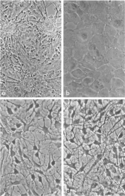
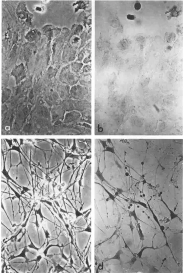
17-day Sprague-Dawley rat fetuses are used. The cerebrums and cerebellums are dissected, pooled and dissociated with trypsin in Tyrode solution free of calcium and magnesium. After seeding in a tissue culture flask, the cells form numerous small aggre- gates out of which two types of cells emerge: the flat epithelial cells which migrate rapidly onto the flask surface forming a thin layer of cell carpet; the bipolar cells (presumably neuroblasts) which migrate slowly and grow only on top of the flat cells (Fig.
la). Before completely migrating out of the aggregates, cable- like interconnections between groups of neuroblasts are common.
Upon subculture and by changing the medium within 24 hours, the neuroblasts are differentially eliminated leaving a culture of homogeneous epithelial cells (Fig. lb). In the experiments re- ported in this article we deal exclusively with these epithelial brain cells. If permitted to reaggregate, the epithelial cells eventually mature into glial cells. However, if left in a mono- layer, they will remain flat for an indefinite time.
In January, 1972, this laboratory (Lim et al., 1972) made the observation that if an extract from adult rat brain is added to a culture of epithelial brain cells, dramatic morphological changes take place. The changes involve (1) the rounding up of individual cells and (2) the outshoot of numerous cell processes which are branched and interconnected between cells (Fig. Ic). Their ap- pearance is characteristic of mature astrocytes. The morphologi- cal changes reach a peak at about 20 hours, after which further process outgrowth occurs if the medium containing the brain ex- tract is renewed every other day (Fig. Id). We routinely carry out the experiment on plastic culture flasks although the cells behave similarly on glass surface. The phenomenon is operational- ly designated as morphological transformation and the factor as the Morphological Transforming Factor. Care should be taken in
EPITHELIAL BRAIN CELLS 463
FIGURE 1 Live phase-contrast photographs demonstrating mor- phological transformation in fetal brain cells. (a] Primary cul- ture showing a mixture of epithelial cells and neuroblast-like cells Qi 150) . Q>) Secondary culture showing the epithelial cells before exposure to brain extract Qi 300). (c) Secondary culture showing the epithelial cells after exposure to brain extract for
464 RAMON LIM et al.
not confusing the term "transformation" in this article with the other connotation of the word as in "malignant transformation,"
The brain extract is prepared by dialyzing the 100,000 x g supernatant of a brain homogenate. In order to confine ourselves to the macromolecules the extracts are always dialyzed before sub- jecting them to any study reported here, although the undialyzed extract produces essentially the same morphological effect.
III. NATURE OF THE EPITHELIAL CELLS
A. Are the Epithelial Cells Indeed Brain Cells?
The epithelial cells used for our studies make up about 50%
of the total cell population dissociated from the brain tissue, the other 50% being neuroblasts. Since in a 17-day rat embryo most of the neuroblasts have already appeared whereas glial cells normally come into existence two weeks postnatally, it is reason- able to assume that a great proportion of these ill-defined cells are glial precursors. Such an assumption has previously been made by other investigators (Shein, 1965; Varon and Raiborn, 1969). We estimate that contamination by fibroblasts does not exceed 5%, based on the abundance of blood vessels embedded in the embryonic brain parenchyma; these blood vessels are the only source of fi- broblasts since we meticulously peel off the meninges and the superficial vascular linings of the brain before dissociation.
This estimate has been corroborated by our inability to obtain the same epithelial cells from other organs of the same embryos, even from those that are rich in fibroblasts such as heart and skeletal muscle. Furthermore, collagen fibers are never observed in the brain cell cultures with electron microscopy. That the
20 hours CX 300}. Cd) Secondary culture showing the epithelial cells after exposure to brain extract for a week, the medium con- taining the extract being changed every other day CX 300).
EPITHELIAL BRAIN CELLS 465
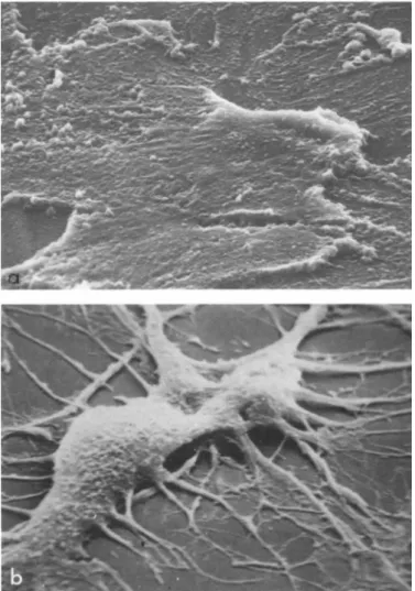
FIGURE 2 Scanning electron microscopy of brain epithelial cells Ca) before and Cb) after morphological transformation by brain extract CX 2500).
epithelial brain cells are indeed developing cells is supported by our unsuccessful attempts to obtain them from the adult brain.
B. Fine Structure of the Cells
With the scanning electron microscope CFig. 2) a confluent monolayer of epithelial brain cells appears as a monotonous con-
466 R A M O N LIM et al.
tinuous sheet. Except for some thickened areas which probably represent cell borders, the cells are devoid of recognizable sur- face landmarks. A great contrast is observed with cells exposed to brain extract. These cells possess a body of definite volume, from which numerous processes and ramifications originate.
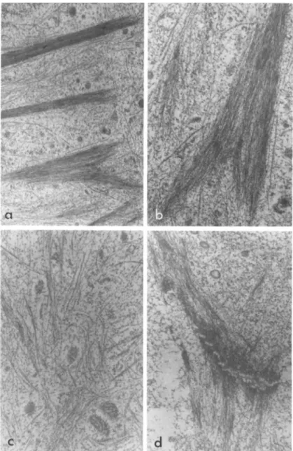
Transmission electron microscopy (Figs. 3 and 4} reveals dif- ferences in intracellular ultrastructure before and after trans- formation. Sheath microfilaments-'- (fibrillar structures that are 50 8 in diameter and arranged in bundles) are abundant in the flat cells but disappear following transformation by brain extract,
ô °
Tonofilaments-1- (100 A in diameter) are found in both types of cells but appear to be more numerous in the transformed cells.
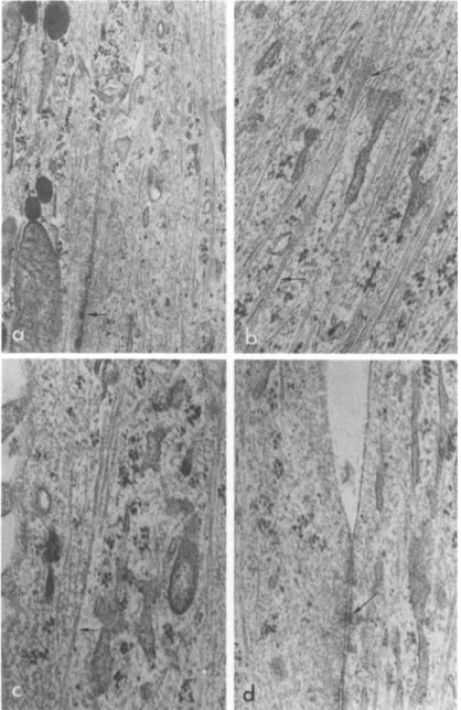
Those seen in the flat cells are usually randomly coiled without a definite orientation whereas those in the transformed cells are arranged in parallel arrays. One distinctive feature of the transformed cells is that their processes are packed with tono-
o
filaments running along the axis. Microtubules (200 A in diam- eter) are a minor component in both cells, but the difference in arrangement and orientation before and after transformation is similar to the tonofilaments. The flat and the transformed cells also differ in cell junctions. The desmosome-like junctions (com- plete with tonofilaments converging on both sides) characteristic of the flat cells are replaced by junctions similar to zonula adhaerentes described by Farquhar and Palade (1963) (see also Peters et al., 1970).
C. Sequential Events During Morphological Transformation
We have studied the morphological changes with cinematography.
Upon exposure of the epithelial cells to brain extract, a latent period covering the first 8 hours is observed, within which the only noticeable change is the clearer separation of the cell bor-
1The classification of the intracellular fibrillar structures in this article follows that of Wessells (1973).
EPITHELIAL BRAIN CELLS 467
FIGURE 3 Transmission electron microscopy of brain epithelial cells before transformation, showing; (a) sheath microfilaments
(X 12,500); Cb) detail of a bundle of sheath microfilaments (X 25,000); (c) tonofilaments with some microtubules (X 25,000); (d) a desmosome-like junction (X 25,000). All stained with phos- photungstic acid.
468 R A M O N LIM et al.
FIGURE 4 Transmissxon electron microscopy of brain epithelial cells after transformation, showing: (a) numerous tonof ilaments in two apposing cell processes; arrow indicates a junction complex between the two processes (X 25,000); (b) detail of tonofilaments
(arrows) (X 50,000); (c) mixture of microtubules farrow) and tono- filaments CX 50,000); (d) detail of a junctional complex (arrow)
EPITHELIAL BRAIN CELLS 469 dera. Between the 12th and the 20th hours, dramatic changes oc-
cur. Many cells suddenly undergo mitotic activity after which the cells maintain a contracted appearance and extrude processes.
All the cells become very active, constantly extending and re- tracting their processes. Since the cells are interconnected, they appear to be engaged in a tug-of-war, as reported earlier by Lumsden and Pomerat C1951) to be characteristic of glia. On the other hand, actual locomotion by the cells from one place to another is minimal. The general histotypic pattern is that of an interconnected cell net showing coordinated movement, as if being pulled from one end of the net (this dynamic relationship is com- mon to glial cell cultures, as opposed to fibroblasts which exhi- bit no visible intercellular coordination). Three days after ex- posure to the extract, movement and mitosis gradually subside while the processes continue to extend. At the end of a week one sees a crisscrossing network with little movement or cell divi- sion.
In a parallel study using isotope incorporation (Table I), we found an increase in the rate of DNA synthesis in cultures exposed to brain extract over the control cells at the 12th hour and the 24th hour points. At the latter time point RNA and protein syn- thesis also increase. The series of events reflects cell divi- sion. A dramatic rise in intracellular cyclic AMP level occurs 4 days after stimulation by the brain extract. The time course of cyclic AMP changes, when taken together with the cinematographic observations on the cells, is consistent with the known inhibitory effects of the agent on cell division (Abell and Monahan, 1973) and motility (Johnson et al., 1972).
Another interesting observation that distinguishes the flat and the transformed cells lies in the ability of some of the lat- ter cells Cabout 20% of the total population) to concentrate
(X 50,000), All stained with osmium tetroxide, uranyl acetate and lead citrate.
eu
<ΗT«
S
ϋ I o\o 00 rH Ï ï üΡ•H [ (Í
pH ϋ + +
CO U rH rH ο Ö
rH ϋ
0 ù
U •H
-P ϋ)
fi φ
L ) 0 - Ñ
c ο\ο o\o ÔP
TJ >1 ιη 00 m
fi CO rH LT)
Ð3 é + é é
fi
H -Η
«J Ö
•P +>
fi
0Q) u
urr eu
er
ft ï
H CO
fi - Η CO
(U Ö
Q) Xi üΡ üΡ (ë»
£ - Ñ CT» CO Ï m
•P C ãΗ in VO
Ö >1 CO I + I I
<d W <
φ s
ϋ fi
φ U-f φ
<4Η ϋ
•rH CO
Û -.Η
CO
ιΗ (d φ CO Ο <Í> ÜΡ <*>
m ï
û • Ñ KD rH m
•H 0 Ö fi >* CO + rH + é é
U < Ù
Η
[•ABLE
CO CO
Ö >i >1
0 tΦ (Φ
•Η ô é ¼ ¼
Åη Jf1 rH
φ ù 0 ft
"-*' φ
rH CO ι—I
φ ϋ
rH rd
•P •
fi
φ •Ρ
0 ϋ
•Η rd φ •Ρ 5η
ft ×! Ö
φ fi
φ • Η
•Ρ (d ϊη
fi
•Η 0
•Ρ -Ρ
fi ¼
Ö Ö
ÎH ft
ϋ φ φ
¼ - Ñ ÎH 0
0 fi
- Ñ •
fi CO - Ñ
Ö rH ϋ
0 rH cd
Ö Ö û - Ñ u
ϋ fi rH Ö
•Η 0 •
5η fi îH
Ö - Ñ - Ç 0 en fi 0 (d H CO u
• Ñ û
fi ϋ
φ Ö 0 φ
û Xi - Ñ u u • Ñ ft
φ φ
ft 0 u (d
470
EPITHELIAL BRAIN CELLS 471
FIGURE 5 Difference in methylene blue uptake by brain epi- thelial cells before and after transformation by brain extract.
The dye was added to the media of live cultures to make a final dilution of 1/50,000 (w/v). (a) and (b): Phase-contrast and light microscopy, respectively, of the flat cells 4 hours after addition of the dye. (c) and Cd): Phase-contrast and light microscopy, respectively, of the transformed cells 1 hour after addition of the dye; arrow indicates one of the cells with "silhouette" cell bodies and beaded cell processes as a result of excessive dye up- take. (X 200).
472 R A M O N LIM et al
methylene blue from the medium when used as a supravital stain (Fig. 51. Since Costero and Pomerat (1951) indicated that the up- take of methylene blue is more of a neuronal property than glial, the significance of this observation is yet to be clarified.
D. Dependency of Morphological Transformation on Cell Passage The transformability of the epithelial brain cells decreases as the number of cell passage increases. On the 5th passage the flat cells are no longer responsive to stimulation by the brain extract. It is not known whether this decreasing transformability is due to the selection in favor of the non-responsive cells or is a result of progressive de-differentiation of all the epithelial cells.
E. Reversibility of Transformation and Temporal Summation of Transforming Effect
Withdrawal of brain extract from the medium leads to the eventual reversion of the transformed cells to the flat cells.
On the other hand, renewing the medium containing the brain ex- tract every other day not only sustains but also augments the out- growing of cell processes. A temporal summation of transforming effect is particularly evident when the cells happen to respond poorly after the first exposure to the brain extract. The need of the continuous presence of the factor indicates a sustaining rather than inducing role with regard to differentiation.
F. Response to Dibutyryl Cyclic AMP and 3-Adrenergic Agents The epithelial cells can be transformed by 1 mM dibutyryl cyclic AMP. However, unlike the brain extract effect which shows a long latency, the effect of dibutyryl cyclic AMP is rapid
(starting within 5 minutes of exposure and reaching a height at about 4 hours) and rather transient. Furthermore, whereas with brain extract cellular retraction and process outgrowth are
EPITHELIAL BRAIN CELLS 473 equally prominent, with, dibutyryl cyclic AMP cellular retraction is the main feature. Cyclic AMP at 1 mM has no effect.
1-Isoproterenol CO.01 mMl transforms the cells in a manner and time course similar to dibutyryl cyclic AMP. The effect is
blocked by the $-blocker dichloroisoproterenol (0.1 mM) but not by the á-blocker phentolamine (0.1 mM). 1-Norepinephrine at 0.1 mM shows only a weak activity compared to isoproterenol. Thus the cells appear to have 3-adrιnergie receptors which utilize cyclic AMP as the mediator, a view consistent with earlier reports that ß-adrenergic agents produce a transient surge in cyclic AMP level in astrocytoma cultures (Clark and Perkins, 1971; Gilman and Nirenberg, 1971), brain slices (Rail and Gilman, 1970; Schultz and Daly, 1973a and 1973b), and dissociated brain cells (Gilman and Schrier, 1972).
G. Response to Variations in Serum Concentration
Complete absence of serum may lead to a transient retraction of some of the epithelial cells in a manner similar to the effect of dibutyryl cyclic AMP. At serum concentrations of 2% or high- er, this spontaneous change is not seen.
The influence of serum on the transforming effect of brain extract is complex. In the presence of 10% serum or higher, the transforming effect of brain extract normally observable at the 20th hour is suppressed or reduced. However, if the same culture is observed over a period of 4 or more days, with renewal of the medium containing brain extract every other day, the response eventually shows up to the same degree in high serum as in low serum. This is true even with serum concentrations as high as 17%.
474 R A M O N LIM et al.
H. Agents Blocking Morphological Transformation
The following agents block the transforming effect of brain extract: cycloheximide C0.1 yg/ml); colchicine (0,1 yg/ml); vin- blastine (0.5 yg/ml); cytochalasin  (5 yg/ml),
IV. NATURE OF THE TRANSFORMING FACTOR
A. Some Initial Doubts
The question arose as to whether the observed morphological transformation is caused by some chemical message contained in the brain extract or simply by changes in physical conditions intro- duced by the extract, such as pH of the medium and the surface texture of the flask. The pH effect is ruled out by our ability to demonstrate the same transformation by brain extract under various pH conditions (Lim and Mitsunobu, 1975). The texture ef- fect is ruled out by the observation that growing the epithelial brain cells in flasks pretreated (one-day preincubation) with brain extract does not result in morphological transformation (un- less fresh brain extract is concomitantly present) and that trans- formed cells, if transferred to a new flask not currently or pre- viously exposed to brain extract, retain the transformed morphol- ogy, thus indicating that the direct contact between the cells and the extract, and not between the flask surface and the ex- tract, is essential for the observed morphological changes. Fur- thermore, other pieces of evidence against the flask surface ef- fect are available: (1) growing the epithelial brain cells in plasma clot or collagen-coated surface, or the direct addition of gelatin and fibrinogen to the culture, does not result in process outgrowth; (2) the brain is one of the organs with the lowest col- lagen content yet its transforming effect is the highest among organs assayed; (3) extracts prepared from rat brains whose plas- ma fibrinogen has been eliminated by saline perfusion do not show
E P I T H E L I A L B R A I N C E L L S 475
a lower transforming activity compared with, extracts from unper- fused brains; C4I no fibrous deposit has ever been detected by scanning electron microscopy and no collagen fiber has ever been observed with transmission electron microscopy on cultures exposed to brain extract; C5Î preincubation of the brain extract with col- lagenase or direct addition of the enzyme into the culture does not block morphological transformation.
Can the transforming effect be attributed to some special nutritional factor contained in brain extract? Since the small molecules have been routinely dialyzed out, this question directs itself to the macromolecules as possible nutrients. We have demonstrated that if the factor is a protein, it is the native molecule and not the amino acid constituents that are responsible for the activity. To prove this we first boiled the dialyzed brain extract for 10 minutes to coagulate the proteins (the mix- ture was now devoid of activity). With the coagulum in place, pronase (2 mg/ml) was introduced and the mixture incubated for 4.5 hours at 37°C until the coagulum cleared up. The solution was again boiled for 10 minutes to coagulate the pronase which was subsequently eliminated by centrifugation. The resulting protein digest was completely inactive for transformation.
The protein digest experiment described above also suggests that the putative transforming factor cannot be a heat-stable small molecule (such as cyclic AMP or prostaglandin) that might have adhered to the proteins, since the pronase digestion would have released them and they would have remained active in the supernatant fluid. The presence of contaminating cyclic AMP that might have exerted the transforming effect was also ruled out by the inability of cyclic AMP phosphodiesterase to suppress the transformation by brain extract when either pre-incubated with the extract or directly added to the culture.
476 RAMON LIM et al.
Â. What the Factor Is Not
Numerous commercially available proteins (Lim and Mitsunobu, 1974) were assayed; none transformed the cells. Other synthetic and natural products free of transforming activity (when tested at 1.2 mg/ml medium) include: polyvinylpyrrolidone, Ficoll, poly- ethylene glycol, hyaluronic acid, chondroitin sulfate, heparan sulfate, yeast RNA, and poly-L-aspartic acid. The following fac- tors, known to affect growth and/or differentiation of some other cells, show no morphological effect on the epithelial brain cells:
Nerve Growth Factor, phytohemagglutinin, concanavalin A, wheat germ agglutinin and pokeweed mitogen. Of these. Nerve Growth Factor was tested on three separate occasions: once with a sample purchased from Wellcome Research Laboratories, once with a sample obtained in our laboratory directly from the submaxillary glands of an adult male mouse, once with a gift supplied by Drs. Ing- Ming Jeng and Ralph A. Bradshaw of Washington University. All gave negative results when tested at a concentration optimum for neurite outgrowth in dorsal root ganglia. Furthermore, a partial- ly purified Morphological Transforming Factor (see below) sent from our laboratory to Drs. Jeng and Bradshaw for radioimmunoas- say and bioassay for Nerve Growth Factor was negative by either criterion. Cyclic AMP at 1 mM and prostaglandin at concentra- tions ranging from 0.01 pg/ml to 10 pg/ml are without effect on our cells (Lim et al., 1973).
C. Biological Distribution of the Transforming Factor
Extracts from rat and pig brains are equally active on fetal rat brain cells. Other rat and pig organs, such as liver, kidney, heart and muscle, also contain significant amounts of the activi- ty. 17-day rat embryos (brain or whole body extract) contain on- ly 1/5 of the activity found in adult brain. The factor is also low in brain tumors such as rat astrocytoma and mouse neuro- blastoma, where less than 1/10 of the activity in adult brain can
E P I T H E L I A L B R A I N C E L L S 477
be detected. Human red blood cells contain only 1/25 of the rat brain activity whereas the activity in microorganisms such as E.
coli. Bacillus subtilis and yeast is practically nil Cless than 1/200 of adult brain). All the comparisons above are made on the basis of extractable protein. The following body fluids, when used at concentrations up to 15% (v/v) in the medium, do not pos- sess any transforming activity: fetal calf serum, bovine serum, horse serum, rat serum, chicken serum and human cerebrospinal flu- id. Thus, the general distribution indicates that the factor is limited to solid organs of normal adult animals. Conditioned medium from the epithelial brain cells, even after a 5-fold con-
centration, does not show any transforming activity.
D. Chemical Properties of the Transforming Factor
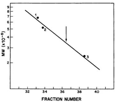
The factor has an apparent molecular weight of 350,000 (Fig.
6). It is resistant to DNAase and RNAase. Incubation with tryp- sin does not destroy nor reduce the activity. However, the fac- tor is completely inactivated by pronase (0.1 mg enzyme/ml) after an overnight incubation under aseptic conditions, indicating that it is a protein. There is no indication as to whether it is a pure or a conjugated protein. Periodate oxidation does not af- fect the transforming activity, suggesting that any carbohydrate moiety, if present, probably does not play an essential role in its function. The factor is resistant to reducing agents and alkylating agents. It has an isoelectric point below neutrality.
Other protein properties include its denaturation by the extremes of pH, by 8 M urea, and by prolonged heating. We have achieved a 400-fold partial purification (Lim and Mitsunobu, 1975) of the factor by ethanol treatment, trypsinization and column chroma- tography with Sephadex G-200 and Sepharose 4B. The purest sample is active at 3 ug protein/ml medium, equivalent to a concentra- tion of 1 x 1 0 ~8 M,
478 RAMON LIM et al.
I 1 1 1 I I I \ \ \ \ \ \
32 34 36 38 40
FRACTION NUMBER
FIGURE 6 Molecular weight determination for the Morphological Transforming Factor on a Sepharose 4B column. 1 = thyroglobulin;
2 = &-galactosidase; 3 = catalase. Arrow indicates the peak of the transforming activity.
V. SUMMARY AND CONCLUSIONS
CD Epithelial brain cells from rat fetuses can be stimulated by a tissue factor to grow out processes after a latency of about 8 to 12 hours,
(2) Most of the cells are glial cells.
(3) The effect of the tissue factor is not due to alteration of the physical conditions of the culture.
C4) The factor is a high molecular weight protein which is active when used in minute amounts.
C5) The cells stimulated by the factor show an increase in DNA, RNA, and protein synthesis at the early time point and an in- crease in cyclic AMP content at the late time point.
C6) The reversibility of transformation and the presence of the factor in adult rather than embryonic tissue suggest that the
E P I T H E L I A L B R A I N C E L L S 479
factor plays a maintenance role in cellular differentiation and morphogenesis,
(7) The factor is present in the brain and several other or- gans of the mature animal.
ACKNOWLEDGMENTS
Dr. Mitsunobu was, and Dr. Troy is Research Associate in Dr.
Limfs laboratory. Dr. Turriff is a Public Health Service Post- doctoral Fellow (NS-05017). This work was supported by U.S. Pub- lic Health Service grants NS-09228, CA-14599 and NS-07376.
REFERENCES
Abell, C. W. and Monahan, T. M. C1973). J. Cell Biol. 59, 549-558.
Clark, R. B. and Perkins, J. P. C1971). Proc. Nat. Acad. Sei. USA.
68, 2757-2760.
Costero, I. and Pomerat, C. M. (1951). Am. J. Anatomy. 89, 405- 439.
Farquhar, M. G. and Palade, G. E. (1963). J. Cell Biol. 17, 375- 412.
Gilman, A. G. and Nirenberg, M. W. (1971). Proc. Nat. Acad. Sei.
USA. 68, 2165-2168.
Gilman, A. G. and Schrier, Â. K. (1972). Molecular Pharma. 8, 410- 416.
Johnson, G. S., Morgan, W. D. and Pastan, I. (1972). Nature. 235, 54-56.
Lim, R., Li, W. K. P. and Mitsunobu, K. (1972). Abstracts of 2nd Annual Meeting, Society for Neuroscience, Houston, Texas, p.
181.
Lim, R., Mitsunobu, K. and Li, W. K. P. (1973). Exp. Cell Res., 79, 243-246.
Lim, R. and Mitsunobu, K. (1974). Science. 185, 63-66.
480 RAMON LIM et ai
Lim, R. and Mitsunobu, Ê, (19751. Biochim. Biophus. Acta, 400, 200-207.
Lumsden, C, E, and Pomerat, C. M, (1951). Exp, Cell Res, 2, 103- 114.
Peters, A., Palay, S. L. and Webster, H. de F. C1970). "The Fine Structure of the Nervous System" Harper and Row, Publishers, New York.
Rail, T. W. and Gilman, A. G. (1970). Neurosciences Res. Prog.
Bull. 8, 239-242.
Schultz, J. and Daly, J. W. (1973a). J. Neurochem. 21, 1319-1326.
Schultz, J. and Daly, J. W. (1973b). J. Biol. Chem. 248, 860-866.
Shein, H. M. (1965). Exp. Cell Res. 40, 554-569.
Varon, S. and Raiborn, C. W. Jr. (1969). Brain Res, 12, 180-199.
Wessels, Ν. Ê. (1973). Neurosciences Res, Prog, Bull. 11, 24-27.