ORIGINAL ARTICLE
Chicken or the Egg: Microbial Alterations in Biopsy Samples of Patients with Oral Potentially Malignant Disorders
Gabor Decsi1&Jozsef Soki2&Bernadett Pap3&Gabriella Dobra3&Maria Harmati3&Sandor Kormondi4&
Tibor Pankotai5&Gabor Braunitzer6&Janos Minarovits7&Istvan Sonkodi1&Edit Urban2&Istvan Balazs Nemeth8&
Katalin Nagy1&Krisztina Buzas3,7
Received: 8 June 2018 / Accepted: 19 July 2018
#Arányi Lajos Foundation 2018
Abstract
Oral carcinogenesis often leads to the alteration of the microbiota at the site of the tumor, but data are scarce regarding the microbial communities of oral potentially malignant disorders (OPMDs). Punch biopsies were taken from healthy and non- healthy mucosa of OPMD patients to analyze the microbiome using metagenome sequencing. In healthy oral mucosa biopsies the bacterial phylaFirmicutes, Fusobacteria, Proteobacteria, ActinobacteriaandBacteroideteswere detected by Ion Torrent se- quencing. The same phyla as well as the phylaFibrobacteresandSpirochaeteswere present in the OPMD biopsies. On the species level, there were 10 bacterial species unique to the healthy tissue and 35 species unique to the OPMD lesions whereas eight species were detected in both samples. We observed that the relative abundance ofStreptococcus mitisdecreased in the OPMD lesions compared to the uninvolved tissue. In contrast, the relative abundance ofFusobacterium nucleatum, implicated in carcinogenesis, was elevated in OPMD. We detected markedly increased bacterial diversity in the OPMD lesions compared to the healthy oral mucosa. The ratio ofS. mitisandF. nucleatumare characteristically altered in the OPMD lesions compared to the healthy mucosa.
Keywords OPMD . Oral microbiome . Metagenome sequencing . Lichen . Leukoplakia
Introduction
Numerous scientific data demonstrate the altered bacterial col- onization of cancerous tissue, but the causality of microbial alteration has not clarified yet.
The role of oral microbes in the development of oral poten- tially malignant disorder (OPMD) and oral squamous cell car- cinoma (OSCC) is periodically reevaluated, since OPMD or OSCC may develop in the absence of the traditional risk factors like smoking, alcohol consumption and betel nut use [1–3].
While the microbiological background of oral squamous cell carcinoma was intensively studied in the last two decades, less attention has been paid to OPMD in this respect [4].
OPMD is a group of disorders of diverse etiologies, fre- quently associated with tobacco consumption and mutations in the DNA of oral epithelial cells. A fraction of OPMD un- dergoes clinical and histomorphological alterations that lead to the development of OSCC. These disorders include leuko- plakia, erythroplakia, oral lichen planus, submucous fibrosis, and actinic cheilitis [5,6]. In addition, inherited cancer syn- dromes such as xeroderma pigmentosum and Fanconi’s
* Krisztina Buzas
kr.buzas@gmail.com; buzas.krisztina@brc.mta.hu
1 Faculty of Dentistry, Department of Oral Surgery, University of Szeged, Tisza Lajos krt. 64, Szeged H-6720, Hungary
2 Albert Szent-Gyorgyi Clinical Centre, Institute of Clinical Microbiology, University of Szeged, Semmelweis u. 6, Szeged H-6725, Hungary
3 Biological Research Centre, Hungarian Academy of Sciences, Temesvari krt. 62, Szeged H-6726, Hungary
4 Albert Szent-Gyorgyi Clinical Centre, Department of Traumatology, University of Szeged, Semmelweis u. 6, Szeged H-6725, Hungary
5 Department of Biochemistry and Molecular Biology, University of Szeged, Kozep fasor 52, Szeged H-6726, Hungary
6 dicomLAB Ltd., Pulz u. 46/b, Szeged H-6724, Hungary
7 Faculty of Dentistry, Department of Oral Biology and Experimental Dental Research, University of Szeged, Tisza Lajos krt. 64, Szeged H-6720, Hungary
8 Department of Dermatology and Allergology, University of Szeged, Szeged H-6720, Hungary
https://doi.org/10.1007/s12253-018-0457-x
anemia are also associated with an increased incidence of malignant tumors, among them oral carcinoma [6].
Leukoplakia is defined as ″A white plaque of ques- tionable risk having excluded (other) known diseases or disorders that carry no increased risk for cancer″.
Leukoplakia is six times more common among smokers than among non-smokers [6]. These lesions are divided into homogenous (simplex) and non-homogenous types.
The non-homogenous types based on the cellular vari- ability are verrucous leukoplakia, nodular leukoplakia and erythroleukoplakia [7]. Proliferative verrucous leu- koplakia (PVL) is a subtype of verrucous leukoplakia that shows resistance to treatment and a high rate of malignant transformation. It is more frequent among el- derly women, in many cases without smoking in the anamnesis. Distinct histopathological changes, like hy- perkeratosis with or without dysplasia, may accompany the transition of PVL to verrucous hyperplasia and verrucous carcinoma [6].
There are conflicting results regarding the association of leu- koplakia and human papillomavirus (HPV) infection [6,8]. The role of torque teno virus (TTV) [9,10], Epstein-Barr virus (EBV) [11] andCandida albicans[8,12–16] in leukoplakia develop- ment and carcinogenesis remains to be clarified, too.
Additionally, it has been also demonstrated that specific bacterial infections like Helicobacter pylori[17] or the intracellular Mycoplasma salivarium[18] could also be involved in this process.
A disbalance in the oral microbial flora can be also accom- panied with leukoplakia and carcinogenesis, as suggested by the association of periodontal inflammation with leukoplakia [19] and a changing bacterial flora in the saliva and on the oral mucosal surfaces of patients with OPMD [20,21].
Lichen planus (LP) is a chronic, idiopathic, inflammatory disease of the oral mucosa or the skin, presenting as a white lesion when it affects the oral cavity (oral lichen planus, OLP).
A crucial aspect of the pathomechanism of OLP is the accu- mulation of CD8+ T lymphocytes under the basal cell layer of the oral mucosa, which causes DNA damage and the apopto- sis of the keratinocytes by antigen-specific cell-mediated im- mune response, and also basement membrane degradation [22–25]. According to the most accepted hypothesis, chronic stimulation from the inflammatory and stromal cells provides the initial signal which lead to the abolished growth control of epithelial cells. Additionally, oxidative stress induced DNA damage could also lead to neoplastic changes, but the initial event leading to this signal cascade activation has not been characterized yet. Based on the increasing evidence viral, fun- gal, and bacterial antigens have all been suggested [26–34] as a potential initiating factor in LP. If there is a relationship between the bacterial flora and OLP, the question is whether the trigger area is in the oral cavity, or at another area of the body, such as the skin, the gastrointestinal tract, the larynx or
the eyes. If oral bacterial infection could initiate OLP devel- opment, it is not clear whether a single bacterial species could initiate the OLP transformations, or is it the interaction of several species during the process? Additionally, the disturbed balance of the normal bacterial flora could also be involved in the initial steps of OLP activation.
In our experimental setup, we examined the oral microbiome of patients diagnosed with OPMD. We compared the microbiome of healthy and non-healthy mucosal surfaces.
Using metagenome sequencing, we detected markedly in- creased bacterial diversity in the OPMD lesions compared to the healthy oral mucosa. In parallel, in the OPMD lesions there was a reduced representation of distinctStreptococcusspecies that dominate the bacterial community of healthy oral mucosa.
Materials and Methods
Patient SelectionPotential participants were interviewed by the clinical coordi- nators at the University of Szeged, Faculty of Dentistry. Every potential participant was informed about the ethical permis- sion of the study and received both written and oral informa- tion on the goals and procedures of this study. The initial participants were selected by volunteering activity and the detailed questionnaire on family anamnesis, chronic diseases, medication-, alcohol-, tobacco- and drug consumption, oral hygiene, and sexual habits was filled out by the volunteers.
Eligibility was determined based on the results of this ques- tionnaire. Female and male subjects over 18 years of age were eligible for the study, provided that they were able to provide signed and dated informed consent and if they did not meet any of the exclusion criteria. As for the patient group, an established diagnosis of OPMD was also a requirement. The exclusion criteria included vital signs outside the acceptable range at the screening visit (i.e., blood pressure > 140/
90 mmHg, oral temperature > 37 °C, heart rate > 100/
min), pregnancy, the potential subject being a sex work- er, topical antibiotic treatment up to 7 days before the screening visit, and the use of the following drugs with- in 2 months before the screening visit: systemic antibi- otics, corticosteroids, cytokines, methotrexate or immu- nosuppressive cytotoxic agents, or large doses of com- mercial probiotics (≥ 108 CFU mL−1 organisms per day). No patients were on specific diet, nor on antibi- otic therapy in the previous 6 months. The clinical char- acteristics and brief medical history of the patient group is given in Table 1.
The study protocol conformed to the Declaration of Helsinki in all respects and was approved by the Institutional Ethics Committee of the University of Szeged (No. 3161).
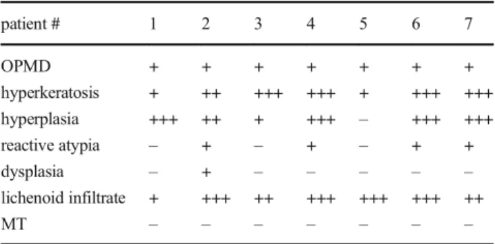
Table1Thebriefmedicalhistoryofpatients patient#1234567 sex♂♂♀/2PM♀/2PM♂♂♀/1PM age60736257855477 clinicaldiagnosisleukoplakia verrucosalabiileukoplakiaproliferativa verrucosagingivaelichenreticularis etatrophicans buccaeetlinguae lichenreticularis ethypertrophicus buccae
lichenreticularis buccaelichenatrophicans buccaelichenreticularis etatrophicans buccae etgingivae pathological diagnosisOPMD-verrucous hyperplasiaOPMD–lichenoid gingivitis withhyperplasia, reactivechanges, togetherwithbasal celldysplasia
OPMD–lichenoid buccitiswith hyperplasiaand reactivechanges OPMD–lichenoid buccitiswith hyperplasiaand reactivechanges OPMD–lichenoid buccitiswithreactive changes OPMD–lichenoid buccitiswithmassive hyperplasiaandreactive changes
OPMD–lichenoid buccitiswith hyperplasiaand reactivechanges systemicdiseasesHT, onychomycosis, MI,cGa,cDu, IHD
HT,hChol,hTGnoHTHT,hypothyr., h.uricemianoHT,DM,RA, h.uricemia, hyperthyr.,hChol allergynonononomedicineallergynono packyearsof smoking1000020202.5 alcohol consumption/dayno2–3unit(wine)nono2unit(wine)nono frequencyof tooth-brushingeveryweekmanytimesadaymanytimesadaymanytimesadayonceadaymanytimesadaymanytimesaday useoforalrinsenomanytimesaday, changednoListerine/CorsodylnoListerine/MeridolListerine sexualactivitynoneinthepast 4yrsactiveactiveactiveactiveactiveactive historyoforal sexualactivitynononoyesnoyesno medicationsatthe timeofthestudy:Asactal,Concor, Pantoprazol, Roxera,Apranax, Tritace Lipidil,Xeter, Chinotal, Nortivan,Agen, Aspirin Protect
ØBetaserc,Nootropil, Prenessa,FrontinTiroxin,Milurit, WarfarinØ1 Abbreviations:OPMD:oralpotentiallymalignantdisorder;PM:postmenopausal;/1/2:parity;HT:hypertension;MI:Myocardialinfarct;cGa:chronicgastritis;cDu:chronicduodenitis;IHD:ischemic heartdisease;hChol:hypercholesterinaemia;hTG:Hypertrigliceridaemia,DM:diabetesmellitus;RA:rheumatoidarthritis;hypothr:hypothryreosis,hyperthr:hyperthyreosis,h.uricemia:hyperuricemia 1 L-Thyroxin,Meloxep,Rosuvastatin,Glucobay,Milurit,Talliton,Frontin,Tramadol,Moxogamma,Metoprolol,Enalapril,Meloxep,Apo-Famotidin,chrom,vitamin-C,vitamin-D,Mg-B6,kurkuma
Tissue Sample Collection for Metagenome Sequencing, Histopathological Assessment
As a classical method for tissue sample collection from muco- sal lesions in the oral cavity, we used punch biopsy. The biopsy was made with punches 4 mm in diameter. We collected 2 identical samples from the intralesional area: one sample was taken for hematoxylin-eosin (HE) staining, and an additional sample for metagenome sequencing. Additionally, we also col- lected a third, control specimen from the ipsilateral healthy mucosa for metagenome sequencing. The specimens contained the mucosa, and submucosal connective tissue. Samples for metagenome sequencing were collected in Eppendorf tubes filled with 20 mM potassium phosphate buffer (pH 7.0), and stored at−20 °C. Tissue samples for HE staining were fixed in
formaldehyde solution, and sent for histopathological assess- ment. Pathological dataset (Table2) included the absence or presence, together with severity of hyperkeratosis, hyperplasia, reactive atypia, dysplasia, and lichenoid infiltrate [35].
Metagenomic DNA Isolation
Total DNA was isolated from patient samples as de- scribed previously, with minor modifications [36].
Briefly, samples (600 μL) were suspended in 650 μL of extraction buffer (100 mM TrisCl, pH 8.0, 100 mM EDTA, pH 8.0, 1.5 M NaCl, 100 mM sodium phos- phate, pH 8.0, 1% CTAB) 3.5 μL proteinase K (20.2 mg mL−1) and incubated horizontally at 37 °C for 45 min, next 80 μL of 20% SDS was added and mixed by inversion for several times with further incu- bation at 60 °C for 1 h. The sample in each tube was mixed thoroughly every 15 min. The particles were col- lected by centrifugation (17,000 g) for 5 min. The su- pernatant was transferred into clean tubes and was mixed with equal quantity of phenol chloroform and isoamyl alcohol (25:24:1) and extracted three times.
DNA was precipitated with 0.7 volume isopropanol, the pellet was washed with 70% ethanol. Crude DNA pellets were dried and dissolved in 50 μL of TE buffer (10 mM Tris-HCl, 1 mM sodium EDTA, pH 8.0).
Metagenomic DNA was quantified using Qubit® 2.0 Fluorometer. Half of total metagenomic DNA from the Table 2 The histopathological characteristics of the biopsies
patient # 1 2 3 4 5 6 7
OPMD + + + + + + +
hyperkeratosis + ++ +++ +++ + +++ +++
hyperplasia +++ ++ + +++ – +++ +++
reactive atypia – + – + – + +
dysplasia – + – – – – –
lichenoid infiltrate + +++ ++ +++ +++ +++ ++
MT – – – – – – –
Abbreviations: (−no, + mild, ++ moderate, +++ severe; OPMD: oral potentially malignant disorder; MT: malignant transformation)
Fig. 1 Representative pictures from OPMD tissue samplesOPMD tissue samples with marked hyperkeratosis (a, b–HyKer), acanthosis and lichenoid infiltrate (a, d–LI). Occasionally there was a marked verrucous hyperplasia with elongated dermal papillae (c–DP). Note
the regenerative basal cell changes (b–RA/regenerative atypia/), in contrast to mild dysplasia (d–DY) with focal acantholysis /HE; OM 112×/
healthy and lesion samples were pooled and stored at
−20 °C for sequencing.
Library Preparation and Sequencing
Ion Torrent PGM Fragment libraries of 200 nt were generated according to the appropriate protocols (Ion Torrent PGM, Life Tech, USA). 1μg pooled metagenomic DNA from each sam- ple was used for library preparation for each sample. DNA shearing and end-repair was achieved by Ion Xpress™Plus Fragment Library Kit, and DNA Purification was carried out by PureLink PCR Purification Kit (Thermo Fisher Scientific, MA). Adapter ligation and nick translation were performed by Ion Shear Plus Reagents Kit (Thermo Fisher Scientific, MA).
Size selection was performed in 2% agarose gel to enrich the 300–350 nt fragments then library amplification was achieved by using Platinum® PCR SuperMix (Thermo Fisher Scientific, MA). ION Library TaqMan qPCR was used for quantitation and Ion Xpress Template 200 ePCR kit was used for the emul- sion PCR. Sequencing was performed on Ion Torrent Personal Genome Machine™using Ion 318 chip. Ion Torrent Personal Genome Machine™sequencing resulted 872,798 sequence
reads for sample 1 (control) with an average read length of 219 ± 71 bases and 644,200 sequence reads for sample 2 (diseased) with an average read length of 220 ± 72 bases.
Quality Assurance
The MG-RAST software performs a QC (quality control) and an automatic normalization of the FASTQ sequence. For the taxonomical analyses, maximum e-value cut-off of 10−15, minimum percent identity cut-off 90% and minimum align- ment length cut-off 40 nt settings were applied. The overall community composition was determined using the M5nr da- tabase. Hits for the eukaryotic data were removed and relative abundance of the bacterial data was calculated.
Swab Sample Collection
We used cotton swabs for collecting bacterial samples. Two individual samples were taken from each patient, one from the surface of the lesion, and another one from the ipsilateral healthy (non-involved) mucosa. The swabs were rolled 4 times over the chosen areas. Then the swabs were placed in anaerobic
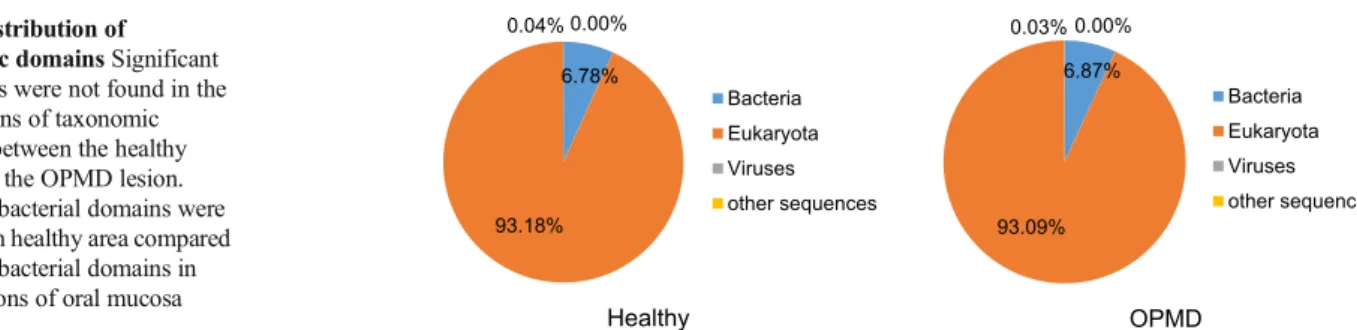
6.87%
93.09%
0.03% 0.00%
Bacteria Eukaryota Viruses other sequences
OPMD Healthy
6.78%
93.18%
0.04% 0.00%
Bacteria Eukaryota Viruses other sequences Fig. 2 Distribution of
taxonomic domainsSignificant differences were not found in the distributions of taxonomic domains between the healthy tissue and the OPMD lesion.
6.78% of bacterial domains were detected in healthy area compared 6.87% of bacterial domains in white lesions of oral mucosa
71%
11%
9%
5%
4%
Firmicutes Fusobacteria Proteobacteria Actinobacteria
Bacteroidetes 67%
9%
8%
7%
5%4%1%
Firmicutes Fusobacteria Proteobacteria Actinobacteria Bacteroidetes Fibrobacteres Spirochaetes
a
b
OPMD Healthy
10 8 35
OPMD Healthy
Fig. 3 Bacterial diversitya Metagenome sequencing revealed that the bacterial diversity in the OPMD biopsies was higher compared to the healthy oral mucosa. Within the Bacteria domainFirmicutes,
Fusobacteria, Proteobacteria, Actinobacteria, Bacteroidetes phyla were present in the healthy oral mucosa. In the OPMD biopsies the same phyla were identified in descending order of relative abundance; however, two additional phyla,Fibrobacteres andSpirochaeteswere observed, too.bMetagenome sequencing detected 18 different bacterial species in healthy tissue and 43 species in the OPMD lesion.
Eight bacterial species were detected in both samples
transport medium (AnaerobeSystems, CA) and sent immedi- ately to the Institute of Clinical Microbiology, University of Szeged, forFusobacterium nucleatumcultivation.
Fusobacterium nucleatumqPCR
DNA templates for qPCR were prepared from the undiluted BHI suspensions of the oral swabs by the QiAmp Stool Mini DNA Kit (Qiagen, Germany) as recommended by the supplier.
Subsequent quantitative RT-PCRs for Fusobacterium nucleatum were done in StepOne RT-PCR instrument (Invitrogen, CA) using 5μL Brilliant II master mix (Agilent), primers FnucF CTTAGGAATGAGACAGAGATG and FnucR TGATGGTAACATACGAAAGG 0.2μL (35 pmole/μL) each, 1μL of template sample and the following cycling conditions:
starting denaturation and hot start activation 95 °C 10 min, 95 °C 15 s, 56 °C 15 s and 72 °C 30 s, 40×; and a melting curve from 72 °C to 95 °C [37] CFUs were calculated comparing the
means of threshold cycles to ones of aF. nucleatum 10-fold serial dilution samples prepared with the same kit.
Data Analysis
For the comparison of the numerical data, the Mann-Whitney U test was used in SPSS 21.0 (IBM, NY).
Results
Sample selection was based on the histological analysis of oral potentially malignant disorder. Figure1shows representative pictures of the OPMD tissue samples with marked hyperker- atosis, acanthosis and lichenoid infiltration. Table1. describes the brief medical history of selected patients (Selection criteria has been found in Materials and methods). Detailed histolog- ical classification summarized in Table2.
OPMD Healthy
32.73%
25.45%
7.27%
3.64%
3.64%
3.64%
3.64%
1.82%
100%
Psychrobacter sp. PRwf-1 Enhydrobacter aerosaccus Haemophilus influenzae Neisseria subflava Neisseria meningitidis Streptococcus oralis Streptococcus infantis Gemella morbillorum Prevotella bergensis Porphyromonas gingivalis Actinomyces urogenitalis Fusobacterium sp. 4_1_13 Fusobacterium sp. 3_1_36A2 Fusobacterium nucleatum Rothia dentocariosa Gemella haemolysans Streptococcus pneumoniae Streptococcus mitis
18.62%
13.79%
5.52%
4.83%
4.14%
4.14%
4.14%
4.14%
0%
10%
20%
30%
40%
50%
60%
70%
80%
90%
100%
100%
Treponema vincentii Simonsiella muelleri Delftia acidovorans Lautropia mirabilis Fusobacterium sp. 7_1 Fusobacterium sp. 3_1_27 Fusobacterium sp. 1_1_41FAA Veillonella sp. oral taxon 158 Veillonella sp. 3_1_44 Eubacterium saburreum Parvimonas micra
Streptococcus thermophilus Streptococcus sp. oral taxon 071 Streptococcus sp. C300 Streptococcus pyogenes Gemella morbillorum Capnocytophaga gingivalis Thermomonospora curvata Actinomyces urogenitalis Actinomyces sp. oral taxon 171 Haemophilus influenzae Leptotrichia buccalis Streptococcus salivarius Streptococcus gordonii Granulicatella adiacens Gemella haemolysans Corynebacterium matruchotii Corynebacterium efficiens Veillonella parvula Streptococcus vestibularis Capnocytophaga ochracea Prevotella melaninogenica Rothia mucilaginosa Streptococcus sp. M334 Streptococcus sanguinis Campylobacter concisus Streptococcus sp. M143 Streptococcus oralis Fibrobacter succinogenes Streptococcus parasanguinis Fusobacterium nucleatum Streptococcus pneumoniae Streptococcus mitis
Fig. 4 Comparison of the detected bacterial speciesMetagenome sequencing detected 18 different bacterial species in healthy tissue and 43 species in the OPMD lesion
First, we determined the ratio of bacterial DNA in our sample. As punch biopsy samples contain a high amount of human tissue, this step helped us to define the limitation of the study. In healthy oral mucosa samples, 6.78% of the sequence reads were annotated to the domain Bacteria. The ratio of bacterial sequences was similar (6.87%) in the pooled OPMD DNA samples (Fig.2).
Metagenome sequencing revealed that the bacterial diversity in the OPMD biopsies was higher compared
to the healthy oral mucosa. Within the Bacteria domain F i r m i c u t e s , F u s o b a c t e r i a , P r o t e o b a c t e r i a , Actinobacteria, Bacteroidetes phyla were present in the healthy oral mucosa. In the OPMD biopsies the same phyla, i.e. Firmicutes, Fusobacteria, Proteobacteria, Actinobacteria and Bacteroidetes were identified in de- scending order of relative abundance; however, two ad- ditional phyla, Fibrobacteres and Spirochaetes were ob- served, too (Fig. 3a).
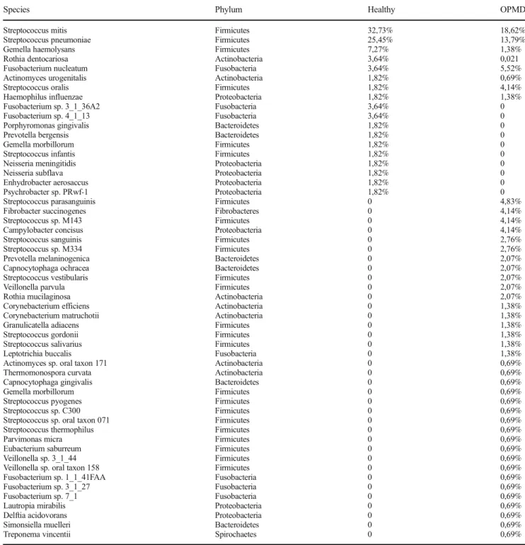
Table 3 Comparison of bacterial diversity in the healthy mucosa and the OPMD lesion
Species Phylum Healthy OPMD
Streptococcus mitis Firmicutes 32,73% 18,62%
Streptococcus pneumoniae Firmicutes 25,45% 13,79%
Gemella haemolysans Firmicutes 7,27% 1,38%
Rothia dentocariosa Actinobacteria 3,64% 0,021
Fusobacterium nucleatum Fusobacteria 3,64% 5,52%
Actinomyces urogenitalis Actinobacteria 1,82% 0,69%
Streptococcus oralis Firmicutes 1,82% 4,14%
Haemophilus influenzae Proteobacteria 1,82% 1,38%
Fusobacterium sp. 3_1_36A2 Fusobacteria 3,64% 0
Fusobacterium sp. 4_1_13 Fusobacteria 3,64% 0
Porphyromonas gingivalis Bacteroidetes 1,82% 0
Prevotella bergensis Bacteroidetes 1,82% 0
Gemella morbillorum Firmicutes 1,82% 0
Streptococcus infantis Firmicutes 1,82% 0
Neisseria meningitidis Proteobacteria 1,82% 0
Neisseria subflava Proteobacteria 1,82% 0
Enhydrobacter aerosaccus Proteobacteria 1,82% 0
Psychrobacter sp. PRwf-1 Proteobacteria 1,82% 0
Streptococcus parasanguinis Firmicutes 0 4,83%
Fibrobacter succinogenes Fibrobacteres 0 4,14%
Streptococcus sp. M143 Firmicutes 0 4,14%
Campylobacter concisus Proteobacteria 0 4,14%
Streptococcus sanguinis Firmicutes 0 2,76%
Streptococcus sp. M334 Firmicutes 0 2,76%
Prevotella melaninogenica Bacteroidetes 0 2,07%
Capnocytophaga ochracea Bacteroidetes 0 2,07%
Streptococcus vestibularis Firmicutes 0 2,07%
Veillonella parvula Firmicutes 0 2,07%
Rothia mucilaginosa Actinobacteria 0 2,07%
Corynebacterium efficiens Actinobacteria 0 1,38%
Corynebacterium matruchotii Actinobacteria 0 1,38%
Granulicatella adiacens Firmicutes 0 1,38%
Streptococcus gordonii Firmicutes 0 1,38%
Streptococcus salivarius Firmicutes 0 1,38%
Leptotrichia buccalis Fusobacteria 0 1,38%
Actinomyces sp. oral taxon 171 Actinobacteria 0 0,69%
Thermomonospora curvata Actinobacteria 0 0,69%
Capnocytophaga gingivalis Bacteroidetes 0 0,69%
Gemella morbillorum Firmicutes 0 0,69%
Streptococcus pyogenes Firmicutes 0 0,69%
Streptococcus sp. C300 Firmicutes 0 0,69%
Streptococcus sp. oral taxon 071 Firmicutes 0 0,69%
Streptococcus thermophilus Firmicutes 0 0,69%
Parvimonas micra Firmicutes 0 0,69%
Eubacterium saburreum Firmicutes 0 0,69%
Veillonella sp. 3_1_44 Firmicutes 0 0,69%
Veillonella sp. oral taxon 158 Firmicutes 0 0,69%
Fusobacterium sp. 1_1_41FAA Fusobacteria 0 0,69%
Fusobacterium sp. 3_1_27 Fusobacteria 0 0,69%
Fusobacterium sp. 7_1 Fusobacteria 0 0,69%
Lautropia mirabilis Proteobacteria 0 0,69%
Delftia acidovorans Proteobacteria 0 0,69%
Simonsiella muelleri Bacteroidetes 0 0,69%
Treponema vincentii Spirochaetes 0 0,69%
On the species level, 18 bacterial species were detect- ed in the healthy tissue, 35 species were unique for the OPMD lesions and eight bacterial species were detected both in healthy oral mucosa and the OPMD biopsy samples (Fig. 3b). Metagenome sequencing showed markedly increased bacterial diversity in the OPMD tis- sue samples. Analysis of bacterial species detected 2.38 fold increasing in diversity of the OPMD lesions (Fig. 4, Table 3).
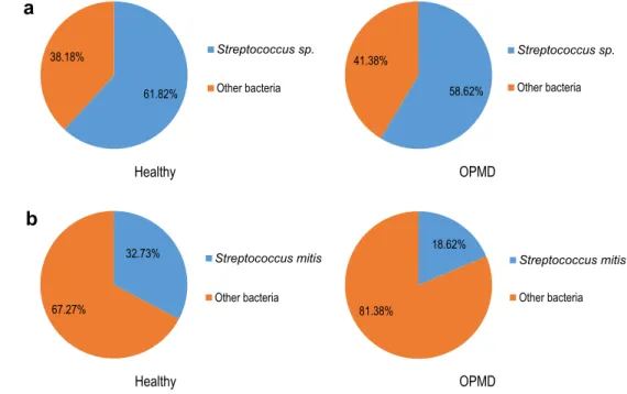
Based on the data of metagenome sequencing, the relative abundance of Streptococcus sp. did not show significant difference between the healthy tissue and the OPMD lesions: it was 61.82% in healthy tissue and 58.62% in OPMD, respectively (Fig. 5a) However, the relative abundance of the Streptococcus mitis was found to be dramatically decreased in OPMD: it was only 18.62%, compared to 32.73% in the healthy tissue (Fig.5b). Moreover, examining all Streptococcusspecies their prevalence in the OPMD lesions was determined to be more diverse compared to the healthy tissue (Table 3).
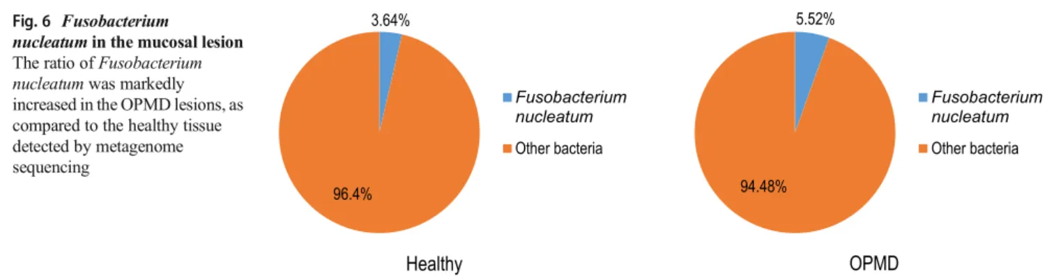
According to the results of metagenome sequencing, the ratio ofFusobacterium nucleatumwas higher in the OPMD lesions (5.52%), compared to the healthy tissue (3.64%) (Fig. 6). Relative abundance of Fusobacterium nucleatum (5.52%) and Streptococcus oralis (4.14%) bacteria were identified in a larger portion among the overlapping bacte- ria in OPMD while 3.64 and 1.82% were observed in healthy tissue, respectively). The abundances of the species Gemella haemolysans(1.38%),and the above mentioned S.
mitis(18.62%) decreased markedly in OPMD as compared to the healthy tissue (7.27% and 32.73%) (Table 3).
35 different bacterial species has been found in the OPMD lesions which has not been presented in the healthy tissue, e.g., Streptococcus parasanguinis, Fibrobacter succinogenes, Campylobacter concisus, S t re p t o c o c c u s s a n g u i n i s , R o t h i a m u c i l a g i n o s a , Prevotella melaninogenica, Capnocytophaga ochracea, S t re p t o c o c c u s v e s t i b u l a r i s , Ve i l l o n e l l a p a r v u l a , C o r y n e b a c t e r i u m e f f i c i e n s , C o r y n e b a c t e r i u m matruchotii, Granulicatella adiacens, Streptococcus gordonii, Streptococcus salivarius, Leptotrichia buccalis, Thermomonospora curvata, Capnocytophaga gingivalis, Streptococcus pyogenes Streptococcus thermophiles, Parvimonas micra, Eubacterium saburreum, Lautropia mirabilis, Delftia acidovorans, Simonsiella muelleri, Treponema vincentii, etc., in descending order of the abundances.
As F. nucleatum was implicated in carcinogenesis in several cancers [38], we verified the results of metagenome sequencing by F. nucleatum-specific qPCR. As Fig. 7 shows, the number of bacteria (CFUs) is significantly higher (P< 0.0001) in samples taken from OPMD lesions than in those from the healthy mucosa. This difference is remarkable, even when one considers the high inter-individual variability of the samples.
Discussion
It was suggested that OPMD was frequently associated with alcohol and tobacco consumption, and affected mainly males in their fifties, but the practice shows that
18.62%
81.38%
Streptococcus mitis Other bacteria 32.73%
67.27%
Streptococcus mitis
Other bacteria
b
Healthy OPMD
58.62%
41.38% Streptococcus sp.
Other bacteria 61.82%
38.18% Streptococcus sp.
Other bacteria
a
Healthy OPMD
Fig. 5 Ratio of Streptococci in the mucosal lesionaThe ratio of Streptococci to all oral bacteria was not significantly different in the healthy tissue compared to the OPMD lesion.bThe ratio of Streptococcus mitisdramatically decreased in the OPMD, as compared to the healthy tissue
a much wider variety of patients are affected [1, 3].
Accordingly, we included a wide range of patients in our sample, without strict limitations on age or sex.
In our study we compared the microbiome and microbiota of healthy and the OPMD tissues of oral cavity.
The association of Fusobacterium nucleatum with oral carcinoma was documented [38, 39]. Moreover, it was suggested that distinct subspecies of F. nucleatum, such as F. nucleatum subsp. polymorphum and F.
n u c l e a t u m s u b s p. v i n c e n t i i, m a y p l a y a n etiopathogenetic role in oral carcinogenesis [40]. A study also described the presence of F. nucleatum in desquamative gingivitis [41]. However, our report shows the first time the presence of Fusobacterium nucleatum in lesion biopsies of OPMD patients.
The finding that F. nucleatum is present in OPMD may be exploited to develop a novel therapeutic strategy of distinct oral disorders such as oral lichen planus. In addition, one may speculate that a targeted antibiotic therapy could be beneficial in preventing the develop- ment of oral cancer in a subset of OPMD patients.
We o b s e r v e d t h a t t h e r e l a t i v e a b u n d a n c e o f Streptococcus mitis decreased dramatically in the patho- logical niche. Since Streptococci are characteristic com- ponents of the oral flora, the quantitative analysis of these bacteria is indispensable for the understanding of pathological processes. Streptococci comprise of a wide
variety of bacterial species that interact with other mem- bers of the oral microbiome. It was suggested that S.
mitis is involved in the maintenance of a healthy oral flora by affecting adhesion and biofilm formation by periodontal pathogens to [42–44].
Therefore, it is an interesting and important finding that the relative abundance of S. mitis in the OPMD lesions decreased to nearly the half of the healthy area.
This observation may possibly be exploited for thera- peutic purposes: similarly to the reconditioning of vag- inal Lactobacillus balance [45], the restitution of S.
mitis niche in OPMD could also have beneficial effects [46].
Taken together, we presented evidence for the alter- ation of microbiome and microbiota of OPMD patients.
We detected an increased bacterial diversity in the OPMD lesions compared to the healthy oral mucosa.
In addition, decreased relative abundance of S. mitis and an increased relative abundance of F. nucleatum may play a role in the transition of OPMDs to oral cancer.
Although our study is not suitable to answer the BChicken or the Egg^ problem in all aspects, but we have found that the bacterial colonization of mucosa has already altered in OPMD tissues. These observa- tions may form the basis of novel therapeutic ap- proaches preventing oral carcinogenesis in a subset of patients with OPMD.
Acknowledgements This work was in part supported by the National Research, Development and Innovation Fund of Hungary, financed under the NKFI-6-K funding scheme (11493 project), GINOP-2.3.2-15-2016- 00015, János Bolyai Research Scholarship of the Hungarian Academy of Sciences and GINOP 2.3.2-15-2016-00011.
Compliance with Ethical Standards
Conflict of Interest No potential conflict of interest was reported by the authors.
Ethical Approval The study protocol conformed to the Declaration of Helsinki in all respects and was approved by the Institutional Ethics Committee of the University of Szeged (No. 3161).
Fig. 7 Fusobacterium nucleatum-specific PCR of the healthy tissue and the OPMDOPMD showed significantly higher colonization byF.
nucleatumcompared to healthy mucosa
5.52%
94.48%
Fusobacterium nucleatum Other bacteria 3.64%
96.4%
Fusobacterium nucleatum Other bacteria
OPMD Healthy
Fig. 6 Fusobacterium
nucleatumin the mucosal lesion The ratio ofFusobacterium nucleatumwas markedly increased in the OPMD lesions, as compared to the healthy tissue detected by metagenome sequencing
References
1. Morse DE, Psoter WJ, Cleveland D et al (2007) Smoking and drinking in relation to oral cancer and oral epithelial dysplasia.
Cancer Causes Control 18(9):919–929
2. Jepsen SA, Closmann JJ (2008) The insidious nature and presenta- tion of oral squamous cell carcinoma in the low-risk population.
Gen Dent 56(1):78–82
3. Llewellyn CD, Linklater K, Bell J et al (2003) Squamous cell car- cinoma of the oral cavity in patients aged 45 years and under: a descriptive analysis of 116 cases diagnosed in the South East of England from 1990 to 1997. Oral Oncol 39(2):106–114
4. Nagy KN, Sonkodi I, Szöke I et al (1998) The microflora associated with human oral carcinomas. Oral Oncol 34(4):304–308 5. Sarode SC, Sarode GS, Tupkari JV (2012) Oral potentially malig-
nant disorders: precising the definition. Oral Oncol 48(9):759–760 6. van der Waal I (2009) Potentially malignant disorders of the oral and oropharyngeal mucosa; terminology, classification and present concepts of management. Oral Oncol 45(4–5):317–323
7. Sonkodi I (2009) Oral and Maxilofacial medicine. Semmelweis Publisher, Budapest
8. Silverman S Jr, Gorsky M (1997) Proliferative verrucous leukopla- kia. A follow-up study of 54 cases. Oral Surg Oral Med Oral Pathol Oral Radiol Endod 84(2):154–157
9. Fehér E, Gáll T, Murvai M et al (2009) Investigation of the occur- rence of torque tenovirus in malignant and potentially malignant disorders associated with human papillomavirus. J Med Virol 81(11):1975–1981
10. Fehér E, Kardos G, Gáll T et al (2011) Comparison of diversity of torque teno virus 1 in different mucosal tissues and disorders. Acta Microbiol Immunol Hung 58(4):319–337
11. Kis A, Fehér E, Gáll T et al (2009) Epstein-Barr virus prevalence in oral squamous cell cancer and in potentially malignant oral disorders in an eastern Hungarian population. Eur J Oral Sci 117(5):536–540 12. Wu L, Feng J, Shi L et al (2013) Candidal infection in oral leuko-
plakia: a clinicopathologic study of 396 patients from eastern China. Ann Diagn Pathol 17(1):37–40
13. Hebbar PB, Pai A, D S. (2013) Mycological and histological associ- ations of Candida in oral mucosal lesions. J Oral Sci 55(2):157–160 14. Dilhari A, Weerasekera MM, Siriwardhana A et al (2016) Candida infection in oral leukoplakia: an unperceived public health problem.
Acta Odontol Scand 74(7):565–569
15. Gainza-Cirauqui ML, Nieminen MT, Novak Frazer L et al (2013) Production of carcinogenic acetaldehyde by Candida albicans from patients with potentially malignant oral mucosal disorders. J Oral Pathol Med. 42(3):243–249
16. Bakri MM, Cannon RD, Holmes AR et al (2014) Detection of Candida albicans ADH1 and ADH2 mRNAs in human archival oral biopsy samples. J Oral Pathol Med 43(9):704–710
17. Kazanowska-Dygdała M, DuśI, Radwan-Oczko M (2016) The presence of helicobacter pylori in oral cavities of patientsn with leukoplakia and oral lichenplanus. J Appl Oral Sci 24(1):18–23 18. Mizuki H, Kawamura T, Nagasawa D (2015) In situ immunohisto-
chemical detection of intracellular mycoplasma salivarium in the epithelial cells of oral leukoplakia. J Oral Pathol Med. 44(2):134–144 19. Meisel P, Holtfreter B, Biffar R et al (2012) Association of periodon-
titis with the risk of oral leukoplakia. Oral Oncol 48(9):859–863 20. Hu X, Zhang Q, Hua H et al (2016) Changes in the salivary micro-
biota of oral leukoplakia and oral cancer. Oral Oncol 56:e6–e8 21. Hernandez BY, Zhu X, Goodman MT et al (2017) Betel nut
chewing, oral premalignant lesions, and the oral microbiome.
PLoS One 12(2):e0172196
22. Srinivas K, Aravinda K, Ratnakar P et al (2011) Oral lichen planus –Review on etiopathogenesis. Natl J Maxillofac Surg 2(1):15–16
23. Payeras MR, Cherubini K, Figueiredo MA et al (2013) Oral lichen planus: focus on etiopathogenesis. Arch Oral Biol 58(9):1057– 1069
24. Georgakopoulou EA, Achtari MD, Achtaris M et al (2012) Oral lichen planus as a preneoplastic inflammatory model. J Biomed Biotechnol 2012:759626
25. Roopashree MR, Gondhalekar RV, Shashikanth MC et al (2010) Pathogenesis of oral lichen planus–a review. J Oral Pathol Med.
39(10):729–734
26. Gorouhi F, Davari P, Fazel N (2014) Cutaneous and mucosal lichen planus: a comprehensive review of clinical subtypes, risk factors, diagnosis, and prognosis. ScientificWorld Journal 2014:742826 27. Bäckman K, Jontell M (2007) Microbial-associated oral lichenoid
reactions. Oral Dis 13(4):402–406
28. Taghavi Zenouz A, Mehdipour M, Jafari Heydarlou M et al (2010) Relationship between Lichen Planus and Helicobacter pylori Infection. J Dent Res Dent Clin Dent Prospects 4(1):17–20 29. Attia EA, Abdel Fattah NS, Abdella HM (2010) Upper gastrointes-
tinal findings and detection of helicobacter pylori in patients with oral lichen planus. Clin Exp Dermatol 35(4):355–360
30. Pourshahidi S, Fakhri F, Ebrahimi H et al (2012) Lack of associa- tion between helicobacter pylori infection and oral lichen planus.
Asian Pac J Cancer Prev 13(5):1745–1747
31. Izol B, Karabulut AA, Biyikoglu I et al (2010) Investigation of upper gastrointestinal tract involvement and H. Pylori presence in lichen planus: a case-controlled study with endoscopic and histo- pathological findings. Int J Dermatol 49(10):1121–1126
32. Hulimavu SR, Mohanty L, Tondikulam NV et al (2014) No evi- dence for Helicobacter pylori in oral lichen planus. J Oral Pathol Med. 43(8):576–578
33. Boorghani M, Gholizadeh N, Taghavi Zenouz A et al (2010) Oral lichen planus: clinical features, etiology, treatment and manage- ment; a review of literature. J Dent Res Dent Clin Dent Prospects.
4(1):3–9
34. Ertugrul AS, Arslan U, Dursun R et al (2013) Periodontopathogen profile of healthy and oral lichen planus patients with gingivitis or periodontitis. Int J Oral Sci 5(2):92–97
35. El-Naggar AK, Chan JKC, Takata T, Grandis JR, Slootweg PJ (2017) Tumours of the oral cavity and mobile tongue In: The fourth edition of the head and neck World Health Organization blue book:
editors' perspectives. Hum Pathol 66:10–12
36. Sharma PK, Capalash N, Kaur J (2007) An improved method for single step purification of metagenomic DNA. Mol Biotechnol 36(1):61–63
37. Periasamy S, Chalmers NI, Du-Thumm L et al (2009) Fusobacterium nucleatum ATCC 10953 requires Actinomyces naeslundii ATCC 43146 for growth on saliva in a three-species community that includes Streptococcus oralis 34. Appl Environ Microbiol 75(10):3250–3257
38. Rubinstein MR, Wang X, Liu W et al (2013) Fusobacterium nucleatum promotes colorectal carcinogenesis by modulating E- cadherin/β-catenin signaling via its FadA adhesion. Cell Host Microbe 14(2):195–206
39. Schmidt BL, Kuczynski J, Bhattacharya A et al (2014) Changes in abundance of oral microbiota associated with oral cancer. PLoS One 9(6):e98741
40. Al-Hebshi NN, Nasher AT, Maryoud MYet al (2017) Inflammatory bacteriome featuring fusobacterium nucleatum and Pseudomonas aeruginosa identified in association with oral squamous cell carci- noma. Sci Rep 7(1):1834
41. Arduino PG, Romano F, Sasia D et al (2017) Subgingival microbi- ota in white patients with Desquamative gingivitis: a cross-sectional study. J Periodontol 88(7):643–650
42. Kreth J, Merritt J, Qi F (2009) Bacterial and host interactions of oral streptococci. DNA Cell Biol 28(8):397–403
43. Van Hoogmoed CG, Geertsema-Doornbusch GI, Teughels W et al (2008) Reduction of periodontal pathogens adhesion by antagonis- tic strains. Oral Microbial Immunol 23(1):43–48
44. Standar K, Kreikemeyer B, Redanz S et al (2010) Setup of an in vitro test system for basic studies on biofilm behavior of mixed- species cultures with dental and periodontal pathogens. PLoS One 5(10):e13135
45. Otsuki K, Imai N (2017) Effects of lactoferrin in 6 patients with refractory bacterial vaginosis. Biochem Cell Biol 95(1):31–33 46. KlarićM, MandićV, LovrićS, e al. (2017) Antimicrobial efficacy
of probiotic-containing toothpastes: an in vitro evaluation. Med Glas (Zenica) 14(1):16–24