Revised manuscript
Final version published in:
Eur. J. Org. Chem. 2018: 2902–2909 (2018)
DOI: 10.1002/ejoc.201800447
Synthesis and Biological Evaluation of Paclitaxel Conjugates Involving Lysosomally Cleavable Linkers and α V β 3 -Integrin Ligands for Tumor Targeting
Paula López Rivas,
[a]Ivan Ranđelović,
[b]André Raposo Moreira Dias,
[a]Arianna Pina,
[a]Daniela Arosio,
[c]Jószef Tóvári,
[b]Gábor Mező,
[d]Alberto Dal Corso,
[a]Luca Pignataro,
[a]Cesare Gennari*
[a,c]Abstract: Two cyclo[DKP-RGD]-PTX (PTX = paclitaxel) and two cyclo[RGDfK]-PTX conjugates containing the lysosomally cleavable Gly-Phe-Leu-Gly (GFLG) linker were synthesized and compared to two cyclo[DKP-RGD]-Val-Ala-PTX conjugates. The conjugates were evaluated for their ability to inhibit biotinylated vitronectin binding to the isolated αvβ3 receptor, retaining good binding affinity, in the same nanomolar range of the free ligands.
Cell viability assays were performed for the six conjugates in the αvβ3+ U87 and in the αvβ3– HT29 cell lines. Loss of potency was observed for all the conjugates, attenuated by the presence of a tetraethylene glycol (PEG-4) spacer. A good Targeting Index (TI
= Relative Potency in the αvβ3+ U87 / Relative Potency in the αvβ3– HT29) was displayed by the conjugates, in particular by cyclo[DKP-RGD]-PEG-4-Val-Ala-PTX 9 (TI = 533). This conjugate was tested in the αvβ3+ U87 cell line in the presence of 50-fold excess free cyclo[DKP-RGD] ligand 2. In this competition experiment, a fivefold decrease of the conjugate cytotoxicity was calculated, suggesting that the conjugate is possibly internalized by an αvβ3 integrin-mediated process.
Introduction
Cytotoxic drugs are characterized by a limited clinical efficacy in the cure of cancer due to a narrow therapeutic window, which means that the difference between the Maximum Tolerated Dose (MTD) and the Minimum Effective Dose (MED) is very small.[1] One of the approaches to overcome this limitation is to increase the selectivity of the cytotoxic agents by conjugation to tumor-targeting devices, which leads to less severe side effects and to the requirement of lower quantities.[2] To date, Antibody-
Drug Conjugates (ADCs) have been successfully used for this objective, combining humanized or chimeric antibodies with cytotoxic drugs, that allow the selective delivery of the antitumor agent into antigen-positive malignant cells. Indeed, four ADCs have been introduced into the market: the well-known KadcylaTM (azo-trastuzumab emtansine) and AdcetrisTM (brentuximab vedotin) and the recently approved MylotargTM (gemtuzumab- ozogamycin) and BesponsaTR (inotuzumab ozogamicin).[1, 3 ] However, several drawbacks are associated to this antibody targeting approach: high manufacturing costs, low tissue diffusion, possible immune system-induced alteration of the drug efficiency, etc..[ 4 ] As an alternative, Small Molecule-Drug Conjugates (SMDCs) have emerged in the last decades, combining small receptor binders with cytotoxic agents. Just as in ADCs, also in SMDCs the choice of a proper linker system is crucial to avoid premature drug release in the blood stream and to achieve selective release of the payload at the tumor site.
One of the possible receptors that can be targeted by SMDCs is integrin αVβ3. This heterodimeric transmembrane receptor is overexpressed in the blood vessels of several human cancers (breast cancer, glioblastoma, pancreatic tumor, prostate carcinoma) but not in the healthy tissues.[ 5 ] Integrin αVβ3
recognizes the tripeptide Arg-Gly-Asp (RGD)[ 6 ] and for this reason a number of peptides and peptidomimetics containing this sequence and displaying high binding affinity to the receptor (in the low nanomolar range) have been prepared.[7] Many of these RGD-peptidomimetics – such as, for example, cyclo[RGDfK] 1 (Figure 1) – have been used for the preparation of tumor-targeting drugs and imaging agents.[ 8,9] In this context, our research group has developed a series of cyclic RGD ligands containing a diketopiperazine scaffold (DKP),[7d-f] among which cyclo[DKP-RGD] 2 (Figure 1) showed a very good binding affinity and selectivity for integrin αVβ3 (Table 1). As a further step, compound 3 (Figure 1) – i.e. an analog of ligand cyclo[DKP-RGD] 2 bearing a primary amino group[ 10 ] – was synthesized and conjugated to different drugs through various types of linker.[10,11] In an initial attempt, we connected ligand cyclo[DKP-RGD] to paclitaxel (PTX) through an ester linkage, but the construct obtained suffered from limited stability in plasma.[10] We then connected ligand cyclo[DKP-RGD] to paclitaxel (PTX) through a more stable carbamate linkage and the lysosomally cleavable Val-Ala (VA) dipeptide linker,[ 12 ] obtaining a cyclo[DKP-RGD]-VA-PTX conjugate.[11b,13] With the latter compound, cell viability assays were run in which a cell line expressing integrin αvβ3 (αvβ3+) was compared with an αvβ3
non-expressing isogenic cell line (αvβ3–). In these experiments, the cyclo[DKP-RGD]-VA-PTX conjugate[11b,13] showed a fairly effective integrin targeting [Targeting Index (TI) up to 9.0], although the potency was slightly reduced compared to free PTX.[11b] In another contribution from our group, the VA linker [a] P. López Rivas, A. Raposo Moreira Dias, A. Pina, Dr. L. Pignataro,
Prof. Dr. C. Gennari
Università degli Studi di Milano, Dipartimento di Chimica Via C. Golgi, 19, 20133 Milan, Italy E-mail: cesare.gennari@unimi.it
http://sites.unimi.it/gennarigroup/
[b] I. Ranđelović, Dr. J. Tóvári
Department of Experimental Pharmacology, National Institute of Oncology
Ráth György u. 7-9., 1122 Budapest, Hungary [c] Dr. D. Arosio
CNR, Istituto di Scienze e Tecnologie Molecolari (ISTM) Via C. Golgi, 19, 20133 Milan, Italy
[d] Prof. Dr. G. Mező
Eötvös Loránd University, Faculty of Science, Institute of Chemistry, MTA-ELTE Research Group of Peptide Chemistry
Pázmány Péter st. 1/A, 1117 Budapest, Hungary
Supporting information for this article is given via a link at the end of the document
was also exploited for the development of cyclo[DKP-RGD]
conjugates with α-amanitin.[11e] Recently, we also explored the use of reductively cleavable disulfide linkers, synthesizing a cyclo[DKP-RGD]-SS-camptothecin conjugate[11d] which, however, did not show any targeting due to the low stability of the α,α- unsubstituted disulfide bond in the cell medium.[14]
Figure 1. A: ligand cyclo[RGDfK] (1). B: ligand cyclo[DKP-RGD] (2) and its functionalized version (3).
Table 1. Inhibition of biotinylated vitronectin binding to the isolated αvβ3 and αvβ5 receptors.
Ligand Structure IC50 (nM)[a] αVβ3 IC50 (nM)[a] αVβ5
1 cyclo[RGDfK] 1.4 ± 0.2 117.5 ± 7.8
2 cyclo[DKP-RGD] 4.5 ± 1.1 149 ± 25
[a] IC50 values were calculated as the concentration of compound required for 50% inhibition of biotinylated vitronectin binding as estimated by GraphPad Prism software. All values are the arithmetic mean ± the standard deviation (SD) of duplicate determinations.
Following the promising preliminary results outlined above,[11b] in this paper we report a more systematic study on the RGD conjugates bearing intracellularly cleavable peptide linkers.
Taking conjugate cyclo[DKP-RGD]-VA-PTX[11b,13] as a reference, we set to assess the influence of the SMDC’s different moieties on integrin binding and cell viability. As shown in Figure 2, keeping the PTX-self-immolative spacer system unchanged, we modified in turn: i) the peptide linker; ii) the spacer connecting the linker to the ligand; iii) the ligand itself. In conjugates 4-7 (Figure 3) we selected the well-known lysosomally cleavable tetrapeptide Gly-Phe-Leu-Gly (GFLG), that has been used in several different delivery systems,[15 ] as alternative to the VA linker (present in conjugates 8 and 9). To replace the glutarate spacer (present in conjugates 4, 5 and 8), we used a PEG-4 chain connected to a triazole ring formed by the CuAAC “click”
reaction[16] (conjugates 6, 7 and 9). Finally, in conjugates 5 and 7 the well-known and easy-to-prepare ligand cyclo[RGDfK][17] was employed as replacement for ligand cyclo[DKP-RGD].
Figure 2. Synopsis of the RGD ligand-PTX conjugates used for the studies reported in this paper.
Figure 3. PTX conjugates: cyclo[DKP-RGD]-GFLG-PTX (4), cyclo[RGDfK]-GFLG-PTX (5), cyclo[DKP-RGD]-PEG-4-GFLG-PTX (6), cyclo[RGDfK]-PEG-4-GFLG- PTX (7), cyclo[DKP-RGD]-VA-PTX (8) and cyclo[DKP-RGD]-PEG-4-VA-PTX (9).
Results andDiscussion
While conjugates 8[11b] and 9[ 18 ] (featuring the VA linker) had already been prepared in our laboratory, the synthesis of compounds 4-7 is reported here. In these new SMDCs, PTX is connected to the C-terminus of the GFLG linker through a para- aminobenzyl carbamate (PABC)-N,N’-dimethylethylenediamine self-immolative spacer, while the N-terminus of the tetrapeptide is connected to the integrin ligand (either cyclo[DKP-RGD] or cyclo[RGDfK] 1, Scheme 1) with a spacer, see Scheme 2. The series is formed by two pairs of analogs (4 / 6 and 5 / 7) differing only for the spacer, which can be a glutarate (compounds 4 and 5) or a triazole-tetraethylene glycol (PEG-4, compounds 6 and 7). Integrin receptor competitive binding assays (Table 2) and cell viability assays (Table 3) were performed for the new GFLG conjugates (4-7) and for the VA conjugates 8 and 9 (Figure 3).[11b,18] Additionally, a competition experiment was performed (Table 4), in which the αVβ3+ U87 cell line was treated with conjugate 9 in the presence of 50-fold excess free ligand cyclo[DKP-RGD] (2).
Synthesis
Synthesis of compounds 1 and 14
The linear protected RGDfK ligand [Fmoc-Asp(OtBu)-D-Phe- Lys(Boc)-Arg(Pbf)-Gly-OH 11, Scheme 1] – precursor of cyclo[RGDfK] 1 – and the Fmoc-protected GFLG linker 14 (Scheme 2) were synthesized manually by solid phase peptide synthesis (SPPS) on 2-chlorotrityl chloride resin using the Fmoc protocol (see the Supporting Information). Cleavage from the resin (using a 8:1:1 CH2Cl2/MeOH/AcOH mixture for 2 h) followed by precipitation in water led to the protected peptides 11 (Scheme 1) and 14 (Scheme 2), which were used in the next step without purification.
Scheme 1. Synthesis of ligand 1. Reagents and conditions: a) i. Fmoc-Gly-OH (1 equiv.), iPr2NEt (3 equiv.), 1:1 CH2Cl2/DMF, 2 h, r.t.; ii. Capping with 7:2:1 CH2Cl2/MeOH/iPr2NEt; b) i. Fmoc-deprotection: 2% DBU, 2% piperidine, DMF, 1 h; ii. Fmoc-AA-OH (3 equiv.), HOBt (4 equiv.), DIC (4 equiv.), 2 h; conditions (b) are repeated for the coupling of every amino acid of the sequence; c) 8:1:1 CH2Cl2/MeOH/AcOH, 2 h, precipitation in water; d) DMF, 20% piperidine, 2 h;
e) 6:4:4 iPr2NEt/BOP/HOBt, 1 mM concentration in DMF, 24 h, precipitation in 5% NaHCO3; f) TFA/thioanisole/EDT/phenol/TIS, 3 h. DBU = 1,8- Diazabicyclo[5.4.0]undec-7-ene; AA = amino acid; BOP = (benzotriazol-1- yloxy)tris(dimethylamino)phosphonium hexafluorophosphate; DIC = N,N’- diisopropylcarbodiimide; EDT = 1,2-ethanedithiol; TIS = triisopropylsilane.
Compound 11 was treated with piperidine to remove the Fmoc protecting group and the resulting free amino acid 12 was cyclized in the presence of a 6:4:4 iPr2NEt/BOP/HOBt mixture in DMF to give compound 13. The cyclization reaction was run under diluted conditions (1 mM) to avoid possible intermolecular side reactions. The protecting groups of 13 were removed with a TFA/thioanisole/EDT/phenol/TIS cocktail to give pure cyclo[RGDfK] (1) after purification using preparative HPLC.
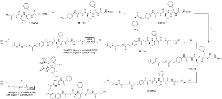
Synthesis of conjugates 4-7
The cyclo[DKP-RGD]-GFLG-PTX and the cyclo[RGDfK]-GFLG- PTX conjugates (compounds 4-7) were synthesized as shown in Scheme 2. The Fmoc-protected GFLG peptide 14 was coupled to 4-aminobenzyl alcohol with HOBt and N,N’- diisopropylcarbodiimide (DIC), leading to the benzylic alcohol 15, which was activated with 4-nitrophenyl chloroformate and coupled to N-Boc-N,N’-dimethylethylenediamine to yield compound 17. The latter served as a common intermediate for the synthesis of all the conjugates, either containing the glutarate or the PEG-4 spacer. In the first case, after Fmoc deprotection of 17 under basic conditions, the free amine was treated with glutaric anhydride to afford compound 18, that was activated as a N-hydroxysuccinimidyl ester and coupled either to functionalized cyclo[DKP-RGD] (3) or to cyclo[RGDfK] (1) at controlled pH to afford intermediates 19a and 19b. The latter compounds were later Boc-deprotected with trifluoroacetic acid (TFA) and coupled to 2’-(4-nitrophenoxycarbonyl)paclitaxel,[11b]
giving the final conjugates 4 and 5.
For the synthesis of the conjugates containing the PEG-4 spacer, compound 17 was Fmoc-deprotected and coupled to 4- pentynoic acid in the presence of HATU, HOAt and iPr2NEt to give alkyne 20, which was later Boc-deprotected with TFA and coupled with 2’-(4-nitrophenoxycarbonyl)paclitaxel affording compound 21. Finally, this compound was subjected to copper- catalyzed azide-alkyne cycloaddition (“click” reaction) with either cyclo[DKP-RGD]-PEG-4-azide (22a)[18] or cyclo[RGDfK]-PEG-4- azide (22b), prepared in two steps from ligands 3 and 1, respectively (see the Supporting Information). The click reaction gave the final conjugates 6 and 7.
Integrin receptor competitive binding assay
Conjugates 4-9 were evaluated for their ability to inhibit biotinylated vitronectin binding to the isolated αVβ3 and αVβ5
receptors. The calculated half-maximal inhibitory concentrations (IC50) are listed in Table 2. Screening assays were performed by incubating the immobilized integrin receptor with solutions of the cyclo[DKP-RGD]-paclitaxel and cyclo[RGDfK]-paclitaxel conjugates at different concentrations (10-12-10-5 M) in the presence of biotinylated vitronectin (1 µg mL-1) and measuring bound vitronectin. Table 2 shows that conjugates 4-9 retain good binding affinity and high selectivity for αvβ3 integrin, with IC50 values in the nanomolar range, only slightly worse compared to those obtained for the free ligands (see Table 1, compounds 1 and 2). These results prompted us to proceed with cell viability assays.
Scheme 2. Synthesis of conjugates 4-7. Reagents and conditions: a) HOBt, DIC, 4-aminobenzyl alcohol, DMF, overnight; b) 4-nitrophenylchloroformate, pyridine, 4:1 THF/DMF, 2 h; c) N-Boc-N,N’-dimethylethylenediamine, iPr2NEt, THF, overnight; d) i. piperidine, DMF, 4 h; ii. glutaric anhydride, DMAP, iPr2Net, DMF, overnight; e) i. DIC, NHS, DMF; ii. 3 (for 19a) or 1 (for 19b), 1:1 DMF/PBS (phosphate-buffered saline, pH 7.3-7.6), overnight; f) i. TFA, CH2Cl2; ii. 2’-(4- nitrophenoxycarbonyl)PTX, iPr2NEt, DMF, overnight; g) i. piperidine, DMF, 4 h; ii. 4-pentynoic acid, HATU, HOAt, iPr2NEt, DMF, overnight; h) 22a or 22b, CuSO4·5 H2O, sodium ascorbate, DMF/H2O. DIC = N,N’-diisopropylcarbodiimide; DMAP = 4-dimethylaminopyridine; NHS = N-hydroxysuccinimide; PTX = paclitaxel.
Table 2. Inhibition of biotinylated vitronectin binding to the isolated αvβ3 and αvβ5 receptors.
Compound Structure IC50 (nM)[a] αVβ3 IC50 (nM)[a] αVβ5
4 cyclo[DKP-RGD]-
GFLG-PTX 54.8 ± 14.0 > 1000[b]
5 cyclo[RGDfK]-
GFLG-PTX 62.6 ± 10.9 649 ± 136
6 cyclo[DKP-RGD]-
PEG-4-GFLG-PTX 42.4 ± 7.4 > 1000[b]
7 cyclo[RGDfK]-PEG-
4-GFLG-PTX 12.1 ± 2.0 473 ± 25
8 cyclo[DKP-RGD]-
VA-PTX 13.3 ± 3.6[c] 924 ± 290[c]
9 cyclo[DKP-RGD]-
PEG-4-VA-PTX 14.8 ± 3.9[d] > 1000[b]
[a] IC50 values were calculated as the concentration of compound required for 50% inhibition of biotinylated vitronectin binding as estimated by GraphPad Prism software. All values are the arithmetic mean ± the standard deviation (SD) of duplicate determinations. [b] Biotinylated vitronectin binding was not completely inhibited in the concentration range tested. [c] See ref. [11b]. [d]
See ref. [18].
Cell viability assays
The antiproliferative activity of conjugates 4-9 was evaluated in two cell lines expressing different levels of αVβ3 integrin. U87 cells (human glioblastoma) were selected as αVβ3+, while HT29 cells (human colorectal adenocarcinoma) were used as αVβ3–.
The expression of αVβ3 integrin on the cell membrane was assessed by flow cytometry (see the Supporting Information), and the results were in good agreement with the literature.[19]
Both cell lines were treated for 96 hours[ 20 ] with different concentrations of free PTX and conjugates 4-9 and the cell viability was evaluated with the MTT assay.
The calculated IC50 values are shown in Table 3. All the conjugates proved less potent than free PTX in both cell lines.[21]
Interestingly, the conjugates containing the PEG-4 spacer (6, 7
and 9) proved 2.4-6.2 times more potent in the αVβ3+ U87 cell line than the conjugates bearing the glutarate spacer (4, 5 and 8). This might be due to improvement of the water solubility and flexibility provided by the tetraethylene glycol spacer, which is known to facilitate binding to the receptor.[22] For all conjugates, the Relative Potencies (RP = IC50 PTX / IC50 Conjugate) in the αVβ3+ U87 cell line are 1-2 orders of magnitude higher than in the αVβ3– HT29 cell line. This means that the loss of potency of the conjugates with respect to PTX is higher when there is no αVβ3 receptor in the cell line. Accordingly, we observed good Targeting Indexes (TI = RP in the αVβ3+ U87 / RP in the αVβ3– HT29) throughout the series (TIs = 30-45, for compounds 4-8), with compound 9 displaying the best value (TI = 533).
Table 3. Evaluation of anti-proliferative activity of free PTX and PTX conjugates 4-9 in U87 and HT29 cell lines.
IC50 (nM)[a]
Comp. U87 (αVβ3+) HT29 (αVβ3–) RPU87[b]
RPHT29[c]
TI[d]
PTX 32.66 ± 21.81 1.82 ± 1.85 1 1 1
4 2031 ± 454 3413 ± 983 0.01608 0.00053 30
5 1250 ± 293.6 2692 ± 676 0.02613 0.000692 38 6 854.7 ± 165.1 1979 ± 252 0.03821 0.0009196 42 7 506.2 ± 113.6 1272 ± 156 0.06452 0.001431 45 8 2686 ± 589 6452±1723 0.01216 0.0002821 43 9 432.6 ± 129.3 12840 ± 2730 0.07550 0.0001417 533 [a] IC50 values were calculated as the concentration of compound required for 50% inhibition of cell viability. Both cell lines were treated with different concentrations of PTX and compounds 4-9 during 96 hours. The samples were measured in triplicate; [b] Relative Potency in U87 cell line (RPU87): IC50
PTX in U87/ IC50 Conjugate in U87; [c] Relative Potency in HT29 cell line (RPHT29): IC50 PTX in HT29/ IC50 Conjugate in HT29; [d] Targeting Index (TI):
RPU87/RPHT29.
To determine whether the observed targeting is mediated by an integrin binding and internalization process, a competition experiment was carried out in the αVβ3+ U87 cell line. Conjugate 9 was tested in the presence of 50-fold excess free ligand cyclo[DKP-RGD] (2), with the aim of blocking integrins on the cell surface (see Table 4).
Table 4. Competition experiments of conjugate 9 in the presence of 50-fold excess cyclo[DKP-RGD] (2) in U87 cell line.
# Compound(s) IC50 (nM)[a]
U87 (αVβ3+)
Inhibition Ratio[b]
Corrected Inhibition
Ratio[c]
1 cyclo[DKP-RGD] (2) 342.8 · 103 ±
94.7 · 103 - -
2 PTX 32.66 ± 21.81 - -
3 PTX + 50-fold excess
cyclo[DKP-RGD] (2) 10.66 ± 4.8 0.33 1
4 9 432.6 ± 129.3 - -
5 9 + 50-fold excess
cyclo[DKP-RGD] (2) 717.5 ± 216.3 1.66 5.03 [a] IC50 values were calculated as the concentration of compound required for 50% inhibition of cell viability. Both cell lines were treated with different concentrations compounds 9 in the presence of 50-fold excess cyclo[DKP- RGD] (2) during 96 hours. The samples were measured in triplicates; [b]
Inhibition Ratio = (IC50 Compound + 50-fold excess 2) / IC50 Compound; [c]
Corrected inhibition Ratio = [(IC50 Compound + 50-fold excess 2) / IC50
Compound] / [(IC50 PTX + 50-fold excess 2) / IC50 PTX].
As expected, the free ligand cyclo[DKP-RGD] (2) proved scarcely toxic (Table 4, entry 1: IC50 = 343 · 103 nM = 343 μM), but induced a distinct change in cell morphology and cell detachment. In the presence of excess cyclo[DKP-RGD] (2), PTX showed a threefold increased toxicity (Table 4, cf. entry 3 and entry 2). On the contrary, with conjugate 9 the presence of excess free ligand 2 induced a moderate increase of IC50 (Table 4, cf. entry 5 and entry 4). Taking into account the effect produced by excess ligand 2 onto free PTX, a fivefold decrease of the conjugate cytotoxicity is calculated (Corrected Inhibition Ratio, see Table 4, entry 5), which might suggest that internalization is mediated by the αVβ3 integrin receptor.
Conclusions
The research reported in this article had the goal to investigate how the biological properties of integrin αvβ3-targeting SMDCs may vary depending on the type of ligand, spacer and linker employed, with a specific focus on the intracellularly cleavable peptide linkers. To this end, two cyclo[DKP-RGD]-GFLG-PTX (4 and 6) and two cyclo[RGDfK]-GFLG-PTX conjugates (5 and 7) were synthesized, the members of each pair differing for the spacer between ligand and peptide linker (glutarate in 4 and 5, triazole-PEG-4 in 6 and 7). Together with the two cyclo[DKP- RGD]-VA-PTX conjugates 8 and 9, compounds 4-7 were used in (i) binding tests on the integrin αvβ3 isolated receptor, and (ii) viability tests using U87 (αVβ3+) and HT29 cancer cells (αVβ3–).
As a general trend, all conjugates showed low-nanomolar affinity for the integrin αVβ3 receptor and reduced potency compared to
free PTX, with lower IC50 values being obtained using the αVβ3+ cells compared to the αVβ3– cells (TI in the 30-45 range for 4-8, TI = 533 for 9). While replacement of the cyclo[DKP-RGD] ligand moiety with cyclo[RGDfK] barely affected the SMDCs’ properties, the conjugates featuring a triazole-PEG-4 spacer proved 2.4-6.2 times more potent against the αVβ3+ cell line than those bearing the glutarate spacer. Replacing the peptide linker impacted on the conjugates’ anti-proliferative effect in a non-easy-to- rationalize manner. The decrease of potency observed when the best compound (9) was incubated with U87 cells in the presence of a large excess of free ligand cyclo[DKP-RGD] (2) suggests that the conjugate is possibly internalized by an integrin αVβ3- mediated process.
From these results it emerges that integrin αVβ3-targeted SMDCs can achieve promising TI values, although substantial improvements in terms of potency are needed in order for them to become therapeutically useful. To this end, modification of the spacer(s) and/or of the type of linker are being currently taken in consideration by our research group.
Experimental Section
All manipulations requiring anhydrous conditions were carried out in flame-dried glassware, with magnetic stirring and under a nitrogen atmosphere. All commercially available reagents were used as received.
Anhydrous solvents were purchased from commercial sources and withdrawn from the container by syringe, under a slight positive pressure of nitrogen. The reactions were monitored by analytical thin-layer chromatography (TLC) using silica gel 60 F254 pre-coated glass plates (0.25 mm thickness). Visualization was accomplished by irradiation with a UV lamp and/or staining with Cerium/Molibdate reagent or ninhydrin.
HPLC purifications and HPLC traces of final products were performed on Dionex Ultimate 3000 equipped with Dionex RS Variable Wavelength Detector (column: Atlantis Prep T3 OBDTM 5 m 19 × 100 mm; flow 15 ml/min unless stated otherwise). The analysis of the integrals and the relative percentage of purity was performed with the software Chromeleon 6.80 SR11 Build 3161. Low resolution mass spectra (MS) were recorded on Thermo Finnigan LCQ Advantage (ESI source), Micro Waters Q-Tof (ESI source) and Bruker Daltonics Microflex LT (MALDI source) instruments. High-resolution mass spectra (HRMS) were performed with a Fourier Transform Ion Cyclotron Resonance (FT-ICR) Mass Spectrometer APEX II & Xmass software (Bruker Daltonics) – 4.7 T Magnet (Magnex) equipped with ESI source, available at CIGA (Centro Interdipartimentale Grandi Apparecchiature) c/o Università degli Studi di Milano. Freeze-drying: the products were dissolved in water and frozen with dry ice. The freeze-drying was carried out at least for 48 h at -50 °C using the instrument 5Pascal Lio5P DGT.
Ligand cyclo[DKP-RGD] (2),[7e] its functionalized analog (3)[10] and N- Boc-N,N’-dimethylethylenediamine[ 23 ] were prepared according to the literature. Also, conjugates 8,[11b] 9[18] and the activated drug 2’-(4- nitrophenoxycarbonyl)paclitaxel[11b] were prepared as previously described. The synthesis and characterization of ligand cyclo[RGDfK] (1), the Fmoc-protected linker 14, intermediates 15-18, intermediate 20 and azide-ligands 22a and 22b are described in the Supporting Information.
Cyclo[DKP–RGD]–Gly-Phe-Leu-Gly-N-[4-[[[(N-(Boc)-N,N’-
dimethylethylenediamine)carbonyl]oxy]methyl]phenyl] (19a).
Compound 18 (11 mg, 0.0119 mmol, 2 equiv.) was dissolved in DMF (500 µL) and cooled at 0 ºC under nitrogen. N-Hydroxysuccinimide (NHS, 1.8 mg, 0.0155 mmol, 2.6 equiv.) and EDC.HCl (3.4 mg, 0.0179 mmol, 3 equiv.) were added and the reaction was stirred at 0 ºC for a 5 min. The reaction was allowed to reach r.t. and stirred overnight under nitrogen atmosphere. Volatiles were then removed in vacuo and the crude was re- dissolved in DMF (375 µL). A solution of compound 3 (5.2 mg, 0.006
mmol, 1 equiv.) in phosphate buffer (375 µL, pH 7.5) was then added.
During the first hours, the pH was kept in the 7.3-7.6 range by adding small aliquots of 0.2 M NaOH. The resulting solution was stirred overnight and then concentrated under vacuum. The crude residue was purified by semipreparative HPLC [Gradient: 100% (H2O + 0.1% CF3COOH) / 0%
(CH3CN + 0.1% CF3COOH) to 25% (H2O + 0.1% CF3COOH) / 75%
(CH3CN + 0.1% CF3COOH) in 15 min; tR (product): 10.5 min]. Yield: 1.41 mg (15%) over 2 steps. HRMS (ESI+): m/z calcd. for [C68H96N17O18]+:1438.712 [M+H]+; found: 1438.710; m/z calcd. for [C68H96N17NaO18]+: 730.856 [M+Na]+; found: 730.851; m/z calcd. for [C68H95N17Na2O18]2+: 741.840 [M+2Na]2+; found: 741.842.
Cyclo[RGDfK]–Gly-Phe-Leu-Gly-N-[4-[[[(N-(Boc)-N,N’-
dimethylethylenediamine)carbonyl]oxy]methyl]phenyl] (19b).
Compound 18 (13 mg, 0.016 mmol, 1.3 equiv.) was dissolved in DMF (350 µL) and cooled at 0 ºC under nitrogen. NHS (2.8 mg, 0.024 mmol, 2 equiv.) and DIC (3.7 µL, 0.024 mmol, 2 equiv.) were added and the reaction was stirred at 0 ºC for 5 min. The reaction was allowed to reach r.t. and stirred overnight under nitrogen atmosphere. Volatiles were then removed in vacuo and the crude was redissolved in DMF (750 µL). A solution of cyclo[RGDfK] 1 (10 mg, 0.012 mmol, 1 equiv.) in phosphate buffer (750 µL, pH 7.5) was then added. During the first hours, the pH was kept in the 7.3-7.6 range by adding small aliquots of 0.2 M NaOH.
The resulting solution was stirred overnight and then concentrated under vacuum. The crude residue was purified by semipreparative HPLC [Gradient: 100% (H2O + 0.1% CF3COOH) / 0% (CH3CN + 0.1%
CF3COOH) to 25% (H2O + 0.1% CF3COOH) / 75% (CH3CN + 0.1%
CF3COOH) in 15 min; tR (product): 11 min]. Yield: 3.15 mg (17%) over two steps. MS (MALDI): m/z calcd. for [C68H99N16O17]+: 1412.61 [M+H]+; found: 1412.2 (HCCA matrix); HRMS (ESI+): m/z calcd. for [C68H98N16NaO17]+: 1433.719 [M+ Na]+; found: 1433.717; m/z calcd. for [C68H99N16Na2O17]2+ 728.360 [M+ 2Na]2+; found: 728.355.
General Procedure for the Boc-deprotection of compounds 19a, 19b and 20.
A half volumen of TFA was added to a 0.03 M solution of intermediates 19a, 19b or 20 in CH2Cl2 and the reaction was stirred at room temperature for one hour. The solvent was evaporated and then the CH2Cl2 was added twice to the residue followed by evaporation under vacuum each each time to afford the amine TFA salt. The crudes were frozen-dried and used without further purification.
Alkyne-Gly-Phe-Leu-Gly-PTX (21). The TFA salt obtained from deprotection of compound 20 (7.5 mg, 0.0108 mmol, 1 equiv.) was dissolved in DMF (320 µL) and cooled at 0 ºC under nitrogen atmosphere. iPr2NEt (7.5 µL, 0.043 mmol, 4 equiv.) and 2’-(4- nitrophenoxycarbonyl)paclitaxel (16.5 mg, 0.0162 mmol, 1.5 equiv.) were added in this order and the mixture was allowed to reach r.t. and stirred overnight. The reaction mixture was diluted with AcOEt and washed with a 1 M aqueous solution of KHSO4 (2 x) and brine (1 x). The organic phase was dried over Na2SO4, concentrated and purified by flash chromatography (eluent: 9:1 CH2Cl2/CH3OH) to afford pure 21. Yield:
10.18 mg (60%). HRMS (ESI+): m/z calcd. for [C84H98N8O22Na]+: 1593.669 [M+Na]+; found: 1593.667; m/z calcd. for [C84H98N8O22Na2]2+: 808.329 [M + 2Na]2+; found: 808.328.
Cyclo[DKP-RGD]-GFLG-PTX (4). The TFA salt obtained from deprotection of compound 19a (6.4 mg, 0.0041 mmol, 1 equiv.) was dissolved in DMF (470 µL) and cooled at 0 ºC under nitrogen atmosphere. iPr2NEt (3.6 µL, 0.0205 mmol, 5 equiv.) and 2’-(4- nitrophenoxycarbonyl)paclitaxel (12.5 mg, 0.0123 mmol, 3.5 equiv.) were added in this order and the mixture was allowed to reach r.t. and stirred overnight. The crude was concentrated, and the residue was purified by semipreparative HPLC [Gradient: 100% (H2O + 0.1% CF3COOH) / 0%
(CH3CN + 0.1% CF3COOH) to 0% (H2O + 0.1% CF3COOH) / 100%
(CH3CN + 0.1% CF3COOH) in 20 min; tR (product): 12.5 min]. Yield: 2.09 mg (26%). MS (MALDI): m/z calcd. for [C111H137N18O31]+: 2219.38 [M+H]+; found: 2220.6 (HCCA matrix), 2219.1 (SA matrix); HRMS (ESI+):
m/z calcd. for [C111H137N18O31Na]2+: 1120.480 [M+1H+Na]2+; found:
1120.479; m/z calcd. for [C111H137N18O31Na2]3+: 754.650 [M+1H+2Na]3+; found: 754.648.
Cyclo[RGDfK]-GFLG-PTX (5). The TFA salt obtained from deprotection of compound 19b (2.5 mg, 0.00163 mmol, 1 equiv.) was dissolved in DMF (190 µL) and cooled at 0 ºC under nitrogen atmosphere. iPr2NEt (2 µL, 0.0115 mmol, 7 equiv.) and 2’-(4-nitrophenoxycarbonyl)paclitaxel (5.8 mg, 0.0057 mmol, 3.5 equiv.) were added in this order and the mixture was allowed to reach r.t. and stirred overnight. The crude was concentrated, and the residue was purified by semipreparative HPLC [Gradient: 100% (H2O + 0.1% CF3COOH) / 0% (CH3CN + 0.1%
CF3COOH) to 0% (H2O + 0.1% CF3COOH) / 100% (CH3CN + 0.1%
CF3COOH) in 20 min; tR (product): 13 min]. Yield: 1.22 mg (33%). MS (MALDI): m/z calcd. for [C111H140N17O30]+: 2192,39 [M+H]+; found: 2193.4 (HCCA matrix); 2194.1 (SA matrix); HRMS (ESI+): m/z calcd. for [C111H140N17O30Na]2+: 1106.993 [M+1H+Na]; found: 1106.991.
Cyclo[DKP-RGD]-PEG-4-GFLG-PTX (6). Compound 21 (3.9 mg, 0.0025 mmol, 1.5 equiv.) and 22b (1.7 mg, 0.0017 mmol, 1 equiv.) were dissolved in a degassed 1:1 water/DMF mixture (170 µL). Degassed aqueous solutions of CuSO4 .
5H2O (0.2 mg, 0.000835 mmol, 0.5 equiv.) and sodium ascorbate (0.198 mg, 0.001mmol, 0.6 equiv.) were added at room temperature and the mixture was stirred overnight at 30 ºC. The solution was concentrated, and the crude residue was purified by semipreparative HPLC [Gradient: 100% (H2O + 0.1% CF3COOH) / 0%
(CH3CN + 0.1% CF3COOH) to 0% (H2O + 0.1% CF3COOH) / 100%
(CH3CN + 0.1% CF3COOH) in 20 min; tR (product): 12.5 min]. Yield:
1.67mg (39%) over two steps. MS (ESI+): m/z calcd. for [C121H153N21O35Na2]2+: 1253.81 [M+H]+, found 1254.06; MS (ESI-): m/z calcd. for [C121H151N21O35]2-: 1229.8 [M-2H]2-, found 1230.0.
Cyclo[RGDfK]-PEG-4-GFLG-PTX (7). Compound 21 (5 mg, 0.0032 mmol, 1.5 equiv.) and 22a (1.84 mg, 0.0022 mmol, 1 equiv.) were dissolved in a degassed 1:1 water/DMF mixture (220 µL). Degassed aqueous solutions of CuSO4 .
5H2O (0.28 mg, 0.0011 mmol, 0.5 equiv.) and sodium ascorbate (0.26 mg, 0.00132mmol, 0.6 equiv.) were added at room temperature and the mixture was stirred overnight at 30 ºC. The solution was concentrated, and the crude residue was purified by semipreparative HPLC [Gradient: 100% (H2O + 0.1% CF3COOH) / 0%
(CH3CN + 0.1% CF3COOH) to 0% (H2O + 0.1% CF3COOH) / 100%
(CH3CN + 0.1% CF3COOH) in 20 min; tR (product): 13 min]. Yield: 4.13 mg (76 %) over two steps. MS (MALDI): m/z calcd. for [C121H157N20O34]+: 2434.65 [M+H]+, found: 2436.0 (HCCA matrix); 2435.7 (SA matrix).
Acknowledgements
We thank the University of Milan for a PhD fellowship (to A.P.) and the European Commission (Marie Skłodowska-Curie ITN MAGICBULLET 642004) for PhD fellowships (to P.L.R., A.R.M.D., and I.R.) and financial support. We also gratefully acknowledge Ministero dell’Università e della Ricerca (PRIN 2015 project 20157WW5EH) for financial support.
Keywords: antitumor agents • peptidomimetics • drug delivery • integrins • linker technology
[1] R. V. J. Chari, M. L. Miller, W. C. Widdison, Angew. Chem. Int. Ed.
2014, 53, 3796-3827; Angew. Chem. 2014, 126, 3872-3904.
[2] T. Lammers, F. Kiessling, W. E. Hennink, G. Storm, J. Control. Release 2012, 161, 175-187.
[3] C. Chalouni, S. Doll, J. Exp. Clin. Cancer Res. 2018, 37:20.
https://doi.org/10.1186/s13046-017-0667-1.
[4] a) N. Krall, J. Scheuermann, D. Neri, Angew. Chem. Int. Ed. 2013, 52, 1384-1402; Angew. Chem. 2013, 125, 1424-1443; b) G. Casi, D. Neri, J. Med. Chem. 2015, 58, 8751-8761.
[5] a) J. S. Desgrosellier, D. A. Cheresh, Nat. Rev. Cancer 2010, 10, 9-22;
b) F. Danhier, A. Le Breton, V. Préat, Mol. Pharm. 2012, 9, 2961-2973 ; c) N. Franceschi, H. Hamidi, J. Alanko, P. Sahgal, J. Ivaska, J. Cell Sci.
2015, 128, 839-852.
[6] M. D. Pierschbacher, E. Ruoslahti, Nature 1984, 309, 30-33.
[7] a) K. E. Gottschalk, H. Kessler, Angew. Chem. Int. Ed. 2002, 41, 3767- 3774; Angew. Chem. 2002, 114, 3919-3927; b) L. Auzzas, F. Zanardi, L. Battistini, P. Burreddu, P. Carta, G. Rassu, C. Curti, G. Casiraghi, Curr. Med. Chem. 2010, 17, 1255-1299; c) T. G. Kapp, F.
Rechenmacher, S. Neubauer, O. V. Maltsev, E. A. Cavalcanti-Adam, R.
Zarka, U. Reuning, J. Notni, H.-J. Wester, C. Mas-Moruno, J. Spatz, B.
Geiger, H. Kessler, Sci Rep. 2017, 7, 39805; d) A. S. M. Ressurreição, A. Vidu, M. Civera, L. Belvisi, D. Potenza, L. Manzoni, S. Ongeri, C.
Gennari, U. Piarulli, Chem. Eur. J. 2009, 15, 12184-12188; e) M.
Marchini, M. Mingozzi, R. Colombo, I. Guzzetti, L. Belvisi, F. Vasile, D.
Potenza, U. Piarulli, D. Arosio, C. Gennari, Chem. Eur. J. 2012, 18, 6195-6207; f) S. Panzeri, S. Zanella, D. Arosio, L. Vahdati, A. Dal Corso, L. Pignataro, M. Paolillo, S. Schinelli, L. Belvisi, C. Gennari, U.
Piarulli, Chem. Eur. J. 2015, 21, 6265-6271.
[8] a) M. Yoshimoto, K. Ogawa, K. Washiyama, N. Shikano, H. Mori, R.
Amano, K. Kawai, Int. J. Cancer 2008, 123, 709-715; b) M. B. Dowling, L. Li, J. Park, G. Kumi, A. Nan, H. Ghandehari, J. T. Fourkas, P.
DeShong, Bioconjugate Chem. 2010, 21, 1968-1977; c) N. Larson, S.
Roberts, A. Ray, B. Buckway, D. L. Cheney, H. Ghandehari, Macromol.
Biosci. 2014, 14, 1735-1747; d) A. Gandioso, E. Shaili, A. Massaguer, G. Artigas, A. González-Cantó, J. A. Woods, P. J. Sadler, V. Marchán, Chem. Commun. 2015, 51, 9169-9172; e) T. Chatzisideri, S. Thysiadis, S. Katsamakas, P. Dalezis, I. Sigala, T. Lazarides, E. Nikolakaki, D.
Trafalis, O. Gederaas, M. Lindgren, Eur. J. Med. Chem. 2017, 141, 221-231.
[9] For recent reviews, see: a) A. Dal Corso, L. Pignataro, L. Belvisi, C.
Gennari, Curr. Top. Med. Chem. 2016, 16, 314-329; b) C. S. Kue, A.
Kamkaew, K. Burgess, L. V. Kiew, L. Y. Chung, H. B. Lee, Med. Res.
Rev. 2016, 36, 494-575; c) D. Arosio, L. Manzoni, C. Corno, P. Perego, Recent Pat Anticancer Drug Discov, 2017, 12, 148-168; d) S.
Katsamakas, T. Chatzisideri, S. Thysiadis, V. Sarli, Future Med. Chem.
2017, 9, 579-604.
[10] R. Colombo, M. Mingozzi, L. Belvisi, D. Arosio, U. Piarulli, N. Carenini, P. Perego, N. Zaffaroni, M. De Cesare, V. Castiglioni, E. Scanziani, C.
Gennari, J. Med. Chem. 2012, 55, 10460-10474.
[11] a) M. Mingozzi, L. Manzoni, D. Arosio, A. Dal Corso, M. Manzotti, F.
Innamorati, L. Pignataro, D. Lecis, D. Delia, P. Seneci, C. Gennari, Org.
Biomol. Chem. 2014, 12, 3288-3302; b) A. Dal Corso, M. Caruso, L.
Belvisi, D. Arosio, U. Piarulli, C. Albanese, F. Gasparri, A. Marsiglio, F.
Sola, S. Troiani, B. Valsasina, L. Pignataro, D. Donati, C. Gennari, Chem. Eur. J. 2015, 21, 6921-6929; c) S. Zanella, M. Mingozzi, A. Dal Corso, R. Fanelli, D. Arosio, M. Cosentino, L. Schembri, F. Marino, M.
De Zotti, F. Formaggio, L. Pignataro, L. Belvisi, U. Piarulli, C. Gennari, ChemistryOpen 2015, 4, 633-641; d) A. Pina, A. Dal Corso, M. Caruso, L. Belvisi, D. Arosio, S. Zanella, F. Gasparri, C. Albanese, U. Cucchi, I.
Fraietta, A. Marsiglio, L. Pignataro, D. Donati, C. Gennari, ChemistrySelect 2017, 2, 4759-4766; e) P. López Rivas, L. Bodero, B.
Korsak, T. Hechler, A. Pahl, C. Müller, D. Arosio, L. Pignataro, C.
Gennari, U. Piarulli, Beilstein J. Org. Chem. 2018, 14, 407-415.
[12] D. Marcin, I. T. Nadya, J. M. Christopher, Curr. Pharm. Des. 2004, 10, 2311-2334
[13] In the present paper, cyclo[DKP-RGD]-VA-PTX is conjugate 8 of Figure 3.
[14] Disulfide groups bearing substituents at the α-positions – currently under investigation – should display higher stability, see: B. A. Kellogg, L. Garrett, Y. Kovtun, K. C. Lai, B. Leece, M. Miller, G. Payne, R.
Steeves, K. R. Whiteman, W. Widdison, H. Xie, R. Singh, R. V. J. Chari, J. M. Lambert, R. J. Lutz, Bioconjugate Chem. 2011, 22, 717-727, and references therein.
[15] a) E. Orbán, G. Mező, P. Schlage, G. Csík, Ž. Kulić, P. Ansorge, E.
Fellinger, H. M. Möller, M. Manea, Amino Acids 2011, 41, 469-483; b) P. Zhang, A. G. Cheetham, L. Lin Lock, H. Cui, Bioconjugate Chem.
2013, 24, 604-613; c) C. Zhang, D. Pan, K. Luo, N. Li, C. Guo, X.
Zheng, Z. Gu, Polym. Chem. 2014, 5, 5227; d) I. Szabó, S. Bősze, E.
Orbán, É. Sipos, G. Halmos, M. Kovács, G. Mező J. Pept. Sci. 2015, 21, 426-435.
[16] a) 1,3-Dipolar Cycloadditions Chemistry, R. Huisgen, Ed., Wiley (New York), 1984, Vol. 1, pp. 1-176; b) R. Huisgen, Pure Appl. Chem. 1989, 61, 613-628; c) H. C. Kolb, M. G. Finn, K. B. Sharpless, Angew. Chem.
Int. Ed. 2001, 40, 2004-2021; Angew. Chem. 2001, 113, 2056-2075; d) H. C. Kolb, K. B. Sharpless, Drug Discov. Today 2003, 8, 1128-1137.
[17] R. Haubner, R. Gratias, B. Diefenbach, S. L. Goodman, A. Jonczyk, H.
Kessler, J. Am. Chem. Soc. 1996, 118, 7461-7472.
[18] A. Raposo Moreira Dias, A. Pina, A. Dal Corso, D. Arosio, L. Belvisi, L.
Pignataro, M. Caruso, C. Gennari, Chem. Eur. J. 2017, 23, 14410- 14415.
[19] a) S. L. Goodman, H. J. Grote, C. Wilm, Biol. Open 2012, 1, 329-340;
b) N. V. Currier, S. E. Ackerman, J. R. Kintzing, R. Chen, M. F.
Interrante, A. Steiner, A. K. Sato, J. R. Cochran, Mol. Cancer Ther.
2016, 15, 1291-1300.
[20] We chose a relatively long incubation time (96 h), since the final release of free PTX involves cyclization to give 1,3-dimethyl-2-imidazolidinone which is the slow step (t1/2 = 8-10 h) of the para-aminobenzyl carbamate (PABC)-N,N’-dimethylethylenediamine cleavage, unpublished results from our laboratory.
[21] A much more dramatic drop of potency was reported when an
“uncleavable” cyclo[DKP–RGD]–PTX conjugate (with nonpeptide linker) is used in cell viability assays, see ref. [11b].
[22] S. Ordanini, N. Varga, V. Porkolab, M. Thepaut, L. Belvisi, A. Bertaglia, A. Palmioli, A. Berzi, D. Trabattoni, M. Clerici, F. Fieschi, A. Bernardi, Chem. Commun. 2015, 51, 3816-3819.
[23] K. Hochdçrffer, K. Abu Ajaj, C. Sch.fer-Obodozie, F. Kratz, J. Med.
Chem. 2012, 55, 7502–7515.
![Figure 3. PTX conjugates: cyclo[DKP-RGD]-GFLG-PTX (4), cyclo[RGDfK]-GFLG-PTX (5), cyclo[DKP-RGD]-PEG-4-GFLG-PTX (6), cyclo[RGDfK]-PEG-4-GFLG- cyclo[RGDfK]-PEG-4-GFLG-PTX (7), cyclo[DKP-RGD]-VA-cyclo[RGDfK]-PEG-4-GFLG-PTX (8) and cyclo[DKP-RGD]-PEG-4-VA-cy](https://thumb-eu.123doks.com/thumbv2/9dokorg/1420328.120217/3.892.82.799.709.1055/figure-conjugates-cyclo-gflg-rgdfk-rgdfk-rgdfk-rgdfk.webp)
![Table 4. Competition experiments of conjugate 9 in the presence of 50-fold excess cyclo[DKP-RGD] (2) in U87 cell line.](https://thumb-eu.123doks.com/thumbv2/9dokorg/1420328.120217/6.892.70.436.310.489/table-competition-experiments-conjugate-presence-fold-excess-cyclo.webp)
