Article
Delineating the Rules for Structural Adaptation of Membrane-Associated Proteins to Evolutionary Changes in Membrane Lipidome
Highlights
d
The two acyl tails in
S. japonicusphospholipids tend to differ by 6–8 carbons
d S. japonicus
but not
S. pombeFAS makes both medium and long-chain fatty acids
d S. japonicus
membranes are more ordered than membranes of its relative
S. pombed
Changes in membrane lipids may drive co-evolution of transmembrane helices
Authors
Maria Makarova, Maria Peter, Gabor Balogh, ..., Eugene Makeyev, Laszlo Vigh, Snezhana Oliferenko
Correspondence
snezhka.oliferenko@crick.ac.uk
In Brief
Makarova et al. show that membranes of related fission yeasts
S. pombeand
S. japonicusare made of structurally distinct phospholipids because of the difference in fatty acid synthase activities.
Bioinformatics and retro-engineering experiments reveal that evolutionary changes in lipid metabolism require adaptation of the membrane-associated proteome.
Makarova et al., 2020, Current Biology30, 367–380
February 3, 2020ª2019 The Author(s). Published by Elsevier Ltd.
https://doi.org/10.1016/j.cub.2019.11.043
Current Biology
Article
Delineating the Rules for Structural Adaptation of Membrane-Associated Proteins
to Evolutionary Changes in Membrane Lipidome
Maria Makarova,1,2,7Maria Peter,3,6Gabor Balogh,3,6Attila Glatz,3James I. MacRae,1Nestor Lopez Mora,4,8 Paula Booth,4Eugene Makeyev,5Laszlo Vigh,3and Snezhana Oliferenko1,2,9,*
1The Francis Crick Institute, 1 Midland Road, London NW1 1AT, UK
2Randall Centre for Cell and Molecular Biophysics, School of Basic and Medical Biosciences, King’s College London, Guy’s Campus, London SE1 1UL, UK
3Institute of Biochemistry, Biological Research Centre, Hungarian Academy of Sciences, Temesva´ri krt. 62, Szeged 6726, Hungary
4Department of Chemistry, King’s College London, Britannia House, London SE1 1DB, UK
5MRC Centre for Developmental Neurobiology, King’s College London, Guy’s Campus, London SE1 1UL, UK
6These authors contributed equally
7Present address: Institute of Microbiology and Infection, College of Medical and Dental Sciences, University of Birmingham, Edgbaston, Birmingham B15 2TT, UK
8Present address: School of Chemistry, University of Edinburgh, David Brewster Road, Edinburgh EH9 3FJ, UK
9Lead Contact
*Correspondence:snezhka.oliferenko@crick.ac.uk https://doi.org/10.1016/j.cub.2019.11.043
SUMMARY
Membrane function is fundamental to life. Each spe- cies explores membrane lipid diversity within a genet- ically predefined range of possibilities. How mem- brane lipid composition in turn defines the functional space available for evolution of membrane-centered processes remains largely unknown. We address this fundamental question using related fission yeasts
Schizosaccharomyces pombeand
Schizos
accharo- myces japonicus. We show that, unlike
S. pombethat generates membranes where both glycerophos- pholipid acyl tails are predominantly 16–18 carbons long,
S. japonicussynthesizes unusual ‘‘asymmet- rical’’ glycerophospholipids where the tails differ in length by 6–8 carbons. This results in stiffer bilayers with distinct lipid packing properties. Retroengi- neered
S. pombesynthesizing the
S.-japonicus-typephospholipids exhibits unfolded protein response and downregulates secretion. Importantly, our protein sequence comparisons and domain swap experi- ments support the hypothesis that transmembrane helices co-evolve with membranes, suggesting that, on the evolutionary scale, changes in membrane lipid composition may necessitate extensive adaptation of the membrane-associated proteome.
INTRODUCTION
Biological membranes are semi-permeable lipid barriers delimit- ing cells and subcellular compartments. By recruiting and scaf- folding specific proteins and protein complexes, membranes serve as platforms for cellular communication, signaling, and
metabolism. Structural and functional properties of membranes depend on their lipid composition [1,2]. The main lipid classes found in membranes are the glycerophospholipids (GPLs), sphingolipids, and sterols. GPLs are further classified based on their variable polar head groups and hydrophobic fatty acyl (FA) tails, which can vary in length and saturation [3]. Membranes differ in lipid composition on multiple scales, suggesting that lipid diversity may be functionally consequential. Specific lipids may form nanoscale membrane domains, distribute asymmetri- cally between leaflets and show preferential enrichment in cellular organelles [4,5]. Lipid diversity may contribute to differ- ences in the physical properties of the lipid bilayer, such as hy- drophobic thickness, lipid packing, and bending rigidity [5].
These in turn can affect the distribution and function of mem- brane-associated proteins [6]. Importantly, the lipid composition varies between species. Thus, understanding the physiological origins and role of membrane lipid diversity in cell biological pro- cesses is a fascinating problem that may also have important biomedical implications.
The fission yeastsSchizosaccharomyces pombe(S. pombe) and Schizosaccharomyces japonicus (S. japonicus) show remarkable differences in fundamental membrane-centered processes such as establishment of polarity and mitotic nuclear envelope (NE) remodeling [7,8]. The different strategies of man- aging the NE have been linked to the distinct regulation of lipid synthesis during the cell cycle in the two species [9,10].
RESULTS
S. pombeandS. japonicusExhibit Profound Differences in the Structure of Membrane Glycerophospholipids To analyze membrane lipid compositions of the two sister spe- cies, we performed shotgun electrospray ionization tandem mass spectrometry (ESI-MS/MS) analysis of total cellular lipid extracts (Table 1 in Data S1). The most abundant membrane Current Biology30, 367–380, February 3, 2020ª2019 The Author(s). Published by Elsevier Ltd. 367 This is an open access article under the CC BY license (http://creativecommons.org/licenses/by/4.0/).
(legend on next page) 368 Current Biology30, 367–380, February 3, 2020
lipids were GPLs, defined by the polar head groups and FA chains at thesn-1andsn-2positions of the glycerol backbone (Figure 1A). We observed subtle differences in the abundance of four major GPL classes, including phosphatidylcholine (PC), phosphatidylethanolamine (PE), phosphatidylinositol (PI), and phosphatidylserine (PS) (Figure 1B). We also detected some variation in the abundance of the minor GPL classes, sphingoli- pids, sterols, and storage lipids, withS. japonicuscontaining less sterols and sphingolipids (Figures 1C,1D, andS1A; Table 1 in Data S1).
Remarkably, we observed differences in the FA chain composition betweenS. pombeandS. japonicus. In line with previous reports, the most abundant molecular species in four major GPL classes inS. pombewere 36:2 and 34:1, where the first number indicates the combined length of FA tails and the second, total number of double bonds in FA tails (Figure 1E;
Tables ii and iii inData S1; [11]). The size profile ofS. pombe GPLs was thus similar to the lipidome of the budding yeast Saccharomyces cerevisiae [12, 13]. However, S. japonicus GPLs exhibited a markedly distinct chemical composition, with abundant 26:0 and 28:0 molecular species (Figures 1E and 1F; Tables ii and iii inData S1). Such a trend toward lower molecular weight species was present in every analyzed lipid class (Table 2 inData S1).
In addition to differences in total length, phospholipid acyl chains were more saturated in S. japonicus compared to S. pombe(Figure 1G; Table 3 inData S1). Despite this, we iden- tified some polyunsaturated GPLs inS. japonicus, which were not detected inS. pombe, suggesting differences in desaturase activity (Figure 1G). This is consistent with the S. japonicus genome encoding a delta-12 desaturase, in addition to the delta-9 desaturase Ole1 [14], common to both fission yeasts and other eukaryotic groups [15].
In order to elucidate the structure of the lower molecular weight GPL species, we performed fragmentation analysis of the major GPL classes inS. japonicus(Figure 1H;Table S1). The resulting mass spectra indicated that C16 and C18 long-chain FAs were
frequently found in combination with the medium-chain FA C10:0, forming asymmetrical structures, with C10:0 located in thesn-2position of the glycerol backbone (see data for an abun- dant PI species C28:0 inFigures 1H and 1I, with full interpretation of data inTable S1; data for the other major GPLs are presented in Table 4 inData S1). We confirmed that C10:0 was found invari- ably in thesn-2position by analyzing the products of thesn-2 specific phospholipase A2 (PLA2) digestion (Figure 1J). The pro- portion of such highly asymmetrical structures, where the two chains differed in length by 6–8 carbon atoms, varied between phospholipid classes, reaching90% for PI (Figure S1B; Tables ii and iv inData S1). We concluded thatS. japonicussynthesizes a large proportion of highly asymmetrical GPLs.
To test whether C10:0 is essential forS. japonicusphysiology, we inhibited endogenous FA synthesis with cerulenin [16] and supplemented bothS. pombeandS. japonicuswith exogenous FAs of different lengths and saturation. As expected, cell growth and survival of both species decreased drastically upon cerulenin treatment. The addition of exogenous long-chain fatty acids (LCFAs) such as oleic (C18:1) or stearic acid (C18:0) was suffi- cient to rescue both parameters inS. pombe(Figures 1K,1L, andS1C). In the same experimental setup, LCFAs failed to rescue cerulenin-induced defects in growth and survival ofS. japonicus.
C10:0 alone was also insufficient, but, when it was added to cer- ulenin-treatedS. japonicus cultures in combination with either C18:1 or C18:0, the growth and survival defects were largely rescued (Figures 1K,1L, andS1D). When supplemented together with C18:1, C10:0 was 4.6 times more efficiently incorporated into GPLs of cerulenin-treatedS. japonicus as compared with S. pombe(Figure S1E; Table 5 inData S1). Together, these re- sults demonstrate thatS. japonicusphysiology relies on the pres- ence of both C10:0 and LCFAs.
Lipid Packing and Membrane Rigidity Are Higher in Membranes Derived fromS. japonicusTotal Polar Lipids Differences in lipid structure may bear on biological functions by altering membrane’s properties. One of the fundamental
Figure 1. S. japonicusRelies on Production of Membrane Glycerophospholipids Exhibiting Pronounced Acyl Chain Asymmetry (A) Generic structure of a GPL molecule.
(B) Relative abundance of the four main GPL classes (phosphatidylcholine (PC), phosphatidylinositol (PI), phosphatidylethanolamine (PE) and phosphatidylserine (PS)) inS. pombeandS. japonicus.
(C) Relative abundance of the indicated lipid classes (phosphatidic acid (PA), cardiolipin (CL), lysophosphatidylcholine (LPC), lysophosphatidylethanolamine (LPE), lysophosphatidylinositol (LPI), lysophosphatidylserine (LPS), ceramide (Cer), inositol phosphoceramide (IPC), mannosyl-inositolphosphoceramide (MIPC), diacylglycerol (DG)) inS. pombeandS. japonicus.
(D) Relative abundance of lanosterol and ergosterol inS. pombeandS. japonicus.
(E) Molecular species composition calculated for the sum of PC, PI, PE, and PS in the two species. The categories shown are defined based on the total number of carbon atoms: total number of double bonds in acyls.
(F) Average combined FA length in PC, PI, PE, and PS in the two species.
(G) Comparison of FA saturation in PC, PI, PE, and PS in the two species.
(H) A diagram of the 28:0 PI species abundant inS. japonicus. Colors and arrows indicate ions formed upon fragmentation.
(I) A typical mass spectrum of the C28:0 PI species indicating ions generated upon fragmentation. Colors match the indicated fragments in (H).
(J) Mass spectrometry analysis of FA content in GPLs before and after PLA2 treatment, which removes FAs specifically from thesn-2 position. Note that C10:0 is not recovered after digestion indicating that it is located at thesn-2 position.
(K) Comparison of growth rates ofS. pombeandS. japonicuscultures under indicated conditions. Growth rates were calculated for an exponential region of the growth curve as a change in OD595per hour.
(L) Survival ofS. pombeandS. japonicuscells grown in Edinburgh minimal media (EMM) supplemented with indicated FAs loaded on BSA. Colony forming units (CFUs) per mL were counted after 48 h of growth.
(B–G) Shown are the mean values ± SD (n = 5). p values are derived from the unpaired parametric t test.
(K–L) Shown are the mean values ± SD (n = 3). p values are derived from the unpaired parametric t test.
See alsoFigure S1andTable S1.
Current Biology30, 367–380, February 3, 2020 369
biophysical parameters is the bending rigidity, which reflects how much energy is needed to deform the membrane from its intrinsic curvature. This energy reflects the bilayer stiffness [17]. We have generated giant unilamellar vesicles (GUVs), which are minimal systems to study the lipidic components of cellular membranes, from S. pombe or S. japonicus total polar lipid fractions (TPLs). In addition to GPLs, TPLs contained3%–
5% sphingolipids (mainly ceramides). These GUVs were then subjected to osmotic stress to induce membrane fluctuations (Figure 2A). Analysis of thermal membrane fluctuations demon- strated that the bending rigidity parameter of membranes assembled from theS. japonicusTPL mixture was approximately 2-fold higher than that originating fromS. pombe GUVs (Fig- ure 2A). This indicated that GPL asymmetry and/or increased FA saturation inS. japonicusmight contribute to higher bilayer stiffness.
Lipid packing is another important biophysical parameter that determines membrane properties [18]. We therefore examined the lipid lateral segregation in the bilayer of S. pombe and S. japonicusGUVs by examining the fluorescence intensity of the lipid tracer Fast DiO, which has a strong preference for the liquid disordered (Ld) over the liquid ordered (Lo) phase [19].
Our analyses revealed that GUVs ofS. pombe at 24C were predominantly in the Ld phase with some GUVs showing
coexistence of Ld and Lo phases (Figures 2B andS2). The pro- portion of the Lo phase decreased with raise in temperature, with mostS. pombemembranes becoming fully disordered at 37C.
In contrast, GUVs assembled fromS. japonicusTPLs exhibited pronounced membrane order at 24C. Increasing temperature resulted in the growth of Ld domains in S. japonicus-derived membranes but phase coexistence was maintained at 37C, which is at the higher end of the physiologically relevant temperature range (Figure 2B). This was in spite of the lower sphingolipid to GPL ratio in this organism, as compared to S. pombe(Figure 1C). Taken together, these results demonstrate that the divergence in GPL structures between the two fission yeasts may have a profound effect on the biophysical properties of their membranes, resulting in higher membrane stiffness and a distinct phase behavior inS. japonicus.
Cytosolic Fatty Acid Synthase Drives C10:0 Synthesis in S. japonicus
We then set out to understand biosynthetic origins of the C10:0 abundance inS. japonicus. Using metabolic labeling followed by gas chromatography-mass spectrometry (GC-MS), we ruled out the possible shortening of LCFAs as a source of this medium- chain fatty acid (MCFA). Inhibition of endogenous FA synthesis by cerulenin and subsequent supplementation with stably Figure 2. Membranes Assembled fromS. japonicusPolar Lipids Exhibit Higher Membrane Stiffness and Lipid Packing
(A) Shown on the left, in a typical experiment GUVs in a 30 to 50mm range were obtained by hydrogel formation and thermal fluctuations were analyzed by light microscopy. Bending rigidities were calculated using fluctuation analysis. Shown on the right, one-dimensional scatterplot summarizing the results of these experiments. n = 8 and 11 forS. pombeandS. japonicus, respectively.
(B) One-dimensional scatterplots summarizing the results of the temperature-dependent Lo and Ld phase separation in GUVs made ofS. pombe(left) and S. japonicus(right) lipids. The Ld/Lo ratios were calculated from the measurements of the areas of distinct intensities at the middle plane of GUVs. Numbers of GUV measurements are indicated underneath each column.
In vitroexperiments were carried out with total polar lipid fractions. Means ± SD are indicated, and the p values are derived from the Kolmogorov-Smirnov test.
See alsoFigure S2.
370 Current Biology30, 367–380, February 3, 2020
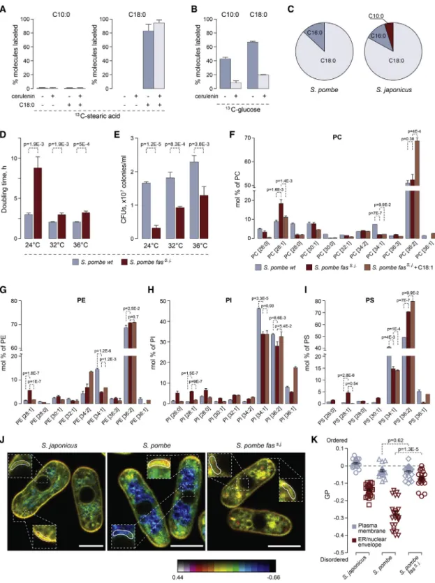
Figure 3. C10:0 Is Synthesized in Cytosol by the FAS Complex
(A) GC-MS analysis of13C-labeled C10:0 and C18:0 FAs extracted fromS. japonicusgrown in the presence of the U-13C-C18:0 for 4 h.
(B) GC-MS analysis of13C-labeled C10:0 and C18:0 FAs extracted fromS. japonicusgrown in the presence of the U-13C-glucose for 4 h.
(C) A pie chart summarizing the results of shotgun mass spectrometry analysis of FA-CoA products generatedin vitroby purified cytosolic FA synthases from S. pombeandS. japonicus. Shown are percentages of acyl-CoA species identified in each reaction.
(D) Doubling times forS. pombewild-type andfass. j.cultures grown at indicated temperatures in the yeast extract with supplements (YES) medium (n =3).
(E) Survival of theS. pombewild-type andfass. j.cells at indicated temperatures, in CFUs per mL of culture.
(F–I) Graphs representing molecular species profiles for PC (F), PE (G), PI (H), and PS (I) inS. pombewild-type,fass. j$andfass. j$cells grown in the presence of C18:1.
Note a frequent increase in 28:1 at the expense of 34:1 species. Shown are the mean values ± SD (n = 3). p values are derived from the unpaired parametric t test.
(legend continued on next page) Current Biology30, 367–380, February 3, 2020 371
labeled stearic acid (13C-C18:0) did not result in the detection of
13C-labeled C10:0 (Figure 3A), which was in line with our obser- vation that supplementation with LCFAs did not rescue S. japonicusgrowth upon fatty acid synthase (FAS) inhibition (Figures 1K–1L). This suggested that C10:0 likely originated throughde novoFA synthesis. Indeed, analysisof S. japonicus cells grown in the presence of 13C-labeled glucose revealed that the rates of synthesis were comparable between C10:0 and other FAs and that all FA production was inhibited by the FAS inhibitor cerulenin (Figures 3B andS3A).
Fungi and animals encode two pathways for de novo FA biosynthesis—located in the mitochondria or the cytosol. Dele- tion of three genes (htd2D,mct1D, andetr1D), whose products are predicted to execute essential steps of mitochondrial FA syn- thesis [20], did not result in changes to the relative abundance of C10:0 inS. japonicus(Figure S3B). The cytosolic fatty acid syn- thase (FAS) is a multi-enzyme complex containing all catalytic activities required for the reaction sequence as discrete func- tional domains [21]. We purified FAS complexes from either S. pombe or S. japonicus and reconstituted FA synthesis in vitro. The FAS activity was monitored by measuring the malonyl-CoA- and acetyl-CoA-dependent rates of NADPH oxidation (Figure S3C; [22]). Mass spectrometry analyses of the reaction products revealed that, in contrast to the S. pombeFAS that produced only C18:0 and some C16:0, the purified S. japonicus enzyme was able to synthesize C10:0 alongside the LCFAs, albeit with a lower yield compared to the steady-state abundance of C10:0in vivo(Figure 3C;Table S2).
We wondered whether the genetic exchange of theS. pombe cytosolic FAS to itsS. japonicuscounterpart would be sufficient to alter the spectrum of FA products and, as a result, structural composition of GPLs. The fission yeast FAS is composed ofa andbsubunits forming ana6b6oligomer [21]. We substituted the open reading frames (ORFs) encoding both theaand theb subunits (fas2 and fas1, respectively) in S. pombe with their S. japonicusorthologs, maintaining the native regulatory con- texts unchanged. Interestingly, replacement of the S. pombe FAS with the corresponding complex fromS. japonicus(fass. j.) led to cells dividing at smaller size (Figure S3D), increased pop- ulation doubling times (Figure 3D), and decreased cell survival (Figure 3E) across the physiological range of temperatures.
The growth defect was particularly pronounced at 24C in both rich and minimal media (Figures 3D andS3E).S. pombe fass. j.
cultures grew at comparable rates in glucose-rich and respira- tory media (Figure S3F) and consumed oxygen similarly to the wild type (Figure S3G), indicating that mitochondrial function was not overtly affected by FAS substitution. Addition of exoge- nous oleic acid (C18:1) toS. pombe fass. j.cultures rescued their doubling times, which suggested that the enhanced C10:0 pro- duction could be at root of slow growth (Figure S3H).
Indeed, we observed an increase in lower molecular weight species (mostly C28:1) for all major GPL classes in the engineered
S. pombestrain (Figures 3F–3I andS3I; Table 6 inData S1). Frag- mentation analyses showed that such lipids contained C18 FAs in thesn-1and C10:0 in thesn-2positions of the glycerol backbone (as an example, see MS/MS spectra for PI C28:1 inFigure S3J.
Interestingly, an increase in the C28:1 species in most classes coincided with decreased abundance of C34:1 lipids containing C18:1 and C16:0 chains (Figures 3F–3I andS3I). This suggested that theS. japonicusFAS was sufficient to increase C10:0 content at the expense of C16:0 and that C10:0 was incorporated in place of a saturated long FA chain intoS. pombeGPLs. Additionally, we detected increased levels of ceramide, phosphatidic acid (PA), sterols, and a slightly increased PC/PE ratio inS. pombeexpress- ing the transplanted S. japonicusFAS (Figures S3K and S3L;
Table 6 inData S1). As expected from its rescue of growth and survival, oleic acid treatment led to the decrease in C10:0-con- taining GPL species inS. pombe fass. j.cells (Figures 3F–3I and S3I). It rescued changes in the abundance of some but not all lipid classes and induced further disbalance in the PC/PE ratio (Fig- ure S3K and S3L).
We explored potential changes in subcellular membrane orga- nization caused by the transplantation ofS. japonicusFAS to S. pombeusing the ratiometric dye di-4-ANEPPDHQ [23]. Curi- ously, the NE-endoplasmic reticulum (ER) membranes became more ordered in S. pombe fass. j. cells, whereas the plasma membrane lipid order did not change. This observation was broadly in line with the measurements ofS. japonicus, which re- vealed a considerably higher NE/ER lipid order as compared to the wild-typeS. pombe(Figures 3J and 3K).
AlthoughS. japonicusFAS was sufficient to synthesize C10:0 bothin vitroandin vivo, the relative amounts of C10:0 were lower compared toS. japonicuscells. This suggests that other factors may enhance the efficiency of C10:0 synthesis in that organism.
The acyl-CoA-binding protein (ACBP) was shown to be involved in FA termination and transport and, when in excess, to shift the FAS product spectrum toward MCFAsin vitro[24]. However, neither deletion nor replacement of the gene encoding the S. japonicusACBP (acb1) with itsS. pombeortholog led to sig- nificant changes in the relative amount of C10:0 as compared to the wild-typeS. japonicus(Figure S3M). This indicated that the generation of C10:0 inS. japonicusdoes not rely on ACBP activ- ity, and other factors will need to be examined in future studies.
Replacement of theS. pombeFatty Acid Synthase with ItsS. japonicusCounterpart Causes UPR Activation and Affects the Secretory Pathway
S. pombe fass. j.cells exhibited global changes in mRNA abun- dance as compared to the wild type, as assessed by RNA sequencing (RNA-seq), both at 24C and 30C. Differentially ex- pressed genes were broadly comparable between the two growth conditions, although we also detected some potential temperature-specific regulation events (Figure S4A; Tables vii and viii inData S1). The largest group of upregulated transcripts
(J) Single-plane pseudocolored generalized polarization (GP) images. Color bar designates the range of GP values where reds show high membrane order and blues show low membrane order. Zoomed regions indicate plasma membrane and NE/ER zones used to quantify mean GP values. Scale bar, 5mm.
(K) A plot representing mean GP values quantified at the plasma membrane region at the tip of the cell and at the NE/ER region in cells of indicated genotypes (22 cells each).
For (A) and (B), 10mM cerulenin was added when indicated. Shown are the means of percentages of13C-labeled FAs ± SD (n = 6).
See alsoFigure S3andTable S2.
372 Current Biology30, 367–380, February 3, 2020
included the core environmental stress-response (CESR) genes (Figures 4A andS4B; Tables vii and viii inData S1). Yet another group of upregulated mRNAs including FA metabolism genes was previously identified as targets of the transcriptional regu- lator of lipid homeostasis Mga2 [25] (9.88-fold enrichment, p = 1.94E-05; Table 7 inData S1).
Of note, we observed a striking enrichment of the gene ontology (GO) term ‘‘transmembrane transport’’ among signifi- cantly downregulated genes (Figures 4A and S4B; Tables vii
and viii inData S1). This suggested a possibility of a global mem- brane-related cellular response to the FAS substitution. Indeed, several downregulated genes, includingpho1encoding a major secreted acid phosphatase [26], were previously identified as targets of the RIDD-type unfolded protein response (UPR) in S. pombe. In this organism, a transmembrane ER-resident ki- nase/endonuclease Ire1 governs the decay of ER-associated mRNAs, working together with the downstream mRNA no-go- decay machinery [27, 28]. An RT-qPCR-based expression Figure 4. Genetic Replacement of the Cytosolic FAS Complex inS. pombewith Its S.japonicusCounterpart Results in UPR Activation and Defects in Secretory Pathway
(A) Differentially expressed genes constituting two largest functional categories inS. pombe fass. j.cells as compared to the wild type. Cells were grown in YES at 24C. AnGeLi suite was used for GO annotations.
(B) qPCR analyses of indicated poly(A) mRNAs inS. pombewild type and wild-type treated with 0.5mg/mL tunicamycin for 1 h to induce the UPR andfass. j.mutant cells.
(C) qPCR analyses ofbip1poly(A) and total mRNA species inS. pombewild type and wild-type treated with 0.5mg/mL tunicamycin for 1 h andfass. j.mutant cells.
(D) Maximum intensity z-projections of spinning-disk confocal images ofS. pombewild-type andfass. j.cells expressing Sec24-GFP. Average fluorescence intensities relative to the wild type are presented on the right. n = 142 and 154 cells for the wild type and the mutant, respectively.
(E) Maximum intensity z-projections of spinning-disk confocal images ofS. pombewild-type andfass. j.cells expressing Sec72-GFP. For each genotype,trans- Golgi cisternae numbers per unit of area are presented on the right. n = 153 and 167 cells for the wild-type and the mutant, respectively.
(F) The efficiency of acid phosphatase secretion measured inS. pombewild-type andfass. j.cells. Shown are the mean values ± SD (n = 3).
Shown in (B) and (C) are the mean values derived from three biological and two technical repeats, normalized to the wild type. p values are derived from the unpaired parametric t test. In (D) and (E), scale bars represent 5mm. Calibration bars are shown for each image.
See alsoFigure S4.
Current Biology30, 367–380, February 3, 2020 373
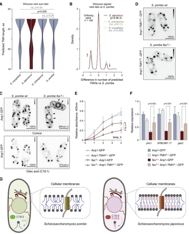
Figure 5. Transmembrane Helices May Have Co-evolved with Membrane Lipids inS. japonicus
(A) A violin plot for distributions of predicted TMH lengths in the four fission yeast species. 1:1:1:1 orthologs of single-span TMH proteins were selected for the analysis. p values are derived from the Wilcoxon rank-sum test.
(B) A density trace graph summarizing pairwise differences in the number of predicted TMHs inS. japonicus,S. octosporus, andS. cryophilus, as compared to S. pombe. Only 1:1:1:1 orthologs ofS. pombesingle-span TMH proteins were included. p values are derived from the Wilcoxon rank-sum test.
(C) Single-plane spinning-disk confocal images of cells expressing Anp1-GFP inS. pombewild-type andfass. j.cells in the absence (n = 205 and n = 153 cells, respectively) or the presence of C18:1 supplementation (n = 109 and n = 97 cells, respectively).
(D) Single-plane spinning-disk confocal images of cells expressing Anp1-TMHS.j.-GFP as a sole copy of the protein (107 cells with endogenous FAS and 110 cells with replaced FAS were counted).
(E) A graph summarizing the results of the acid phosphatase secretion assay inS. pombecultures of indicated genotypes. Shown are the mean values ± SD (n = 3).
(legend continued on next page) 374 Current Biology30, 367–380, February 3, 2020
analysis of a number of Ire1 targets, including those identified in our RNA-seq datasets, revealed a significant decrease in their steady-state mRNA levels inS. pombe fass. j.cells as compared to the wild type (Figures 4B andS4C).
As expected, similar, albeit more pronounced downregulation was observed in cells treated with the inducer of ER stress, tuni- camycin (Figures 4B andS4C). Unlike other UPR targets, the bip1mRNA is stabilized by the cleavage of its poly(A) tail, sup- porting activity of this major chaperone upon ER stress [27]. In line with this, we observed a decline in poly(A)bip1mRNA but an overall message stabilization inS. pombe fass. j.mutants (Fig- ure 4C). Importantly,ire1deletion was synthetically lethal with
fass. j.replacement. Together, these data are consistent with
the hypothesis that substituting the S. pombe FAS with the S. japonicuscomplex leads to a chronic activation of the UPR and that survival of the retroengineered cells relies on the pres- ence of this ER homeostasis pathway.
We reasoned that the presence of the unusual C10:0-contain- ing asymmetrical membrane GPLs and increase in ER mem- brane order inS. pombe fass. j.cells may impact on the organi- zation and function of the secretory pathway, where lipid composition is tightly regulated [29]. Indeed, we observed a drastic decrease in the fluorescence intensity of the transitional ER marker Sec24-GFP [30] (Figures 4D andS4D). Furthermore,
thefass. j. mutant cells exhibited a reduced number oftrans-
Golgi cisterna (Figure 4E). Consistent with UPR-dependent downregulation ofpho1mRNA (Figure 3D) and general defects in ER and Golgi organization, S. pombe fass. j. cells were severely defective in acid phosphatase secretion (Figure 4F).
Thus, introduction of asymmetrical phospholipids toS. pombe leads to pronounced defects in functions of the secretory pathway.
Evidence for Evolutionary Adaptation of Transmembrane Domains to Membrane Lipid Composition
Membrane composition and physical properties may impose constraints on the structure of transmembrane parts of integral membrane proteins [31–33]. The comparisons of predicted transmembrane helices (TMHs) in all orthologous groups of transmembrane proteins betweenS. pombeand other fission yeast species (1:1:1:1 orthologs) showed that predicted S. japonicusTMHs contained many small non-polar amino acids and fewer large non-polar residues compared to the rest of fission yeasts (Figures S5A–S5C). Interestingly, the pairwise comparison of predicted TMHs in single-TMH proteins revealed a statistically significant enrichment of relatively short TMHs in S. japonicus(Figure 5A; Table 9 inData S1). For a number of S. japonicusproteins, TMHs failed to be predicted with confi- dence altogether (Figure 5B; Table 9 inData S1). Thus, it appears that transmembrane domains of at least some orthologous pro- teins inS. japonicusdiverged greatly compared to those in other
fission yeasts. Such differences might be a result of structural adaptation of TMHs inS. japonicusto profound changes in the FA composition of membrane phospholipids.
We reasoned that S. pombe proteins, orthologs of which exhibit TMH shortening or poor TMH prediction inS. japonicus as compared to other fission yeast species, might be particularly vulnerable to the introduction of the S. japonicus FAS to S. pombeand, hence, an increase in GPL acyl chain asymmetry.
To test this hypothesis, we visualized the subcellular localization of four TMH-divergent candidates, predicted to localize to various membrane sub-compartments. A mannosyltransferase complex subunit Anp1 that normally localizes tocis-Golgi [30]
was mis-targeted inS. pombe fass. j.cells, partitioning clearly be- tween the ER and Golgi (Figure 5C). Correction of cellular FA composition by exogenous oleic acid (C18:1) led to the reversal of this mis-localization pattern (Figure 5C). The mis-localization was not due to UPR engagement as Anp1-GFP did not relocalize to the ER in tunicamycin-treated cells (Figure S5D). The Anp1-in- teracting protein Gmh5/Mnn10 [34] was also retained in the ER in S. pombe fass. j.cells (Figure S5E), suggesting that the localiza- tion and function of this entire complex could be affected. Curi- ously, the hydrophobic helix in S. japonicusGmh5 does not reach the TMH prediction threshold, unlike itsS. pombecounter- part (Table 9 in Data S1). Mis-targeting phenotypes were observed for two other protein candidates that were predicted to have a shorter TMH or failed to reach the TMH prediction cut- offs inS. japonicus. The ER-lipid droplet diacylglycerol trans- ferase Lro1 [35] became strongly enriched at the perinuclear ER while the fluorescence intensity of the GFP-tagged ER- plasma membrane tethering protein Tcb2 increased and it was now detected throughout the entire ER network [36] (Figure S5E).
The predicted TMH in Anp1 was 18 amino acids inS. japonicus as compared to 20 amino acids inS. pombeand 23 amino acids in other fission yeasts (Figure S5F). Despite this difference, endogenous Anp1 proteins exhibited a typical Golgi localization in bothS. pombeandS. japonicus(Figures 5C andS5G). To establish whether mis-localization of Anp1 inS. pombe fass. j.
cells could be driven by a mismatch between its TMH structure and the rewired membrane lipid composition, we engineered an Anp1 chimeras in S. pombe by replacing the native TMH with its shorterS. japonicusversion and keeping the rest of the protein intact (Figure S5F). Strikingly, this was sufficient to restore the Golgi localization pattern of Anp1 in S. pombe fass. j$cells (Figure 5D). The swap did not affect Anp1 Golgi local- ization in the wild type, indicating that theS. japonicusTMH is compatible with both types of membranes.
We did not observe significant differences in the growth rates between the Anp1-GFP and the Anp1-TMHS. j.-GFPS. pombe fass. j.cultures (Figure S5H). Yet, recovering Anp1 Golgi localiza- tion by the TMH swap led to a significant improvement in acid phosphatase secretion efficiency inS. pombe fass. j.cells (Fig- ure 5E). Sincepho1is a target of the UPR pathway (Figure 4B),
(F) A graph summarizing the results of qPCR analyses of indicated poly(A) mRNAs. Shown are the mean values derived from three biological and two technical repeats, normalized to the wild type. p values are derived from the unpaired parametric t test.
(G) A diagram summarizing our hypothesis on co-evolution of transmembrane helices and membrane lipids inS. pombeandS. japonicus.
In (C) and (D), scale bars represent 5mm. Calibration bars are shown for each image. Included are the percentages of cells in a population exhibiting the indicated phenotypes. In (E) and (F), p values are derived from the unpaired parametric t test.
See alsoFigure S5.
Current Biology30, 367–380, February 3, 2020 375
we tested whether this could be due to a higher abundance of the pho1mRNA. Indeed, the steady-state levels of pho1 poly(A) mRNA and other UPR targets were partially rescued in S. pombe fass. j.cells expressing Anp1-TMHS. j.-GFP (Figure 5F).
Thus, restoring the localization of the Anp1-containing manno- syltransferase complex to Golgi is sufficient to ameliorate at least some of the defects induced by grafting theS. japonicusFAS to S. pombe.
DISCUSSION
We show thatS. japonicus, a sister species to a well-studied model organism S. pombe, synthesizes abundant structurally asymmetrical phospholipids, where the medium acyl chain C10:0 is linked to thesn-2 position of the glycerol backbone and thesn-1 position is occupied by a long, usually saturated FA (Figure 1).In vitro, membranes assembled fromS. japonicus TPL mixtures are more rigid and ordered (Figure 2) [37]. The FA tails ofS. japonicusphospholipids are considerably more satu- rated (Figure 1E) and therefore are likely to form closely packed and more ordered bilayers. A large difference in the lengths of two acyl chains of individual molecules may result in partial interdigitation of FAs, also leading to an increase in the strength of lateral interactions and tighter packing [38, 39], although interdigitation is likely dynamic in liquid phase [40,41].In vivo, membrane biophysical properties may be further modified by sterols and sphingolipids. Of note, in spite of being more ordered, S. japonicusmembranes have a lower sphingolipid/GPL ratio as compared toS. pombe(Figure 1C). The caveat ofin vitroexper- iments using TPL fractions is that the different abundance of subcellular membrane compartments might contribute to the observed differences in membrane biophysics. However, S. japonicusendomembranes are also more orderedin vivo(Fig- ures 3J and 3K), suggesting that their unique GPL architecture might have a major impact on membrane properties. Further work will be required to assess how asymmetrical lipids are par- titioned within the cell and the bilayer and how this affects cellular physiology.
The molecular changes underlying production of large amounts of C10:0 alongside the LCFAs and assembly of asym- metrical phospholipids might have allowedS. japonicusto colo- nize new ecological niches. Both FA desaturation and sterol biosynthesis—normally required for maintaining membrane fluidity—are oxygen-demanding processes [42,43]. Curiously, anaerobically grown budding yeast synthesize asymmetrical MCFA-containing GPLs [44]. Similarly, defective desaturation upregulates the synthesis of MCFAs even in aerobic conditions [45]. This suggests that phospholipid asymmetry might provide an alternative way of maintaining membrane physico-chemical properties when FA desaturation and/or sterol synthesis is not possible. In line with possible adaptation to anaerobic lifestyle, S. japonicusdoes not respire, grows well in the absence of ox- ygen, and undergoes robust transitions between yeast and bur- rowing hyphae [46–49]. Phospholipid asymmetry and FA tail saturation both reduce membrane permeability to water, small solutes, and/or oxygen [50–53], which might provide additional advantages in the wild. Budding yeast and S. pombe utilize specialized gene expression programs executed by the mem- brane-sensing transcription factor Mga2 [54] to sustain FA
desaturation in low-oxygen conditions [25,55]. In the future, it will be of interest to assess the wiring of this network in S. japonicus, given a markedly distinct physiology of the organism.
S. japonicusFAS exhibits overall sequence conservation with other fungal FA synthases (seeSTAR Methodsfor correctedfas1 ORF), although it does have a number of potentially interesting substitutions to be analyzed in future. The budding yeast muta- genesis experiments suggest that the evolutionary acquisition of new FAS functionality may not necessarily require a large number of steps [56]. Importantly, other cellular factors may in- fluence the spectrum of FAS products. An interesting possibility could be the different ratios between acetyl- and malonyl-CoA in S. japonicusandS. pombe. Relative depletion of malonyl-CoA results in the enzyme favoring priming over elongation [56] and increases the production of shorter FAs, whereas hyperactiva- tion of the enzyme responsible for malonyl-CoA synthesis shifts FA distribution toward longer lengths [57]. Curiously, the mRNA for this enzyme, cut6 [58], is significantly upregulated in S. pombecells expressingS. japonicusFAS (Tables vii and viii inData S1), perhaps as a part of a Mga2-based compensation mechanism triggered by elevated C10:0 levels. Other cellular ad- aptations are likely required to efficiently handle C10:0 alongside LCFAs, including modifications of lipid remodeling enzymes.
Our bioinformatics analyzes indicate that manyS. japonicus transmembrane proteins might have experienced the need to adapt to changes in cellular lipid composition by reducing an average residue volume or shortening and/or remodeling the TM domains (Figures 5A, 5B, andS5A–S5C). TMDs span the hydrophobic core of the lipid bilayer, and therefore specific membrane FA composition may impose constraints on their sequences [32,33]. TMDs of single-spanning ER andcis-Golgi proteins tend to be shorter than those residing in the later com- partments of the secretory pathway [31]. Curiously, most single- pass TM proteins that exhibit shortened TMDs inS. japonicusas compared to other fission yeasts are predicted to function largely at the ER, ER-organelle contacts, andcis-Golgi (Table 9 inData S1). The membranes of the broad ER territory tend to exhibit looser lipid packing [18], and thus their protein residents could be particularly sensitive to an increase in membrane order.
In addition to reducing the hydrophobic length, other properties of the TMDs may facilitate their insertion and/or folding in well-ordered membranes—perhaps explaining the statistically significant increase in poorly predicted TMHs in S. japonicus single-pass TM proteins versus their orthologs in other fission yeasts.
Supporting the hypothesis that TMHs co-evolve with mem- branes [31–33], the replacement of the TMH inS. pombeAnp1 with a shorterS. japonicusversion rescues its mis-targeting in S. pombe expressing the S. japonicus FAS (Figure 5D) and partially relieves downregulation of UPR targets in this genetic background (Figures 5E and 5F). Anp1 is a key player in the assembly of the mannosyltransferase complex, which takes place in the ER before trafficking to Golgi [34,59]. It is possible that ER retention of this complex impacts folding of proteins destined for secretion through improper glycosylation events, activating UPR signaling. Yet, given the complexity of observed cellular phenotypes, the physiological consequences of intro- ducing theS. japonicusFAS toS. pombeare likely not limited 376 Current Biology30, 367–380, February 3, 2020
to this particular pathway. Poor growth and stress-response activation in fass. j. cells (Figures 3D, 3E,4A, S3E, andS4B) may be attributed not only to downregulation of a large number of membrane transporters but also the changes in membrane biophysical properties (Figures 3J and 3K), which could affect signaling through the cellular stress pathways [60, 61]. Ire1 may also sense bilayer stress directly [62–64]. Given the reliance ofS. japonicuson C10:0-containing largely saturated phospho- lipids, it will be interesting to see whether this organism might have rearranged the structural features of Ire1 allowing it to sense ER lipid composition.
Our work provides new insights into the generation of phos- pholipid diversity in evolution and its impact on cellular biology connected to membrane functions. We propose that evolu- tionary changes in membrane FA composition may necessitate rapid adaptation of the transmembrane domains (Figure 5G).
The two fission yeast species with their divergent lifestyles constitute an ideal experiment of Nature, study of which may lead to a conceptually new understanding of the relationship be- tween the underlying metabolic makeup and the evolution of cellular properties.
STAR+METHODS
Detailed methods are provided in the online version of this paper and include the following:
d KEY RESOURCES TABLE
d LEAD CONTACT AND MATERIALS AVAILABILITY
d EXPERIMENTAL MODEL AND SUBJECT DETAILS
d METHOD DETAILS
B Molecular genetics and strain husbandry B Cell growth and survival measurements B ESI-MS-based lipidomic analysis B Phospholipase A2 digestion
B GUVs preparation in DexPEG hydrogel films B Bending rigidity measurements
B Electroformation of GUVs and labeling with the FAST DIO
B Metabolic labeling B GC-MS
B FAS purification and FA synthesisin vitro B Fatty acid synthase assay – product analysis B Image acquisition and analysis
B Measurements of membrane lipid orderin vivo B RNA isolation
B RNA sequencing
B Reverse Transcription and Real-Time Quantitative PCR (RT-qPCR)
B Measurements of oxygen consumption B Acid phosphatase secretion assay B Bioinformatics
d QUANTIFICATION AND STATISTICAL ANALYSIS
d DATA AND CODE AVAILABILITY
SUPPLEMENTAL INFORMATION
Supplemental Information can be found online athttps://doi.org/10.1016/j.
cub.2019.11.043.
ACKNOWLEDGMENTS
We are grateful to the Oliferenko lab for discussions and D. Owen (King’s Col- lege London) for suggestions on the manuscript. Many thanks are due to V. Christodoulou (The Francis Crick Institute) for help with FAS purification, A. Le Marois (The Francis Crick Institute) for advice on GUV preparation, and M. Grininger (University of Frankfurt) for advice on FAS assays. Our work was supported by the Francis Crick Institute, which receives its core funding from Cancer Research UK (FC001002), the UK Medical Research Council (FC001002), and the Wellcome Trust (FC001002) and the Wellcome Trust Se- nior Investigator Award (103741/Z/14/Z) to S.O.
AUTHOR CONTRIBUTIONS
M.M. conceived and performed cell biological, biochemical, and biophysical experiments; generated strains; analyzed data; and co-wrote the manuscript.
M.P., G.B., A.G., and L.V. designed, performed, and interpreted all lipidomics experiments except GC-MS; J.I.M. performed and analyzed the GC-MS ex- periments. N.L.M. and P.B. performed and interpreted measurements of membrane bending rigidity. E.M. performed bioinformatics analyses. M.P., G.B., A.G., L.V., J.I.M., N.L.M., and E.M. edited the manuscript. S.O.
conceived and interpreted experiments and wrote the manuscript.
DECLARATION OF INTERESTS
The authors declare no competing interests.
Received: August 27, 2019 Revised: October 31, 2019 Accepted: November 13, 2019 Published: January 16, 2020 REFERENCES
1.Harayama, T., and Riezman, H. (2018). Understanding the diversity of membrane lipid composition. Nat. Rev. Mol. Cell Biol.19, 281–296.
2.Bogdanov, M., Dowhan, W., and Vitrac, H. (2014). Lipids and topological rules governing membrane protein assembly. Biochim. Biophys. Acta 1843, 1475–1488.
3.van Meer, G., Voelker, D.R., and Feigenson, G.W. (2008). Membrane lipids: where they are and how they behave. Nat. Rev. Mol. Cell Biol.9, 112–124.
4.Sezgin, E., Levental, I., Mayor, S., and Eggeling, C. (2017). The mystery of membrane organization: composition, regulation and roles of lipid rafts.
Nat. Rev. Mol. Cell Biol.18, 361–374.
5.Antonny, B., Vanni, S., Shindou, H., and Ferreira, T. (2015). From zero to six double bonds: phospholipid unsaturation and organelle function.
Trends Cell Biol.25, 427–436.
6.Bogdanov, M., Sun, J., Kaback, H.R., and Dowhan, W. (1996). A phospho- lipid acts as a chaperone in assembly of a membrane transport protein.
J. Biol. Chem.271, 11615–11618.
7.Russell, J.J., Theriot, J.A., Sood, P., Marshall, W.F., Landweber, L.F., Fritz-Laylin, L., Polka, J.K., Oliferenko, S., Gerbich, T., Gladfelter, A., et al. (2017). Non-model model organisms. BMC Biol.15, 55.
8.Gu, Y., and Oliferenko, S. (2019). Cellular geometry scaling ensures robust division site positioning. Nat. Commun.10, 268.
9.Yam, C., He, Y., Zhang, D., Chiam, K.H., and Oliferenko, S. (2011).
Divergent strategies for controlling the nuclear membrane satisfy geomet- ric constraints during nuclear division. Curr. Biol.21, 1314–1319.
10.Makarova, M., Gu, Y., Chen, J.S., Beckley, J.R., Gould, K.L., and Oliferenko, S. (2016). Temporal regulation of lipin activity diverged to account for differences in mitotic programs. Curr. Biol.26, 237–243.
11.Peter, M., Glatz, A., Gudmann, P., Gombos, I., To¨ro¨k, Z., Horva´th, I., Vı´gh, L., and Balogh, G. (2017). Metabolic crosstalk between membrane and stor- age lipids facilitates heat stress management in Schizosaccharomyces pombe. PLoS ONE12, e0173739.
Current Biology30, 367–380, February 3, 2020 377
12.Shui, G., Guan, X.L., Low, C.P., Chua, G.H., Goh, J.S., Yang, H., and Wenk, M.R. (2010). Toward one step analysis of cellular lipidomes using liquid chromatography coupled with mass spectrometry: application to Saccharomyces cerevisiae and Schizosaccharomyces pombe lipidomics.
Mol. Biosyst.6, 1008–1017.
13.Ejsing, C.S., Sampaio, J.L., Surendranath, V., Duchoslav, E., Ekroos, K., Klemm, R.W., Simons, K., and Shevchenko, A. (2009). Global analysis of the yeast lipidome by quantitative shotgun mass spectrometry. Proc.
Natl. Acad. Sci. USA106, 2136–2141.
14.Stukey, J.E., McDonough, V.M., and Martin, C.E. (1990). The OLE1 gene of Saccharomyces cerevisiae encodes the delta 9 fatty acid desaturase and can be functionally replaced by the rat stearoyl-CoA desaturase gene.
J. Biol. Chem.265, 20144–20149.
15.Rhind, N., Chen, Z., Yassour, M., Thompson, D.A., Haas, B.J., Habib, N., Wapinski, I., Roy, S., Lin, M.F., Heiman, D.I., et al. (2011). Comparative functional genomics of the fission yeasts. Science332, 930–936.
16.Moche, M., Schneider, G., Edwards, P., Dehesh, K., and Lindqvist, Y.
(1999). Structure of the complex between the antibiotic cerulenin and its target, beta-ketoacyl-acyl carrier protein synthase. J. Biol. Chem.274, 6031–6034.
17.Elani, Y., Purushothaman, S., Booth, P.J., Seddon, J.M., Brooks, N.J., Law, R.V., and Ces, O. (2015). Measurements of the effect of membrane asymmetry on the mechanical properties of lipid bilayers. Chem.
Commun. (Camb.)51, 6976–6979.
18.Bigay, J., and Antonny, B. (2012). Curvature, lipid packing, and electro- statics of membrane organelles: defining cellular territories in determining specificity. Dev. Cell23, 886–895.
19.Baumgart, T., Hunt, G., Farkas, E.R., Webb, W.W., and Feigenson, G.W.
(2007). Fluorescence probe partitioning between Lo/Ld phases in lipid membranes. Biochim. Biophys. Acta1768, 2182–2194.
20.Kastaniotis, A.J., Autio, K.J., Keratar, J.M., Monteuuis, G., Makela, A.M., Nair, R.R., Pietikainen, L.P., Shvetsova, A., Chen, Z., and Hiltunen, J.K.
(2017). Mitochondrial fatty acid synthesis, fatty acids and mitochondrial physiology. Biochim. Biophys. Acta Mol. Cell Biol. Lipids1862, 39–48.
21.Jenni, S., Leibundgut, M., Boehringer, D., Frick, C., Mikola´sek, B., and Ban, N. (2007). Structure of fungal fatty acid synthase and implications for iterative substrate shuttling. Science316, 254–261.
22.Wieland, F., Renner, L., Verfu¨rth, C., and Lynen, F. (1979). Studies on the multi-enzyme complex of yeast fatty-acid synthetase. Reversible dissoci- ation and isolation of two polypeptide chains. Eur. J. Biochem. 94, 189–197.
23.Owen, D.M., Rentero, C., Magenau, A., Abu-Siniyeh, A., and Gaus, K.
(2011). Quantitative imaging of membrane lipid order in cells and organ- isms. Nat. Protoc.7, 24–35.
24.Schjerling, C.K., Hummel, R., Hansen, J.K., Borsting, C., Mikkelsen, J.M., Kristiansen, K., and Knudsen, J. (1996). Disruption of the gene encoding the acyl-CoA-binding protein (ACB1) perturbs acyl-CoA metabolism in Saccharomyces cerevisiae. J. Biol. Chem.271, 22514–22521.
25.Burr, R., Stewart, E.V., Shao, W., Zhao, S., Hannibal-Bach, H.K., Ejsing, C.S., and Espenshade, P.J. (2016). Mga2 transcription factor regulates an oxygen-responsive lipid homeostasis pathway in fission yeast.
J. Biol. Chem.291, 12171–12183.
26.Elliott, S., Chang, C.W., Schweingruber, M.E., Schaller, J., Rickli, E.E., and Carbon, J. (1986). Isolation and characterization of the structural gene for secreted acid phosphatase from Schizosaccharomyces pombe. J. Biol.
Chem.261, 2936–2941.
27.Kimmig, P., Diaz, M., Zheng, J., Williams, C.C., Lang, A., Arago´n, T., Li, H., and Walter, P. (2012). The unfolded protein response in fission yeast mod- ulates stability of select mRNAs to maintain protein homeostasis. eLife1, e00048.
28. Guydosh, N.R., Kimmig, P., Walter, P., and Green, R. (2017). Regulated Ire1-dependent mRNA decay requires no-go mRNA degradation to main- tain endoplasmic reticulum homeostasis inS. pombe. eLife.https://doi.
org/10.7554/eLife.29216.
29.Jackson, C.L., Walch, L., and Verbavatz, J.M. (2016). Lipids and their traf- ficking: an integral part of cellular organization. Dev. Cell39, 139–153.
30.Vjestica, A., Tang, X.Z., and Oliferenko, S. (2008). The actomyosin ring re- cruits early secretory compartments to the division site in fission yeast.
Mol. Biol. Cell19, 1125–1138.
31.Sharpe, H.J., Stevens, T.J., and Munro, S. (2010). A comprehensive com- parison of transmembrane domains reveals organelle-specific properties.
Cell142, 158–169.
32.Lorent, J.H., Diaz-Rohrer, B., Lin, X., Spring, K., Gorfe, A.A., Levental, K.R., and Levental, I. (2017). Structural determinants and functional consequences of protein affinity for membrane rafts. Nat. Commun.8, 1219.
33.Quiroga, R., Trenchi, A., Gonza´lez Montoro, A., Valdez Taubas, J., and Maccioni, H.J. (2013). Short transmembrane domains with high-volume exoplasmic halves determine retention of Type II membrane proteins in the Golgi complex. J. Cell Sci.126, 5344–5349.
34.Jungmann, J., Rayner, J.C., and Munro, S. (1999). The Saccharomyces cerevisiae protein Mnn10p/Bed1p is a subunit of a Golgi mannosyltrans- ferase complex. J. Biol. Chem.274, 6579–6585.
35.He, Y., Yam, C., Pomraning, K., Chin, J.S., Yew, J.Y., Freitag, M., and Oliferenko, S. (2014). Increase in cellular triacylglycerol content and emer- gence of large ER-associated lipid droplets in the absence of CDP-DG synthase function. Mol. Biol. Cell25, 4083–4095.
36.Manford, A.G., Stefan, C.J., Yuan, H.L., Macgurn, J.A., and Emr, S.D.
(2012). ER-to-plasma membrane tethering proteins regulate cell signaling and ER morphology. Dev. Cell23, 1129–1140.
37.Roux, A., Cuvelier, D., Nassoy, P., Prost, J., Bassereau, P., and Goud, B.
(2005). Role of curvature and phase transition in lipid sorting and fission of membrane tubules. EMBO J.24, 1537–1545.
38.Hui, S.W., Mason, J.T., and Huang, C. (1984). Acyl chain interdigitation in saturated mixed-chain phosphatidylcholine bilayer dispersions.
Biochemistry23, 5570–5577.
39.Xu, H., and Huang, C.H. (1987). Scanning calorimetric study of fully hydrat- ed asymmetric phosphatidylcholines with one acyl chain twice as long as the other. Biochemistry26, 1036–1043.
40.Schram, V., and Thompson, T.E. (1995). Interdigitation does not affect translational diffusion of lipids in liquid crystalline bilayers. Biophys. J.
69, 2517–2520.
41.Ali, S., Smaby, J.M., Momsen, M.M., Brockman, H.L., and Brown, R.E.
(1998). Acyl chain-length asymmetry alters the interfacial elastic interac- tions of phosphatidylcholines. Biophys. J.74, 338–348.
42.Aguilar, P.S., and de Mendoza, D. (2006). Control of fatty acid desatura- tion: a mechanism conserved from bacteria to humans. Mol. Microbiol.
62, 1507–1514.
43.Shimizu, I., and Katsuki, H. (1975). Effect of temperature on ergosterol biosynthesis in yeast. J. Biochem.77, 1023–1027.
44.Meyer, F., and Bloch, K. (1963). Metabolism of stearolic acid in yeast.
J. Biol. Chem.238, 2654–2659.
45.Proudlock, J.W., Haslam, J.M., and Linnane, A.W. (1971). Biogenesis of mitochondria. 19. The effects of unsaturated fatty acid depletion on the lipid composition and energy metabolism of a fatty acid desaturase mutant of Saccharomyces cerevisiae. J. Bioenerg.2, 327–349.
46.Bulder, C. (1963). On Respiratory Deficiency in Yeasts. Volume PhD (University of Delft).
47.Kaino, T., Tonoko, K., Mochizuki, S., Takashima, Y., and Kawamukai, M.
(2018). Schizosaccharomyces japonicus has low levels of CoQ10synthe- sis, respiration deficiency, and efficient ethanol production. Biosci.
Biotechnol. Biochem.82, 1031–1042.
48.Okamoto, S., Furuya, K., Nozaki, S., Aoki, K., and Niki, H. (2013).
Synchronous activation of cell division by light or temperature stimuli in the dimorphic yeast Schizosaccharomyces japonicus. Eukaryot. Cell12, 1235–1243.
378 Current Biology30, 367–380, February 3, 2020