Dementia with Lewy bodies – a clinicopathological update
János Bencze1,2, Woosung Seo3, Abdul Hye4,5, Dag Aarsland5,6,7, Tibor Hortobágyi2,6,7,8
1 Department of Pathology, Faculty of Medicine, University of Debrecen, Debrecen, Hungary
2 MTA‐DE Cerebrovascular and Neurodegenerative Research Group, Department of Neurology, University of Debrecen, Debrecen, Hungary
3 Department of Surgical Sciences, Radiology, Uppsala University, Uppsala, Sweden
4 Maurice Wohl Clinical Neuroscience Institute, Institute of Psychiatry, Psychology and Neuroscience, King’s College London, London, UK
5 NIHR Biomedical Research Centre for Mental Health & Biomedical Research Unit for Dementia at South London
& Maudsley NHS Foundation, London, UK
6 Department of Old Age Psychiatry, Institute of Psychiatry Psychology and Neuroscience, King’s College London, London, UK
7 Centre for Age‐Related Medicine, SESAM, Stavanger University Hospital, Stavanger, Norway
8 Institute of Pathology, Faculty of Medicine, University of Szeged, Szeged, Hungary
Corresponding author:
Tibor Hortobágyi MD PhD DSc FRCPath EFN ∙ Institute of Pathology ∙ Faculty of Medicine ∙ University of Szeged ∙ Szeged ∙ Állomás utca 1 ∙ H‐6725 ∙ Hungary
hortobagyi.tibor@med.u‐szeged.hu
Submitted: 31 December 2019 Accepted: 06 February 2020 Published: 18 February 2020
Abstract
Dementia is one of the major burdens of our aging society. According to certain predictions, the number of patients will double every 20 years. Although Alzheimer’s disease (AD), as the most frequent neurodegenera‐
tive dementia, has been extensively analysed, less is known about dementia with Lewy bodies (DLB). Neuropa‐
thological hallmarks of DLB are the deposition of intracellular Lewy bodies (LB) and Lewy neurites (LN). DLB belongs to the α‐synucleinopathies, as the major component of these inclusions is pathologically aggregated α‐
synuclein. Depending on the localization of LBs and LNs in the central nervous system cognitive and motor symptoms can occur. In our work, we will systematically review the possible etiology and epidemiology, patho‐
logical (both macroscopic and microscopic) features, structural and functional imaging findings, with a special emphasis on the clinico‐pathological correlations. Finally, we summarize the latest clinical symptoms‐based diagnostic criteria and the novel therapeutic approaches. Since DLB is frequently accompanied with AD pathol‐
ogy, highlighting possible differential diagnostic approaches is an integral part of our paper. Although our pre‐
sent knowledge is insufficient, the rapid development of diagnostic and research methods provide hope for better diagnosis and more efficient treatment, contributing to a better quality of life.
Keywords: α‐synuclein, Biomarkers, Diagnostic criteria, Clinico‐pathological correlation, Dementia with Lewy bodies
Review
Abbreviations
AA, Alzheimer’s Association; AD, Alzheimer’s dis‐
ease; Aβ, Amyloid‐beta; APOE, Apolipoprotein E gene;
BNE, BrainNet Europe Consortium; BOLD, Blood oxygen level dependent; ChAT, Choline‐acetyltransferase; ChAT‐
I, Acetyl‐cholinesterase inhibitors; CR, Creatinine; CSF, Cerebrospinal fluid; DLB, Dementia with Lewy bodies;
DMN, Default mode network; DSM, Diagnostic Statistical Manual; fMRI, Functional MRI; FP‐CIT, [123I] 2ß‐
carbomethoxy‐3b‐(4‐iodophenyl)‐N‐(3‐fluoropropyl) nortropane; GBA, Glucosylceramidase‐beta gene; iLBD, Incidental Lewy body disease; LB, Lewy‐body; LBDA, Lewy Body Dementia Association; LN, Lewy neurite;
MAPT, Microtubule associated protein tau gene; MDS, Movement Disorders Society; MRI, Magnetic resonance imaging; NAA, N‐acetyl aspartate; NCD, Neurocognitive disorder; NF, Neurofilament; NIA, National Institute on Aging; NMDAR, N‐methyl‐D‐aspartate receptor; PD, Parkinson’s disease; PDD, Parkinson’s disease dementia;
PET, Positron emission tomography; PSD95, Density protein 95; REM, Rapid‐eye‐movement; SCARB2, Scav‐
enger Receptor Class B Member 2; SNARE, SNAP (Solu‐
ble NSF Attachment Protein) Receptor; SNCA, α‐
synuclein gene; SPECT, Single‐photon emission comput‐
ed tomography; SSRI, Selective serotonin reuptake in‐
hibitors; UPR, Unfolded protein response; WML, White matter lesions; ZnT3, Zinc transporter 3
Epidemiology and etiology
Dementia with Lewy bodies (DLB) is the second most common primary neurodegenerative dementia.
The etiology is mainly unknown, however, in certain cases there is strong evidence of genetic background. A few papers have reported families with accumulating occurrence of cognitive impairment throughout genera‐
tions1. The vast majority of the familial cases are traced to α‐synuclein (SNCA) gene alterations, particularly to E46K mutation2. Five candidate genes, including APOE, GBA, MAPT, SNCA and SCARB2, are considered as a sig‐
nificant risk factor for DLB, nevertheless, further genetic studies are needed3. Although it mostly appears in older age, dementia is not strictly associated with aging. Rare‐
ly it may occur under the age of 65 and even in young adulthood. Surprisingly in the case report of a teenager, the clinical symptoms and post‐mortem pathological findings were consistent with those of in DLB4. The prev‐
alence of the disease is not clearly established. Accord‐
ing to a comprehensive analysis of epidemiological data, the prevalence is 4.2% in community based and 7.5% in clinical studies, while the incidence is 3.8% and grows linearly with aging5, while the prevalence of DLB among patients with dementia is probably around 15%6.
Pathologic background
1. Macroscopic observation
Many of the general pathologic features resemble those in Parkinson’s disease (PD). The brain weight is often within the normal limits, mild cortical atrophy of the frontal lobe, neuromelanin pigment loss in the sub‐
stantia nigra and locus coeruleus are noted. In the case of severe concomitant AD pathology, atrophy of the temporal and parietal regions is more evident. In con‐
trast to AD, the general brain atrophy is less prominent in DLB along with the relatively preserved temporal lobe and hippocampus7.
2. Microscopic features
Lewy bodies (LBs), the key pathological findings in DLB, were initially described by Friedrich Lewy in 19128. However, LBs are characteristic of other neurodegenera‐
tive diseases including PD. Their typical appearance, stained with conventional hematoxylin‐eosin, is a central spherical eosinophilic core surrounded by a peripheral halo situated intracellularly, causing dislocation of sub‐
cellular organs. These features are regularly found in brainstem predominant LB formation. The rest of the brain expresses rather irregular LBs without the typical peripheral halo (Figure 1).
Histopathologically, the dominant component of the central core is α‐synuclein, while the peripheral halo consists of several ubiquitinated proteins9. Lewy neu‐
rites (LNs), as an additional hallmark of α‐
synucleinopathies, are abnormal thickened neurites containing filaments corresponding to those in LBs10. Interestingly, in experimental mouse models mutant SNCA inoculation of wild type mouse causes LB/LN‐like pathology, supporting the pathognomonic role of the protein11. The background of LB genesis is still undiscov‐
ered, although numerous hypotheses have been pro‐
posed trying to explain the proper mechanism. Accord‐
ing to the aggresome hypothesis, LB formation is origi‐
nally a neuroprotective process, facilitating the cells to remove harmful proteins12. However, failure of the ag‐
gresome formation may occur, leading to excessive LB development13. Other authors presume that autophagy dysfunction is responsible for the neuronal loss14. A recent study suggests that the increased unfolded pro‐
tein response (UPR) activation has an important role in the LB pathology15. Nevertheless, according to Tompkins et al. neurons burdened with LBs are less apoptotic16. Moreover, the downregulation of tyrosine hydroxylase enzymes protects against toxic products of dopamine oxidation17. These findings confirm the theory that α‐
synuclein is physiologically involved in the maintenance of cell homeostasis
Figure 1. Dementia with Lewy bodies (DLB) specific pathologi‐
cal changes are shown with hematoxylin and eosin (HE) stain‐
ing (Panel A) and α‐synuclein immunohistochemistry (ICH) (Panel B). Cortical‐type Lewy bodies (LBs) are eosinophilic intracellar neuronal inclusions which dislocate the nucleus (Panel A, black star). α‐synuclein IHC highlights LBs (white star, Panel B) and LNs (arrowhead, Panel B).
The first standardized criterion assessing the con‐
nections between pathological findings and DLB was made by Kosaka et al. in 1984. Based on the anatomical distribution of pathology they determined three differ‐
ent subtypes: i) brainstem predominant LBs (commonly in PD) ii) limbic (transitional) LBs and iii) diffuse cortical LBs18. Later the Consortium on DLB International Work‐
shop has improved the original assignment and pub‐
lished a more detailed instruction emphasizing the im‐
portance of the diagnostic procedure. The widely used immunohistochemical markers, α‐synuclein (the most specific) and p62 serve the accurate pathological diagno‐
sis19. The latest McKeith diagnostic consensus criteria recommends a semiquantitative grading of 10 different brain regions (dorsal motor nucleus of Vagus, locus co‐
eruleus, substantia nigra, nucleus basalis of Meynert, amygdala, transentorhinal and cingulate gyri, temporal‐, frontal‐ and parietal lobes) based on the lesion density instead of the previously used LB counting method.
Moreover, it suggests two additional categories, the amygdala‐predominant and the olfactory bulb only DLB.
According to the distribution of LB pathology on α‐
synuclein immunostained slides, the scoring system
distinguishes four stages: 1 – mild (sparse LBs or LNs); 2 – moderate (1< LBs in a low power field and sparse LNs);
3 ‐ severe (4≤ LBs and scattered LNs in a low power field); 4 – very severe (numerous LBs and numerous LNs).
Finally, as a synthesis of score and localization it ranks the seen pathology into one out of the three previously mentioned subtypes20. Besides the McKeith staging and subtyping, the other widely used evaluating technique is the Braak staging21. The authors proposed to assess the severity of Lewy‐type pathology labelled by α‐synuclein immunostaining in 13 different brain areas (dorsal motor nucleus of Vagus, locus coeruleus, raphe, substantia nigra, CA2 region of hippocampus, nucleus basalis of Meynert, transentorhinal‐, cingulate‐ and insular gyri, temporo‐occipital‐, temporal‐, frontal‐ and parietal lobes). Considering that neither McKeith nor Braak pro‐
tocol could reach more than 80% inter‐observer agree‐
ment; the original methods have been modified by Lev‐
erenz et al.22 and Müller et al.23, respectively. However, their results did not lead to a significant improvement in the scoring systems. Thus, in 2009 the BrainNet Europe Consortium (BNE) revised their former assignments and suggested modifications, to reach a better inter‐
observer agreement. Using the original McKeith and Braak staging, but eliminating their major pitfalls and obstacles as well as introducing the amygdala predomi‐
nant category, BNE’s novel strategy resulted in above 80% agreement in both typing and staging of α‐synuclein pathology24. Beach et al. also published their unified staging system with the aim of dividing every subject with Lewy‐type α‐synuclein pathology into a well‐
defined neuropathological group25. In 2009 they used McKeith26 and Braak21 staging systems and failed to categorize individuals with olfactory bulb or limbic‐
predominant LB pathology. Investigating 10 standard brain regions they could classify all patients with PD, DLB, incidental Lewy body disease (iLBD) and AD with concomitant LB pathology into one of the following stages: I Olfactory Bulb Only; IIa Brainstem‐
predominant; IIb Limbic‐predominant; III Brainstem and Limbic; IV Neocortical. Moreover, they found strong correlation between the progression through these stages and the severity of nigrostriatal degeneration, cognitive impairment and motor dysfunction. It should be noted that olfactory bulb only and amygdala‐
predominant subtypes are also included in the current McKeith criteria20. As mentioned above, AD pathology is frequently encountered in DLB. Approximately 80% of patients have diffuse amyloid‐beta (Aβ) plaques and 60%
have neurofibrillary tangles with varying severity in the entorhinal cortex and rarely in the neocortex. Some of these cases meet the pathological criteria of AD27. In contrast, “pure” neuropathological form of DLB is less common. Autopsy series suggest that the frequency of this entity is approximately 25% of all DLB cases28,29.
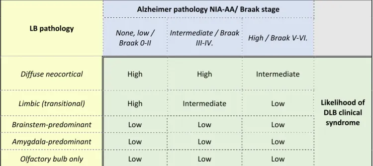
LB pathology
Alzheimer pathology NIA‐AA/ Braak stage
None, low /
Braak 0‐II
Intermediate / Braak
III‐IV. High / Braak V‐VI.
Diffuse neocortical High High Intermediate
Likelihood of DLB clinical
syndrome
Limbic (transitional) High Intermediate Low
Brainstem‐predominant Low Low Low
Amygdala‐predominant Low Low Low
Olfactory bulb only Low Low Low
Table 1. Likelihood of Dementia with Lewy bodies (DLB) clinical syndrome resulting from the assessment of Alzheimer’s‐type and Lewy body pathology (LB= Lewy body; DLB=Dementia with Lewy bodies; NIA‐AA = National Institute on Aging – Alzheimer’s Association) [Modified from McKeith et al.20]
Theoretically, the likelihood to manifest DLB clinical syndrome is directly proportional to the severity of LB pathology and inversely proportional to the severity of AD pathology. The McKeith classification integrates the assessment of concomitant AD pathology by National Institute on Aging and Alzheimer’s Association (NIA‐
AA)30, the Braak criteria and the type of LB pathology20. Table 1 shows the probability of pathological findings in relation to DLB clinical syndrome. It is likely that not only the α‐synuclein pathology is responsible for the cogni‐
tive decline, but plaques and phosphorylated tau pro‐
teins also contribute to the overall deficit31.
Cognitive impairment is also a common hallmark of DLB and Parkinson’s disease dementia (PDD). These two neurocognitive disorders share several clinical and neu‐
ropathological features, i.e. they are both characterized by cortical and subcortical α‐synuclein/LB, β‐amyloid and tau pathologies32.
According to Jellinger et al., a common pathophys‐
iology in DLB and PDD is synaptic dysfunction due to aggregation of α‐synuclein in the presynapses. This re‐
sults in disruption of axonal transport and neurotrans‐
mitter deprivation leading to neurodegeneration32. In‐
terestingly, there are some morphologic differences as well. DLB seems to show higher load of β‐amyloid and tau in several brain regions primarily in striatum, cortex, claustrum, amygdala and putamen compared to PDD32. Jellinger et al. also describes that α‐synuclein distribu‐
tion is different in DLB and PDD where α‐synuclein load
was highest in hippocampal subarea CA2 and in amygda‐
la in DLB, whereas in PD it is highest in the cingulate cortex. Moreover, nigral neuronal loss is more marked in PDD which ultimately results in dopaminergic upregula‐
tion32. A simple interpretation of above findings could support the current diagnostic criteria where DLB is associated with early cognitive impairment including memory problems, which differs from PDD with mainly motor impairment in the early phase.
In reality, it is not easy to define DLB based upon only the histological findings, thus in uncertain cases, anamnestic clinical data has invaluable support for the diagnostics. It is important to highlight that the listed strategies are not absolute or perfect diagnostic criteria, but rather useful schemes to predict the clinical syn‐
drome of DLB based on the observed pathological find‐
ings. As mentioned above, a major weakness of current guidelines is the low inter‐observer agreement; there‐
fore further research is needed to assess the accuracy of the current clinical diagnostic criteria versus neuropa‐
thology similar to studies assessing the 2005 criteria in this respect33.
3. Synaptic alterations
In connection with the dopaminergic neuronal loss of substantia nigra, dopamine transporter level is de‐
creased in the striatum in DLB, although not to the same extent observed in PD34. The choline‐acetyltransferase
(ChAT) levels are lower than in patients with AD, who suffer from similarly severe dementia35,36. Interestingly, Dynamin‐1, which takes part in the regulation of synap‐
tic transmission, shows significantly decreased levels in the prefrontal cortex in parallel with the severity of cognitive decline37. In addition, the amount of zinc transporter 3 (ZnT3) and postsynaptic density protein 95 (PSD95), are significantly reduced in DLB compared to aged‐controls and patients with AD38,39. It should be noted that at the beginning of the disease, an upregula‐
tion of soluble NSF attachment protein receptors (SNARE) complex is observed, probably as a compensa‐
tory response to the synaptic loss. However, during the progression of DLB synaptotagmin, synapsin and synap‐
tophysin, proteins involved in the SNARE complex steadily disintegrate, contributing further to synaptic dysfunction40,41. Recent studies have revealed that more than 90% of α‐synuclein aggregates are located at the presynapses leading to neurotransmitter deficits42,43. These findings suggest that degeneration of postsynaptic neurons may result from the loss of their inputs. This theory serves as a potential explanation for the DLB‐
specific clinical symptoms as well as raises the possibility of future curative treatments by pharmaceutical modifi‐
cation of neurotransmission.
4. Incidental Lewy‐body disease (iLBD)
In iLBD, patients have histologically detectable α‐
synuclein deposits in their brain, without presenting any clinical symptoms. Researchers accept that iLBD, which appears in approximately 8‐17% of the clinically normal 60+ years old patients, is the early stage of PD or DLB44,45. Confirming this hypothesis, it is frequently ob‐
served in patients who represent the prodromes of PD, including olfactory dysfunction or bowel frequency46,47. The presence of neuronal loss is usually minimal in these cases contrary to PD or DLB48. Distribution of α‐synuclein pathology in iLBD is particularly predictive, i.e. the brain‐
stem predominant subtype frequently results in PD, while the cortical predominant subtype often leads to DLB44.
Clinical symptoms
The new Diagnostic Statistical Manual (DSM‐5) (published in 2013) created a new neurocognitive disor‐
der (NCD) group, replacing the former ‘dementia, deliri‐
um, amnestic and other cognitive disorders’ category, that were used in the previous DSM‐IV handbook. The definition emphasizes that NCD is a progressive, ac‐
quired decline; which implies that disorders occurring at birth or at the early stage of cognitive development are
excluded from this category. As an advantage, the new handbook removed the stigmatizing debilitating term, dementia. Within the NCD there are two subcategories:
major and mild neurocognitive disorders based upon the severity of decline. For the diagnosis, six main fields are examined by DSM‐5: complex attention, executive func‐
tion, learning and memory, language, perceptual motor or social cognition49.
In the case of DLB, patients show both cortical and subcortical progressive dementia symptoms. Character‐
istic features comprise of attention and spatial percep‐
tion disorder, dysexecutive syndrome, fluctuating cogni‐
tive performance lasting from minutes to days. Among the psychiatric alteration the most frequent is the visual hallucination, although anxiety, apathy or organised delusions could also be detected.50,51. Interestingly, ad‐
vanced cerebral amyloid angiopathy and small vessel disease are associated with psychosis in AD and not in DLB52. Depression is more common in patients with DLB than in patients with AD. It may accompany with mild dementia, although more frequent in cases of advanced DLB53,54. The vast majority of the patients, from the be‐
ginning or during the progression, show extrapyramidal signs, such as action tremor, gait disturbances, rigidity or changes in facial expressions55. Rapid‐eye‐movement (REM) parasomnia is frequently diagnosed in DLB, char‐
acterised by vivid, often frightening dreams and complex purposeful motor activity. Since it often appears in dif‐
ferent neurodegenerative disorders, REM parasomnia might be an early herald of these diseases56. In DLB the autonomic nervous system dysfunction is more severe than in AD. Patients usually complain of dizziness, falls or loss of consciousness. Orthostatic hypotension, cardio‐
inhibitory carotid sinus hypersensitivity and urinary incontinency often accompany with DLB57. Pathogno‐
monic feature is the neuroleptic hypersensitivity, trig‐
gered by even a small dose of drug, leading to severe parkinsonism20. A 5‐year prospective cohort study pub‐
lished by Rongve et al. has reported that the progression of cognitive decline from mild to severe stage is more rapid in patients with DLB than in patients with AD58.
Many clinical symptoms of DLB cannot be ex‐
plained only by the intracerebral localization of LBs and LNs33. Neurotransmitter depletion, synaptic alterations, concomitant AD‐type pathology and metabolic changes highly influence the clinical presentation of DLB, and correlate with the severity of cognitive decline6,42,59,60. Moreover, a recent paper has reported association of serum potassium levels with cognitive decline in DLB, specifically in patients not using any medications that affect serum K+ levels61. Reduced perfusion and dyscon‐
nectivity of the occipital lobe may contribute to visual hallucinations and disturbance of visuospatial orienta‐
tion62,63. REM sleep behavior disorder (RBD) is also asso‐
ciated with striatal dopamine depletion64. Vegetative dysfunctions (i.e. orthostatic hypotension) may result from the deposition of LBs in the autonomic ganglia65. Decreased ZnT3 levels were also identified in patients with depression, providing a novel therapeutic tar‐
get38,66. Interestingly, several clinical symptoms of DLB are transient in nature, however LBs and LNs perma‐
nently occur in the brain. Probably, metabolic disturb‐
ances67, hormonal alterations68, circadian rhythm69 and comorbidities such as high blood pressure70 can affect the clinical appearance of DLB.
Radiologic features
Besides the clinical symptoms, the most significant diagnostic tool in the identification of DLB is the rapidly developing imaging techniques. The numerous reacha‐
ble modalities allow both structural and functional ex‐
amination.
Magnetic resonance imaging (MRI) examination Regarding the structural changes, the literature is not consistent: some studies have noted significant at‐
rophy of the insular, frontal, inferior parietal, temporal or occipital cortex71, whereas others have found only a minimal volumetric decrease in the frontal and parietal lobe, or in the territory of hypothalamus, basal forebrain and midbrain 72. However, these alterations are not specific to DLB and might appear in AD. Although differ‐
entiation from AD is usually based on the absence of medial temporal atrophy, its presence cannot rule out the diagnosis of DLB73. Sabattoli et al. have found atrophic changes in the anterior CA1, CA2/3 hippocam‐
pus, subiculum and presubiculum in DLB. This pattern differs significantly from those in AD74. According to an MRI study, the annual progression rate of cortical atro‐
phy is approximately 2x higher in patients with AD72. The role of white matter lesions (WML) in DLB, including loss of myelin, axonal damage and gliosis is not consistent.
Presumably, WMLs are implicated in vascular dementia rather than being specific feature of neurodegenerative disorders75. MRI spectroscopy provides a chance to indi‐
rectly assess the neuronal and glial function by measur‐
ing the key metabolites. N‐acetyl aspartate (NAA) and creatinine (CR) are two widely used markers in the char‐
acterization of central nervous system metabolism. Wat‐
son et al. have found that the NAA/CR ratio is relatively preserved in DLB in comparison to in AD76.
Functional imaging
Although, task‐related functional MRI (fMRI) has been performed in very few cases of DLB, a paper has
reported reduced activity in response to movement activity compared with aged controls and patients with AD77. Alternatively, researchers have examined the func‐
tional activity during rest. The resting‐state network shows increased activity during rest and decreased activ‐
ity during cognitive tasks. The most investigated resting‐
state network to date is the default mode network (DMN), including prefrontal cortex, posterior cingulate gyrus, medial temporal lobe and precuneus. In these areas the task‐related deactivation during the colour and motion tasks were decreased in DLB, but it was not sig‐
nificantly different from AD78. A technique for evaluation of functional resting state connectivity is based on the functional alterations in blood oxygen level dependent (BOLD) activity. Galvin et al. have noted increased rest‐
ing state connectivity between precuneus seeding re‐
gions, inferior parietal cortex and putamen, with de‐
creased connectivity between medial prefrontal, fron‐
toparietal operculum and visual cortex79. Taking into account that the results in DLB significantly differed from those of in AD, the method can be considered in the in vivo differential diagnosis.
The real value of perfusion imaging techniques in the identification of DLB is not fully consistent. The vast majority of the authors described occipital hypoperfu‐
sion as a hallmark of the disease on single‐photon emis‐
sion computed tomography (SPECT) images, whereas the specificity of the method is variable depending on the study80. [123I] 2ß‐carbomethoxy‐3b‐(4‐iodophenyl)‐
N‐(3‐fluoropropyl) nortropane (FP‐CIT) could be useful to discover DLB in early stages before the full spectrum of clinical symptoms evolve, moreover it reliably identi‐
fies neurobiological changes in the dopaminergic sys‐
tem81. In DLB there is markedly reduced tracer uptake in the regions of caudate and putamen reflecting the se‐
verely affected dopaminergic system. Strong connection has been found between striatal FP‐CIT uptake and cer‐
tain clinical symptoms, such as depression, anxiety, apa‐
thy and daytime somnolence82.
Positron emission tomography (PET) is probably a more sensitive functional imaging technique than SPECT in different dementias. Ishii et al. have compared the two methods and found that PET is more reliable in the detection of occipital and parietal lobe hypometabo‐
lism83. Besides the dopaminergic system, cholinergic transmission also has a crucial role in the pathomecha‐
nism of DLB, for instance in the alteration of memory, attention or arousal. Significant cholinergic neuronal loss and reduced ChAT activity are noted extensively in the cortical and subcortical regions, including both nicotin‐
ergic and muscarinergic systems, compared to AD36. Shimada et al. have shown reduced ChAT enzymatic activity in the medial occipital cortex with relatively preserved temporal activity in DLB, in contrast to AD84.
The fact that EEG is involved in the McKeith criteri‐
on further justifies its diagnostic relevance. Compared to AD the detectable slower background activity and more diffuse slow‐wave activity probably reflect the severe cholinergic deficit in DLB85.
Biomarkers
It is difficult to find specific biomarkers of DLB that would allow us to discriminate neuropathologically
‘pure’ forms of the disease from cases with concomitant AD pathology86, as that majority of DLB cases show a combination of α‐synuclein, tau and β‐amyloid patholo‐
gies87,88. However, novel biomarkers such as reduced electroencephalography activity89 and the detection of RBD90 also have some value.
Recent DLB biomarker research has focused on targets that are primarily related to AD. As in AD, CSF levels of Aβ1‐42 are decreased in DLB91, although tau seems to show an opposite relationship from AD92. That said tau has been shown to be higher in DLB when com‐
pared to PD and PDD87,93,94.
DLB is considered α‐synucleinopathy alongside with PD and PDD. When α‐synuclein was discovered as a major component of LB this initiated a string of studies investigating α‐synuclein as a biomarker in CSF95,96. In general, some groups have reported reduced α‐
synuclein97,98 while others have shown contradictory results99,100. These discrepancies may arise from the nature of α‐synuclein expression. α‐synuclein appears in four isoforms, (α‐syn98, α‐syn112, α‐syn126 and α‐
syn140), based on alternative splicing of exon 3 and 5, and the largest isoform, α‐syn140, is the most abundant isoform in the brain101,102. Nonetheless, to overcome this issue several groups have tried using a combination of antibodies to quantify ‘total’ α‐synuclein103. In addition to this, many are convinced that oligomeric α‐synuclein is potentially a more pathogenic form104 and efforts have been made to measure this in both CSF and in plasma105–
107.
In addition to α‐synuclein and AD biomarkers, oth‐
er potential biomarkers such as neurofilaments (NF) have been investigated108. NFs are components within a cell that assists in maintaining the structural integrity. It has been shown that NFs are associated with AD and other degenerative disorders109–111, however that is not the case with DLB. NFs seem to provide only a general hint of neuronal and axonal dysfunction without provid‐
ing any differential value to separate DLB from other disorders. On the other hand, other isoforms of NFs112 do exist and would have to be further investigated in DLB.
Since there is greater involvement of the dopamin‐
ergic and serotonergic neurotransmitters in DLB com‐
pared to AD, several groups have investigated metabo‐
lites from these pathways. In combination with CSF Aβ1‐
42, reduced levels of 5‐ hydroxyindolacetic acid and 3‐
methoxy‐4‐hydroxyphenylethyleneglycol have been found in DLB compared with AD104,113.
In summary, current studies suggest that DLB is in‐
termediate to AD and PD, such that biomarkers from AD and PD have been tested in DLB with moderate suc‐
cess114. Likewise, new diagnostic proteins may be dis‐
covered in the future with further proteomic studies which could provide a better differentiation of DLB from other closely related neurodegenerative disorders115–117.
Diagnostic criteria
Considering the heterogeneous phenotype and the frequently associated AD pathology, the clinical symp‐
tom‐based diagnosis of DLB is rather difficult. To date, there is no consensus on the guidelines for assessing clinical symptoms of DLB. A recent paper58 recommends the following rating strategies: for evaluating cognitive decline – Clinical Dementia Rating scale118; for estimat‐
ing fluctuating cognition – Clinician Assessment of Cogni‐
tive Fluctuations119 or Mayo Fluctuation Question‐
naire120; for investigating REM parasomnia ‐ Mayo Sleep Questionnaire121; for rating parkinsonism – Unified Par‐
kinson’s Rating Scale122; for testing disability ‐ Rapid Disability Rating Scale‐2123; for diagnosing visual halluci‐
nations or other psychiatric disorders – Neuropsychiatric Inventory124; for measuring effects of comorbidities – Cumulative Illness Rating Scale118. Additional difficulty is to make a distinction, if it exists, between DLB and Par‐
kinson’s disease dementia. The question, whether DLB and PDD are different entities or the same one, is still debated. Although there are a few morphologic differ‐
ences between them (e.g. cortical spreading of LBs or rate of neuronal loss in SN)125, differential diagnosis is rather based on the temporal sequence of symptoms. If dementia occurs 1 year after the onset of extrapyrami‐
dal motor signs, it is considered as PDD, otherwise if dementia has proceeded or presented within 1 year after movement disorder, the diagnosis should be DLB126. The latest McKeith diagnostic criteria define probable and possible DLB based on the clinical symp‐
toms and biomarkers (Figure 2)20. The presence of de‐
mentia, memory impairment and deficit of attention, executive functions and visuospatial orientation is essen‐
tial for diagnosis. Core symptoms are frequently ob‐
served in DLB clinical syndrome; in the lack of at least one core feature, the probable DLB diagnosis
Figure 2.: Symptoms and biomarkers contribute to the diagnosis of probable or possible Dementia with Lewy bodies (DLB).
Essential symptoms are mandatory for diagnosis of DLB. Core clinical symptoms are characteristic to DLB, while supportive symptoms can confirm the decision by clinicians. Indicative biomarkers are frequently observed in DLB, while supportive biomarkers can facilitate the decision‐making procedure. Important to note that clinicians cannot establish the diagnosis of probable DLB based on the bi‐
omarkers. (Recategorized or newly added features are highlighted in blue colour; REM = Rapid eye movement; DAT = Dopamine trans‐
porter; MIBG = Metaiodobenzylguanidine; PSG = Polysomnography; RBD = REM sleep behavior disorder; SPECT = Single‐photon emis‐
sion computed tomography; PET = Positron‐emission tomography; EEG = Electroencephalography; ↓ = decrease). [Adapted from McKeith et al.20]
cannot be established. The development of diagnostic techniques revealed that REM sleep behaviour disorder is more characteristic of LBD than it was previously thought. Therefore, McKeith et al. recategorized RBD from supportive to core features20. Supportive features may help the clinicians; however, these symptoms are not specific to DLB and their presence is not required for the diagnosis of neither probable nor possible DLB. The refreshed criteria emphasize the importance of disease‐
specific biomarkers. One indicative biomarker itself is sufficient for possible DLB diagnosis. If one indicative biomarker associates with one core symptom, the diag‐
nosis is probable DLB.
Therapy
There are two different therapeutic approaches:
pharmacologic and non‐pharmacologic. Although there is no curative treatment strategy, the adequate therapy may slow the disease progression with the chance of a better quality of life.
Non‐pharmacologic interventions
At the first signs of DLB or mild NCD, installation of the below mentioned interventions are recommended.
Changes in the dietary habit, Mediterranean diet, and regularly performed social and mental tasks as well as physical activities could reduce the rate of progres‐
sion127. Personalized cognitive rehabilitation trainings with focus on the declined field, improve both quality of life and memory128.
Pharmacologic treatments
The frequently noted extrapyramidal signs should be treated with the smallest effective dose of levodopa, to avoid the worsening of psychiatric symptoms129. Se‐
lective serotonin reuptake inhibitors (SSRIs) are widely used and considered as effective medications for de‐
pression130. In the case of REM sleep behaviour disorder, clonazepam and melatonin also have beneficial effect131 Acetyl‐cholinesterase inhibitors (ChAT‐I) have benefits in the treatment of psychiatric disturbances such as visual hallucination, delusions, behaviour disorders or apathy.
Despite gastrointestinal side effects (i.e. vomiting, diar‐
rhoea) these drugs are usually well tolerated20. If ChAT‐Is (rivastigmine, donepezil) are not effective, the use of atypical neuroleptic drugs (i.e. clozapine) might be inevi‐
table, but typical neuroleptics should be avoided to minimize the possibility of severe neuroleptic hypersen‐
sitivity reaction132. ChAT‐Is are more effective in patients
with DLB compared to patients with AD. These drugs improve the cognitive functions, reduce fluctuation, decrease the progression and in addition patients score better on neuropsychological tests133. N‐methyl‐D‐
aspartate receptor (NMDA‐R) antagonist memantine is also useful in the treatment of cognitive decline134. Lucza et al. have suggested treatment with ChAT‐Is in mild or mid‐severe dementia and memantine combined with high dose of rivastigmine (13.5 mg) in severe demen‐
tia135.
Summary
Despite the continuously growing incidence of de‐
mentia, which could be the most prevalent disease with‐
in 20 years in the industrialized world, our present knowledge is still insufficient to fully comprehend the underlying pathomechanism. This is particularly true for DLB, which is the second most common neurodegenera‐
tive dementia. Etiology of the disorder, its connection with aging, underlying pathomechanisms of clinical signs and imaging findings are still in need for further elucida‐
tion. Unfortunately, the definitive diagnosis is possible only by post‐mortem histopathological examination and not by in vivo techniques (apart from a low percentage of cases). The concomitant Alzheimer’s‐type and vascu‐
lar pathology raises issues regarding diagnostic clarity and accuracy136. Future research should be multidiscipli‐
nary and should include pathological, proteomic, genetic and epigenetic approaches to identify the key factors of
the disease and reveal the correlation between clinical symptoms, radiological alterations and pathological findings. Development of imaging techniques brings the possibility of in vivo diagnosis, which implies adequate treatments in early stages, a crucial advancement to ensure better quality of life. Longitudinal cohort studies are also required to provide detailed prognostic infor‐
mation that are essential for planning a more effective and cost‐efficient treatment protocol.
Author’s contribution
Authors contributed equally to the work.
Funding
Supported by the ÚNKP‐19‐3 New National Excel‐
lence Program of the Ministry of Innovation and Tech‐
nology and EFOP‐3.6.3‐VEKOP‐16‐2017‐00009 (J.B.);
GINOP‐2.3.2‐15‐2016‐00043, Hungarian Brain Research Program (2017‐1.2.1‐NKP‐2017‐00002), NKFIH SNN 132999, SZTE ÁOK‐KKA No. 5S 567 (A202) and DE ÁOK Research Fund (T.H.). This paper represents independent study partly funded by the National Institute for Health Research (NIHR) Biomedical Research Centre at South London and Maudsley NHS Foundation Trust and King’s College London. The views expressed are those of the author(s) and not necessarily those of the NHS, the NIHR or the Department of Health and Social Care.
References
1. Tsuang, D. W. et al. Familial dementia with Lewy bodies: a clinical and neuropathological study of 2 families. Arch. Neurol. 59, 1622–30 (2002).
2. Zarranz, J. J. et al. The new mutation, E46K, of alpha‐synuclein causes Parkinson and Lewy body dementia. Ann. Neurol. 55, 164–73 (2004).
3. Bras, J. et al. Genetic analysis implicates APOE, SNCA and suggests lysosomal dysfunction in the etiology of dementia with Lewy bodies.
Hum. Mol. Genet. 23, 6139–46 (2014).
4. Takao, M. et al. Early‐onset dementia with Lewy bodies. Brain Pathol. 14, 137–147 (2004).
5. Hogan, D. B. et al. The prevalence and incidence of dementia with Lewy bodies: A systematic review. Can. J. Neurol. Sci. 43, S83–S95 (2016).
6. Aarsland, D. Cognitive impairment in Parkinson’s disease and de‐
mentia with Lewy bodies. Park. Relat. Disord. 22, S144–S148 (2016).
7. Love, J., Kalaria R. Dementia. in: Love, S. et al. Greenfield’s Neuropa‐
thology. 9th ed., pp. 858‐973, CRC Press (2015).
8. Lewy, F. H. Paralysis agitans. I. Pathologische Anatomie. in: Handb.
der Neurol. 920–958, Springer, Berlin (1912).
9. Wakabayashi, K. et al. The Lewy body in Parkinson’s disease: mole‐
cules implicated in the formation and degradation of alpha‐synuclein aggregates. Neuropathology 27, 494–506 (2007).
10. Spillantini, M. G. et al. alpha‐Synuclein in filamentous inclusions of Lewy bodies from Parkinson’s disease and dementia with Lewy bodies.
Proc. Natl. Acad. Sci. U. S. A. 95, 6469–73 (1998).
11. Luk, K. C. et al. Modeling Lewy pathology propagation in Parkin‐
son’s disease. Park. Relat. Disord. 20, S85‐7 (2014).
12. Olanow, C. W. et al. Lewy‐body formation is an aggresome‐related process: a hypothesis. Lancet Neurol. 3, 496–503 (2004).
13. Alghamdi, A. et al. Reduction of RPT6/S8 (a Proteasome Compo‐
nent) and Proteasome Activity in the Cortex is Associated with Cogni‐
tive Impairment in Lewy Body Dementia. J. Alzheimers Dis. 57, 373–
386 (2017).
14. Cuervo, A. M. et al. Impaired degradation of mutant alpha‐
synuclein by chaperone‐mediated autophagy. Science 305, 1292–5 (2004).
15. Baek, J. H. et al. Unfolded protein response is activated in Lewy body dementias. Neuropathol. Appl. Neurobiol. 42, 352–365 (2016).
16. Tompkins, M. M. et al. Contribution of somal Lewy bodies to neu‐
ronal death. Brain Res. 775, 24–9 (1997).
17. Mori, F. et al. Relationship among alpha‐synuclein accumulation, dopamine synthesis, and neurodegeneration in Parkinson disease substantia nigra. J. Neuropathol. Exp. Neurol. 65, 808–15 (2006).
18. Kosaka, K. et al. Diffuse type of Lewy body disease: progressive dementia with abundant cortical Lewy bodies and senile changes of varying degree‐‐a new disease? Clin. Neuropathol. 3, 185–92 (1984).
19. Kuusisto, E. et al. Morphogenesis of Lewy bodies: dissimilar incor‐
poration of alpha‐synuclein, ubiquitin, and p62. J. Neuropathol. Exp.
Neurol. 62, 1241–53 (2003).
20. McKeith, I. G. et al. Diagnosis and management of dementia with Lewy bodies. Neurology 89, 88–100 (2017).
21. Braak, H. et al. Staging of brain pathology related to sporadic Parkinson’s disease. Neurobiol. Aging 24, 197–211 (2003).
22. Leverenz, J. B. et al. Empiric refinement of the pathologic assess‐
ment of Lewy‐related pathology in the dementia patient. Brain Pathol.
18, 220–4 (2008).
23. Müller, C. M. et al. Staging of sporadic Parkinson disease‐related alpha‐synuclein pathology: inter‐ and intra‐rater reliability. J. Neuropa‐
thol. Exp. Neurol. 64, 623–8 (2005).
24. Alafuzoff, I. et al. Staging/typing of Lewy body related α‐synuclein pathology: a study of the BrainNet Europe Consortium. Acta Neuropa‐
thol. 117, 635–652 (2009).
25. Beach, T. G. et al. Unified staging system for Lewy body disorders:
Correlation with nigrostriatal degeneration, cognitive impairment and motor dysfunction. Acta Neuropathol. 117, 613–634 (2009).
26. McKeith, I. G. et al. Diagnosis and management of dementia with Lewy bodies: Third report of the DLB consortium. Neurology 65, 1863–
1872 (2005).
27. Jellinger, K. A. et al. Impact of coexistent Alzheimer pathology on the natural history of Parkinson’s disease. J. Neural Transm. 109, 329–
39 (2002).
28. Barker, W. W. et al. Relative frequencies of Alzheimer disease, Lewy body, vascular and frontotemporal dementia, and hippocampal sclerosis in the State of Florida Brain Bank. Alzheimer Dis. Assoc. Dis‐
ord. 16, 203–212 (2002).
29. Kosaka, K. Diffuse lewy body disease in Japan. J. Neurol. 237, 197–
204 (1990).
30. Hyman, B. T. et al. Longitudinal assessment of AB and cognition in aging and Alzheimer disease. Ann. Neurol. 69, 181–192 (2011).
31. Howlett, D. R. et al. Regional multiple pathology scores are associ‐
ated with cognitive decline in Lewy body dementias. Brain Pathol. 25, 401–408 (2015).
32. Jellinger, K. A. et al. Are dementia with Lewy bodies and Parkin‐
son’s disease dementia the same disease? BMC Med. 16, (2018).
33. Skogseth, R. et al. Accuracy of clinical diagnosis of dementia with lewy bodies versus neuropathology. J. Alzheimer’s Dis. 59, 1139–1152 (2017).
34. Piggott, M. A. et al. Striatal dopaminergic markers in dementia with Lewy bodies, Alzheimer’s and Parkinson’s diseases: Rostrocaudal distribution. Brain 122, 1449–1468 (1999).
35. Perry, E. K. et al. Cholinergic correlates of cognitive impairment in Parkinson’s disease: comparisons with Alzheimer’s disease. J. Neurol.
Neurosurg. Psychiatry 48, 413–21 (1985).
36. Tiraboschi, P. et al. Cholinergic dysfunction in diseases with Lewy bodies. Neurology 54, 407–411 (2000).
37. Vallortigara, J. et al. Dynamin1 concentration in the prefrontal cortex is associated with cognitive impairment in Lewy body dementia.
F1000Research 3, 108 (2014).
38. Whitfield, D. R. et al. Assessment of ZnT3 and PSD95 protein levels in Lewy body dementias and Alzheimer’s disease: Association with cognitive impairment. Neurobiol. Aging 35, 2836–2844 (2014).
39. Bereczki, E. et al. Synaptic proteins predict cognitive decline in Alzheimer’s disease and Lewy body dementia. Alzheimer’s Dement. 12, 1149–1158 (2016).
40. Vallortigara, J. et al. Decreased Levels of VAMP2 and Monomeric Alpha‐Synuclein Correlate with Duration of Dementia. J. Alzheimer’s Dis. 50, 101–110 (2015).
41. Bereczki, E. et al. Synaptic markers of cognitive decline in neuro‐
degenerative diseases: A proteomic approach. Brain 141, 582–595 (2018).
42. Schulz‐Schaeffer, W. J. The synaptic pathology of α‐synuclein aggregation in dementia with Lewy bodies, Parkinson’s disease and Parkinson’s disease dementia. Acta Neuropathol. 120, 131–143 (2010).
43. Bridi, J. C. et al. Mechanisms of α‐Synuclein induced synaptopathy in parkinson’s disease. Front. Neurosci. 12, (2018).
44. Frigerio, R. et al. Incidental Lewy body disease: do some cases represent a preclinical stage of dementia with Lewy bodies? Neurobiol.
Aging 32, 857–63 (2011).
45. Auning, E. et al. Early and presenting symptoms of dementia with Lewy bodies. Dement. Geriatr. Cogn. Disord. 32, 202–208 (2011).
46. Ross, G. W. et al. Association of olfactory dysfunction with inci‐
dental Lewy bodies. Mov. Disord. 21, 2062–2067 (2006).
47. Abbott, R. D. et al. Bowel movement frequency in late‐life and incidental Lewy bodies. Mov. Disord. 22, 1581–6 (2007).
48. DelleDonne, A. et al. Incidental Lewy body disease and preclinical Parkinson disease. Arch. Neurol. 65, 1074–80 (2008).
49. American Psychiatric Association: Diagnostic and statistical manual of mental disorders. American Psychiatric Publishing, Arlington, USA.
(2013).
50. Walker, Z. et al. Lewy body dementias. Lancet 386, 1683–1697 (2015).
51. Majer, R. et al. Behavioural and psychological symptoms in neu‐
rocognitive disorders: Specific patterns in dementia subtypes. Open Med. 14, 307–316 (2019).
52. Vik‐Mo, A. O. et al. Advanced cerebral amyloid angiopathy and small vessel disease are associated with psychosis in Alzheimer’s disease. J. Neurol. Neurosurg. Psychiatry 90, 728–730 (2019).
53. Fritze, F. et al. Depressive symptoms in Alzheimer’s disease and Lewy body dementia: A one‐year follow‐up study. Dement. Geriatr.
Cogn. Disord. 32, 143–149 (2011).
54. Fritze, F. et al. Depression in mild dementia: Associations with diagnosis, APOE genotype and clinical features. Int. J. Geriatr. Psychia‐
try 26, 1054–1061 (2011).
55. Aarsland, D. et al. Comparison of extrapyramidal signs in dementia with Lewy bodies and Parkinson’s disease. J. Neuropsychiatry Clin.
Neurosci. 13, 374–379 (2001).
56. Ferini‐Strambi, L. et al. REM Sleep Behavior Disorder (RBD) as a marker of neurodegenerative disorders. Arch. Ital. Biol. 152, 129–146 (2014).