2018
Doctoral Thesis for the Hungarian Academy of Sciences Karri Lamsa, Ph.D.
Regulation of cortical activity through inhibitory interneuron plasticity
Department of Physiology, Anatomy and Neuroscience, University of Szeged
and
The Hungarian Academy of Sciences Neuroscience Program
dc_1581_18
Table of Contents
1. Summary of the scientific work ... 3
2. Introduction ... 3
3. Methods ... 3.1. Experiments on rodent acute brain slices ... 4
3.2. Experiments in vivo rat ... 7
3.3. Human brain slices ... 8
3.4. Anatomical analyses and identification of neuron types ... 10
4. Results and discussion ... 4.1. GABAergic interneurons exhibit various forms of synaptic plasticity and some are specific to interneuron type ... 11
4.2. Identified interneuron types show the learning-related long-term plasticity in vivo ... 17
4.3. Human cortical microcircuits show evolutionarily conserved interneuron plasticity with specific features ... 19
4.4. Interneuron long-term plasticity produces permanent changes in local network activity both in the rodent and in the human ... 22
5. Conclusions ... 24
6. References ... 24
7. Other scientific activities ... 7.1. Original research articles forming the base of application ... 29
7.2. Most important talks at scientific conferences ... 29
7.3. Supervised Ph.D. students ... 30
7.4. Lectures given in doctoral programs ... 30
7.5. Training of postdoctoral fellows ... 30
7.6. Organization of domestic and international symposia ... 30
7.7. Memberships in editorial or review boards ... 31
7.8. Memberships in scientific committees ... 31
7.9. Scientific awards ... 31
7.10 Personal research grants and fellowships ... 31
7.11 Grant reviewing activities ... 31
7.12 Public outreach ... 32
8. Summary of the most significant results ... 32
9. Original research articles ... 33
1. Summary of the scientific work
In this thesis, I will summarize recent developments in the field of “synaptic long-term plasticity in the cortical GABAergic interneurons” and discuss our research team's contribution to the topic. Indeed, we among other laboratories have demonstrated during the past decade that 1) excitatory glutamatergic synapses targeting the GABAergic interneurons in the hippocampus and the neocortex exhibit various different forms of activity-induced learning-related long-term plasticity (Lamsa et al. 2005; Lamsa et al. 2007a; Lamsa et al.
2007b). Importantly, we have demonstrated that the plasticity forms are often specific to interneuron type; anatomically specialized GABAergic interneurons exhibit distinct long-term plasticity mechanisms and require specific neuronal activity patterns for the plasticity induction (Oren et al. 2009; Nissen et al. 2010). 2) We have shown that the interneuron plasticity reported in acute brain slice preparations also occurs in the intact brain in vivo, and that the plasticity regulation in vivo brain is complex (Lau et al. 2017). 3) We have proven that common interneuron types exhibiting synaptic long-term plasticity in a rodent brain show plasticity in the human neocortex although with specific features. 4) Both in the rodent and in the human cortex, the interneuron plasticity strongly modifies the local neuronal network activities permanently changing signal transmission though polysynaptic circuits (Lamsa et al.
2005; Szegedi et al. 2016; Szegedi et al. 2017). In addition, our results indicate that interneuron plasticity is required to maintain high temporal precision of principal cells' signal processing in the cortex in the face of learning (Kullmann and Lamsa 2007; Kullmann and Lamsa 2011b).
2. Introduction
Salient and contextual information in the brain is encoded in firing of neurons as neuronal ensembles, and GABAergic (γ-aminobutyric acid -releasing) inhibitory interneurons play a pivotal role in this process. The activated neuronal ensembles (often referred to as engrams) are thought to represent means carrying relevant stored pieces of information, and they are promptly re-organized by learning (Tonegawa et al., 2015; Poo et al., 2016; Buzsaki and Llinas, 2017). The re-organisation of engrams is at least partly manifested by long-term synaptic plasticity between the excitatory glutamatergic pyramidal neurons (Lisman, 2017). However, it has been poorly understood whether and how long-term plasticity in GABAergic inhibitory
interneurons contributes to this process (McBain et al., 1999; Kullmann and Lamsa, 2011b). It is well established that glutamatergic excitatory neurons exhibit synaptic and non-synaptic long-term plasticity forms. In contrast, the GABAergic inhibitory neurons were initially considered rigid and unchangeable with a hypothesis that their function may not exhibit learning-associated permanent changes (McBain and Maccaferri, 1997; McBain et al., 1999;
Ross and Soltesz, 2001). Yet, a past decade in the research of neocortical and hippocampal microcircuits has revealed sophisticated plasticity forms in synapses to the GABAergic inhibitory neurons (Kullmann and Lamsa, 2011a). Several research groups including ours have independently demonstrated that the GABAergic neurons undergo a wide range of synaptic and non-synaptic activity-induced plasticity processes (Laezza et al., 1999; Alle et al., 2001;
Perez et al., 2001; Lamsa et al., 2005; Pelkey et al., 2005; Lamsa et al., 2007b; Lu et al., 2007;
Galvan et al., 2010; Sambandan et al., 2010; Peterfi et al., 2012; Griguoli et al., 2013; Le Roux et al., 2013; Camire and Topolnik, 2014; Zarnadze et al., 2016; Nicholson and Kullmann, 2017).
A remarkable feature in their plasticity is – in terms of its induction and expression – that it often (although not always) differs from that known to exist in the excitatory principal neurons (Kullmann and Lamsa, 2007; Pelkey and McBain, 2008; Bartos et al., 2011; Galvan et al., 2011; Kullmann et al., 2012; Topolnik, 2012). This thesis will shortly review the topic and explain how our research team activity has participated to the exciting and timely scientific endeavor of the learning-related cortical GABAergic interneuron plasticity. In four main chapters, I will review current understanding of the synaptic long-term plasticity in the interneurons summarizing (1) its induction and the mechanisms explored in vitro slice preparation, and (2) present evidence for the plasticity in vivo brain. In following chapters (3 and 4), I will elaborate the topic from the rodents (which are the most commonly used experimental animals in cellular neuroscience research) to the human cortex. This research thesis focuses on the learning-associated plasticity specifically in the excitatory synaptic input to the GABAergic neurons.
3. Methods
3.1. Experiments on rodent acute brain slices
Three- to four- week old male Sprague-Dawley rats were killed by cervical dislocation and decapitated. The brain was rapidly removed and placed in ice-cold (0 to +4°C) sucrose cutting
solution containing (mM): 75 sucrose, 87 NaCl, 2.5 KCl, 0.5 CaCl2, 7 MgCl2, 1.0 NaH2PO4, 25 NaHCO3, 25 glucose, pH 7.4, bubbled with 95 % O2 / 5 % CO2 (Lamsa et al. 2005; Lamsa et al.
2007b; Oren et al. 2009; Nissen et al. 2010; Szabo et al. 2012). Transverse hippocampal slices (350 μm thickness) were cut using a vibrating microtome (Leica VT 1000S, Leica Microsystems, Germany). Slices were kept submerged at 32°C in the sucrose solution for 20- 25 min before being transferred to an interface chamber where they were maintained in Earle's Balanced salt solution (EBSS) (Gibco-Invitrogen) with 3 mM Mg2+ and 1 mM Ca2+ at room temperature (20–25°C) for at least 60 min before starting experiments. Hippocampal or somatosensory neocortex slices (Szegedi et al. 2016) were placed in a recording chamber (Luigs & Neumann, Germany) mounted on the stage of an upright microscope (Olympus BX51WI, Japan), where they were held under a nylon mesh grid and superfused at 3-8 ml min−1 with artificial cerebrospinal fluid (ACSF) at 31 – 33°C. The ACSF contained (mM): NaCl (119), KCl (2.5), CaCl2 (2.5), MgSO4 (1.3), NaH2PO4 (1.25), NaHCO3 (26), glucose (11); final pH 7.4 (equilibrated with 95% O2 / 5% CO2) (except in Szegedi et al. 2016 where 3 mM Ca2+ with 1.5 mM Mg2+ was used instead). A cut was made between the CA3 and CA1 subfields in the hippocampus to prevent the spread of bursting activity. Slices were visualized using a 20x immersion objective with 2–4x zoom and infra-red differential interference contrast (DIC) optics. In the study using mice the hippocampal slices were similarly processed and maintained and prepared from heterozygous αCaMKII T286A-mutants (backcrossed into a hybrid C57BL6 – 129/Sv genetic background and interbred to obtain homozygous and wild- type (WT) littermates) (Lamsa et al. 2007a). Genotyping was carried out by PCR analysis with DNA obtained from tail biopsies on postnatal day 21, the day of weaning (Lamsa et al. 2007a).
Male mice homozygous for αCaMKII T286A and WT littermates were used in the experiments.
Somatic perforated-patch microelectrode recordings (Lamsa et al. 2005; Lamsa et al. 2007a;
Lamsa et al. 2007b; Oren et al. 2009; Nissen et al. 2010; Szabo et al. 2012) were made from neurons in CA1 area. Electrodes were prepared from borosilicate glass capillaries (GC150F, 1.5 mm o.d., Harvard Apparatus, UK) pulled on a Sutter microelectrode puller (Novato, CA, USA).
Pipette resistance was typically 8–18 MΩ for perforated patch, and 4-7 MΩ for whole cell recordings. A Multiclamp 700B amplifier was used for recording (Molecular Devices, CA, USA).
Infra red DIC images of the cell at different magnification (20x, 40x and 80x) were obtained with a CCD camera (Till Photonics, Germany) during electrophysiological experiments.
Gramicidin stock solution (100 mg ml−1, Sigma) was prepared in dimethyl sulfoxide daily. The
pipette filling solution containing gramicidin was prepared by diluting the stock solution 1:1000 in potassium gluconate pipette solution. The pipette solution contained (mM):
potassium gluconate (145), NaCl (8), KOH-HEPES (20-25), EGTA (0.2), and QX-314 Br (1-5); pH 7.2, osmolarity 295 mOsm l−1. The electrode tip was filled with gramicidin-free filtered potassium gluconate solution. The series resistance was continuously monitored throughout the experiment, and recordings were started when it was < 150 MΩ. Bridge balance and pipette capacitance compensation were adjusted throughout the recordings. The presence of QX-314 in the filling solution allowed for detection of inadvertent patch rupture.
Suprathreshold depolarizing current steps were injected intermittently to evoke action potentials. Failure to generate action potentials indicated membrane rupture in which case the experiment was aborted. Upon completion of perforated patch recordings, the pipette was slowly retracted under infra red DIC observation. Once the pipette detached from the cell it was rapidly withdrawn from the slice. Next, the same cell was approached with a new pipette and re-patched in whole-cell configuration. Infra red images obtained during the perforated patch recording and the whole-cell recordings were compared to verify that the same cell was re-patched.
The whole-cell filling solution contained (mM) CsCl (135), KOH-HEPES (10), BAPTA (10), NaCl (8), Mg-ATP (2), GTP (0.3), and QX-314 Br (5); pH 7.2, 290 mOsm (Lamsa et al. 2005; Lamsa et al. 2007a; Lamsa et el. 2007b; Oren et al. 2009; Nissen et al. 2010; Szabo et al. 2012).
Spermine tetrahydrochloride (0.5 mM, Tocris) was included in some studies in the filling solution to maintain polyamine-mediated rectification of AMPA/kainate receptors during whole cell recording. In addition, neurobiotin (0.3-0.5%, Alomone labs) or biocytin (0.3-0.5%, Sigma-Aldrich) was included in the solution for post hoc anatomical analysis of cells. Pipette capacitance compensation was applied in the cell-attached configuration before membrane rupture. Series resistance was not compensated during voltage clamp recordings, but regularly monitored with small hyperpolarizing voltage steps (−5 mV). Data were not corrected for junction potentials.
Monosynaptic excitatory postsynaptic potentials (EPSPs) or excitatory postsynaptic currents (EPSCs) were evoked by alternately stimulating in the CA1 area at 0.067 Hz via two concentric bipolar electrodes (o.d. 125 μm, FHC, ME USA), connected to constant current isolated stimulators (DS3, Digitimer UK, 20–200 μA, duration 50 μs). The stimulators were controlled
by a custom data acquisition program (LabView, National Instruments) or by pClamp 10 software (Axon Instruments). Evoked EPSPs were recorded from the resting membrane potential or in some experiments during a brief (500 ms) hyperpolarizing step (5–10 mV) to avoid action potential generation. For LTP induction one of the pathways was stimulated at 100 Hz for 1 s, delivered twice with a 20 s interval. Simultaneously, the postsynaptic cell was voltage clamped (1200-2000 ms) at −70 – −90 mV or at 0 mV (somatic potential). Miniature EPSCs (mEPSCs) were recorded in 120 s sweeps, with the seal test monitored in between sweeps (Oren et al. 2009). The first recordings were made after 15 min after breaking through into the whole cell configuration to allow for stabilization of the cell input resistance
In all recordings data were low-pass filtered (4 - 5 kHz) and acquired at 10 - 20 kHz on a PC for offline analysis. Data was analysed using LabView, pClamp 10 or in Igor Pro (Wavemetrics, USA). The GABA receptor blockers picrotoxin (100 μM) and CGP55845 (1 μM) were added to the extracellular solution. Where indicated, the NMDA receptor antagonist DL-2-Amino-5- phosphonovaleric acid (DL-APV, 100 μM) or glutamate receptor antagonists philanthotoxin- 433 (PhTx, 10 μM) was also included. All drugs were applied via superfusion (3-5 ml/min).
Tetrodotoxin (TTX 1 μM) was present during mEPSC recordings. Chemicals were purchased from Sigma and drugs were purchased from Tocris Cookson (Bristol, UK) or Ascent Scientific (Weston-super-Mare, UK).
3.2. Experiments in vivo rat
Experiments were carried out on adult male (weight 280–350 g) Sprague–Dawley rats (Charles River, UK) according to the Animal Scientific Procedures Act, 1986 (UK) using a heating mattress (37.5 ± 0.5 °C) with an external abdominal temperature measurement probe with feedback to the heating pad. Anesthesia was induced with isoflurane (4 % v/v in O2) and maintained by a single intraperitoneal (i.p.) injection of urethane (1.25–1.3 mg/kg in 0.9 % saline, i.p.). Ketamine (30 mg/kg i.p.) and xylazine (3 mg/kg i.p.) were given at the start of the procedure and in supplementary small doses during recording to maintain anesthesia. Saline- based glucose solution (5 % v/v glucose) was injected subcutaneously (2 ml/2 h) to compensate for fluid loss during the experiment. A rostrocaudal incision was performed to expose the skull, and surgical windows were made above the right and left dorsal hippocampal CA1 areas with a dental drill. A wall of dental cement was built to protect the
openings and saline was applied regularly to the exposed brain surface. For accurate measurement of penetration depth, saline solution was drained before inserting electrodes into the brain. The windows were covered with warm paraffin wax once the electrodes were lowered into the brain.
Microelectrodes were pulled from borosilicate glass capillaries (GC120F-10, Harvard Apparatus, UK) and were filled with 1.5–3 % (w/v) neurobiotin (Vector Laboratories, UK) in 0.5 M NaCl. The recording electrodes were lowered into the brain at 20 µm/s, and into the hippocampus at 5 µm/s using a micro drive holder (EXFO-8200 IMMS, Canada) and a computer-controlled 0.5 µm-stepping interface. Stereotaxic co-ordinates for the recording electrodes were: 3.0 mm posterior to Bregma (±0.3 mm), 3.6 mm from midline (±0.5 mm), and depth 2.2 mm (±0.3 mm). The electrode resistance was 15–21 MΩ. Following extracellular recording, the electrode was moved into juxtacellular position and the recorded cells were modulated by applying a series of +10 to +50 nA square pulses of 200 ms duration in 30 s episodes for 2–3 minutes continuously (Lau et al. 2017). We verified that the action potential properties (extracellular spike kinetics) of the modulated cell corresponded to the action potential properties recorded during plasticity experiment. This labeling procedure was followed by a period from 1 to 5 hours (Lau et al. 2017), which allowed for the diffusion of neurobiotin inside the modulated cells. Signal was amplified 1000× (10×, head-stage amplifier, Axon Instruments, USA; 100×, NL-106, DigitimerTM, UK) and band-pass filtered between 0.3 and 300 Hz for local field potentials (LFP) and between 300 Hz and 5 kHz for detection of single spikes. The LFP and single neuron activity were acquired at 1 and 19.841 kHz, respectively using Spike2 (version 7.0; Cambridge Electronic Design, UK). Concentric bipolar stimulating electrodes (125 µm tip diameter, FHC Inc., USA) were stereotaxically placed in the left hippocampal CA1 area 3.0–3.2 mm posterior and 3.0–4.0 mm lateral to Bregma and at 2.1–2.5 mm depth from the cortical surface (Lau et al. 2017). Single-shock stimulation (100 µs, 150–600 µA) was delivered every 5 s using current isolator stimulator (DS3; Digitimer, UK) to elicit spikes. The train of theta-burst stimulation (TBS) for plasticity induction consisted of 20 bursts (at 200 ms intervals) of five stimuli at 100 Hz.
3.3. Human brain slices
All procedures were performed according to the Declaration of Helsinki with the approval of the University of Szeged Ethical Committee and Regional Human Investigation Review Board (ref. 75/2014). Human neocortical slices were derived from material that had to be removed to gain access to the surgical treatment of deep-brain tumors from the left and right frontal, temporal, and parietal regions with written informed consent of the patients prior to surgery.
The patients were 10–85 y of age (mean ± SD = 50 ± 4 y), including 17 males and 14 females.
The tissue obtained from underage patients was provided with agreement from a parent or guardian. The resected samples were cut from the frontal and temporal lobes of left or right hemisphere. Anesthesia was induced with intravenous midazolam and fentanyl (0.03 mg/kg, 1–2 lg/kg, respectively). A bolus dose of propofol (1–2 mg/kg) was administered intravenously. The patients received 0.5 mg/kg rocuronium to facilitate endotracheal intubation. After 2 min, the trachea was intubated and the patient was ventilated with O2/N2O mixture (a ratio of 1:2). Anesthesia was maintained with sevoflurane at monitored anesthesia care volume of 1.2–1.5. After surgical removal, the resected tissue blocks were immediately immersed in ice-cold standard solution containing (in mM): 130 NaCl, 3.5 KCl, 1 NaH2PO4, 24 NaHCO3, 1 CaCl2, 3 MgSO4, 10 D(+)-glucose, and saturated with 95% O2 and 5%
CO2. Slices were cut perpendicular to cortical layers at a thickness of 350 μm with a microtome (Microm HM 650 V) and were incubated at room temperature (20–24°C) for 1 h in the same solution. The solution used during electrophysiology experiments was identical to the slicing solution, except it contained 3 mM CaCl2 and 1.5 mM MgSO4. Recordings were performed in a submerged chamber (perfused 8 ml/min) at approximately 36–37°C. Cells were patched using water-immersion 20× objective with additional zoom (up to 4x) and infrared differential interference contrast video microscopy. Micropipettes (5–8 MOhm) were filled with intracellular solution for whole-cell patch-clamp recording (in mM): 126 K- gluconate, 8 KCl, 4 ATP-Mg, 0.3 Na2–GTP, 10 HEPES, 10 phosphocreatine (pH 7.20; 300 mOsm) with 0.3% (w/v) biocytin. Current and voltage clamp recordings were performed with Mutliclamp 2B amplifier (Axon Instruments), low-pass filtered at 6 kHz (Bessel filter). Series resistance (Rs) and pipette capacitance were compensated in current clamp mode and pipette capacitance in voltage clamp mode. Rs was monitored and recorded continuously during the experiments. The recording in voltage clamp mode was discarded if the Rs was higher than 25 ΩM or changed more than 20%. Extracellular stimulation was applied with a concentric bipolar electrode (125 μm tip diameter, FHC Inc., US) positioned on L2–3. Paired pulse stimuli (50 μs, with 50 ms interval, intensity range from 20 to 300 μA) were delivered every 15 s with
current isolator stimulator (Model DS3, Digitimer, UK). Compound EPSCs in were confirmed by observing less than 100 pA increases in the evoked EPSC amplitude when gradually increasing stimulation intensity.
3.4. Anatomical analyses and identification of neuron types
Neurons were filled with neurobiotin (Vector Labs, UK) or biocytin (Sigma, UK) during whole cell recordings (at least 30 min). Slices were fixed overnight at 4°C in a solution containing 4 % paraformaldehyde, 0.05 % glutaraldehyde and 0.2 % picric acid in 0.1 M sodium phosphate buffer (PB). The next day, slices were washed thoroughly in 0.1 M phosphate-buffer and stored in PB plus 0.05 % sodium azide (BDH, UK) at 4°C. For re-sectioning, slices were embedded and fixed in 20 % gelatin and re-sectioned at 60-70 μm thickness using a vibrating microtome (Leica VT1000S, Leica Microsystems, Germany). The sections were washed once in 0.1 M PB, and several times in 50 mM Tris-buffered saline (TBS, Sigma, UK) with 0.3% Triton X- 100, and then incubated for at least 5 hrs with Alexa Fluor 488-labeled streptavidin (Invitrogen, UK, diluted 1:1000) in TBS with 0.3 % Triton X-100. Human brain slices were permeabilized using a freeze and thaw procedure. Sections were mounted in Vectashield (Vector Laboratories, Burlingame, CA) under coverslips, and examined with a fluorescent microscope. Images were captured using a CCD camera (C4747–95; Hamamatsu Photonics, Hamamatsu, Japan) with an appropriate filter set and analyzed using the Openlab 4.0.4 image analysis software package (Improvision, Coventry, UK).
Images were constructed from Z-stacks using ImageJ 1.42 software (NIH, USA) and inverted to show the cell on white background; NeuronJ program was used for neurite tracing at a preset line thickness and quantification. Epifluorescent images were taken with the Zeiss AxioImager.Z1 microscope (Zeiss HE38 filter, 40x or 63x oil-immersion objective) using AxioVision software, and digital micrographs were constructed from Z-stacks with ImageJ software. Micrographs were not manipulated selectively, only brightness and contrast of the whole stacked image was adjusted. Interneuron types were identified as described in the original publications (Lamsa et al. 2007b; Oren et al. 2009; Nissen et al. 2010; Szabo et al.
2012; Szegedi et al. 2016; Lau et al. 2017; Szegedi et al. 2017). For immunofluorescence experiments, sections were processed as described in the original publications.
Some sections for cell structure illustrations (cells in Oren et al., 2009; Szegedi et al. 2016; Lau et al. 2017) were further incubated in a solution of conjugated avidin-biotin horseradish peroxidase (HRP; 1 : 300; Vector Labs) in Tris-buffered saline (TBS, pH = 7.4) at 4 °C overnight.
Sections were post-fixed with 1 % OsO4 in 0.1M PB. After several washes in distilled water, sections were stained in 1 % uranyl acetate, dehydrated in ascending series of ethanol.
Sections were infiltrated with epoxy resin (Durcupan) overnight and embedded on glass slices.
Three-dimensional light microscopic reconstructions from sections were carried out using Neurolucida system with 100 x objective (Olympus BX51, Olympus UPlanFI, Hungary). Images were collapsed in z-axis for illustration.
Sections from some axo-axonic cells were prepared for electron microscopic analysis (Nissen et al. 2010). After fixation and re-sectioning of slices (as above), selected sections were washed in 0.1M PB and then stored in 0.05% sodium azide with 0.1M PB. Following cryoprotection with sucrose and freeze-thaw to enhance penetration of reagents, the cells were revealed with HRP reaction (ABC Elite kit, Vector Laboratories; 0.05% DAB, Sigma, UK;
0.01% H2O2). The sections were treated with 1% OsO4 (in PB; TAAB Laboratory Equipment Ltd, UK) and 1% aqueous uranyl acetate, dehydrated and embedded in epoxy resin (Durcupan, Fluka, UK).
4. Results and discussion
4.1. GABAergic interneurons exhibit many forms of synaptic plasticity and some are specific to interneuron types
Retrospectively, we can conclude that a large source of disagreement on whether the excitatory synapses onto interneurons undergo long-term plasticity stemmed from the experimental paradigms chosen to elicit plasticity, and from the diversity of GABAergic interneuron types. When initially testing a hypothesis on synaptic long-term potentiation (LTP) and –depression (LTD) in interneurons, it was assumed that the GABAergic neurons would undergo plasticity similar to that is seen in the principal pyramidal cells (for discussion, see McBain and Maccaferri (1997). However, later studies have revealed that many GABAergic interneuron subpopulations (but not all, see for instance Lamsa et al., 2005; Lamsa
et al., 2007a; Le Roux et al., 2013) fail to show the classic NMDA (N-methyl-D-aspartate) glutamate receptor-mediated synaptic LTP and LTD occurring in the pyramidal neurons (Kullmann and Lamsa, 2007). Instead, most GABAergic interneurons exhibit the LTP or the LTD with different induction mechanisms that require activation of metabotropic glutamate receptors (mGluRs), calcium-permeable α-amino-3-hydroxy-5-methyl-4-isoxazolepropionic acid -glutamate receptors (CP-AMPARs) or voltage-gated calcium channels (Galvan et al., 2011; Kullmann and Lamsa, 2011b; Pelkey et al., 2017). The LTP and LTD reported in interneurons in different brain regions in a rodent are summarized in Figure 1.
Another important reason explaining why no consensus existed for a long time with the interneuron plasticity results (see McBain and Maccaferri, 1997; Kullmann and Lamsa, 2011b) is the diversity of cortical GABAergic interneuron types (Ascoli et al., 2008; Klausberger and Somogyi, 2008). Cortical GABAergic neurons are currently classified into at least five different major subclasses whose specific features already emerge during early ontogenic development (for a review, see Pelkey et al., 2017). Thus, the pioneering studies examining synaptic plasticity in the GABAergic cortical neurons expected the interneurons to behave in this regard as a relatively homogenous group, akin to what had been observed with the principal neurons (Buzsaki and Eidelberg, 1982; Maccaferri and McBain, 1996; McMahon and Kauer, 1997; Cowan et al., 1998; Mahanty and Sah, 1998). Yet, later it was demonstrated that distinct interneuron subpopulations as well as different afferent pathways to an individual interneuron can strongly differ in their plasticity features (Lei and McBain, 2004; Lamsa et al., 2005; Nissen et al., 2010; Sambandan et al., 2010; Le Roux et al., 2013; Galvan et al., 2015;
Zarnadze et al., 2016). Consequently, various early attempts to address the question whether the synaptic long-term potentiation exists in the GABAergic cells produced variable and inconclusive results (for a review see McBain and Maccaferri 1997). As more recent studies have shown, it is crucial that the cortical interneurons are tested for the hypothesis as distinct subgroups rather than as an entity (Lei and McBain, 2004; Kullmann and Lamsa, 2011b; Le Roux et al., 2013).
Work from many laboratories, including seminal work of prof. Peter Somogyi in the University of Oxford (UK), has demonstrated that hippocampal GABAergic interneurons represent various specialized cell types (Somogyi and Klausberger, 2005). Consequently, in the hippocampal CA1 area (field 1 in hippocampal area named as Cornu Ammonis) alone there are roughly twenty different GABAergic interneuron types (Klausberger and Somogyi, 2008). The hippocampus with its clearly identified GABAergic cell types allowed us to test a hypothesis whether synaptic long-term plasticity in interneurons was actually cell-type specific. Our results at least partly explained the previously inconsistent outcome of the interneuron plasticity experiments.
In 2005, we published a research article with prof. Dimitri Kullmann (University College London, UK) demonstrating that the hippocampus shows a clear spatial pattern for one
A
B
C
LTP and LTD in hippocampus
LTP and LTD in visual cortex
LTP and LTD in somatosensory cortex
LTP and LTD in striatum LTP in
amygdala LTP and
LTD in DCN
PV+
CCK+
SCA BC PC AAC BC BiS OLM
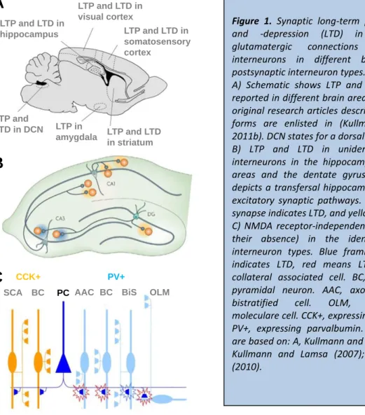
Figure 1. Synaptic long-term potentiation (LTP) and -depression (LTD) in the excitatory glutamatergic connections to GABAergic interneurons in different brain areas and postsynaptic interneuron types.
A) Schematic shows LTP and LTD as has been reported in different brain areas in a rodent. The original research articles describing the plasticity forms are enlisted in (Kullmann and Lamsa, 2011b). DCN states for a dorsal cochlear nucleus.
B) LTP and LTD in unidentified GABAergic interneurons in the hippocampal CA1 and CA3 areas and the dentate gyrus (DG). Schematic depicts a transfersal hippocampal slice with four excitatory synaptic pathways. Blue shading in a synapse indicates LTD, and yellow shows LTP.
C) NMDA receptor-independent LTP and LTD (or their absence) in the identified CA1 area interneuron types. Blue framing of a synapse indicates LTD, red means LTP. SCA, Schaffer collateral associated cell. BC, basket cell. PC, pyramidal neuron. AAC, axo-axonic cell. BiS, bistratified cell. OLM, oriens-lacunosum moleculare cell. CCK+, expressing cholecystokinin.
PV+, expressing parvalbumin. Modified images are based on: A, Kullmann and Lamsa (2011b); B, Kullmann and Lamsa (2007); C, Lamsa et al.
(2010).
specific type of LTP (NMDAR-dependent) among the CA1 area interneurons (Lamsa et al., 2005).
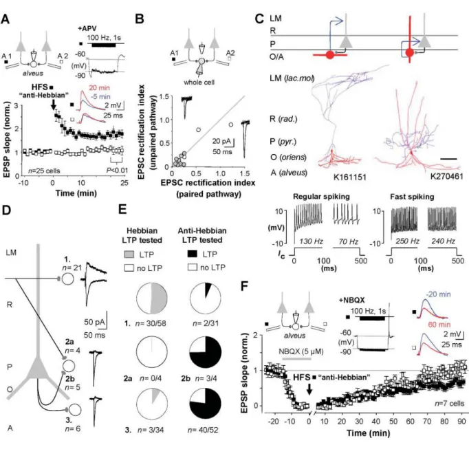
Figure 2. Hippocampal fast-spiking interneurons in the CA1 area exhibit a specific type of synaptic LTP that requires calcium-permeable AMPA receptors (CP-AMPARs) and a different induction pattern than the LTP in pyramidal cells.
A) Application of high-frequency stimulation to a synaptic pathway (black symbols) when a postsynaptic interneuron is hyperpolarized elicits the NMDAR-independent LTP, which is pathway specific (open symbols show control pathway). B) The glutamatergic synapses in the interneurons have CP-AMPARs indicated by EPSC inward rectification (rectification index below 1). C) Two visualized and identified cells showing the LTP; OLM cell and a fast- spiking basket cell. Scale 200 μm D) CP-AMPARs and the NMDAR-independent LTP are common in interneurons located in strata pyramidale (P) and oriens (O), and rare in GABAergic cells in strata radiatum (R) and locunosum- moleculare (LM). E) The NMDAR-dependent and the independent LTP occur in distinct CA1 area interneuron subpopulations. Note that not all tested interneurons show either type of LTP. F) The NMDAR-independent "anti- Hebbian" LTP requires CP-AMPARs. Transient blockade of AMPARs (by NBQX, horizontal bar) during the high- frequency stimulation prevents LTP in interneurons. Image adapted from Lamsa et al. (2007b).
We discovered that when using an associative pre- and postsynaptic discharge pairing protocol (identical to what is commonly used for LTP induction in pyramidal cells), less than half of the tested postsynaptic GABAergic cells showed LTP, while a majority failed to show plasticity. We reported these findings in two separate articles published in Nature Neuroscience (Lamsa et al., 2005) and The Journal of Physiology (Lamsa et al., 2007a), the former describing the phenomenon in interneurons and the latter uncovering its induction mechanism downstream to the NMDAR activation (which we found is different from that existing in pyramidal cells, since in the interneurons a beta isoform of Ca2+/calmodulin- dependent protein kinase II -enzyme is required for LTP, whereas in the CA1 pyramidal cells alpha isoform is necessary for the long-term potentiation).
The bimodal expression pattern of the NMDAR-mediated LTP among the CA1 area interneurons encouraged us to approach an anatomy specialist, prof. Somogyi, and suggest a collaboration project to test whether the plasticity in hippocampal interneurons is associated with anatomically specialized interneuron types. We set a simple hypothesis: testing two common LTP induction protocols, we investigated whether there is a correlation between the plasticity result and an identified postsynaptic interneurons type? This required rigorous post hoc anatomical and immunohistochemical analyses of the electrophysiologically investigated postsynaptic neurons. We first focused on the hippocampal O-LM interneuron type (named as Oriens-Lacunosum Moleculare interneuron because its axon characteristically occupies these layers) (McBain et al., 1994) in the CA1 area to test this idea. The O-LM cell was a good candidate to study this question, because there was already evidence in the literature showing that LTP often occurs in interneurons with soma in stratum oriens layer of the CA1 area (Perez et al., 2001). The O-LM interneuron somata locate in stratum oriens. Indeed, we were able to demonstrate that the O-LM cells consistently show LTP, which is addition was mechanistically different from the LTP in pyramidal cells. We published these findings in two research articles first showing the novel type of LTP occurring in many CA1 area interneurons (but not in pyramidal cells) and then demonstrating that it requires the activation of postsynaptic calcium-permeable AMPA receptors and group I metabotropic glutamate receptors (Lamsa et al., 2007b).
In addition, we demonstrated in the articles that the synapses to interneurons with this type of LTP do not show the conventional "pyramidal cell-like LTP" that requires the glutamatergic
NMDA receptors. Details of the main findings published in Science (Lamsa et al. 2007b) are shown in Figure 2. The second research article published in The Journal of Neuroscience (Oren et al., 2009), was released two years later when I already had moved to Oxford University as an independent research group leader. In that paper we further demonstrated that the O-LM cells consistently exhibit the CP-AMPAR-dependent LTP form.
Both these studies utilized a technically challenging experimental approach – a sequential recording from a postsynaptic interneuron with two separate micropipettes – in which the
Figure 3. Synaptic long-term plasticity in the hippocampal interneurons is specific to an interneuron type.
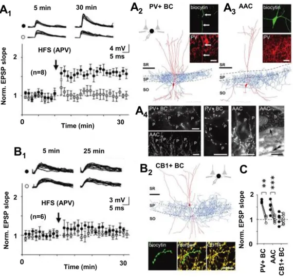
A) The NMDAR-independent LTP exists in PV+ basket (BC) and axo-axonic cells (AAC), but not in B) CCK- expressing interneuron types in the CA1 area. A1) LTP induced by the high-frequency stimulation (HFS) in the presence of NMDAR blocker APV. Black symbols show the HFS-treated pathway, open symbols indicate control pathway. A2) A visualized PV+ BC and A3) an AAC showing the LTP. Scale bar in the cell reconstruction 100 μm, and in the confocal micrographs 20 μm. A4) Confocal images of the CA1 stratum pyramidale (first four from left, scale 20 μm) illustrate the BC and the AAC axon terminals used to identify the cell types. Right: electron micrograph showing AAC synapse (arrow) received by nearby pyramidal cell axon initial segment. Scale 0.25 μm. B1) CCK-expressing CA1 interneurons fail to show long-term plasticity with identical protocol. B2) A visualized sample CCK BC (scale bar 100 μm) with characteristic cannabinoid receptor type 1 (CB1R, scale 10 μm) expression on the axon. C) Summary of baseline-normalized EPSP potentiation after the HFS in the treated (black symbols) pathway versus the control pathway (open symbols) in the three different CA1 interneuron types. Image modified from Nissen et al. (2010).
0
0
postsynaptic cell was first recorded using a perforated patch -method to minimize dilution of the intracellular contents. It was crucial for stable long-lasting recordings that allowed testing subsequent plasticity protocols in the same neuron. For anatomical identification of postsynaptic cell type, the recorded neurons were re-patched with a conventional whole-cell micropipette, and the cells were filled with a marker molecule (neurobiotin or biocytin) for their post hoc visualization. The results played a key role in our review article published the same year in Nature Reviews Neuroscience (Kullmann and Lamsa, 2007).
This project generated two further research articles both released in The Journal of Neuroscience and published with Prof. Somogyi while I was in Oxford. We showed that the CP-AMPAR-dependent LTP occurs not only in O-LM cells but in addition in other neurons expressing parvalbumin (PV) (note: some O-LM cells also exhibit this marker although weakly, see Ferraguti et al., 2004), more specifically basket cells and in axo-axonic cells, and in interneurons with no PV but with neuronal nitric acid synthase (nNOS) (Nissen et al., 2010;
Szabo et al., 2012). On the contrary, this plasticity was absent in the CA1 area interneurons expressing cholecystokinin (CCK) but not PV (Nissen et al., 2010). Key results of these findings are illustrated in Figure 3. In addition, we demonstrated that the O-LM cells and the nNOS- expressing ivy cells both exhibit CP-AMPARs and the CP-AMPAR-dependent LTP because they lack the glutamate AMPA receptor subunit 2 (GluA2) (Szabo et al., 2012). Interestingly, later studies have revealed that interneuron types with CP-AMPARs are mostly derived from the same developmental brain area during early ontogenesis (Akgul and McBain, 2016). Hence, we speculate that the specific type of plasticity is already programmed in the interneurons during early ontogenesis.
4.2. Identified interneuron types show the learning-related long-term plasticity in vivo
Although in vitro slice preparation studies enabled the detailed investigation of interneuron plasticity mechanisms in many identified cell types – because the method easily allows long and stable recording from identified cells – it still remained open whether the same cell types would similarly show plasticity in the intact brain of a living animal. Interestingly, some publications already existed showing indirect evidence for the activity-induced long-term potentiation and -depression in interneurons of the hippocampal CA1 area (Buzsaki and Eidelberg, 1982; Dupret et al., 2013). In these articles, which utilized extracellular recording of
spiking activity of unidentified CA1 area interneurons, it was demonstrated that spike coupling of the presynaptic pyramidal cells (or their fibers) and the interneurons in a rat hippocampus was permanently strengthened or weakened by either a common LTP-induction paradigm (applying repetitive extracellular electrical stimulation, see Buzsaki and Eidelberg, 1982) or following spatial learning tasks (Dupret et al., 2013). However, neither of these studies did or was able to identify the postsynaptic interneuron types for methodological reasons.
Hence, we next investigated the long-term plasticity of synaptic excitatory drive of anatomically identified CA1 area interneurons in vivo. To optimize long-term stability of the recordings, the rats were anaesthetized during experiments (by combination of urethane, xylasine and ketamine). Rather than using a multichannel electrode (Dupret et al., 2013), we studied the interneuron spiking probability with an extracellularly juxtapositioned glass micropipette in response to microelectrode stimulation of afferent glutamatergic projection
A
1A
2B
1B
2C
1C
2D
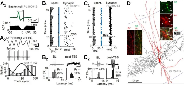
Figure 4. Fast-spiking PV+ basket cells (BCs) show long-term plasticity in a rat hippocampus in vivo. A) PV+ BCs in urethane-anaesthetized rats show characteristic spontaneous firing at low frequency (Klausberger et al., 2003).
A1) Extracellularly recorded BC spikes in the CA1 area exhibit fast kinetics. Spontaneous firing shows no autocorrelative pattern. A2) The spontaneous firing occurs at low frequency and it is characteristically phase- locked to descending phase of the local field potential (LFP) theta oscillation cycle (Klausberger et al. 2003). B-C) High-frequency stimulation (HFS) induces either LTD- or LTP-like change in the single pulse stimulation-evoked spiking probability of the PV+ BCs. B1) A BC fires (“synaptic”) with a 5-10 ms delay to the contralateral site hippocampal CA1 stimulation, and the HFS induces a long-term decrease in the evoked spike probability. Note that spontaneous firing activity (shown left with bars) remains unaltered. B2) Individual traces and histograms summarize the reduced single shock-evoked basket cell firing probability after the HFS. Data shown in baseline and 30 min following the HFS. Scale 0.2 mV, 5 ms. C1-C2) Similar experiment from another PV+ BC showing long- term potentiation of the evoked spike probability. D) One recorded BC (with LTP) illustrated with immunoreactions. NB, neurobiotion. PV, parvalbumin. CB1R, cannabinoid receptor type 1. Image adapted from Lau et al. (2017).
fibers. The juxtacellular glass microelectrode recording method allowed us to label the studied cells with neurobiotin for post hoc anatomical analyses and identify interneuron type (Lau et al., 2017). We focused the study on the postsynaptic fast-spiking PV+ basket cells and the non fast-spiking nNOS+ (immunopositive for neuronal nitric oxide synthase) ivy cells, which we and others had previously shown in the in vitro slice preparation to exhibit robust LTP by the high-frequency stimulation (Alle et al., 2001; Nissen et al., 2010; Szabo et al., 2012;
Campanac et al., 2013; Le Roux et al., 2013).
Similar to the results by Dupret et al. (2013) as well as Buzsaki and Eidelberg (1982), we found that the fast-spiking CA1 interneurons can generate either LTP or LTD following high- frequency glutamatergic fiber activity. Interestingly, when identifying the interneuron types we found that both the PV+ basket cells as well as the ivy cells could generate either LTP or LTD in these conditions. Because the results differed from what we had previously observed in slice preparations (in vitro the LTP was consistently generated in both of these CA1 interneuron types), we hypothesized that the direction of plasticity in vivo might be regulated by the underlying brains state in the anaesthetized animal defined by the local field potential oscillation pattern at the time when the plasticity was induced (see Kullmann and Lamsa, 2007). However, we found that neither did the occurrence of predominant theta (4-8 Hz) oscillation or slow wave (1 Hz) activity manage to explain whether the LTP or the LTD was generated in these interneurons (Lau et al., 2017).
Hence, we suggested that the complex plasticity results in vivo could possibly emerge from variable underlying modulatory effects of monoaminergic, cholinergic or endocannabinoid system in the experiments. Indeed, such modulations have been reported with pharmacological agents in slice preparations (Peterfi et al., 2012; Griguoli et al., 2013), and in intact brain such effects could be generated endogenously. Our results in vivo rat hippocampus were first reported in Brain Structure and Function (Lau et al., 2017) and the key findings are illustrated in Figure 4.
4.3. Human cortical microcircuits show evolutionarily conserved interneuron plasticity with specific features
We asked whether the interneuron plasticity reported in the rodent occurs similarly in the human brain, or if there are specific plasticity features in the human cortex not present in a rat or mice? The question is highly relevant, since recent studies have demonstrated that the human neocortical microcircuits are not identical to rodents but show various specializations in the intrinsic neuronal and synaptic functions (Molnar et al., 2008; Blazquez-Llorca et al., 2010; Defelipe, 2011; Testa-Silva et al., 2014; Eyal et al., 2016; Molnar et al., 2016; Sousa et al., 2017). Many of these adaptations are either enhancing the temporal signal processing or the spatial propagation of neuronal activity in the human neocortex. There is an emerging idea that some of these features may have evolved during the human evolution to enhance the brain computational power (DeFelipe et al., 2002; Lourenco and Bacci, 2017; Sousa et al., 2017). However, it had remained unknown whether the plasticity of GABAergic inhibitory circuits also showed specific functional features in the human cortex.
We investigated the question in acute slices prepared from neocortical tissue samples resected in a deep brain oncology or aneurism surgery in order to have the access to subcortical pathological target (Molnar et al., 2008; Szegedi et al., 2017). Such samples represent the closest to healthy control tissue, since the patients are typically operated with a short delay from the first symptoms and they lack systematic and persistent pre-medication (unlike the epilepsy patients) (Lourenco and Bacci, 2017). Importantly, the resected neocortical tissue samples in the operations locate far from the pathological target. For clarity, we have systematically reported in our studies the operated patient age, gender and their primary clinical diagnosis leading to the operation (Szegedi et al., 2016; Szegedi et al., 2017).
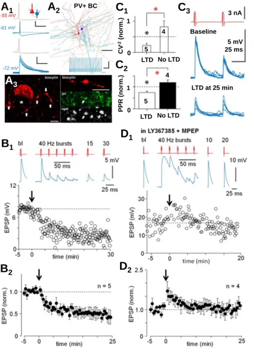
Whole-cell recordings from the layer 2-3 PV+ basket cells revealed a robust LTD in their glutamatergic afferents by the high-frequency bursting of the fibers. The LTD was similarly generated by extracellular stimulation in a rat and in the human (Szegedi et al. 2016). In both cases, the LTD showed presynaptic expression site and it was blocked by antagonist of the group I metabotropic glutamate receptors. The results are well in line with previous reports in rodents (Yazaki-Sugiyama et al., 2009) showing that metabotropic receptors mediate the LTD in the fast-spiking cortical interneurons (Lu et al., 2007; Peterfi et al., 2012).
Yet, about 15 % of pyramidal cell to fast-spiking interneuron connections in the human neocortex layer 2-3 exhibit very large monosynaptic glutamatergic EPSPs (VLEs, average amplitude 13 mV) elicited by single pyramidal cell spike (Molnar et al., 2008; Komlosi et al., 2012; Szegedi et al., 2016; Szegedi et al., 2017). The VLEs are often suprathreshold hence representing a microcircuit feature not occurring in a rat and being possibly specific to the human neocortex (Molnar et al., 2016). Importantly, we found that the strong synaptic VLE- connections are able to trigger the mGluR-dependent LTD independently; a high frequency bursting activity of just single pyramidal cell triggers the LTD, which in a rat required simultaneous co-activation of multiple glutamatergic fibers. The LTD in the single cell connections in the human is shown in detail in Figure 5. In our research article published in
A
1A
2A
3C
1C
2C
3B
1B
2D
1D
2Baseline
LTD at 25 min 5 mV 25 ms 3 nA 4
5
5 4
PPR (norm.)
1
0 0 1
CV-2(norm.)
-55 mV -61 mV
-72 mV
LTD
LTD
No LTD
No LTD
PV+ BC Figure 5. Fast-spiking PV+ basket cells (BCs) show synaptic long-term plasticity in human neocortex with specific features. Unlike rodents, the human neocortex contains very strong glutamatergic synapses that connect the layer 2-3 pyramidal cells (PCs) specifically to GABAergic interneurons (Molnar et al., 2008). These VLE connections (Very Large EPSPs) are based on synapses with multivesicular transmitter release (Molnar et al., 2016) and the EPSPs evoked in the BCs are often suprathreshold (Szegedi et al., 2017). A1) Visualized PC (red) to PV+ BC (blue; scale 40 mV, 20 ms) connection with a suprathreshold VLE (top). Bottom: post- synaptic hyperpolarization reveals the VLE.
Blue trace scale 4 mV, 20 ms. A2) Illustration of the cell pair (PC red, BC blue). Scale 100 μm. Inset shows the BC high-frequency firing pattern (scale 60 mV, 100 ms). A3) The BC axon (used to identify the cell) in the layer 2- 3. Left: The boutons (arrow) are arranged around a nearby cell soma (asterisk).
Confocal micrographs show the boutons are immunopositive for vgat and PV. Right: Scale bar 5 μm. B) PC firing with high-frequency bursts (5 pulses at 40 Hz, 40 repeats) induces LTD in VLEs synapses. B1) A sample experiment. B2) Mean and s.e.m. of 5 experiments. C) LTD has presynaptic expression site indicated by the EPSP amplitude coefficient of variation (C1) and paired-pulse ratio (C2) in LTD. C3) Paired- pulse EPSPs in baseline and in LTD. D) LTD is blocked by antagonist of group I mGluRs. D1) A sample experiment. D2) Mean and s.e.m.
of 4 experiments. Images adapted from Szegedi et al. (2016) and (2017).
PLoS Biology, we speculated that the VLE synapses – with their multivesicular glutamate release (Molnar et al., 2016) – may be sufficient to activate perisynaptic mGluRs critical for the LTD in postsynaptic PV+ cells. In the rat neocortex the mGluR activation requires spill-over glutamate released from several simultaneously active adjacent synapses (Rusakov et al., 1999).
Altogether, we found that similar plasticity – in terms of its induction by the high-frequency afferent fiber bursting and the pharmacological sensitivity – is induced in a rat and in the human neocortex glutamatergic synapses to PV+ basket cells. However, the strong VLE connections between two individual neurons in the human can trigger the plasticity independently. The results suggest an evolutionarily conserved mechanism for the interneuron plasticity in the mammalian neocortex, but reveal microcircuit level specializations between the species in the learning-related interneuron plasticity.
4.4. Interneuron long-term plasticity produces permanent changes in local network activity both in the rodent and in the human
Since GABAergic interneurons play a pivotal role in organizing the space and the time of cortical ensemble activity (see Introduction), we finally investigated whether the interneuron plasticity was sufficient to alter the activity in neuronal networks. First, we investigated this in the rodent hippocampus inducing the long-term plasticity in the CA1 area circuitry so that LTP was either restricted to postsynaptic pyramidal cells, or that it in parallel also occurred in the GABAergic interneurons.
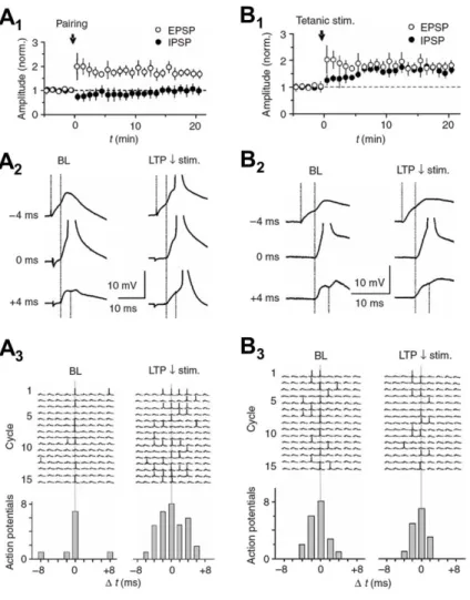
The results summarized in Figure 6 show that LTP in both the pyramidal cells and in the disynaptic GABAergic inhibition (i.e. LTP in interneurons) is required to preserve the high temporal fidelity of the CA1 pyramidal cells' input-output transformation (meaning the temporal accuracy how synaptic inputs from the CA3 area are integrated in the CA1 cells to generate their action potential firing) following CA3 area high-frequency bursting (Lamsa et al., 2005). In other words, we demonstrated that LTP in GABAergic interneurons is needed to preserve fast co-incidence detection in the excitatory signal transmission from the CA3 to CA1 area in the face of learning and LTP in pyramidal neurons. The results stress the importance of GABAergic interneuron long-term plasticity during hippocampal learning processes.
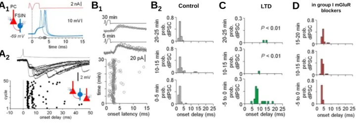
Correspondingly, in the human neocortex the layer 2-3 single pyramidal cell spike -evoked network activity (called complex events or the ensembles) allowed us to test if the plasticity in pyramidal cell-to-interneuron synapses was able to modify the evoked network activity.
Indeed, we found that in parallel with the mGluR-dependent LTD in the fast-spiking PV+
basket cells, there was a change in the complex event pattern evoked; the PV+ basket cells, which are characteristically activated at the earliest phase of the complex events (Szegedi et al., 2017), were silenced in the evoked ensembles by the mGluR-dependent LTD (Szegedi et al., 2016). These results show that also in human neocortex the long-term plasticity in PV+
interneurons leaves a permanent imprint in the network activity pattern evoked in the local circuitry.
Figure 6. Synaptic plasticity in hippocampal interneurons permanently modifies input integration in the CA1 area pyramidal cells (PCs). A) LTP restricted between PC synapses compromises temporal fidelity of the input- output operation in the CA1 circuit. A1) LTP in single- shock (extracellular stimulation) -evoked monosynaptic EPSP, but not in disynaptic IPSP in a CA1 pyramidal cell.
The LTP was induced by low-frequency (1 Hz) stimulation of the pathway paired with postsynaptic depolarization (timing indicated by “pairing”). A2) Temporal summation of two such pathways. In baseline, the PC generates action potential only when the two input pathways are active precisely at the same time (lag 0 ms). Following the LTP, the spike generation occurs in a wider time window.
A3) Consecutive cycles pairing the two pathways with different lags (from -8 ms to + 8 ms) in baseline and following the LTP. Histograms summarize widening of the time window for spike generation after the LTP. B) Parallel LTP in monosynaptic EPSP and disynaptic IPSP preserves narrow time window for spike generation in the CA1 pyramidal cells. B1) High-frequency extracellular stimulation (“tetanic stim.”) results in LTP of both the EPSP and the disynaptic IPSP. B2) Sample traces showing temporal integration of two input pathways with EPSP and disynaptic IPSP. Both in the baseline and LTP, the spike generation only occurs when the two input pathways are activated with zero lag. A3) Fifteen consecutive cycles in baseline and in the LTP in one experiment showing the input integration in a CA1 PC with different lag in their activation (from -8 ms to + 8 ms). Adapted from Lamsa et al. (2005).
5. Conclusions
Various laboratories during the past decade have shown that learning-related and activity- induced long term plasticity occurs not only in the cortical pyramidal cells but in addition in the GABAergic inhibitory interneurons. Our laboratory has contributed to this endeavor showing that glutamatergic excitatory fibers undergo long-term plasticity in specialized anatomically identified cortical interneuron types in a rodent as well as in the human. We have demonstrated that same cell types that exhibit the plasticity in vitro slice preparations, do also show LTP and LTD in vivo rodent brain. Importantly, in these interneurons the cellular mechanisms and the induction pattern of LTP often differ from that know in pyramidal cells.
The interneuron-specific plasticity mechanisms may reflect their different physiological firing pattern in learning processes, such as in the hippocampus during spatial learning tasks (Klausberger and Somogyi, 2008).
6. References
Akgul G, McBain CJ (2016) Diverse roles for ionotropic glutamate receptors on inhibitory interneurons in developing and adult brain. The Journal of Physiology 594:5471-5490.
B1 B2 Control C LTD D
A1
A2
in group I mGluR blockers
Figure 7. Long-term plasticity in interneurons alters the neuronal ensemble activation in the human neocortex. A1) Single spike in layer 2-3 pyramidal cell (PC) elicits a large amplitude EPSP (VLE) in fast-spiking interneuron (FSIN). A2) In parallel a PC spike triggers a complex event with di- and polysynaptic IPSPs in a nearby pyramidal cell. Plot shows 50 consecutive complex events triggered by the PC. Note that first wave of IPSPs is similarly time-locked to PC spike as the basket cell firing in A1. B1) The first wave of inhibition in complex events plotted for 30 consecutive events (recorded in voltage clamp). B2) Histogram plotting the first IPSCs in control conditions in the 30 cycles. C) Same high-frequency PC bursting pattern that elicits the LTD in VLE synapses onto basket cells, causes a permanent suppression of the first wave of the complex event. Note that the time-locked early IPSC (bottom) disappears after the PC bursting (middle and upper histograms). D) The suppression of the early complex event activity is blocked by group I mGluR antagonist, similar to the LTD in PC to BC synapses shown in Figure 5. Images adapted from Szegedi et al. (2016) and (2017).
Alle H, Jonas P, Geiger JR (2001) PTP and LTP at a hippocampal mossy fiber-interneuron synapse. Proceedings of the National Academy of Sciences of the United States of America 98:14708-14713.
Ascoli GA et al. (2008) Petilla terminology: nomenclature of features of GABAergic interneurons of the cerebral cortex. Nature Reviews Neuroscience 9:557-568.
Bartos M, Alle H, Vida I (2011) Role of microcircuit structure and input integration in
hippocampal interneuron recruitment and plasticity. Neuropharmacology 60:730-739.
Blazquez-Llorca L, Garcia-Marin V, DeFelipe J (2010) GABAergic complex basket formations in the human neocortex. The Journal of Comparative Neurology 518:4917-4937.
Buzsaki G, Eidelberg E (1982) Direct afferent excitation and long-term potentiation of hippocampal interneurons. Journal of Neurophysiology 48:597-607.
Buzsaki G, Llinas R (2017) Space and time in the brain. Science 358:482-485.
Camire O, Topolnik L (2014) Dendritic calcium nonlinearities switch the direction of synaptic plasticity in fast-spiking interneurons. The Journal of Neuroscience 34:3864-3877.
Campanac E, Gasselin C, Baude A, Rama S, Ankri N, Debanne D (2013) Enhanced intrinsic excitability in basket cells maintains excitatory-inhibitory balance in hippocampal circuits. Neuron 77:712-722.
Cowan AI, Stricker C, Reece LJ, Redman SJ (1998) Long-term plasticity at excitatory synapses on aspinous interneurons in area CA1 lacks synaptic specificity. Journal of
Neurophysiology 79:13-20.
Defelipe J (2011) The evolution of the brain, the human nature of cortical circuits, and intellectual creativity. Frontiers in Neuroanatomy 5:29.
DeFelipe J, Alonso-Nanclares L, Arellano JI (2002) Microstructure of the neocortex:
comparative aspects. Journal of Neurocytology 31:299-316.
Dupret D, O'Neill J, Csicsvari J (2013) Dynamic reconfiguration of hippocampal interneuron circuits during spatial learning. Neuron 78:166-180.
Eyal G, Verhoog MB, Testa-Silva G, Deitcher Y, Lodder JC, Benavides-Piccione R, Morales J, DeFelipe J, de Kock CP, Mansvelder HD, Segev I (2016) Unique membrane properties and enhanced signal processing in human neocortical neurons. eLife 5.
Ferraguti F, Cobden P, Pollard M, Cope D, Shigemoto R, Watanabe M, Somogyi P (2004) Immunolocalization of metabotropic glutamate receptor 1alpha (mGluR1alpha) in distinct classes of interneuron in the CA1 region of the rat hippocampus. Hippocampus 14:193-215.
Galvan EJ, Cosgrove KE, Barrionuevo G (2011) Multiple forms of long-term synaptic plasticity at hippocampal mossy fiber synapses on interneurons. Neuropharmacology 60:740- 747.
Galvan EJ, Perez-Rosello T, Gomez-Lira G, Lara E, Gutierrez R, Barrionuevo G (2015) Synapse- specific compartmentalization of signaling cascades for LTP induction in CA3
interneurons. Neuroscience 290:332-345.
Galvan EJ, Cosgrove KE, Mauna JC, Card JP, Thiels E, Meriney SD, Barrionuevo G (2010) Critical involvement of postsynaptic protein kinase activation in long-term potentiation at hippocampal mossy fiber synapses on CA3 interneurons. The Journal of Neuroscience 30:2844-2855.
Griguoli M, Cellot G, Cherubini E (2013) In hippocampal oriens interneurons anti-Hebbian long-term potentiation requires cholinergic signaling via alpha7 nicotinic acetylcholine receptors. The Journal of Neuroscience 33:1044-1049.
Klausberger T, Somogyi P (2008) Neuronal diversity and temporal dynamics: the unity of hippocampal circuit operations. Science 321:53-57.