European Journal of Medical Genetics 63 (2020) 104027
Available online 3 August 2020
1769-7212/© 2020 The Authors. Published by Elsevier Masson SAS. This is an open access article under the CC BY license
(http://creativecommons.org/licenses/by/4.0/).
Clinical and genetic findings in Hungarian pediatric patients carrying chromosome 16p copy number variants and a review of the literature
Anna Lengyel
a,*, ´ Eva Pinti
a, Henriett Pik ´ o
b, Eszter J ´ avorszky
c, Dezs ˝ o David
d,
Mariann Tihanyi
e, Eva G ´ onczi ¨
a, Eszter Kiss
a, Zsuzsa T ´ oth
a, K ´ alm ´ an Tory
c, Gy orgy Fekete ¨
a, Ir ´ en Haltrich
aaII Department of Pediatrics, Semmelweis University, Budapest, Hungary
bI Department of Internal Medicine, Semmelweis University, Budapest, Hungary
cI Department of Pediatrics, Semmelweis University, Budapest, Hungary
dDepartment of Human Genetics, National Health Institute Dr. Ricardo Jorge, Lisbon, Portugal
eDepartment of Genetics, Zala County Hospital, Zalaegerszeg, Hungary
A R T I C L E I N F O Keywords:
Copy number variations Chromosome 16p
Neurodevelopmental disorders Behavioral disorders Intellectual disability
A B S T R A C T
The short arm of chromosome 16 (16p) is enriched for segmental duplications, making it susceptible to recurrent, reciprocal rearrangements implicated in the etiology of several phenotypes, including intellectual disability, speech disorders, developmental coordination disorder, autism spectrum disorders, attention deficit hyperac- tivity disorders, obesity and congenital skeletal disorders. In our clinical study 73 patients were analyzed by chromosomal microarray, and results were confirmed by fluorescence in situ hybridization or polymerase chain reaction. All patients underwent detailed clinical evaluation, with special emphasis on behavioral symptoms. 16p rearrangements were identified in 10 individuals. We found six pathogenic deletions and duplications of the recurrent regions within 16p11.2: one patient had a deletion of the distal 16p11.2 region associated with obesity, while four individuals had duplications, and one patient a deletion of the proximal 16p11.2 region. The other four patients carried 16p variations as second-site genomic alterations, acting as possible modifying genetic factors. We present the phenotypic and genotypic results of our patients and discuss our findings in relation to the available literature.
1. Introduction
Congenital developmental anomalies and neurodevelopmental dis- orders (NDDs) are frequently caused by recurrent, reciprocal micro- deletions and microduplications (Deshpande and Weiss, 2018;
Kaminsky et al., 2011). These genomic disorders arise from non-allelic homologous recombination (NAHR) following the misalignment of segmental duplications (SDs) (Antonacci et al., 2010; Chen et al., 2014;
Linardopoulou et al., 2005; Marques-Bonet and Eichler, 2009; Shaw and Lupski, 2004; Stankiewicz and Lupski, 2002; Tai et al., 2016). Chro- mosome 16 has one of the highest percentages [(approximately 10% of its sequence, ~7.8 megabase pairs (Mb) (Martin et al., 2004)] of SDs amongst the human chromosomes, clustered along the short arm (16p).
This predisposes 16p to rearrangements, and the resultant recurrent copy number variations (CNVs) are implicated in various genomic dis- orders (Cooper et al., 2011; Girirajan et al., 2012; Itsara et al., 2009;
Kanduri et al., 2016; Sahoo et al., 2011; Sanders et al., 2011; Stefansson et al., 2014; Weiss et al., 2008).
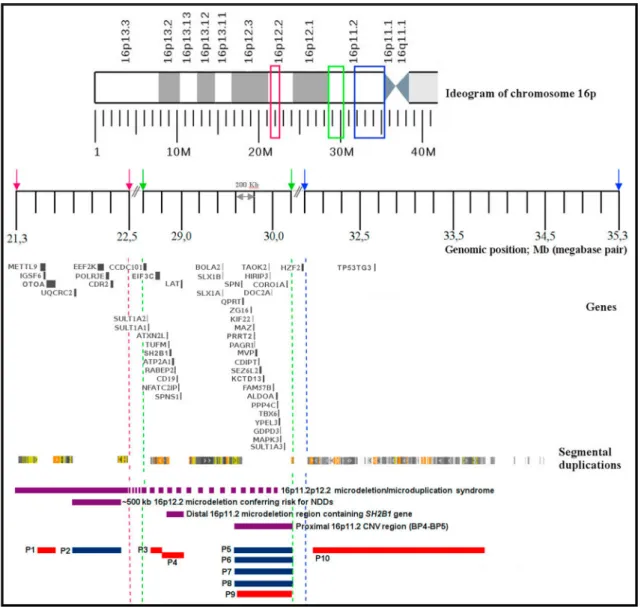
The most frequently affected recurrent CNV region is the proximal breakpoint 4–5 (BP4–BP5) region of 16p, associated with a micro- deletion syndrome (OMIM #611913) and a reciprocal microduplication syndrome (OMIM #614671). The region is approximately 600 kilobase pairs (Kb) in size, located from genomic position ~29.6 to ~30.2 Mb (all genomic positions in the manuscript are according to assembly GRCh37/hg19). In close proximity lies a 95 Kb segment encompassing BOLA2 (OMIM *613183; a gene suspected to be relevant in early em- bryonic development), which has undergone Homo sapiens-specific expansion relatively recently in human evolution, and has been sug- gested to predispose the BP4–BP5 region to recurrent rearrangement (Nuttle et al., 2016). The CNVs are associated with a wide variety of neuropsychiatric phenotypes, growth abnormalities, skeletal abnor- malities and other, less frequent congenital anomalies (Table 1). CNVs of
* Corresponding author. Semmelweis University, II. Dept. of Pediatrics, Cytogenetic Laboratory, 1094, Budapest, Tuzolto utca 7-9, Hungary.
E-mail address: lengyel.anna1@med.semmelweis-univ.hu (A. Lengyel).
Contents lists available at ScienceDirect
European Journal of Medical Genetics
journal homepage: www.elsevier.com/locate/ejmg
https://doi.org/10.1016/j.ejmg.2020.104027
Received 7 January 2020; Received in revised form 10 July 2020; Accepted 25 July 2020
this region were initially ascertained in autism spectrum disorder (ASD) populations: an estimated 0.28–1% of patients carry BP4–BP5 CNVs (Fernandez et al., 2010; Hanson et al., 2015; Kumar et al., 2008; Walsh and Bracken, 2011; Weiss et al., 2008), and subsequent large studies identified the recurrent CNVs in 0.6–0.7% of patients with NDDs (Rosenfeld et al., 2010; Shinawi et al., 2010; Steinman et al., 2016). In large 16p11.2 CNV populations 90% or more of carrier children were found to have neuropsychiatric diagnoses; the prevalence of ASD has been reported between 15 and 25%, and in most cohorts the frequency of autistic-like features is even higher (Green Snyder et al., 2016; Han- son et al., 2015). The proximal CNVs are furthermore associated with developmental coordination disorder (DCD), specific speech disorders, epilepsy, and an increased prevalence of sacral dimples (often with atypical features, which may signify the presence of occult spinal dys- raphism) (D’Angelo et al., 2016; Green Snyder et al., 2016; Hanson
et al., 2015, 2010; Owen et al., 2018; Shinawi et al., 2010; Steinman et al., 2016; Zufferey et al., 2012). Comparing the reciprocal CNVs, duplication carriers are more likely to have tremor, attention deficit hyperactivity disorder (ADHD) and other behavioral problems (aggres- sion, outbursts, etc.), and they have an increased risk for psychosis and severe intellectual disability (ID). Deletion patients, on the other hand, have a higher incidence of ASD and speech and language disorders (Giaroli et al., 2014; Green Snyder et al., 2016; Hanson et al., 2015;
Shinawi et al., 2010; Steinman et al., 2016). The prevalence of behav- ioral disorders shows consistent increase with age in both groups (Ber- nier et al., 2017). Furthermore, the reciprocal CNVs cause mirror phenotypes in terms of body mass index (BMI), head circumference (HC) and brain volume: deletion carriers present with obesity, significantly increased HC and global increased brain size; meanwhile duplication carriers have a tendency towards being underweight and microcephalic, Table 1
Main phenotypic features of the proximal (BP4–BP5) 16p11.2 rearrangements.
Phenotype Proximal 16p11.2 duplication Proximal 16p11.2 deletion
Frequency P5 P6 P7 Phenotype Frequency P9
Growth
Short stature/Reduced BMI/Failure to thrive in infancy VFHPO – – – Obesity OHPO +
Head and neck
Microcephaly 13–17%a – – – Macrocephaly 17%a –
Sparse eyelashes and/or eyebrows VFHPO – + + Hypertelorism OHPO –
Hypertelorism VFHPO – + + Other minor abnormalities of the eyes variable –
Other minor abnormalities of the eyes variable + – – Eye convergence difficulties 11%a –
Eye convergence difficulties 20%a + – – Broad forehead FHPO –
Microtia VFHPO – – – Malar flattening FHPO –
Other minor abnormalities of the ears variable + + + Other facial minor anomalies variable –
Thin upper lip vermillion VFHPO – – –
Other facial minor anomalies variable + + +
Skeletal abnormalities
Scoliosis OHPO – – – Scoliosis OHPO –
Arachnodactyly VFHPO – – – Hand polydactyly OHPO –
Other abnormalities of the fingers and/or toes variable + + – Other abnormalities of the fingers and/or toes variable +
Neurology and behavior
Intellectual disability 30.5%b/40%c + + + Intellectual disability 10%d +
Motor Delay VFHPO + + + Global Developmental Delay VFHPO +
Speech Delay 32%c + + + +
Speech articulation difficulties 30%a/19%c – n.a. n.a. Speech articulation difficulties 79%a –
Phonological Processing Disorder 56%d –
Developmental Coordination Disorder 47%c – – – Developmental Coordination Disorder 58%d –
Abnormal agility 25%a – – – Abnormal agility 47%a –
Muscular hypotonia ~40%a + + + Muscular hypotonia ~50%a –
Muscle weakness 23–32%a + – – Muscle weakness 17–22%a –
Tremor 18–43%a – – – Tremor 5–8%a –
Hyperreflexia 32%a – – – Hyperreflexia 13%a –
Hyporeflexia 31%a – – – Hyporeflexia 48%a –
Seizures/Epilepsy 26–29%a – – + Seizures/Epilepsy 22–27%a –
Neuroimaging abnormalities (EEG/CT/MRI) 40/31/55%a + n.a. n.a. Neuroimaging abnormalities (EEG/CT/MRI) 54/28/26%a –
Reduced brain volume + n.a. n.a. Overgrowth in posterior fossa –
Thinner corpora callosa – n.a. n.a. Thicker corpora callosa –
Ventriculomegaly + n.a. n.a. Chiari I malformation –
Attention Deficit Hyperactivity Disorder 24%c – – – Attention Deficit Hyperactivity Disorder 19%d –
Autism Spectrum disorder 19%c – + + Autism Spectrum disorder 26%d –
Anxiety Disorder 18%c – – – Anxiety Disorder 6%d –
Mood Disorder OHPO – – – Mood Disorder +
Additional features
Caf´e-au-lait spots 31%a – – – Caf´e-au-lait spots 30%a –
Sacral dimple 28%a – – – Sacral dimple 34%a –
Congenital diaphragmatic hernia OHPO – – – Congenital diaphragmatic hernia OHPO –
Heart defects infrequent – – – Heart defects OHPO –
Developmental differences of the urinary tract infrequent – – – Developmental differences of the urinary tract +
Abnormalities of the digestive system infrequent + – – Abnormalities of the digestive system OHPO –
The phenotypic features of the presented patients are listed. P5 is currently 3 years old, therefore evaluation of developmental and behavioral symptoms is subject to change. n.a., not available; BMI, body mass index; EEG, electroencephalography; MRI, magnetic resonance imaging; CT, computer tomography; HPO, Human Phenotype Ontology (https://hpo.jax.org/app/); VFHPO, listed as very frequent (83–99%) in HPO; FHPO, listed as frequent (30–79%) in HPO; OHPO, listed as occasional (5–29%) in HPO.
aSteinman et al. (2016).
b D’Angelo et al. (2016).
cGreen Snyder et al. (2016).
dHanson et al., 2015.
and they have reduced brain volume measurements (Bijlsma et al., 2009; Jacquemont et al., 2011; Qureshi et al., 2014; Shinawi et al., 2010; Steinman et al., 2016; Tabet et al., 2012; Walters et al., 2010). The deletion is more often associated with abnormally shaped, thicker corpora callosa, overgrowth in the posterior fossa, Chiari I malforma- tions and tonsillar ectopia, whereas duplication carriers are more likely to have thinner corpora callosa, decreased white matter volume and ventriculomegaly (Owen et al., 2018).
Another recurrent region is the distal BP2–BP3 16p11.2 region, which is approximately 220 Kb spanning ~28.8–29.0 Mb. Phenotypes associated with the microdeletion include obesity, generalized over- growth, various NDDs [ID, global developmental delay (DD), delayed speech and language development, ASD, seizures, ADHD], and less often congenital anomalies of other organ systems (Bachmann-Gagescu et al., 2010; Barge-Schaapveld et al., 2011; Bochukova et al., 2010). A ten- dency towards increased HC was also observed, along with mirroring BMI and HC reduction in the reciprocal duplication (Loviglio et al., 2017b).
Our clinical study focused on a cohort of 73 Hungarian children referred to genetic counseling with the combination of non-syndromic minor craniofacial anomalies, mild to moderate ID/DD, and other var- iable neuropsychiatric symptoms. All were analyzed by array compar- ative genomic hybridization (array CGH) after various other methods failed to uncover genetic etiologies. A surprisingly large portion of our cohort (9/73, and an additional parent) proved to carry CNVs of the short arm of chromosome 16, therefore we will focus on this subgroup of patients below.
2. Methods
Karyotypes were determined for all patients by analysis of 20 Giemsa-stained metaphases each from standard 72-h peripheral blood lymphocyte cultures. Array CGH was performed on Roche/NimbleGen system [NimbleGen Array (CGX 1.4 M); NimbleGen MS 200 Microarray Scanner (Roche NimbleGen Inc., Madison, WI, USA); patients P1–P4, P10]; on Agilent qChip Post microarray [60K; Genomic Workbench 7.0 analysis software; P5 and P9]; and on Agilent 180K oligo-array [Amadid 023363; Genomic Workbench Lite Edition 6.5 analysis software (Agilent Technologies, Santa Clara, CA, USA); P6–P8]. Array CGH results were validated by FISH using 16p11.2 KCTD13 548 kb and 16p11.2 ATXN2L- LAT probes (Agilent SureFISH) in 100–100 metaphase and interphase cells; or by polymerase chain reaction (PCR). Deletion/duplication of 16p11.2 (genomic positions 29.6–30.2 Mb) was confirmed by quanti- tative multiplex PCR of short fluorescent fragments (QMPSF) analysis with the primers listed in Supplementary File 1, according to the pro- tocol of NPHP1-QMPSF (Javorszky et al., 2017) with modifications (Carrington et al., 2015). Copy numbers were calculated based on the normalized peak areas following intra- and inter-sample normalization (Supplementary File 2). Parental origin was determined by linkage analysis, carried out with microsatellite markers between genomic po- sitions 31.3 and 35.2 Mb of region 16p11.2. Fragment analysis was performed on ABI 3500 Genetic Analyzer (Applied Biosystems, Foster City, CA, USA), and results were evaluated using Peak Scanner Software 2 (Thermo Fischer Scientific, Waltham, MA, USA).
3. Clinical descriptions
Patient 1 [P1; DECIPHER (Database of Chromosomal Imbalance and Phenotype in Humans Using Ensembl Resources) #411579] was referred to genetic counseling with a clinical diagnosis of ASD when he was 10 years old. His perinatal anamnesis was unremarkable. Somatic and psychomotor development was normal until the age of 2 years, at which time he presented sudden and striking developmental regression with the complete loss of expressive speech. He has macrocephaly and a tendency towards self-injurious behavior. Neurologic examination included a negative EEG and brain MRI. Family history is negative for
NDDs and congenital anomalies.
Patient 2 (P2) was referred at the age of three years with psycho- motor delay, feeding difficulties, loss of speech, generalized muscular hypotonia and hypoplasia of the musculature, ataxia and dyssynergia, stereotypy and bruxism. Her perinatal anamnesis was normal. Stra- bismus, sparse eyebrows and eyelashes, epicanthus, short neck, low posterior hairline and joint hypermobility were noted upon physical examination. Brain MRI showed signs of abnormal myelination. EEG, EMG and ENG tests were negative, as were metabolic screenings, MLPA analyses for Prader-Willi and Rett syndromes, and direct sequencing of MECP2 gene. Family history is negative.
Patient 3 (P3, DECIPHER #411594; Fig. 1A) was diagnosed at the age of three years with delayed speech and language development (vocabulary consisted of about five words, receptive speech was less impaired), minor anomalies, polydactyly and gait imbalance. He was born at term with normal parameters from a pregnancy complicated by oligohydramnios. He had a high-pitched cry as an infant, and feeding difficulties due to severe muscular hypotonia. Blepharophimosis, small and low-set ears with abnormal morphology, single transverse palmar crease, joint hypermobility, genu valgum and pes planus were noted. He has mild conductive hearing impairment in his right ear. Family history is negative.
Patient 4 (P4, DECIPHER #411578; Fig. 1B) was referred at two years of age with the suspicion of Prader-Willi syndrome due to psy- chomotor delay, generalized muscular hypotonia, small hands and feet, genu varum, strabismus, severe obesity and polyphagia. She was born after 39 weeks of gestation complicated by maternal diabetes, and impending fetal asphyxia necessitating C-section. She weighed 5500 g and had Apgar scores of 7/8. At age two years her BMI was 32.8 kg/cm2, while her current BMI is 45.7 kg/cm2. Her weight and BMI curves have steadily been well above the age and sex matched 97th percentile.
Additional findings were elevated serum triglyceride levels and normal bone density measurements. Psychological evaluation using the Brunet- L´ezine test estimated one month delay in gross motor development, two months delay in hand-eye coordination, 13 months delay in social in- teractions and 17 months delay in speech development. Her develop- mental quotient was 74 at 25 months of age. FISH analyses targeting SNRPN gene and the 1p36 deletion, as well as methylation analysis for Prader-Willi syndrome, were negative. Both parents are overweight.
Patient 5 (P5, DECIPHER #411596) had normal birth parameters and perinatal adaptation. Bilateral cryptorchidism was observed upon birth. He had apneic episodes and feeding difficulties in infancy, but exhibited average psychomotor development until he was 5 months old, when he lost the ability to hold his head, roll over and crawl. Around this time he was transported to a hospital due to dehydration, generalized lymphoedema, hepatomegaly, fluctuations in consciousness, horizontal nystagmus and mild facial asymmetry. Brain CT revealed cerebral at- rophy and ventriculomegaly, but extensive examinations ultimately failed to explain his acute condition. Clinical genetic consultation noted a wide and depressed nasal bridge, epicanthus, high palate, facial asymmetry, clinodactyly of the 3.-5. toes on both feet, frontal bossing and low-set, protruding ears. P5 is currently 3 years old with global DD and absent speech. He has yet to undergo official psychological evalu- ation, but the mother reports a recent change in behavior (he stopped reacting to his name, has abnormal temper tantrums, started presenting abnormal eating behavior, is not interested in toys and has a short attention span). P5’s parents are healthy, but the paternal grandmother and uncle have abnormal aggressive and impulsive behavior.
Patients 6 and 7 are a pair of brothers born to a non-consanguineous couple. The index patient was the younger child, Patient 6 (P6), who was referred to genetic counseling due to psychomotor delay, muscular hy- potonia and minor facial anomalies (prominent forehead, downslanted palpebral fissures, hypertelorism, sparse medial eyebrows, low-set, protruding ears and downturned corners of mouth). He was born after 41 weeks of gestation, his birth parameters and perinatal adaptation were normal. He has mild ID and autism. His older brother, Patient 7
(P7), has similar facial characteristics with additional micrognathia and strabismus. He was born at term and had asymptomatic neonatal hypoglycaemia. His NDDs include epilepsy, autism, poor attention span and aggressive behavior. Their mother (P8) was healthy at the time of first referral, but has since died from cancer. She gave birth to seven children from three separate partners. We were unable to officially evaluate the mother from a neuropsychiatric standpoint, but the refer- ring physicians perceived that she could have a behavioral disorder.
Mutation analysis for fragile X syndrome was negative for both boys and
their parents. The father and the three full siblings are reported healthy;
the family is not in touch with the two half-siblings.
Patient 9 (P9) was referred due to unilateral renal agenesis and global DD. He was born with normal parameters from a pregnancy complicated by hypertension. The solitary right kidney is structurally and functionally normal. He had feeding difficulties in infancy, but started gaining weight when he was 9 years old. His BMI (27.8 kg/m2) is currently above the 97th percentile for his age and sex, while his height is at the 10th percentile. His weight is unmanageable due to abnormal Fig. 1. Phenotypes of Patients 3, 4 and 10. A: Pa- tient 3’s phenotype is dominated by speech delay, dyslalia, cognitive disability and conductive hearing loss. His somatic development is normal. B: Patient 4’s phenotype is consistent with the distal 16p11.2 deletion: obesity, polyphagia and developmental delay (speech and social interactions are most severely affected). She also has strabismus, relatively small hands and feet, and a tibia varus deformity. C:
This child’s phenotype is dominated by an articula- tion disorder, autism spectrum disorder and attention deficit hyperactivity disorder. Additional features include sensorineural hearing loss and scoliosis.
eating behavior. He had encopresis up to age 8 years, and has mild ID, short attention span, impaired ability to form peer relationships, ste- reotypical hand movements and body rocking, strange habits (e.g. he refuses to say the “o” sound when reading aloud), and occasional aggressive behavior. According to thorough psychiatric evaluation, although he shows signs of both ADHD and ASD, his symptoms are likely the result of ID, short attention span and childhood emotional disorder.
His father has multiple sclerosis; family history is otherwise negative.
Patient 10 (P10, DECIPHER #411577; Fig. 1C) was referred with sensorineural hearing impairment, scoliosis, anal and tracheal stenosis, minor anomalies (joint hypermobility, 2–3 toe syndactyly and clino- dactyly of the 5th fingers) and motor delay. He was born at term with a weight of 4500 g and length of 56 cm, and his Apgar scores were 9/10.
He suffered clavicle fractures during delivery. His psychomotor and cognitive development is normal, but he has speech articulation diffi- culties that require logopedic therapy. His current behavioral disorders include an impaired ability to form peer relationships and abnormal temper tantrums. Pediatric psychiatric evaluation diagnosed ASD and ADHD. Valproic acid (methylphenidate was rejected due to competitive sport activity) has led to noticeable improvement in his behavior. MRI of the brain was normal, meanwhile EEG showed frontotemporal epileptic lesions. His mother and his three brothers are dyslexic. One of the brothers has unilateral hearing impairment and unilateral renal hypoplasia.
The clinical findings of the patients are summarized in Table 2.
Informed consent was obtained from all patients, and additional permit to publish identifiable photographs was obtained from the parents of P4 and P10.
4. Results of array CGH
We identified six pathogenic 16p CNVs in patients whose clinical phenotypes correspond to the literature, and in four further patients the 16p deletion/duplication co-occurred with another CNV. Array CGH results are detailed in Table 3; the proximal and distal recurrent 16p CNV regions and our patients’ chromosomal abnormalities are visual- ized on Fig. 2. Importantly, none of the presented patients have under- gone whole exome/genome sequencing, therefore the possibility of unidentified pathogenic single nucleotide variants is considerable.
4.1. Pathogenic chromosome 16p CNVs
In the case of P4 array CGH revealed a 215.8 Kb loss of the distal BP2–BP3 16p11.2 region (16:28,824,802–29,040,571). Parental FISH examinations with a corresponding probe confirmed the maternal origin of the deletion. P4 has an additional likely benign deletion of 2q37 (2:242,855,645–243,030,854; 175.2 Kb). In patient P5 we identified a 569.0 Kb large duplication of proximal 16p11.2 (16:29,620,689–30,190,568), which corresponds to a pathogenic gain of the recurrent BP4–BP5 CNV region. The duplication was confirmed by FISH (Fig. 3A); inheritance of the CNV is uncertain: P5’s mother was proven to not be a carrier by PCR, but the father refused testing. A similar 574.6 Kb large gain of 16p11.2 (16:29,624,765–30,199,351) was detected in two brothers (P6, P7) and their mother (P8). Array CGH was normal for the father. Finally, P9 presented a deletion of the BP4–BP5 region (16:29,656,684–30,190,568; 533.8 Kb). The loss was confirmed by PCR, and linkage analysis excluded its presence in the mother; the father refused testing.
4.2. Multiple CNVs with 16p involvement
Array CGH revealed two CNVs in P1: a 2.441 Mb large pathogenic duplication of 15q11.1q11.2 (15:20,207,363–22,671,977), and a 173.1 Kb large deletion of 16p12.2 (16:21,566,709–21,739,799), which was interpreted as a variant of unknown significance (VUS). P2 has two CNVs as well, a 1.824 Mb large likely benign deletion of 5q13.2
(5:68,830,621–70,654,255) and a VUS duplication of 16p12.2 (16:21,953,152–22,480,514; 526.9 Kb). P3 carries a 102.1 Kb, likely benign deletion of 16p11.2 (16:28,722,783–28,824,859), and an addi- tional 110.8 Kb deletion on Xq28 (X:153,409,765–153,520,551), clas- sified as VUS. The proximal breakpoint of P3’s 16p11.2 loss is directly adjacent to the distal breakpoint of the BP2–BP3 recurrent region. The families of P1–P3 did not agree to further testing; therefore we were unable to perform segregation analyses. P10 is a carrier of two deletions:
a 1.85 Mb loss of 16p11.2 (16:31,980,001–33,825,000; Fig. 3B), and a 110.8 Kb deletion of Xq28 (X:153,409,765–153,520,551). The 16p11.2 deletion was confirmed by PCR, and was proven to have arisen de novo by linkage analysis of the family (Fig. 3C and D).
Supplementary File 3 summarizes the results of our database search relating to the altered regions of P1–P3 and P10; and Supplementary File 4 lists the OMIM genes encompassed in their CNVs.
5. Discussion
5.1. Pathogenic recurrent CNV regions of chromosome 16p11.2
The herein presented patients P5–P9 carry CNVs of the proximal BP4–BP5 region of 16p. Table 1 summarizes the phenotypic features of the proximal reciprocal CNVs, integrating the symptoms of patients from our cohort, and information from recent literature and online databases.
(P8, mother of P6–P7, is not included due to limited available infor- mation.) P5’s neurodevelopmental phenotype is in line with the dupli- cation syndrome (OMIM #614671), however it remains unclear if (or to what extent) the CNV could be responsible for his previously detailed acute multi-system disease. The brothers (P6 and P7) show typical fea- tures associated with the recurrent 16p11.2 duplication as well; P7 presented a more severe phenotype with epilepsy. Their mother never had formal neuropsychiatric evaluations; however her suspected behavioral and social problems could be attributed to a milder form of the duplication syndrome. Patient P9’s phenotype corresponds to the reciprocal chromosome 16p11.2 deletion syndrome (OMIM #611913).
Patient P4’s phenotype is likewise consistent with the distal 16p11.2 microdeletion syndrome (OMIM #61344). P4 also has a deletion over- lapping with the 2q37 microdeletion syndrome (OMIM #600430), characterized by mild-moderate ID/DD, brachymetaphalangy, short stature, obesity, hypotonia and characteristic facies (which P4 does not resemble). However, her loss is much smaller than the deletions of previously reported patients and does not include the HDCA4 gene (OMIM *605314), proposed to be a critical gene within the micro- deletion (Aldred et al., 2004; Williams et al., 2010). We suggest P4’s phenotype was predominantly caused by the 16p11.2 deletion (inheri- ted from her mother, who is obese, but mentions no history of NDDs);
however a modifying effect of the 2q37 microdeletion cannot be excluded.
Several genes within the regions have been implicated in the asso- ciated phenotypes. A smaller, ~118 Kb deletion within the proximal recurrent region co-segregated with ASD/autistic features in a three generation family, refining a possible critical region for ASD that in- cludes three candidate genes: MVP, SEZ6L2, and KCTD13 (Crepel et al., 2011). SEZ6L2 (OMIM *616667) has been suggested to have a role in the modulation of neuronal differentiation (Boonen et al., 2016). MVP (OMIM *605088) has been linked to ADHD through transcriptome-wide association and mRNA expression profile analyses (Qi et al., 2019); has been shown to have a role in synaptic plasticity (Ip et al., 2018), and is known to interact with PTEN (OMIM *601728), a gene implicated in ASD/ID with macrocephaly (Loviglio et al., 2017b; Yu et al., 2002).
KCTD13 (OMIM *608947) encodes a protein that is a substrate-specific adapter of a BTB-CUL3-RBX1 E3 ubiquitin ligase complex, which targets small GTPase RhoA for ubiquitination and degradation, and is necessary for normal synaptic transmission (Chen et al., 2009). The gene has been suggested to drive the mirror microcephaly/macrocephaly phenotypes in a zebrafish study (Golzio et al., 2012). This experimental finding was
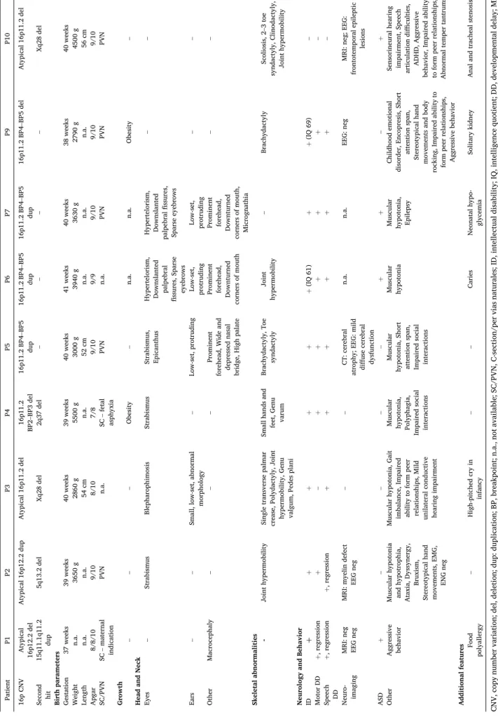
Table 2 Clinical findings of the presented patients. Patient P1 P2 P3 P4 P5 P6 P7 P9 P10 16p CNV Atypical 16p12.2 del Atypical 16p12.2 dup Atypical 16p11.2 del 16p11.2 BP2–BP3 del 16p11.2 BP4–BP5 dup 16p11.2 BP4–BP5 dup 16p11.2 BP4–BP5 dup 16p11.2 BP4–BP5 del Atypical 16p11.2 del Second hit 15q11.1q11.2 dup 5q13.2 del Xq28 del 2q37 del – – – – Xq28 del Birth parameters Gestation 37 weeks 39 weeks 40 weeks 39 weeks 40 weeks 41 weeks 40 weeks 38 weeks 40 weeks Weight n.a. 3650 g 2860 g 5500 g 3000 g 3940 g 3630 g 2790 g 4500 g Length n.a. n.a. 54 cm n.a. 52 cm n.a. n.a. n.a. 56 cm Apgar 8/8/10 9/10 8/10 7/8 9/10 9/9 9/10 9/10 9/10 SC/PVN SC – maternal indication PVN n.a. SC – fetal asphyxia PVN n.a. PVN PVN PVN Growth – – – Obesity – n.a. n.a. Obesity – Head and Neck Eyes – Strabismus Blepharophimosis Strabismus Strabismus, Epicanthus Hypertelorism, Downslanted palpebral fissures, Sparse eyebrows Hypertelorism, Downslanted palpebral
fissures, Sparse eyebrows – – Ears – – Small, low-set, abnormal morphology – Low-set, protruding
Low-set, protruding Low-set, protruding
– – Other Macrocephaly – – – Prominent forehead, Wide and depressed nasal bridge, High palate
Prominent forehead, Downturned
corners of mouth
Prominent forehead, Downturned corners of mouth, Micrognathia
– – Skeletal abnormalities - Joint hypermobility Single transverse palmar crease, Polydactyly, Joint hypermobility, Genu valgum, Pedes plani
Small hands and feet, Genu varum
Brachydactyly, Toe syndactyly Joint hypermobility – Brachydactyly Scoliosis, 2–3 toe syndactyly, Clinodactyly, Joint hypermobility Neurology and Behavior ID þ+++++(IQ 61) ++(IQ 69) – Motor DD +, regression +– +++++– Speech DD +, regression +, regression ++++++– Neuro- imaging MRI: neg EEG neg MRI: myelin defect EEG neg – – CT: cerebral atrophy; EEG: mild diffuse cerebral dysfunction n.a. n.a. EEG: neg MRI: neg; EEG: frontotemporal epileptic lesions ASD +– – – – ++– + Other
Aggressive behavior
Muscular hypotonia and hypotrophia, Ataxia, Dyssynergy, Bruxism, Stereotypical hand movements, EMG, ENG neg Muscular hypotonia, Gait imbalance, Impaired ability to form peer relationships, Mild unilateral conductive hearing impairment Muscular hypotonia, Polyphagia,
Impaired social interactions Muscular hypotonia, Short attention span, Impaired social interactions Muscular hypotonia Muscular hypotonia, Epilepsy
Childhood emotional disorder, Encopresis, Short attention span, Stereotypical hand movements and body rocking, Impaired ability to form peer relationships, Aggressive behavior
Sensorineural hearing impairment, Speech articulation difficulties, ADHD, Aggressive behavior, Impaired ability to form peer relationships, Abnormal temper tantrums Additional features Food polyallergy – High-pitched cry in infancy – – Caries Neonatal hypo- glycemia Solitary kidney Anal and tracheal stenosis CNV, copy number variation; del, deletion; dup: duplication; BP, breakpoint; n.a., not available; SC/PVN, C-section/per vias naturales; ID, intellectual disability; IQ, intelligence quotient; DD, developmental delay; MRI, magnetic resonance imaging; CT, computer tomography; EEG, electroencephalography; EMG, electromyography; ENG, electroneurography; neg, negative; ASD, autism spectrum disorder; ADHD, attention deficit hy- peractivity disorder.
Table 3
Copy number variations identified in the presented patients.
Patient Chromosome Band Type Start breakpoint (bp) End breakpoint (bp) Size (Kb) Origin Classification Disorder (OMIM#)
P1 16p12.2 Del 21,566,709 21,739,799 173.1 n.a. VUS –
15q11.1q11.2 Dup 20,207,363 22,671,977 2441.5 n.a. P 608636
P2 16p12.2 Dup 21,953,152 22,480,514 527.4 n.a. VUS –
5q13.2 Del 68,830,621 70,654,255 1823.6 n.a. LB –
P3 16p11.2 Del 28,722,783 28,824,859 102.1 n.a. LB –
Xq28 Del 153,409,765 153,520,551 110.8 n.a. VUS –
P4 16p11.2 Del 28,824,802 29,040,571 215.8 mat P 613444
2q37 Del 242,855,645 243,030,854 175.2 n.a. LB 600430
P5 16p11.2 Dup 29,620,689 30,190,568 569.0 n.a.a P 614671
P6 16p11.2 Dup 29,624,765 30,199,351 574.6 mat P 614671
P7 16p11.2 Dup 29,624,765 30,199,351 574.6 mat P 614671
P8 16p11.2 Dup 29,624,765 30,199,351 574.6 n.a. P 614671
P9 16p11.2 Del 29,656,684 30,190,568 533.9 n.a.a P 611913
P10 16p11.2 Del 31,980,001 33,825,000 1845.0 d.n. VUS –
Xq28 Del 153,409,765 153,520,551 110.8 n.a. VUS –
bp, basepairs; Kb, kilobase; OMIM, Online Inheritance in Man (https://www.omim.org/); Del, deletion; Dup, duplication; n.a., not available; mat, maternal; d.n., de novo; VUS, variant of unknown significance; P, pathogenic; LB, likely benign.
aFor P5 and P9 maternal inheritance was excluded, but the fathers refused testing.
Fig. 2. Recurrent copy number variations of the short arm of chromosome 16 and the rearrangements of the presented patients. The purple bars denote four recurrent CNV regions; red bars indicate deletions, blue bars indicate duplications. (For interpretation of the references to color in this figure legend, the reader is referred to the Web version of this article.)
not reproduced in a subsequent study; Kctd13 reduction did, however, lead to a decreased functional synapse number and reduced synaptic transmission in mutant mice. Furthermore, reduced synaptic trans- mission correlated with increased levels of Ras homolog gene family, member A (RhoA - a substrate of the aforementioned ubiquitin ligase complex), and was reversed by RhoA inhibition, which might prove therapeutically relevant in the future (Escamilla et al., 2017). Possibly relevant in regards to seizures is the PRRT2 gene [OMIM *614386 (Scheffer et al., 2012)], which is linked to a benign epilepsy syndrome (OMIM *605751).
The gene suspected to play a role in the obesity phenotype associated with the distal microdeletion is SH2B1 (OMIM *608937), encoding a cytoplasmic adaptor protein for various members of the tyrosine kinase receptor family, and postulated to enhance hypothalamic leptin sensi- tivity, thus regulating energy balance, body weight, peripheral insulin sensitivity and glucose homeostasis. Alterations of SH2B1 lead to leptin and insulin resistance, polyphagia, obesity and type 2 diabetes mellitus in mice and humans (Ren et al., 2007; Rui, 2014). LAT (OMIM *602354) – encoding a protein that is part of a complex required for T-cell development and signaling, and likely has an important role in neuro- genesis - was proposed as a major driver gene of the mirror HC pheno- type in the BP2–BP3 rearrangements. Moreover, co-injection of LAT and KCTD13 seems to have an additive effect on zebrafish HC, providing
evidence for genetic interactions between the distal and the proximal recurrent 16p11.2 CNV regions (Loviglio et al., 2017a,b).
5.2. Variants of unknown significance in 16p
Patient 1 has a 16p12.2 microdeletion containing the 3′part of OTOA (OMIM *607038), a key gene implicated in the pathomechanism of the recurrent 16p11.2p12.2 microdeletion syndrome (OMIM #613604).
Defects of OTOA have been implicated in autosomal recessive and non- syndromic hearing loss, and the hearing impairment occasionally seen in 16p11.2p12.2 microdeletion patients. This syndrome is further charac- terized by variable minor anomalies, ID/DD, muscular hypotonia, feeding difficulties, recurrent ear infections, congenital heart defects and behavioral problems, but ASD is uncharacteristic (Battaglia et al., 2009; Hempel et al., 2009; Okamoto et al., 2014). P1’s clinical presen- tation does not adhere to this (normal hearing, no history of hearing loss in his family, present ASD), and his CNV is much smaller than the typical 16p11.2p12.2 microdeletion syndrome. A nearly identical deletion has recently been reported in a patient referred with short stature, dyslexia, pectus excavatum, kyphosis and facial minor anomalies. She inherited it from her deaf and severely speech impaired mother, who carried the deletion in homozygous form (Tassano et al., 2019). Deletions of similar size and gene content can also be found in DECIPHER (Firth et al., 2009), Fig. 3. Illustrations of the preformed genetic tests. A: Duplication of proximal 16p11.2 validated by fluorescence in situ hybridization; the three red signals in each cell correspond to the 548 kb sized 16p11.2 region identified by a KCTD13 specific FISH probe. B-D: Genetic test results for Patient 10. B: Array CGH results showing a 1.85 Mb large deletion between genomic positions 16:31,980,001–33,825,000. C: Haplotypes of the family using microsatellite markers of the proximal 16p11.2 region. D: Electropherograms of the rs74671405 microsatellite marker. (For interpretation of the references to color in this figure legend, the reader is referred to the Web version of this article.)
with great variability in reported phenotypic features, however the majority have at least one NDD listed (see Supplementary File 3 for patient IDs and phenotypes). P1’s other CNV is associated with the 15q11q13 duplication syndrome (OMIM #608636) characterized by normal morphological findings in the majority of patients, learning difficulties, global DD, language impairment, seizures and ASD.
Patient 2’s 16p12.2 duplication encompasses four OMIM genes: the cancer related EEF2K (*606968) and CDR2 (*117340) genes, the POLR3E (*617815) gene with DNA dependent RNA polymerase activity and the disease causing UQCRC2 gene (*191329). The latter encodes a protein that is part of the ubiquinol-cytochrome c reductase complex, and its mutations are responsible for mitochondrial complex III defi- ciency. DECIPHER catalogues 8 duplication cases with similar break- points and nearly identical gene content (Supplementary File 3). At least one NDD is listed in the phenotypic information for six out of the eight cases, with ASD being the most common (4/8). P2 carries an additional large deletion of chromosome 5q13.2, including 6 OMIM genes: OCLN (*602876), SERF1A (*603011), NAIP (*600354), GTF2H2 (*601748), and spinal muscular atrophy (OMIM #253400) associated SMN1 (*600354) and SMN2 (*601627). DECIPHER catalogues overlapping cases with NDDs (Supplementary File 3). There is a nearly identical Gold Standard Variant (gssvL99909) listed in the Database of Genomic Var- iants (DGV) (MacDonald et al., 2014) with an overall allele frequency of 1.54%.
Patient 3’s 16p11.2 deletion encompasses one non-disease associ- ated OMIM gene, EIF3C (*603916). A search of ClinVar database (Landrum et al., 2018) yields three smaller deletions involving EIF3C, classified as benign. The DGV lists a gold standard variant (gssvL43531) involving only this gene (approximately 40% overlap with P3’s CNV) with a frequency of 8.82%. All other ClinVar records that overlap P3’s deletion, and overlapping DECIPHER cases as well, either include the BP2–BP3 pathogenic region, or are much larger in size. P3’s proximal breakpoint is virtually identical with P4’s distal breakpoint (~28,824, 800). Phenotypic comparison of P3 and P4 shows reverse findings regarding BMI (Fig. 1A and B). The CNV seen in P3 is directly upstream of the recurrent BP2–BP3 microdeletion and SH2B1 gene, and thus his lack of the typical obese phenotype further consolidates the pathogenic role of the distal 16p11.2 region and the genes it encompasses. P3 has an additional deletion on Xq28, involving the genes encoding the red [OPN1LW (OMIM *300822)] and green [OPN1MW (OMIM *300821)]
photopigments. Various genetic defects of these genes cause color vision abnormalities (Deeb, 2005), which, to our knowledge, have not been reported in association with the Xq28 CNV. Patient 10 carries an iden- tical Xq 28 deletion. As of yet, we were unable to perform formal color vision testing in the presented patients.
P10’s 16p deletion contains one OMIM gene, TP53TG3 (*617482), which is unlikely to be haploinsufficient according to the DECIPHER score (%HI =94.39), and is not associated with any known disease or phenotype. A nearly identical deletion is a DGV Gold Standard Variant (gssvL43686), with an overall frequency of 1.35%. We have found two similar deletions in patients from international databases (excluding those that also encompass one or both of the pathogenic recurrent 16p11.2 regions; Supplementary File 3). ClinVar database records a VUS (nssv581526), which is nearly identical to P10’s deletion with micro- cephaly and global DD listed in the phenotypic information. DECIPHER contains one deletion classified as likely pathogenic (#328130;
16:30,361,048–33,660,219). The reported features include delayed speech and language development, global brain atrophy, global DD, ID, gait disturbance, tremor and seizures. This patient has no other CNV, however the gene content in their 16p11.2 deletion is much higher compared to P10’s. One notable gene is FUS (OMIM *137070), whose mutations are associated with amyotrophic lateral sclerosis 6, with or without frontotemporal dementia (OMIM #608030) and hereditary essential tremor 4 (OMIM #614782) (Merner et al., 2012; Vance et al., 2009). FUS has high haploinsufficiency potential according to DECI- PHER and could be a plausible key gene in this patient’s gait disturbance
and tremor. Another potentially relevant gene is STX1B (OMIM
*601485), which is associated with early onset generalized epilepsy with febrile seizures plus, type 9 (OMIM #616172) (Schubert et al., 2014), thus linking the deleted region to seizure disorders. Although these two patients from the databases are not directly comparable to P10, they each presented with various NDDs, and speech development was affected in all three. This could suggest that the disturbance of more proximal 16p11.2 regions might compromise normal neuro- development, especially in regards to speech and language, possibly due to altered chromatin interactions.
We believe that the phenotypes of these four patients could be, at least partially, attributed to their co-occurring CNVs, in line with the second hit model, which states that multiple CNVs (including VUS) can have additive, often exacerbating effects on clinical presentation. The second hit genomic imbalances are frequently inherited, with maternal preference (Girirajan et al., 2010; Redaelli et al., 2019). Girirajan et al.
also observed an eight-fold increased risk of DD in children carrying two large CNVs (Girirajan et al., 2012). P1’s phenotype can be attributed to his large 15q11 duplication, however he is at the severe end of the phenotypic spectrum, therefore we believe his second hit 16p12.2 microdeletion plausibly contributed to the striking clinical manifesta- tion of his NDD. P2 carries a VUS duplication of 16p12.2 and a large likely benign 5q13 deletion that have both been sporadically reported in association with NDDs. The added affect of these less serious alterations could contribute to P2’s developmental and behavioral phenotype. The cases of P3 and P10 are less straightforward, as one or both of their respective CNVs could be common variants. It is worth mentioning however, that common genetic variants have been shown to contribute to the risk and variability of severe NDDs in a genome-wide association study (Niemi et al., 2018).
6. Conclusions
Our cohort of patients with chromosome 16p rearrangements pre- sented individually variable neuropsychiatric phenotypes with a broad spectrum of severity. The presented carriers of the BP2–BP2 and BP4–BP5 CNVs corroborate literature data. The neighboring breakpoints identified in Patients 3 and 4 support the pathogenicity of the distal 16p11.2 microdeletions. Patients 1, 2, 3 and 10 highlight the ongoing difficulties surrounding the interpretation of genetic variation, genetic counseling and anticipatory management. Nevertheless, we present these four cases as examples of co-occurring CNVs conferring risk for neuropsychiatric phenotypes, primarily concerning speech and language.
Declaration of competing interest
The authors declare that they have no conflict of interest. This research did not receive any specific grant from funding agencies in the public, commercial, or not-for-profit sectors.
All authors contributed to conception, genetic examinations and counseling, data analysis and interpretation. Anna Lengyel prepared the manuscript. All authors read and approved the manuscript.
Acknowledgments
We would like to express our gratitude towards the patients and their families, and the referring physicians.
Appendix A. Supplementary data
Supplementary data to this article can be found online at https://doi.
org/10.1016/j.ejmg.2020.104027.