ray diffractometry ( m -XRD): A useful technique for the characterization of small amounts of clay minerals
IVETT KOV ACS
1* , TIBOR N EMETH
1,2, GABRIELLA B. KISS
2and ZSOLT BENK O
31Institute for Geological and Geochemical Research, Research Centre for Astronomy and Earth Sciences, Budapest, Hungary
2Department of Mineralogy, Faculty of Sciences, E€otv€os Lorand University, Budapest, Hungary
3Institute for Nuclear Research, Debrecen, Hungary
Received: August 13, 2019 • Accepted: December 5, 2019 Published online: October 1, 2020
ABSTRACT
The laboratory micro X-ray diffraction (m-XRD) technique is a suitable method to study mineralsin- situin whole-rock specimens without any sample preparation or in polished thin sections, and even in small amounts in powdered form. The micro X-ray diffraction method uses the conventional, closed- tube X-ray generator, but modifications were needed in the diffraction column, sample holder and detector in order to achievem-XRD capability.
In this paper, we present a case study of the capillary method used inm-XRD on hydrothermal clay mineral assemblages that formed in the Velence Mts (Hungary). The capillary method inm-XRD has many advantages in the investigation of small amounts of clay minerals: (1) easy and rapid preparation of randomly oriented, powdered samples; (2) rapid measurements; (3) accurate diffraction patterns. By using the capillary method, the formation of preferred orientation can be eliminated; thus the (hkl) reflection of the clay minerals can be precisely measured. Illite polytype quantification and the inves- tigation of (060) reflection of clay minerals can be used satisfactorily inm-XRD.
Hydrothermal clay mineral assemblages are indicative of temperature and pH. Their examination can determine the physicochemical parameters of the hydrothermal fluids that interacted with the host granite in the Velence Mts. The analyzed hydrothermal clay minerals from the western part of the mountains suggest lower temperatures (150–2008C) and intermediate pH conditions. In contrast, the clay mineral assemblages’characteristics for the eastern part of the mountains indicate more intense argillization and higher temperatures (∼2208C) and intermediate pH conditions.
KEYWORDS
micro-XRD, capillary method, clay minerals, illite polytype, Velence Mts
INTRODUCTION
X-ray diffraction (XRD) is a widely-used method to specify the mineralogical composition (phase identification) of natural as well as artificial materials. For a mineralogical study, if a sufficient amount material is available, conventional powder diffraction can be performed either on powdered mineral or rock samples which can be separated by various methods (hand-picking, gravitationally or by centrifugation) from their original host media. However, in many cases, due to technological reasons (similar density of mineral phases, e.g. feldspars), specific mineralogy (e.g. mixtures of clay minerals) or low quantity of the questionable mineral phases (e.g. results of low-grade alteration), preparation of the sample for
Central European Geology
64 (2021) 1, 1–7 DOI:
10.1556/24.2020.00005
© 2020 The Author(s)
ORIGINAL ARTICLE
*Corresponding author. Institute for Geological and Geochemical Research, Research Centre for Astronomy and Earth Sciences, Buda€orsiut 45, H-1112, Budapest, Hungary.
E-mail:kovacs.ivett@csfk.mta.hu
conventional XRD analysis is difficult. Occasionally, due to the high value of the objects, no or only very limited sam- pling is permitted (e.g. archaeological artefacts or planetary materials). Structural characterization of clay minerals–e.g.
illite polytypism– is also challenging, as the conventional sample preparation methods often result in preferred orientation in the sample. In order to overcome the weak- nesses of the conventional XRD technique, the laboratory micro X-ray diffraction (
m
-XRD) technique has been developed. The new method is suitable for examining minerals in-situ in (1) whole-rock specimens without any sample preparation or (2) in polished thin sections, and (3) even in small amounts of powdered form. Advantages of the in-situm
-XRD method over conventional XRD has been demonstrated by a selected set of applications in geology and environmental sciences (see e.g.Tissot 2003; Flemming et al.2005; Flemming 2007), in archaeometry (see e.g. Benedetti et al. 2004; Nel et al. 2006; Bontempi et al. 2008; Swider 2009; Mozgai et al. 2019) and in planetary sciences (see e.g.
Round et al. 2010; Izawa et al. 2011).
Identification of mineralogy, polytypism, crystallinity and proportion of the components in clay mineral assem- blages is indispensable in the studies of diagenetic and anchimetamorphic systems, as well as in the investigation of (ore-forming) hydrothermal systems. Clay mineral assem- blages and the crystal structure (ordering, crystallinity, pol- ytypism) of the individual clay species are indicative of the temperature, pH, as well as of the fluid-rock interaction. In spite of its significance, the link between the alteration product and the altered mineral can hardly be established by the combination of optical microscopy and conventional XRD.
In this paper, case studies are presented of the applica- tion of
m
-XRD on hydrothermal clay assemblages that formed in a Permian granite intrusion during the Alpine cycle. First, we give an introduction into them
-XRD tech-nique; then we demonstrate the suitability of the capillary method of
m
-XRD instrument in the investigation of clay minerals, when only a very limited amount of sample ma- terial is available orin-situmeasurements are required.The m -XRD technique and the capillary method
The laboratory
m
-XRD method uses the conventional, closed-tube X-ray generator, but in order to achievem
-XRDcapability, modifications were needed in the diffraction column, sample holder and detector. Microfocus tubes have been used for a long time to obtain transmission images, for which the focus size was set to a few
m
m in order to increase spatial resolution. In spite of these efforts, adequate X-ray output cannot be obtained with such settings, and the method is insufficient to use in X-ray diffraction (Takumi and Maeyama 2015). However, in the past decades, X-ray tubes with a few tens ofm
m focus have become available; this allows scientists to obtain a usable diffraction pattern from samples even in the 10m
m range.In
m
-XRD, a two-dimensional (2D) detector is used with a larger detection area, such as Imaging Plate (IP), ChargeCoupled Device (CCD), Complementary Metal Oxide Semiconductor (CMOS) and Position Sensitive Proportional Counter (PSPC). All these types have their own advantages.
The detector surface can be curved (IP) or flat (CCD, CMOS, PSPC). The curved detectors are normally designed for fixed sample-to-flat-detector distances, while flat de- tectors have the flexibility to be used at different sample-to- detector distances, so as to choose between higher resolution at greater distance or higher angular coverage at short dis- tance. IP has a larger detection area than CCD, CMOS and PSPC. If the measurements are in fixed positions, they cannot be captured by a small area detector, and it is necessary to use a detector with a large detection area such as an IP (Takumi and Maeyama 2015).
A new RIGAKU D/MAX RAPID II diffractometer was purchased by the Institute for Geological and Geochemical Research (HAS), which is a unique combination of a MicroMax-003 third-generation microfocus, sealed-tube X-ray generator and a curved imaging plate detector. The diffractometer is operated with CuKaradiation generated at 50 kV and 0.6 mA. Different types of collimators can be used (10, 30, 50, 100, 300, 500, 800
m
m) depending on the size of the measured area. A built-in CCD camera was used to select the measurement area and for precise positioning of the sample at a downward angle of 458. The detector system uses a curved IP, which is placed on the inner surface of a cyl- inder that surrounds the u-axis at the center, allowing the recording of a 2D diffraction image over a broad 2qrange.The IP is read by a laser-scanning readout system in about 1 min. 2DP RIGAKU software is used to record the diffraction image from the laser readout and the operator can deter- mine the area to integrate for a 2qversus intensity plot. This plot is read into RIGAKU PDXL 1.8 software for data interpretation.
One of the disadvantages of the method is that due to the geometry of the instrument and the sample holder, the ob- ject/sample may cover certain areas of the imaging plate detector depending on its actual position in the diffraction geometry. Therefore, some higher dhkl values cannot be detected, and to achieve the totally random orientation is often difficult. These limitations make the interpretation difficult in the case of some sample types (e.g. clay minerals).
In order to overcome this limitation, the powdered samples for the micro-diffraction measurements are encapsulated in a borosilicate-glass capillary, with a diameter of 0.3 mm and wall thickness of 0.01 mm, by a vertical manual charging process. Then, the capillary is analyzed by the micro- diffractometer in transmission mode with a beam spot diameter of 100
m
m. In each measurement, 0.5–1 mg of sample is placed in the funnel-end of the capillary and the sample is tapped into the narrow portion. The sample stage with thefilled capillary is aligned before each measurement.The best technique for aligning is to adjust theXandYaxis on the sample stage and rotate the4-axis. Orientation of the minerals can be prevented in the samples during the mea- surement by the rotation of the sample stage from 08to 3608 of the 4-axis, keeping the u-axis fixed at 08 (Fig. 1). The
c-axis isfixed at 458relative to theu-axis. The measurement time ranges between 3 and 10 min.
NEWMOD II and WIDFIRE software were used to analyze interstratified clay mineral samples and the illite polytypism.
CASE STUDY: HYDROTHERMAL CLAY MINERAL ASSEMBLAGES IN THE VELENCE MTS.
The Velence Mountains along the Periadriatic–Balaton Lineament in the western part of the Carpathian basin are a Permian, deeply eroded granite intrusion that have been affected by several hydrothermal processes during the Permian, Triassic and the Paleogene (Fig. 2a). The hydro- thermal fluid-flow events can be characterized by different physicochemical properties (Molnar 1997, 2004; Benko et al.
2012; Toth 2017; Kovacs et al. 2019). The alteration zones often overlap spatially, and the elder mineral assemblages are locally overprinted by the products of the younger hy- drothermal systems.
Four localities, representing different argillic alteration assemblages, are examined in this study. Two localities in the western segment of the intrusion represent the low-tem- perature (80–1508C), regional, Triassic hydrothermal event.
The high-temperature (220–340 8C) Paleogene alteration zones are confined in some E–W structural zones in the
eastern segment of the intrusion and are represented by two selected localities (Fig. 2b). In earlier studies (Benko et al.
2012; Toth 2017; Kovacs et al. 2019), the mineralogical composition of bulk samples was determined by conven- tional XRD measurements and other petrographic methods, but with the conventional XRD technique we could not define the alteration of the single mineral particles. The granite affected by the Triassic hydrothermal alteration contains illite, kaolinite and smectite, while the granite affected by the Paleogene fluid flow contains only illite, based on conventional XRD measurements.
Several bulk samples were collected from the four areas, from which 3–4 cm-thick rock slices were made to facilitate further investigation. Each sample was divided into morphological components based on appearance (color, texture and the occurrence of argillization) under a binoc- ular microscope. Plagioclase, alkali feldspar and clay min- erals (in the fissure) were separated from each bulk sample.
The capillary powder samples were prepared by hand-picked separation (scraped with a spatula) under a binocular mi- croscope and homogenized using an agate mortar.
Units of the intrusion affected by the Triassic regional fluid flow
In both localities (Aranybulla Quarry and Karacsony Quarry) affected by the Triassic fluid flow, the alkali feldspar is macroscopically fresh, whereas the plagioclase and biotite are altered to greenish-white clay minerals (Fig. 3a and b).
Intense and relatively broad peaks at 10 and 5 A clearly indicate that illite is the predominating phase in these samples, based on the
m
-XRD. Besides illite, there are peaks at 7.1 and 3.57A, which suggest the presence of kaolinite.The weak 15 A reflection indicates the presence of some smectite and the asymmetric peak at 10A suggests randomly interstratified illite/smectite, containing a 15–20% swelling component based on the NEWMODE II calculation method. The peak position of the (060) reflection of smectite suggests the presence of dioctahedral smectite (beidellite, montmorillonite). Alkali feldspar from Karacsony Quarry contains wisps of illite (Fig. 3e and f).
Units of the intrusion affected by the Paleogene fluid flow
The structurally-controlled Paleogene hydrothermal alter- ation in the granite is much more intense than the regional pervasive Triassic hydrothermal circulation. Except for the rock-forming quartz and some remnants of alkali feldspar, yellowish-white and white fine-grained material replace the plagioclase and biotite (Fig. 3c and d). In a quarry that represents the distal zone of the hydrothermal system (Sukoro barite excavation), alkali feldspar is poorly altered, and the other minerals are replaced by illite (Fig. 3g). During illite polytype quantification, the measured
m
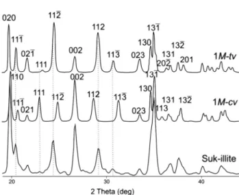
-XRD diffrac- tion patterns of the samples were compared to the WILD- FIRE-calculated diffraction pattern. Several diagnostic polytype peaks of illite occur between 20–328 2q. The 1M character of the layers is suggested by the peak positions and Fig. 1.RIGAKU D/MAX RAPID II micro X-ray diffractometer set-up
the relative peak intensities of the reflections. The measured 11l reflections and intensity distributions are located be- tween the pure 1M trans-vacant (tv) and 1M cis-vacant (cv) illite structures (Fig. 4and Table 1).
According toDrits (2003) and Zviagina et al. (2007)this is represented by the possible interstratification oftvandcv layers.
Based on the
m
-XRD results, the analyzed alkali feldspar pseudomorphs from the other quarry (Nadap Quarry) consist of smectite and illite, based on the intense peaks at 15 A and 10A. However, the white material contains only illite (Fig. 3h). Them
-XRD pattern shows diagnostic reflections at 3.88Að113Þ, 3.73A (023), 3.49Að114Þ, 3.20A (114), 2.98A (025) and 2.86A (115), the appearance of which suggest the 2M1character of the layers (Fig. 5).Formation conditions of the hydrothermal clay minerals inferred from the mineral assemblages and the polytypism
The western samples can be characterized by the weakest argillization. According to White and Hedenquist (1990), illite indicates relatively high temperatures (>200 8C) and neutral to acidic conditions in hydrothermal environments.
Kaolinite forms under acidic conditions (pH 2–7) and at temperatures between 100 and 220 8C (Reyes 1990;
Hedenquist et al. 2000). Smectite forms at relatively low temperature (<1508C) and neutral condition but beidellite is stable at a higher temperature than montmorillonite in hy- drothermal environments (Yamada et al. 1991; Yamada and Nakazawa 1993). Based on the above-mentioned facts, the studied hydrothermal clay minerals represent a narrow Fig. 2.(a) Location of the Velence Mts (Hungary) in the Alpine–Carpathian region afterCsontos and V€or€os (2004); (b) Geologic map of the
Velence Mts. afterHorvath et al. (2004)
stability field of formation. The analyzed clay minerals indicate low formation temperature (150–200 8C) and in- termediate pH conditions.
In contrast to the western part, where only Triassic hydrothermal process caused argillic alteration, in the eastern part both the Triassic and Paleogene hydrothermal Fig. 3.(a–d) Macroscopic feature of the granite samples and the location of the sampling form-XRD (blue and black dotted shapes); (a) Aranybulla Quarry; (b) Karacsony Quarry; (c) Sukoro barite excavation; (d) Nadap Quarry; (e–h) Micro-XRD patterns of granite samples
(Ilt–illite, Kln–kaolinite, Smc–smectite, Qz–quartz, Kfs–alkali feldspar, Pl–plagioclase)
processes developed clay mineral alteration paragenesis.
Polytypism of illite may provide information about the thermodynamic conditions of the mineralizing fluid (Kraus et al. 1999; Bove et al. 2002). In hydrothermal systems, with increasing temperature and pressure, polytypism of illite shows the following sequence from 1Md, 1Mto 2M1(Inoue et al. 1988). However, the polytype determination can be difficult when measuring clay mineral assemblages because the (hkl) diffraction peaks interfere with other clay minerals and with the characteristic peaks of alkali feldspar and plagioclase (Velde and Hower 1963). Therefore, determi- nation of the illite polytypism can only be performed with certainty in the eastern area. Polytypism of illite from the eastern part of the mountains can be characterized by 1M and 2M1polytype. The analyzed clay minerals and the illite polytypism indicate higher formation temperature (∼220 8C) than in the western part of the mountains. This result agrees with the findings of previous studies, which deter- mined the physicochemical properties of the hydrothermal fluid flow systems (Molnar 1997, 2004; Benko et al. 2012;
Toth 2017; Kovacs et al. 2019).
CONCLUSIONS
X-ray diffraction is the basic technique for identification of clay minerals. The capillary method in
m
-XRD has many advantages in the investigation of small amounts of clay minerals: (1) easy and rapid preparation of randomly ori- ented, powdered samples; (2) rapid measurements; (3) cor- rect diffraction patterns. By using the capillary method, formation of the preferred orientation can be eliminated;thus the (hkl) reflection of the clay minerals can be measured.
Illite polytype quantification and the investigation of (060) reflection of clay minerals can be performed satisfactorily by using
m
-XRD. As proven by the samples of the Velence Mts, the capillary method inm
-XRD is a useful tool for identifi- cation of very small amounts of clay minerals analyzed in- situ. Since clay mineral assemblages and the crystal structure of the individual clay species are indicative of the tempera- ture and pH, them
-XRD method proved to be an excellent method for determining the physicochemical parameters of the hydrothermalfluids that interacted with the host granite in the Velence Mts. The analyzed hydrothermal clay minerals from the western part of the mountains suggest lower tem- peratures (150–2008C) and intermediate pH conditions. The clay mineral assemblages indicate more intense argillization and higher temperatures (∼220 8C) characteristic of the eastern part of the mountains.REFERENCES
Benedetti, D., Valetti, S., Bontempi, E., Piccioli, C., and Depero, L.E.
(2004). Study of ancient mortars from the Roman Villa of Pollio Fig. 5.Calculated XRD patterns of 2Millite andm-XRD pattern of
illite from the Nadap Quarry
Fig. 4.Calculated XRD patterns of 1M-tv, 1M-cvillite andm-XRD pattern of illite from the Sukoro barite excavation
Table 1.Reflection indices,dhklvalues and relative peak intensities in the simulated XRD pattern for structural models 1M-tvand 1M-
cv(Zviagina et al. 2007) and thedhklvalues and relative peak intensities in them-XRD pattern of illite from Sukoro barite
excavation (Suk-illite)
1M-tv 1M-cv Suk-illite
hkl dhkl
(A) I (%) hkl
dhkl
(A) I (%)
dhkl
(A) I (%)
020 4.502 97 110 4.460 100 4.480 100
111 4.337 50 111 4.299 21 4.343 35
021 4.106 29 021 4.104 11 4.100 3
111 3.823 4 111 3.879 49 3.874 1
112 3.638 100 112 3.580 39 3.646 54
022 3.344 32 022 3.343 82 3.339 62
112 3.067 80 112 3.119 43 3.069 43
113 2.910 21 113 2.862 41 2.915 2
123 2.676 26 023 2.675 12 2.676 1
130 2.588 51 130 2.589 56 2.583 24
131 2.567 81 131 2.561 88 2.561 88
220 2.550 36 113 2.506 9 2.481 4
202 2.474 9 131 2.460 13 2.445 11
131 2.446 13 132 2.379 27 2.390 19
132 2.396 21 114 2.317 1 2.357 3
201 2.361 13 221 2.243 14 2.243 6
Felice in Sorrento (Naples). Applied Physics A: Materials Sci- ence & Processing, 79: 341–345.
Benko, Zs., Molnar, F., Pecskay, Z., Nemeth, T., and Lespinasse, M.
(2012). The interplay of the Paleogene magmatic-hydrothermal fluidflow on a Variscan granite intrusion: age and formation of the barite vein at Sukoro, Velence Mts, W-Hungary. Bulletin of the Hungarian Geological Society, 142: 45–58.
Bontempi, E., Benedetti, D., Massardi, A., Zacco, A., Borgese, L., and Depero, L.E. (2008). Laboratory two-dimensional X-ray microdiffraction technique: a support for authentication of an unknown Ghirlandaio painting. Applied Physics A: Materials Science & Processing, 92: 155–159.
Bove, D.J., Eberl, D.D., McCarty, D.K., and Meeker, G.P. (2002).
Characterization and modeling of illite crystal particles and growth mechanisms in zoned hydrothermal deposit, Lake City, Colorado. American Mineralogist, 87: 1546–1556.
Csontos, L. and V€or€os, A. (2004) Mesozoic plate tectonic recon- struction of the Carpathian region. Palaeogeography, Palae- oclimatology, Palaeoecology, 210: 1–56.
Drits, V.A. (2003). Structural and chemical heterogeneity of layer silicates and clay minerals. Clay Minerals, 38: 403–432.
Flemming, R.L. (2007). Micro X-ray diffraction (mXRD), a versatile technique for characterization of Earth and planetary materials.
Canadian Journal of Earth Sciences, 44: 1333–1346.
Flemming, R.L., Salzsauler, K.A., Sherriff, B.L., and Sidenko, N.V.
(2005). Identification of scorodite infine-grained, high-sulfide, arsenopyrite mine-waste using micro X-ray diffraction. Cana- dian Mineralogist, 43: 1243–1254.
Hedenquist, J.W., Arribas, A.R., and Gonzales-Urien, E. (2000) Exploration for gold deposits. In: Hagemann, S.G. and Brown, P.E. (Eds): Reviews in Economic Geology, Vol. 13, Gold in 2000. Society of Economic Geologists, Littleton, pp. 245–277.
Horvath, I., Darida-Tichy, M., Dudko, A., Gyalog, L., and Ódor, L.
(2004). Geology of the Velence Hills and the Balatonf}o. – Explanatory Book of the Geological Map of the Velence Hills (1:
25 000). Geological Institute of Hungary, Budapest, pp. 153–316.
Inoue, A., Velde, B., Meunier, A., and Touchard, G. (1988) Mechanism of illite formation during smectite-to-illite con- version in a hydrothermal system. American Mineralogist, 73:
1325–1334.
Izawa, M.R.M., Flemming, R.L., Banerjee, N.R., and McCausland, P.J.A. (2011). Micro-X-ray diffraction assessment of shock stage in enstatite chondrites. Meteoritics & Planetary Science, 46: 638–651.
Kovacs, I., Nemeth, T., Kiss, B.G., Kis, K.V., Toth,A., and Benk o, Zs. (2019). Rare aluminium phosphates and sulphates (APS) and clay mineral assemblages in silicified hydraulic breccia hosted by Permian granite (Velence Mts., Hungary) as in- dicators of high sulfidation type epithermal system. Mineralogy and Petrology, 113: 217–228.
Kraus, I., Chernyshev, I.V., Sucha, V., Kovalenkere, V.A., Lebedev, V.A., and Samajova, E. (1999). Use of illite for K/Ar dating of hydrothermal precious and base metal mineralization in Cen- tral Slovak Neogene volcanic rocks. Geologica Carpathica, 50/5:
353–364.
Molnar, F. (1997) Contributions to the genesis of molybdenite in the Velence Mts.: Mineralogical andfluid inclusion studies on the mineralization of the Retezi Adit. Bulletin of the Hungarian Geological Society, 127: 1–17.
Molnar, F. (2004). Characteristics of Variscan and Palaeogenefluid mobilization and ore forming processes in the Velence Mts., Hungary: A comparative fluid inclusion study. Acta Miner- alogica Petrographica, 45: 55–63.
Mozgai, V., Bajnoczi, B., Mrav, Zs., Kovacsoczy, B., and Toth, M.
(2019). Application of a laboratory micro-X-ray diffractometer (RIGAKU DMAX RAPID II) in the archaeometric analysis of archaeological artefacts – Case studies of metal objects.
Archeometriai M}uhely, 16: 29–42.
Nel, P., Lau, D., and Hay, D. (2006) Non-destructive micro-X-ray diffraction analysis of painted artefacts: determination of detection limits for the chromium oxide-zinc oxide matrix.
Nuclear Instruments and Methods in Physics Research B, 251:
489–495.
Reyes, A.G. (1990). Petrology of Philippine geothermal systems and the application of alteration mineralogy to their assessment.
Journal of Volcanology and Geothermal Research, 43: 279–309.
Round, S., Flemming, R.L., Beausoleil, Y., and McCausland, P.J.A.
(2010). Classification of meteorites by olivine unit cell using micro X-ray diffraction. 73rd Annual Meteoritical Society Meeting, Meteoritics and Planetary Science Supplement, New York, Abstract 5230.
Swider, J.R. (2009). Powder micro-XRD of small particles. Powder Diffraction, 25: 68–71.
Takumi, Y. and Maeyama, M. (2015) Micro X-ray diffraction of cultural properties. Rigaku Journal, 31: 1–6.
Tissot, R.G. (2003) Microdiffraction applications utilizing a two- dimensional proportional detector. Powder Diffraction, 18:
86–90.
Toth, A. (2017). A meleg-hegyi hidroterm as breccsa vizsgalata (Investigation of the Meleg Hill hydrothermal breccia). Msc Thesis. E€otv€os Lorand University, Budapest, p. 95 (in Hun- garian).
Velde, B. and Hower, J. (1963). Petrological significance of illite polymorphism in Paleozoic sedimentary rocks. American Mineralogist, 48: 1239–1254.
White, N.C. and Hedenquist, J.W. (1990). Epithermal environ- ments and styles of mineralization: variations and their causes, and guidelines for exploration. Journal of Geochemical Explo- ration, 36: 445–474.
Yamada, H. and Nakazawa, H. (1993). Isothermal treatments of regularly interstratified montmorillonite–beidellite at hydro- thermal conditions. Clays and Clay Minerals, 41: 726–730.
Yamada, H., Nakazawa, H., Yoshioka, K., and Fujita, T. (1991).
Smectites in the montmorillonite-beidellite series. Clay Min- erals, 26: 359–369.
Zviagina, B.B., Sakharov, B.A., and Drits, V.A. (2007). X-ray diffraction criteria for the identification oftrans- andcis-vacant varieties of dioctahedral micas. Clays and Clay Minerals, 55:
467–480.
Open Access. This is an open-access article distributed under the terms of the Creative Commons Attribution 4.0 International License (https://creativecommons.org/
licenses/by/4.0/), which permits unrestricted use, distribution, and reproduction in any medium, provided the original author and source are credited, a link to the CC License is provided, and changes–if any–are indicated. (SID_1)