Lesson 4 - Biomechanical characteristics of tendons and ligaments
This curriculum has been made at the University of Szeged, and supported by the European Union. Project identity number: EFOP-3.4.3-16-2016-00014.
This lesson contains 10 screens teaching text, 6 zoomable figures, and 5 videos. This lesson requires approximately 2 - 4 hours of study but can vary depending on the student.
Tendons, ligaments and joint capsules are fibrous, dense regular collagenous connective tissues that play essential role in joint motion (9).
Function of tendons and ligaments
Tendons do not actively produce motion but attach muscles to bones and serve as the force transmitting units (4, 9). Additionally, tendons enable the muscles to be at an optimal distance from the joints, helps to maintain the optimal muscle length (9).
Ligaments connect bone with bone and provide mechanical stability of the joints. These components of the musculoskeletal system guide joint motions, and prevent excessive motions, which exceed the physiological range of movement (9).
Ligaments and tendons are mostly highly innerved and have a significant role in weight-bearing and postural control. Golgi tendon
organ, as a
neurotendinous proprioceptive
sensory receptor provides with a feedback on the forces present within tendons and thus adapts the intensity of these forces(15).
Please watch this video below about
collagen and function of tendons and ligaments:
https://www.youtube.com/watch?v=rFYj1fN62T0
Composition of tendons and ligaments
Both tendons and ligaments have low vascularity and cellularity. The tissue has almost no vessels and the nutrition as well as oxygen are supplied at the vascularized myotendinous and osteotendinous junctions (4).
Although tendons and ligaments are composed of the same basic components, their exact composition and arrangement of matrix macromolecules differs to provide the specific mechanical properties required by each structure to function effectively (13).
Both tendons and ligaments are made up of mainly 20% cellular material and 80% extracellular matrix.
Extracellular matrix consists water (70%) and solids (30%). Solid components are collagen, ground substance and a little elastin (9).
Extracellular components:
Water.
Type I collagen serves the 70% of dry weight. Ligament have a higher ratio of fibrillar Type I collagen to Type III collagen (elastic). Thus, ligament’s mechanical properties provide it with not only the flexibility but also the tensile properties to resist force.
Elastin - Tendons and extremity ligaments have very little protein elastin. Although, there are elastic ligaments of human body in which the proportion of elastic fibers is substantial, for example the ligamentum flavum.
Lipids.
Proteoglycans play important roles in the physiology and biomechanical
function of tendons and ligaments through their involvement in regulation of structure and maintenance of extracellular matrix (6).
Cellular component:
Fibroblast is the main cell type in both tendons and ligaments. Their main function is to maintain the structural integrity of the connective tissue by continuously secreting extracellular matrix proteins like collagens, glycosaminoglycans, and glycoproteins.
Compared to tendons, ligaments have lower percentage of collagen, and higher percentage of proteoglycans and water.
Structure
Structure of ligaments and tendons is characterized by regular sequence of collagen fiber bundles and fascicles.
Bundles and fascicles are surrounded by a fine connective tissue sheaths. Tendon units are encased in epitenon, which reduces friction with neighboring tissues. The endotenon fibrils invest tendon fibers and bind fibers together (7).
The arrangement of collagen fibers differs between tendons and ligaments and is adapted to the function of each structure.
In the tendon the collagen fibrils are aligned parallel to each other and to the long axis of the tendon (Fig 1), whereas in the ligament the fibrils are not as uniformly orientated to, allow for multiaxial loading patterns (Fig 2) (9).
Therefore, ligaments are organized in a dense but loose-packed network unlike tendon that have an orderly, parallel, linearly packed arrangement. This facilitates tendons to handle high unidirectional tensile loads during activity whilst
ligaments generally sustain tensile loads in a predominant direction but also bear smaller tensile loads in other directions.
Fig. 1 Parallel collagen bundles arrangement of tendons.
Fig. 2 Nearly parallel collagen bundles arrangement of ligaments.
Structure of insertion into bone – design of enthesis
The organization of the insertion into bone is similar in ligaments and tendons. These parts of ligaments and tendons are classified into four distinct zones of tissue with varying mechanical properties and functions (1) There are two types of entheses according to the type of tissue present at the skeletal attachment site. Fibrous entheses are characterized by dense fibrous connective tissue at the tendon-bone interface and are common in tendons that attach to diaphyses of long bones. These entheses usually occur over large surface areas and are characterized by mineralized collagen fibers.
The majority of entheses in the human musculoskeletal system are fibrocartilaginous. Fibrocartilaginous entheses attach to
bone through a layer of fibrocartilage which acts as a transition from the fibrous tendon tissue (1).
At the end of the tendon the parallel collagen fibers are interlocked to fibrocartilage.
Fibrocartilage zone contains unmineralized and mineralized parts, and then fibrocartilage unites into cortical bone (9).
Biomechanical properties of tendons and ligaments
Result of collagenous proteins, water, and the interactions between collagens and proteoglycans, ligaments and tendons have viscoelastic properties (14).
The main mechanical properties of tendons and ligaments are strength and stiffness. Due to their parallel fiber arrangement, tendons and ligaments are principally adapted to resist tensile loads. Two main factors determine the strength response of a ligament or tendon under loading: their size and shape, and the speed of loading (15).
Tendons, via their strength, able to transmit the high forces resulting from muscle contraction without notable energy loss. Besides tendon, other connective tissues also act as modulators of force output.
The flexibility of tendons and ligaments allows them to accommodate a wide range of joint movement and their compliance allows elastic energy to be stored and returned to reducing the metabolic energy cost of locomotor movement. Thus, these elastic properties help to increase the efficiency of locomotion by storing and releasing energy.
Several studies observed catapult-like mechanism in muscle–tendon units during vertebrate animal jumping (i.e. frog) and demonstrated that catapult mechanisms may be employed in sub-maximal jumps (2, 3, 10).
Additionally, catapult-like mechanism has been observed in muscle–tendon units of the human lower limb during walking and
jumping (5).
Tendons, while passive elements of the musculoskeletal system, are a key energy manager in locomotion.
The ligaments are pliant and more flexible than tendons but are strong and resist to tensile loading.
Biomechanical behavior of tendons and ligaments
As you remember, tendons and ligaments have viscoelastic properties, therefore the relationship between stress and strain for a tendon depends on the time of displacement or load.
There are three major characteristics of a viscoelastic material of tendons and ligaments (11).
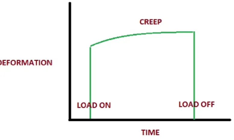
Creep - Increasing deformation under constant load (Fig. 3).
Fig. 3 Creep.
Please watch this short video about creep, below:
https://www.youtube.com/watch?v=rz7qUVdVAWs
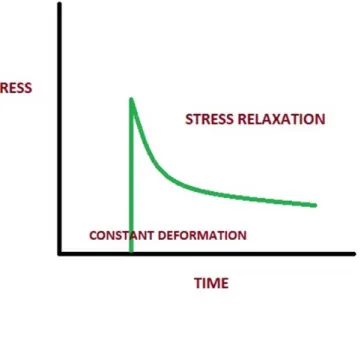
Stress relaxation - Stress acting upon a tendon will eventually reduce under a constant deformation. (Fig. 4).
Fig. 4 Stress relaxation.
Please watch this short video about stress relaxation, below:
https://www.youtube.com/watch?v=IJP92CCVjAk
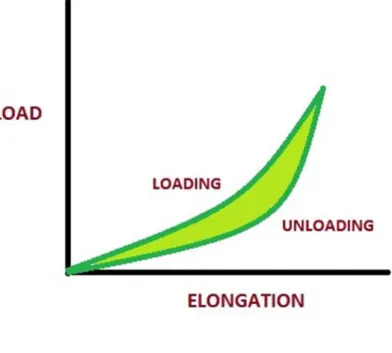
Hysteresis - When a viscoelastic material is loaded and unloaded, the unloading curve is different from the loading curve. The difference between the two curves represents the amount of energy that is lost during loading. (Fig. 5).
Fig. 5 Hysteresis.
Please watch this short video about hysteresis, below:
https://www.youtube.com/watch?v=v90tPg88-Qs
The stress-strain behaviors of ligaments are comprised of a toe-region, a linear-region, a plastic and a failure region (Fig.6).
Fig.6 Stress-stain curve for ligaments and tendons.
The first is the toe region, in which the crimped collagen fibers straightened out due to elongation. At the end of this region tissue is elongated with a small increase in loads. Made primarily of collagen fibers, tendons and ligaments will return to their normal lengths when unloaded (region of elasticity). However, there is an elastic limit after which the tendon or ligament will not return to resting length (region of plasticity). This region represents the early micro failures of the greatly stretched fiber bundles. Maximum load results in complete failure (9). Stresses due to normal daily activities are generally considered to be in the toe-region and possibly in the early portion of the linear-region (9).
Please watch this short video about stress-strain curve for ligaments and tendons, below:
https://www.youtube.com/watch?v=LaIVBd_wJ_M
Mechanism of tendons and ligaments injury
Tendons and ligaments as skeletal force transmitters are exposed to some of the most extreme mechanical demands in the human body. Mechanical stresses can put tendon tissues at risk of damage, and overloading may cause a common clinical tendon condition named tendinopathy. There are two basic fatigue failure processes. Creep failure may occur due to loading at constant stress, while cyclic loading failure appears due to continuous and repeated application of a load (13). Overuse injuries occur when repetitive loading created damage in the tendon or ligament faster than it can be repaired.
According to the degree of severity, ligament and tendon injuries are classified clinically in three ways. The first category is characterized by insignificant symptoms, some pain is felt but there is no joint instability. The second category of injury produces severe pain and joint instability can be detected due to partial ligament rupture. In the third category complete joint instability occurs, most of the collagen fibers are ruptured, severe pain is generated during the trauma then the pain decreases (9).
Tendons and ligaments are poorly vascularized, limiting their potential to self-repair and heal in the case of injury (15).
Effect of aging on ligaments and tendons – structural and functional changes
With aging, various functions of the body gradually deteriorate. It also includes the musculoskeletal system.
The changes associated with increasing age result in a decline in the structure and function of human tendons and ligaments. Age correlates with decrease in the number and activity of cells. Structurally, collagen fibers increase in diameter, lose tensile strength, and become tougher with increasing age. Age also affects tendon and ligament blood flow and the number of capillaries per unit of surface area (8).
Aging tendons and ligaments are increasingly cross-linked resulting in altered viscoelastic properties, with potentially increased risk for micro-damage accumulation and onset of tendon/ligament disease.
Effect of mobilization and immobilization on ligaments and tendons
Tendons and ligaments show the capacity to adapt their structure and mechanical properties to the functional demands. Several studies have found that physical training increases the tensile strength of tendons and ligaments (9), although the tensile strength, elastic stiffness, and total weight of the tendon or ligament decrease due to immobilization.
Several studies demonstrated that prolonged immobilization is disadvantageous to ligaments and tendons. The effects of immobilization are reversible, but the effects of reconditioning took a longer time to show that of immobilization (9).
Study questions:
TRUE/FALSE questions
Read each statement below carefully. Choose the T if you think a statement is TRUE. Choose the F if you think the statement is FALSE.
1. With aging, various functions of the body gradually improve. T or F 2. The tendons are pliant and more flexible than ligaments. T or F 3. The collagen fiber arrangement of tendons and ligaments is same. T or F 4. Both tendons and ligaments have low vascularity and cellularity. T or F 5. The main mechanical properties of tendons and ligaments
are extensibility and laxity. T or F
Matching questions
In this exercise, you have to match each word with a definition.
1. Toe region 2. Creep failure
3. Hysteresis
4. Fibrocartilaginous entheses
5. Fibroblast
6. Region of elasticity
7. Creep
8. Stress relaxation
A. Increasing deformation under constant load.
B. Stress acting upon a tendon will eventually reduce under a constant deformation.
C. In which the crimped collagen fibers straightened out due to elongation.
D. It is the main cell type in both tendons and ligaments.
E. These attach to bone through a layer of fibrocartilage which acts as a transition from the fibrous tendon tissue.
F. Tendons and ligaments will return to their normal lengths when unloaded.
G. It represents the amount of energy that is lost during loading.
H. It may occurs due to loading at constant stress.
References
1. Apostolakos J et al (2014): The enthesis: a review of the tendon-to-bone insertion. Muscles Ligaments Tendons J. 4(3): 333–342.
2. Astley HC, Roberts TJ (2012): Evidence for a vertebrate catapult: elastic energy storage in the plantaris tendon during frog jumping. Biol Lett. 8(3): 386–389.
3. Astley HC, Roberts TJ (2014): The mechanics of elastic loading and recoil in anuran jumping. J Exp Biol. 217(Pt 24):4372-8.
4. Buschmann J, MG Bürgisser: Biomechanics of Tendons and Ligaments, Tissue Reconstruction and Regeneration. 2017, Woodhead Publishing, Pages 3-29.
5. Farris DJ, Lichtwark GA, Brown NAT, Cresswell AG (2016): The role of human ankle plantar flexor muscle–tendon interaction and architecture in maximal vertical jumping examined in vivo. J Exp Biol 219:528-534.
6. Halper J (2014): Proteoglycans and diseases of soft tissues. Adv Exp Med Biol. 802:49-58.
7. Kannus P (2000): Structure of the tendon connective tissue. Scand J Med Sci Sports. 10: 312–320.
8. Kannus P, Paavola M, & Józsa L (2005). Aging and degeneration of tendons. In Tendon Injuries: Basic Science and Clinical Medicine (pp. 25-31). Springer London.
9. Nordin M, Frankel VH: Basic Biomechanics of the Musculoskeletal System. 2014. Wolters Kluwer Health; 4th Edition
10. Roberts TJ, Azizi E (2011): Flexible mechanisms: the diverse roles of biological springs in vertebrate movement. J Exp Biol. 1; 214(Pt 3):353-61.
11. Robi K, Jakob N, Matevz K, Matjaz V (2013). Chapter 2: The Physiology of Sports Injuries and Repair Processes. In Hamlin M (Ed.), ISBN: 978-953-51-1031-6
12. Rumian AP, Wallace AL, Birch HL (2007): Tendons and Ligaments Are Anatomically Distinct But Overlap in Molecular and Morphological Features—A Comparative Study in an Ovine Model. J Ortho Res 25(4):458–64
13. Snedeker JG, Foolen J (2017): Tendon injury and repair – A perspective on the basic mechanisms of tendon disease and future clinical therapy Acta Biom 63:18-36.
14. Wang JHC, Guo Q, Li B. (2012): Tendon Biomechanics and Mechanobiology - A Minireview of Basic Concepts and Recent Advancements. J Hand Ther, 25(2):133–141
15. Weintraub W: Tendon and Ligament Healing: A New Approach to Sports and Overuse Injury. 2003 Paradigm Publications (MA); 2th edition