Spatial clonal evolution leading to ibrutinib resistance and disease progression in chronic lymphocytic leukemia
Ibrutinib has dramatically altered the therapeutic land- scape of chronic lymphocytic leukemia (CLL) with remarkable responses in relapsed or refractory as well as in previously untreated CLL harboring TP53 aberra- tions.1,2 Despite durable responses in the majority of
cases, approximately 20% of patients experience disease progression initiated by secondary therapy resistance. In the majority of patients progressing on ibrutinib, Bruton tyrosine kinase (BTK) or phospholipase Cg2 (PLCG2) resistance mutations predate the manifestation of clinical progression by up to 15 months,3 making early and sensi- tive detection of resistance mutations of major clinical importance. Longitudinal studies including CLL patients undergoing Richter transformation have shed some light on the temporal aspects of clonal evolution processes
haematologica 2018; 103:e38
C ASE R EPORTS
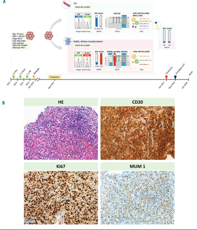
Figure 1. Detailed illustration of the clinical and genetic events at ibrutinib relapse and histological features of the transformed disease.(A) Illustrated are the main clinical and genetic events with a focus on the genetic heterogeneity observed at the time of ibrutinib relapse. (B) Morphological features and immunophe- notype of the transformed diffuse large B-cell lymphoma. The images show hematoxylin and eosin, CD20, Ki67 and MUM1 stain (objective: 20x). ddPCR: digital droplet PCR; FCM: flow-cytometry; FISH: Fluorescence in situ hybridization; MLPA: Multiplex ligation-dependent probe amplification; RFC: rituximab-fludarabine- cyclophosphamid; RCVP: rituximab-cylcophosphamide-vincristine-prednisolone; RB: rituximab-bendamustine; SCT: stem cell transplantation.
A
B
leading to ibrutinib resistance,4-6 however, the spatial het- erogeneity with mutations residing in various environ- mental niches has not yet been reported in detail. Here, we dissect an example of a CLL patient treated with ibru- tinib displaying spatial heterogeneity in terms of the ibru- tinib resistance mutations, and we are able to document, for the first time, an example of ibrutinib-driven spatial convergent evolution leading to disease progression and transformation.
A 47-year-old man was diagnosed with a RAI stage II CLL with unmutated IGHV gene segment and 13q dele- tion in 90% of peripheral blood cells in August 2012.
With an active and bulky disease, the patient required
treatment and, starting in September 2012, he underwent three lines of chemoimmunotherapy (6 cycles of ritux- imab-fludarabine-cyclophosphamide (RFC), 5 cycles of rituximab-cylcophosphamide-vincristine-prednisolone (RCVP) and 5 cycles of rituximab-bendamustine (RB)), with the last cycle of RB administered in August 2015.
The FCR treatment resulted in complete remission (CR), with partial remission achieved in response to both RCVP and RB regimens. The relapsed/refractory disease was subsequently treated with ibrutinib monotherapy (420 mg/day). Del(17p) and TP53 mutation analyses were performed before all lines of therapies with nega- tive results. The patient displayed a stable partial
haematologica 2018; 103:e39
C ASE R EPORTS
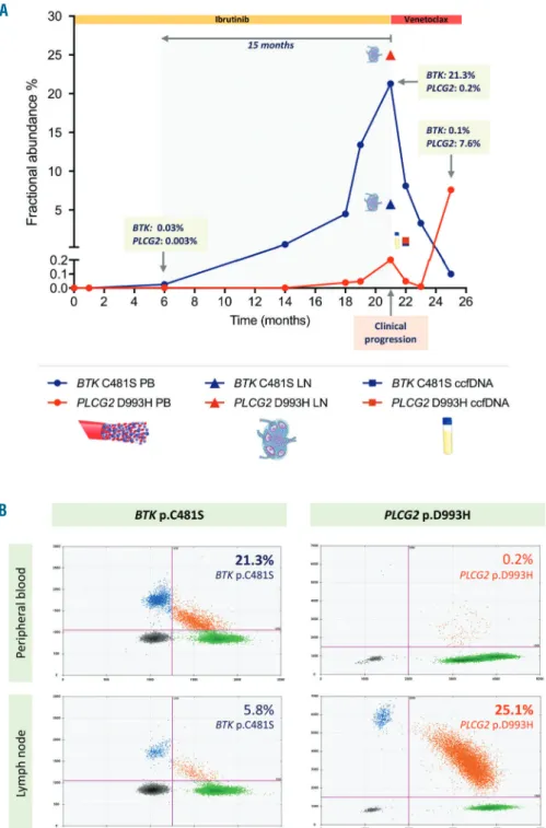
Figure 2. Spatiotemporal dynam- ics of the BTKand PLCG2muta- tions during ibrutinib treatment.
(A) Illustrated are the fractional abundances of the BTKp.C481S and PLCG2p.D993H mutations in peripheral blood, lymph node and circulating cell-free DNA (ccfDNA) during the course of the disease, as defined by digital droplet PCR
(ddPCR) assays. (B)
Representative ddPCR plots illus- trating the different representation of the BTKp.C481S and PLCG2 p.D993H mutations in peripheral blood and lymph node. The muta- tion burden was expressed as frac- tional abundance (FA) with the FA calculated as the ratio between the number of mutant DNA mole- cules and the number of mutant plus wild type molecules. The FA values were normalized to the CLL cell content as defined by flow cytometry. ccfDNA: circulating cell- free DNA.
A
B
response on ibrutinib for 21 months until the emergence of simultaneous lymphadenomegaly and lymphocytosis with ibrutinib resistance. Core biopsy was taken from an enlarged lymph node, and histology examination revealed Richter transformation to diffuse large B-cell lymphoma (DLBCL) (Figure 1A). The clonally related lymphoma was positive for CD20, BCL2, MUM1 and c- MYC, and negative for CD10, CD30 and BCL6, with a Ki67 index of 70% (Figure 1B). In addition to a common 13q deletion, 6q deletion and gain 8q were identified exclusively in the lymph node by multiplex ligation- dependent probe amplification (Figure 1A). The patient was subsequently treated with venetoclax (ramping dosage from 20 mg to 400 mg daily within 5 weeks). Due to progression after three months, high-dose salvage chemotherapy was administered (2 cycles of R-ESHAP:
rituximab + etoposide, cytarabine, cisplatin and methyl- prednisolone), followed by an autologous stem cell trans- plantation. The patient achieved complete remission assessed by CT scan and laboratory tests, but after a short, uneventful post-transplantational period, he relapsed and died on day 66 after transplantation, in July 2018.
To dissect the dynamics of mechanisms leading to spa- tial heterogeneity and ultimately conferring ibrutinib resistance, we analysed serial samples collected at ten different timepoints during the complete disease course (Figure 2A). Genomic DNA was isolated from ficoll-sep- arated peripheral blood mononuclear cells and native lymph node tissue using the Qiagen AllPrep kit (Qiagen).
Circulating cell-free DNA (ccfDNA) was isolated with QIAmp Circulating Nucleic Acid Kit (Qiagen) from peripheral blood samples collected in PAXgene Blood ccfDNA tubes (Qiagen). The fraction of tumor cells (CD5+/CD19+cells) was determined by flow cytometry.
Bidirectional Sanger sequencing of BTKexons 11, 15, 16, and PLCG2 exons 12, 19, 20, 24, 27 and 30 was per- formed on an ABI3500 genetic analyser (ThermoFisher).
Targeted ultra-deep next-generation sequencing (NGS) analysis covering the whole coding regions of the BTK and PLCG2genes was performed using a TruSeq Custom Amplicon approach on a MiSeq platform (Illumina). The same approach was used to perform NGS analysis of the hotspots of ATM, BCOR, BIRC3, EGR2, NOTCH1, NFK- BIE, MYD88, SF3B1 and TP53genes. Abundances of the PLCG2p.P993H and BTKp.C481S variants were quanti- fied using custom assays on a QX200 droplet digital PCR system (ddPCR, BioRad). The IGHV mutation status was determined according the most recent European Research Initiative on CLL (ERIC) recommendation.7The study was conducted in accordance with the Declaration of Helsinki.
The clonal relationship between the CLL at diagnosis, at the time of ibrutinib resistance and following Richter transformation was confirmed by identifying an identical IGHgene rearrangement and unmutated IGHV1-69 gene segment in all three specimens analysed.
Sanger sequencing revealed a canonical BTK p.C481S mutation in the peripheral blood at the time of relapse following ibrutinib treatment. Intriguingly, a PLCG2 p.D993H mutation was detected in the lymph node spec- imen, with the absence of the BTK p.C481S mutation present in peripheral blood, suggesting convergent evolu- tion in terms of the BTKand PLCG2variants with both commonly associated with ibrutinib resistance. To fur- ther scrutinize this spatial heterogeneity, BTK and PLCG2 mutations were screened by ultra-deep NGS which confirmed the Sanger sequencing data and, in addition to the dominant PLCG2p.D993H variant, iden-
tified the BTKp.C481S mutation as a minor clone in the lymph node, with the BTKp.C481S mutation remaining the exclusive variant in the peripheral blood (Figure 1A and 2A).
Next, we tested the presence of these mutations in the ccfDNA isolated at the time of ibrutinib relapse using a highly sensitive ddPCR approach. Notably, both muta- tions were identified in the ccfDNA with a VAF of 1%
(Figure 2A), supporting previous findings that ccfDNA may well represent a reliable source for screening BTK/PLCG2 mutations in patients with CLL undergoing targeted therapy.8
The scrutiny of clonal heterogeneity observed between the peripheral blood and lymph node compartment was extended to the most frequent mutation targets known in CLL by performing an ultra-deep NGS analysis of the ATM, BCOR, BIRC3, EGR2, NOTCH1, NFKBIE, MYD88, SF3B1and TP53genes. In addition to the common ATM p.Phe2393Val, NOTCH1 p.Gln2409* and SF3B1 p.Leu840Trp mutations identified in both compartments, the BIRC3p.Ser566fs and EGR2p.Ala132Thr mutations were exclusively identified in the peripheral blood (Figure 1A). The other genes showed a wild-type genotype.
Recent studies suggest that driver mutations in CLL most likely emerge at a pre-leukemic multipotent hematopoietic progenitor stage, with at least a fraction of somatic mutations occurring before disease onset.9,10The spatial clonal heterogeneity may thus develop from a pre- existing diverse mutation repertoire by differential selec- tive pressure of the treatment and microenvironmental effects in different anatomical niches. Although none of the previous studies detected BTKor PLCG2mutations prior to ibrutinib therapy, in silicoanalyses suggest that mutations even in these two genes may exist as early genetic alterations within a diverse cell population.4,11
To dissect the temporal and spatial dynamics of BTK and PLCG2mutant subclones, ddPCR analysis was per- formed on ten sequential peripheral blood samples col- lected during the course of the disease and a lymph node specimen obtained at the time of relapse, as summarized in Figures 1A and 2A. The BTKand PLCG2variants were absent in the pre-ibrutinib peripheral blood sample, how- ever, their emergence predated the clinical progression by 15 months, with the BTKand PLCG2mutations becom- ing detectable at month 6 on ibrutinib therapy with minor VAFs of 0.03% and 0.003%, respectively (Figure 2A). We observed a gradual clonal expansion of these variants in the sequential samples, demonstrating an explicit clonal selection under the selective pressure of the treatment. Of note, the ddPCR assay identified the PLCG2p.D993H mutation as a minor clone (VAF: 0.2%), previously unseen in the peripheral blood sample by NGS at the time of ibrutinib relapse. At this timepoint, the PLCG2 p.D993H mutation represented a major clone (VAF: 25%) in the lymph node sample, with a concurrent minor BTKp.C481S clone (VAF: 5.8%) (Figure 2A). The two subclones displayed differential sensitivity to the subsequent venetoclax therapy, most likely conferred by Richter transformation present in the lymph node com- partment. In the peripheral blood, we observed a reduc- tion of the BTKp.C481S positive subclone accompanied by expansion of the subclone harbouring PLCG2 p.D993H which was previously dominant in the lymph node (Figure 2A). With these dynamic changes in the sub- clonal architecture, the disease progressed and ultimately led to the death of the patient.
Clonal evolution is a major driving force of relapse and progression of cancer, including haematological malig- nancies.12 Several studies have traced the effect of treat- haematologica 2018; 103:e40
C ASE R EPORTS
ment on temporal evolutionary trajectories of CLL in the context of standard as well as targeted therapies and identified profound subclonal heterogeneity and active clonal selection in the majority of cases.6,13,14 However, the spatial aspect of subclonal dynamics has not been appreciated until recently. Here, we presented a unique case of CLL, developing ibrutinib resistance via multiple routes simultaneously, with the observed spatial hetero- geneity leading to progression and aggressive transforma- tion of the disease. This case highlights the importance of genetic profiling of multiple affected sites as investiga- tions restricted to peripheral blood may underestimate the repertoire of clinically relevant genetic alterations present in the patient.
Richárd Kiss,1Donát Alpár,1Ambrus Gángó,1
Noémi Nagy,1Ediz Eyupoglu,1Dóra Aczél,1András Matolcsy,1 Judit Csomor,1Zoltán Mátrai2and Csaba Bödör1
1MTA-SE Momentum Molecular Oncohematology Research Group, 1st Department of Pathology and Experimental Cancer Research, Semmelweis University, Budapest and 2Department of Haematology and Stem Cell Transplantation, St. István and St László Hospital, Budapest, Hungary
Correspondence: bodor.csaba1@med.semmelweis-univ.hu doi:10.3324/haematol.2018.202085
Funding: this work was funded by the Momentum grant (LP- 95021) and the János Bolyai Research Scholarship of the Hungarian Academy of Sciences, the KH17-126718, NVKP_16-1-2016-0004, NVKP_16-1-2016-0005 and K_16 #119950 grants of the Hungarian National Research, Development and Innovation Office (NKFIH) and the ÚNKP-18-4-SE-62 and ÚNKP-18-3-I-SE-48 grants of the Ministry of Human Capacities. MLPA probemixes and reagents were kindly provided by MRC-Holland (Amsterdam, The Netherlands).
Information on authorship, contributions, and financial & other disclo- sures was provided by the authors and is available with the online version of this article at www.haematologica.org.
References
1. Farooqui MZ, Valdez J, Martyr S, et al. Ibrutinib for previously untreated and relapsed or refractory chronic lymphocytic leukaemia
with TP53 aberrations: a phase 2, single-arm trial. Lancet Oncol.
2015;16(2):169-176.
2. O'Brien S, Furman RR, Coutre S, et al. Single-agent ibrutinib in treat- ment-naive and relapsed/refractory chronic lymphocytic leukemia: a 5-year experience. Blood. 2018;131(17):1910-1919.
3. Ahn IE, Underbayev C, Albitar A, et al. Clonal evolution leading to ibrutinib resistance in chronic lymphocytic leukemia. Blood.
2017;129(11):1469-1479.
4. Burger JA, Landau DA, Taylor-Weiner A, et al. Clonal evolution in patients with chronic lymphocytic leukaemia developing resistance to BTK inhibition. Nat Commun. 2016;7:11589.
5. Kadri S, Lee J, Fitzpatrick C, et al. Clonal evolution underlying leukemia progression and Richter transformation in patients with ibrutinib-relapsed CLL. Blood Advances. 2017;1(12):715-727.
6. Landau DA, Sun C, Rosebrock D, et al. The evolutionary landscape of chronic lymphocytic leukemia treated with ibrutinib targeted therapy. Nat Commun. 2017;8(1):2185.
7. Rosenquist R, Ghia P, Hadzidimitriou A, et al. Immunoglobulin gene sequence analysis in chronic lymphocytic leukemia: updated ERIC recommendations. Leukemia. 2017;31(7):1477-1481.
8. Albitar A, Ma W, DeDios I, et al. Using high-sensitivity sequencing for the detection of mutations in BTK and PLCgamma2 genes in cel- lular and cell-free DNA and correlation with progression in patients treated with BTK inhibitors. Oncotarget. 2017;8(11):17936-17944.
9. Agathangelidis A, Ljungstrom V, Scarfo L, et al. Highly similar genomic landscapes in monoclonal B-cell lymphocytosis and ultra- stable chronic lymphocytic leukemia with low frequency of driver mutations. Haematologica. 2018;103(5):865-873.
10. Damm F, Mylonas E, Cosson A, et al. Acquired initiating mutations in early hematopoietic cells of CLL patients. 2014;4(9):1088-1101.
11. Komarova NL, Burger JA, Wodarz D. Evolution of ibrutinib resist- ance in chronic lymphocytic leukemia (CLL). Proceedings of the National Academy of Sciences of the United States of America.
2014;111(38):13906-13911.
12. Landau DA, Carter SL, Getz G, Wu CJ. Clonal evolution in hemato- logical malignancies and therapeutic implications. Leukemia.
2014;28(1):34-43.
13. Landau DA, Carter SL, Stojanov P, et al. Evolution and impact of sub- clonal mutations in chronic lymphocytic leukemia. Cell.
2013;152(4):714-726.
14. Schuh A, Becq J, Humphray S, et al. Monitoring chronic lymphocytic leukemia progression by whole genome sequencing reveals hetero- geneous clonal evolution patterns. Blood. 2012;120(20):4191-4196.
haematologica 2018; 103:e41