based on the Hungarian practical notes entitled
"Mikrobiológiai Laboratóriumi Gyakorlatok"
entitled "Mikrobiológiai Laboratóriumi Gyakorlatok"
Authors of the Hungarian and English versions: Erika M. Tóth, Andrea K. Borsodi, Tamás Felföldi, Balázs Vajna, Rita Sipos and Károly Márialigeti
Authors of the Hungarian version: Csaba Romsics, Judit Makk, Katalin Jáger, Márton Palatinszky and Éva Ács Editors of the English version: Erika M. Tóth and Károly Márialigeti
Language assistant: Attila Náfrádi
Copyright © 2013 Eötvös Loránd University
This book is freely available for research and educational purposes. Reproduction in any form is prohibited without written permission of the owner.
Made in the project entitled "E-learning scientific content development in ELTE TTK" with number TÁMOP-4.1.2.A/1-11/1-2011-0073.
Consortium leader: Eötvös Loránd University, Consortium Members: ELTE Faculties of Science Student Foundation, ITStudy Hungary Ltd.
1. PREFACE ... v
2. WORK IN A MICROBIOLOGICAL LABORATORY ... 1
2.1. Biological safety level categories and the airborne route of pathogen transmission ... 1
2.2. The principle of containment, the setup and basic pieces of equipment of a microbiological laboratory ... 2
2.3. Biological safety cabinets ... 4
2.4. Behaviour and work in a microbiological laboratory ... 5
2.4 1. What to do in case of biological spill (involving BSL2 microorganisms) ... 7
2.4.2. Decontaminating hygienic hand wash and personal decontamination ... 7
2.4.3. Emergency and first aid guide ... 8
3. STERILISATION AND DISINFECTION ... 10
3.1. Procedures of sterilisation ... 10
3.1.1. Sterilisation by heat ... 10
3.1.2. Sterilisation by radiation ... 12
3.1.3. Filter sterilisation ... 12
3.1.4. Sterilisation by chemicals ... 13
3.2. Procedures of disinfection ... 13
3.3. Control of the efficacy of sterilisation equipment ... 14
3.4. Determination of the microbiological efficacy of disinfectants ... 15
4. SAMPLING METHODS IN MICROBIOLOGY ... 17
4.1. Sampling for diagnostic purposes ... 17
4.2. Sampling from various environments ... 17
4.2.1. Collection of air samples ... 17
4.2.2. Collection of soil samples ... 19
4.2.3. Collection of water samples ... 20
4.2.4. Sampling the surface of objects ... 21
4.2.5. Hygienic control of the hands of operators ... 22
5. INTRODUCTION TO THE USE OF PRACTICAL LABORATORY MICROSCOPES ... 24
5.1. Bright-field light microscopy ... 24
5.2. Fluorescence microscopy ... 26
6. CELL- AND GERM-COUNTING METHODS ... 27
6.1. Determination of cell counts with microscope ... 27
6.2. PCR-based cell counts ... 30
6.3. Determination of germ counts based on cultivation ... 30
6.3.1. CFU-counting techniques ... 30
6.3.2. The membrane filter technique ... 33
6.3.3. The end point dilution technique (MPN method) ... 34
7. STRAIN CULTURE AND CULTIVATION-BASED TECHNIQUES ... 36
7.1. Demonstration of microbes in the environment ... 36
7.2. Preparation of microbiological culture media ... 38
7.3. Basic bacterial cultivation techniques ... 42
7.3.1. Enrichment ... 42
7.3.2. Spread plate and pour plate methods ... 44
7.3.3. Isolation and streak plate technique ... 44
7.3.4. Anaerobic cultivation techniques ... 47
7.3.5. Transfer, maintenance and storage of pure cultures ... 50
7.4. Pheno- and genotypic characterisation of bacterial strains ... 54
7.4.1. Colony- and cell morphology, staining procedures ... 54
7.4.2. Study of bacterial enzymes ... 66
7.4.3. Physiological and ecological studies on bacterial strains ... 75
7.4.4. Study of the effect of antimicrobial compounds and antibiotics ... 79
7.4.5. Chemotaxonomical studies of bacterial strains ... 87
7.4.6. Bacterial species identification based on 16S rDNA sequence homology ... 92
8. ANALYSIS OF THE MICROBIOLOGICAL QUALITY OF DIFFERENT ENVIRONMENTS ... 104
8.1. Microbiology of surface waters and wastewaters, hygienic control ... 104
8.2. Soil examinations ... 108
8.3. Examination of microorganisms participating in the nitrogen cycle ... 110
8.4. Examination of microorganisms participating in the sulphur cycle ... 115
9. FERMENTATION PROCESSES IN BIOTECHNOLOGY ... 117
10. DATA ANALYSIS – TAXOMETRICS ... 121
11. BASIC ALGOLOGICAL METHODS ... 123
12. SUPPLEMENTARY MATERIAL ... 125
12.1 Supplementary exercises ... 125
12.2. Taxon information sheets with supplementary figures ... 133
12.3. Test results with supplementary figures ... 145
13. LIST OF EXERCISES ... 151
14. APPENDIX ... 155
14.1. Using LABOVAL 4 type microscopes for bright-field light microscopic observations ... 155
14.2. Culture media used in the practical ... 155
14.3. Dyes, reagents and solutions used in the practical ... 168
14.4. Tables ... 172
15. WORKING DEFINITIONS ... 175
16. REVIEW QUESTIONS ... 182
17. REFERENCES ... 222
The scientific community has always played distinguished attention to explorers of the remote quarters of our Earth, and to the description of their natural history. The founding fathers of microbiology, Louis Pasteur (1822- 1895), Ferdinand Kohn (1828-1898) and Robert Koch (1843-1910), just to mention a few, were not yet born, or just started school, when Alexander von Humboldt (1769-1869) set sail to America and returned with an amazing collection of plants and fossils (1799-1804). The same is true in the case of Charles Darwin (1809-1882), who participated in an expedition circumnavigating our Globe on board the Beagle survey barque (1831-1836), described his observations, and returned with several plants and animals, etc.
This kind of work based on animated exploratory, descriptive data gathering in the field of microbiology started in the 1870’s by the development of culture methods. By then, Darwin had already concentrated on his explanatory work on biogenesis and constituted his hypotheses on the origin of species. This work initiated an enormous series of hypothesis-driven studies. However, in scientific research, this kind of ambiguity (data-driven exploratory and descriptive studies versus hypothesis-driven explanatory and interpretative research) has always been characteristic.
With the development of the novel genome-based molecular approaches, a new era of diversity exploration has started in microbiology. “Good old hypotheses” based on strain culture studies got turned around, but new data are not yet adequate to reach satisfactory explanations. It is intriguing to participate in this variegation of microbi- ological studies by either exploring the diversity, or explaining the scientific background of environmental obser- vations.
This practical guide collects and explains the most basic techniques used in general microbiology. Mastering these methods will help the students in many other practical disciplines that apply the techniques of aseptic work, steril- isation and disinfection, or work with laboratory cultures. The series of practical exercises is compiled mainly ac- cording to the logic of the exploration and description of the microbial diversity of an environment. Thus, it starts with the description of a microbiological laboratory, preparatory work (sterilisation, etc.), environmental sampling, the microscopic investigation of samples, the methodology of culture and phenotypic characterisation of strains, and the basic molecular identification techniques. Finally, applied microbiological techniques are described briefly, like practises to characterise microbes participating in the various cycles of elements, or the basic techniques of microbiological qualification of water, and some essential biotechnologies.
The description of the practical exercises is built up similarly. They start with a short introduction describing the principle, then the object of the investigation (i.e. strains or environmental sample), and the applied tools and in- struments are listed. The followed procedure is described in the end. The practical sessions in basic microbiology at Eötvös Loránd University, Faculty of Science are organized on a weekly basis throughout the semester. Thus, where possible, culture incubations last for a week (even when it is not the optimal duration) and students get many preparations pre-arranged (e.g. 24-hour cultures). On the contrary, advanced practical exercises (like in molecular microbial ecology) are organized into week-long blocks, thus these practical exercises are arranged accordingly.
There is every reason to expect a rapid change in basic microbiological laboratory methodology in the near future.
The electronic edition makes frequent modifications possible. The authors will use this opportunity to delete, change, expand or insert exercises in due time.
MICROBIOLOGICAL LABORATORY
Safety in a microbiological laboratory substantially differs from that in other (chemical or physical, etc.) laborat- ories because, in addition to hazardous chemicals, substances and operations that pose a laboratory work-related risk, there is a risk of infection when working with microbes. The presence of, and working with infectious agents and materials in a microbiological laboratory, i.e. the potential of acquiring laboratory-associated infection, assumes the application of hierarchical control methods. These control measures first take into account the knowingly or unknowingly (e.g. as is the case in environmental microbiology) handled infectious agents; the approval of labor- atory practices and safety equipment used (good laboratory practices [GLP]; containment approach), and the level by which laboratory workers are aware of the risk of infection (behavioural factors). Therefore, safety programs and safety management are organised with these questions in mind.
Epidemiologic analysis of (laboratory acquired) infections (including not only symptomatic infections, but similarly nonsymptomatic seroconversions as well) made the constitution of risk categories among microbes possible (based on health effects, means of spreading, routes of entry, etc.); and biological safety level requirements in laboratories (containment measures combining laboratory practices, safety equipment and design) in order to prevent the ex- posure of the operator, their colleagues and the broader environment.
The most common ways of exposure to infectious agents are percutaneous inoculation (through injuries caused by sharp contaminated objects and animal bites, scratches, etc.), aerosol inhalation (as a result of spills, or caused by sprays associated with work procedures, e.g. vortexing; the mere opening of a Petri dish culture of a sporulating fungus; work with lyophilised cultures, etc.), and ingestion (e.g. during mouth pipetting, or by eating or drinking in the laboratory). Since the infectious dose of a disease-causing agent is vital, the higher concentration of microor- ganisms associated with certain research procedures (e.g. cultivation) increases the risk. “Infectious dose” is the number of microbial cells that cause an acute infection in humans. E.g. certainVibrio choleraestrains cause a disease when ingesting only 10 cells, whereas with someEscherichia colistrains, >106cells are needed “per os”
for disease induction.
In the laboratory, researchers, assistants and students are exposed to the highest risk; however, one has to take into account the exposure of the cleaning, dishwasher and maintenance staff. The aforementioned laboratory workers are usually assumed to be healthy individuals in risk assessments. However, there are health status conditions, which increase the risk of infection. Different life phases, some (even chronic) diseases and the use of certain medications influence the host’s defence (e.g. pregnancy with the threat of foetal or congenital infection; allergic hypersensitivity, immunodeficiency caused by e.g. diabetes mellitus, cancer chemotherapy, etc.). Moreover, working in a laboratory can result in allergic reactions (e.g. to spore proteins of actinobacteria).
When talking about (microbiological) laboratory in its broadest context, an environmental microbiologist will also consider field trips, the collection and on-site investigation of samples in their natural environment. It is easy to imagine the risk of infection at a communal sewage treatment plant or at a waste deposition site, not to mention other obviously infectious events like the sampling of cadavers/carcasses.
2.1. Biological safety level categories and the airborne route of pathogen transmission
The grouping of microorganisms into four biological safety level (BSL) categories is mainly based on the severity of the disease they cause and their transmission route, since airborne transmission (i.e. transmission via aerosol) is the most difficult to controll. Laboratory facilities and the required laboratory techniques and practices are sim- ilarly classified into four safety levels according to the agent. Organisms in BSL 1 are not known to cause any disease in healthy adults. Working with them needs practically no aerosol containment. BSL 1 facilities are adequate for teaching laboratories at post-secondary or undergraduate training level. In such laboratories, only a sink to wash hands for decontamination is required. Microbes in BSL 2 group are transmitted with ingestion, or via contact with mucous membranes (or by accidental self-injection), however their high concentration in aerosols may result in transmission (high infectious dose at droplet infection). Thus, in the case of working with such microbes, aerosol-
generating laboratory practices have to be contained with the use of an adequate biological safety cabinet (BSC).
Personal protective equipment should be used when appropriate (laboratory coats, splash-protecting glasses and goggles, gloves, etc.). Naturally, washing hands for decontamination is a requirement. Adequate waste collection and decontamination facilities must be available (biohazard waste collecting bags and boxes, containers with mi- crobicide liquid for used pipettes and other consumables, terminal decontamination autoclave, etc.). Microbes ranked as BSL 3 cause disease in humans and explicitly spread airborne (with low infectious dose). In this case, all of the activities with materials that are as much as suspected to be contaminated have to be performed in adequate BSC. Access to the laboratory must be controlled, and adequate ventilation systems are needed to minimise the risk of the release of infectious aerosols. Microbes or samples that are verified or only supposed to have a high risk of causing serious or even fatal disease in humans, independently of the transmission route, are categorized as BSL4. In BSL4 facilities, the highest-level BSCs are used or/and the laboratory personnel is protected by special ventilated suites.
Not only the microorganisms themselves and the infection pose biological hazard, but the metabolic products of the microorganisms are similarly of concern (e.g. toxins, biotransformation products, such as vinyl chloride).
Special care has to be taken to control the (occupational) exposure to such compounds.
Special safety measures regulate biotechnological applications and the use of recombinant technologies especially when large-scale (> 10 L) applications are used. When considering recombinant techniques, well-characterized non-pathogenic hosts should be used, where the presence of incidental events can be excluded. Inserts should be similarly well characterized, free of “harmful” genes. Vectors should be as small as possible in size so they are unable to transfer DNA to wild-type hosts.
Since BSL categories strictly relate to the airborne pathogens and the airborne route of pathogen transmission, it is advisable to briefly summarize the ways by which aerosol is formed in laboratories. Most bacteria and yeast grown in the laboratory on solid media form butyrous cohesive masses, making it unlikely to form aerosol when the culture container is opened. On the contrary, sporulating (conidiospore-forming) bacterial and fungal colonies pose a hazard of spore aerosol formation for example with the mere opening of a Petri dish. For this reason, in the case of such cultures grown for prolonged periods, lids should be taped, not to be opened before prior examination for sporulation (presence of aerial hyphal forms), and should only be opened in BSCs. On the other hand, the ma- nipulation of cultures like subculturing (e.g. the ignition of an inoculating loop), preparation of suspensions (by e.g. vortexing), centrifuging suspensions/broth cultures, pipetting, using blender type homogenizers, etc. are all procedures where small liquid droplets (aerosol) containing (infectious) cells (materials) may form. The larger particles (> 150 µm) readily drop, dry and form dust and thus contaminate bench top and floor surfaces. Particles smaller than 150 µm in diameter will most possibly evaporate before reaching the ground, forming “droplet nuclei”, which may hover for long periods. Droplet nuclei may even penetrate tissue facemasks. All microbes that are de- siccation resistant (e.g.Staphylococcus,Mycobacteriumspp., sporulating microbes) are of stressed importance since they remain alive for longer periods. Their UV tolerance further increases the risk of infection. It is recom- mended to work with the risk of aerosol/droplet nuclei formation in BSCs, and used contaminated materials (e.g.
pipette tips, tubes) should be carefully submerged into disinfectant. The risk of formation of “droplet nuclei” con- taining infectious dust, especially neccessitates the thorough, regular, disinfective cleaning of surfaces in a micro- biological laboratory.
The prevention of aerosol formation is an important aspect in the development of good laboratory practice measures.
Thus, when subculturing e.g.Mycobacterium tuberculosis,in spite of using ordinary loops and a gas burner, rather the use of electric incinerators or the application of disposable loops is required. Similarly, centrifugation (especially high-speed centrifugation) should be made in aerosol-proof safety tubes/containers, and even the rotors should be tightly covered.
2.2. The principle of containment, the setup and basic pieces of equipment of a microbiological laboratory
The conduct of work with infectious agents assumes the application of containment practices. Primary containment or primary barriers are the first line of defence encapsulating the infectious agent (animal, person, sample, etc.).
The culture vessel (e.g. a cotton-plugged test tube) is the primary containment to isolate (enclose) a(n infectious) culture. Opening the vessel intentionally (e.g. to subculture) or unintentionally (due to inadvertent handling) results in exposure, when other means of primary containment (e.g. directed air flow in a BSC, together with high-efficiency particulate air [HEPA] filter in the exhaust pipe or the charcoal filters built in the air path) will keep the agent as close to the site as possible. In the case of unintentional spill, infected surfaces need decontamination using prescribed techniques (disinfection protocols). Thus, primary barrier systems i. minimise the infectious volume, ii. ensure a safe environment for procedures with infectious agents, and iii. provide decontamination measures.
Primary containment is complemented with personal protective equipment (PPE) to further prevent human contact (e.g. skin contact, inhalation) with the agent. The most common personal protective equipment includes laboratory clothing (laboratory coats or body suits), aprons, gloves, eye protection, respiratory protection, head covers, shoe covers, etc. Adequate PPE should be selected to provide protection not only from biological hazards, but also to reduce the exposure to chemical and physical agents involved (e.g. toxic substances or radioactive material). To give an example, disposable rubber surgical gloves adequate to prevent contamination of the hands with infectious material are normally degraded by the solvent xylene, or do not protect from the carcinogenic DNA stain ethidium- bromide. Thus, in such cases, nitrile gloves should be selected. Another example is that disposable polyester “wrap around” gowns, due to their continuous solid front, give adequate splash protection but the material will melt on contact with heat. 100% cotton laboratory coats are flame resistant and nonreactive to many chemicals, but usually have front buttoning, leaving outdoor clothes exposed to contamination.
While primary barriers are designed to protect the personnel and the immediate laboratory environment from contamination, the elements of secondary containment protect the external environment of the laboratory from contamination. This can be achieved by a combination of facility design and operational practices.
Concerning facility design, the most important elements are simplicity and clarity, easy maintenance and operation.
The allocation of adequate space to the planned operations on a long-term basis usually helps. The design of the building and of the laboratories should help keep hazards away from laboratory personnel, restrict the hazard to the smallest affected area, help the treatment of hazardous situations and clean up. Thus, microbiological research (and educational) laboratories should be physically clearly separated from ancillary laboratories (e.g. scullery, media preparation, sterilisation rooms), support rooms (environmental room, room for special instrumentation), storage rooms, and the administrative area, though positioned as close to each other as possible. Separate resting and eating facilities and male/female changing rooms/showers and toilets for the personnel should be available.
Corridors must not be used as storage areas or as secondary laboratory workspaces. This is necessary not only to decrease contamination hazard, but also to meet fire rating criteria.
A typical two-window (two-module) laboratory is depicted in Fig. 1. There is enough space allotted for basic laboratory operations and for equipment (e.g. microscope, bench top centrifuges, water bath, PCR). The built-in cupboards below the benches, and the upper shelves and cupboards serve to store chemicals, laboratory vessels, utensils in immediate use (a laboratory is not a warehouse). There is a sink and a lavatory near the door, their door opens outside of the laboratory. Doors should be minimum 1 m wide to allow access of anticipated equipment.
There is a BSC located where minimum external air currents are expected: furthest from the door and the heat- ing/cooling fans.
BSL 3 and 4 laboratories have more strict design regulations (e.g. fixed windows, changing rooms with hygienic shower, separate access facility with two sets of doors for interchange, air lock systems, etc.) but these are out of the scope of this practical guide.
The finishing of surfaces in the laboratory is designed to be easy to clean. Walls, floors and ceilings should be water resistant. Utility (etc.) penetrations should be sealed or capable of being sealed. Care should be taken to prevent the surfaces from being continuously wet (e.g. because of vapour precipitation or dripping at refrigerators, sinks) since wet surfaces help (infectious) biofilm formation. Laboratory furniture should be compact, firm and durable, and stand on feet to help cleaning. Bench tops should be resistant to acids, alkalis, solvents and moderate heat.
Laboratory benches should include adequately mounted access to electricity (220/240 V and 360/400 V similarly), gas and water beyond the sink. Laboratory work is made extremely easy when there is a built-in, continuous access to pure (reagent grade) water (e.g. distilled or reverse osmosis treated water), cooling water, vacuum and space for the operation of chemical hoods or at least exhaust trunks (for working with toxic volatile/dusting chemicals).
However, utilities have to be assembled with adequate traps to prevent the spread of infections (e.g. in centralized
vacuum systems). In extreme cases, rather special local appliances should be used. Chemical safety equipment includes emergency shower, eyewash stations, and vented storage for flammable/corrosive chemicals. When no adequate environmental rooms are available, storage equipment include incubators, refrigerators, and freezers (with temperature monitoring and alarm signals). Sterilisation equipment (autoclaves, dry air sterilisers, etc.) is of primary concern including also a dedicated instrument for terminal sterilisation of infected/contaminated materials before disposal.
Animal care facilities usually belong to the (micro)biological research/education environment. Even their basic setup falls out of the scope of this practical guide.
Microbiological laboratories should best be separated from areas with unrestricted access in the building. Controlled access using card-key electronic monitoring of laboratory sections is optimal. Moreover, care should be taken at the design of ventilation and air conditioning systems of microbiological laboratories. First, these systems should be adequately separated not to transmit contaminated air to the environment and to control cross-contamination among laboratories; second, air currents should not disturb safe laboratory practices. Laboratories should have adequate waste-handling and disposal provisions and regulations (including transitional storage and handling of general waste, biomedical waste, infectious waste, chemical waste, etc.). Vermin and rodent control are part of the management routine.
Fig. 1. Microbiological laboratory layout.Typical microbiological laboratory with ideal location of biological safety cabinet: 1. heating/air diffuses, 2. biological safety cabinet, 3. table, 4. chair, 5. sink.
2.3. Biological safety cabinets
In the early times of microbiology, the most dangerous (i.e. techniques with aerosol formation) microbiological laboratory operations (like subculturing, handling suspensions, centrifuging, etc.) were restricted to separated chambers within the laboratory, supplied with UV air disinfection (“inoculating rooms”), as a form of primary containment. Increased efforts to strengthen chemical laboratory work safety led to the development of safety hoods. Parallel efforts to protect against (micro)biological exposure led to the development of BSCs (a closed cabinet designed for easy decontamination, with a view screen protecting from splashing and splintering, and continuous air flow through the cabinet with exhaust decontamination). Today, BSCs are manufactured in three general classes, and are combined with HEPA filters, providing clean (“particle free”, near-sterile air) work benches due to laminar airflow. The use of HEPA filters assumes the presence of internal fans in BSCs to provide adequate, directed airflow in the cabinet. BSCs may be ducted (connected to an exhaust) or not ducted. Exhaust air is usually similarly decontaminated with the use of HEPA filters; and in some instances, activated carbon filters may be built into the internal and/or exhaust air stream to adsorb toxins or other hazardous biological/chemical agents.
Chemical hoods are built to protect the worker and the environment (air current, and exhaust filtration/dilution).
BSCs equipped with HEPA filters similarly protect the product/biological procedure (aseptic/sterile operation in the workspace) and provide primary containment (keeping contaminated air in the cabinet).
Class 1 cabinets protect only the operator, but give no protection to the product/procedure. Laboratory air is sucked through the cabinet and the apparatus is equipped with a decontaminated exhaust (with HEPA/activated carbon filters) to the environment. It has to be mentioned at this point that horizontal-flow clean-air benches often used in biological laboratories to protect the product/procedure (e.g. cell culture) provide no operator protection; on the contrary, it may expose the operator to infectious, toxic, allergenic etc. materials deriving from the product. Such devices are not suitable in microbiological laboratories!
Non-ducted class 2 cabinets with HEPA filtered exhaust air fed back into the laboratory are the most common in BSL 1 and 2 microbiological laboratories. These provide protection to the operator and the work simultaneously.
In case of class 2a cabinets, the air-blower recirculates 70 % of the air through a HEPA filter into the workspace, while 30 % is forced through another HEPA filter back into the laboratory air. On the suction side, 30 % fresh air enters at the fringe of the workbench (with 0,4 m/s inflow velocity) preventing the contamination of the laboratory, together with 70 % contaminated air from the workspace. This way, the cabinet provides through the HEPA filter a downward particle free (nearly sterile) laminar air stream over the work-bench, and maintains a continuous air intake through the opening of the view window/work opening (which should not be higher than 20-30 cm during work). The cabinet is sensitive to the disruption of the vertical laminar airflow by objects or devices causing turbu- lence. E.g. the upward airflow caused by gas burners can cause faults, as can the littering of the workbench with unnecessary objects.
Class 2b BSCs maintain a higher fringe inflow air velocity (0,5 m/s), and have ducted exhaust outside the building.
This class BSCs have several subtypes, including also total exhaust cabinets for work with carcinogens.
Class 3 BSCs are maximum containment instruments used in BSL4 laboratories. They are e.g. ducted glove boxes with HEPA filtered clean-air workbenches, where materials are transferred into the work area through an interchange.
The workspace of the BSC is usually equipped with UV lamps, which enable disinfection at layoff, and have op- tional access to public/laboratory services (gas, vacuum, pressure air, water, etc.) They are constructed leak-proof, of corrosion resistant materials to withstand the chemicals generally used, and the terminal decontamination with formaldehyde vapour at filter change, and other service/repair operations.
BSCs, or very clean areas may be built around equipment with contamination hazard (e.g. high-speed centrifuges, fermenters, autoclaves, blenders, animal experiments).
2.4. Behaviour and work in a microbiological laboratory
Good laboratory practices with responsible behaviour at work can prevent most work-related exposures to infectious agents. The basic biosafety practices, compulsory at biosafety level two laboratories and recommended at biosafety level one, are as follows.
• Students should get appropriate training on the potential hazards and prevention measures at the start of the laboratory course, which should be refreshed when applying techniques of (infection) risk. For laboratory per- sonnel, entry training and annual updates are necessary (e.g. policy changes, additional training). Training should be documented.
• Access to the laboratory is limited to formal students and laboratory personnel, especially when work with cultures is in progress. Persons who are at increased risk of infection (e.g. pregnant women, nursing mothers, immuno- compromised people) may be excluded from laboratory work.
• Use 100% cotton laboratory coat/wrap-around gown, and/or other personal protective equipment. Gloves should be worn, particularly when the skin on the hands is injured. Laboratory safety glasses, face shield, aprons should be worn when appropriate. Comply with the indication of the laboratory supervisor! Personal protective equipment and clothing should be removed and left in the laboratory before leaving to non-laboratory areas. Protective clothing is either disposed of or laundered in the department/institution.
• Confine long hair and loose clothing. Wear closed shoes in the laboratory. Nails should be kept clipped and neat.
• Do not work alone in the laboratory if the conducted procedures are hazardous. Always make a work plan when working with hazardous materials. Always handle infectious materials with extreme care. Respect the requirements of the spill control and clean-up instructions. All procedures should be performed carefully to minimise aerosol formation.
• Decontaminate the workbench before and after work.
• Do not pipette by mouth, use pipetting devices. Do not drip contents from the pipette, touch a surface with the tip instead to let the contents slowly run out. When pipetting infectious material, do not “blow out” the pipette (tip). Avoid the use of sharp objects (syringe, needles, broken glass, scalpels, etc.)
• Do not speak when working with cultures, and do so only when necessary. Avoid practical jokes or other con- fusing behaviour, which may distract other students/personnel.
• Wash your hands regularly (especially following work). Do not eat, drink, smoke or apply cosmetics in the laboratory; do not take pen and ink or any other objects into your mouth. Do not store food in the laboratory.
Do not handle contact lenses in the laboratory. Persons wearing contact lenses should wear eye protection (protective safety glasses/goggles).
• Cultures, stocks and wastes should be decontaminated (e.g. autoclaving) before disposal. Materials decontaminated outside the (teaching) laboratory should be collected in leak-proof well-built containers, and closed for transport to the decontamination site (autoclaving).
• Spills and accidents should be immediately reported to the laboratory supervisors.
• Chemicals and equipment should be always properly labelled and the work area should always be kept tidy, not cluttered. Place items in use in the logical order of the applied procedure.
• Aseptic work with cultures in a microbiological laboratory is practically always performed with gas burners on.
The opening of glass culture vessels, metal caps, etc. are flamed between operations.
Be aware that in a microbiological laboratory, an extremely broad range of chemical hazards is represented because of the variety of chemicals used (solvents, acids, bases, carcinogens, mutagens, etc.). Be sure of the proper use of chemicals!
At laboratory/department level, a chemical inventory should be kept, and purchase should be based on real necessity.
Minimise the use of hazardous chemicals. Material safety data sheets of chemicals should be readily available.
Only the most important basic laboratory and “chemical hygiene” measures are summarized below.
• Be aware of the safety hazard categories of the chemicals indicated by hazard warning and identity symbols on the containers (e.g. acutely toxic material, oxidising agent, inflammable material). Use only the smallest aliquots possible in the laboratory. Avoid skin contact with chemicals, do not smell vessels.
• When using electrically powered laboratory instruments, observe the directions for safe use!
• Provide appropriate ventilation, when working with volatile materials, use chemical hoods.
• Before commencement of work, locate the places of safety equipment in the laboratory (e.g. emergency shower, eyewash, first aid kit). Always respect the warning at areas/equipment where special protective equipment should be used!
• Do not discharge mercury and other heavy metal containing materials and flammable liquids into the sink.
Comply with waste disposal regulations.
• Be extremely careful with hot instruments (autoclave, dry heat sterilisers, etc.) and when working with dry ice or liquid nitrogen. They may cause burns. Drying and ignition ovens, other hot plates should be operated over insulating underplates. Use adequate protective equipment when working with vacuum or pressure air (e.g. fil- tration).
• When using UV light (e.g. at DNA-based studies evaluated with transilluminators), always wear eye/face pro- tection.
• Comply with the directions summarized on the instruction sheet when using special laboratory apparatus (e.g.
autoclaves, centrifuges, vacuum drying apparatus). In case of doubt, ask the instructor!
• Comply with the regulations when using compressed gas cylinders. Cylinders should always be secured in an upright position. Do not store cylinders in the laboratory (when empty or not in use). Comply with the regulations related to the instrument (e.g. oxygen should never come in contact with grease and oil).
• In microbiological laboratories, working with open flame (gas burner/surface sterilisation by flaming with eth- anol) is a daily routine. Keep flammable materials away from the flame!
• Emergency telephone numbers, location signs (exits, first aid equipment, , etc.), special warning signs (e.g.
biohazard, use of UV protection masks) should be adequately posted.
• An adequate fire extinguisher must be available. Locate the location of the fire extinguisher in the laboratory, and get familiar with its use. In case of extensive fire, call the fire brigade, their number in Hungary is 105 or 112. Proceed as in section 2.4.3.
• When strong acid or alkali contaminates the skin, immediately rinse it with plenty of water. In the case of acids, use 3 % sodium-hydrogen carbonate (sodium bicarbonate) , in case of alkalis use 3 % boric acid solution to neutralise, and then wash your hands with soap or treat the burns (see 2.4.3.). If acid squirts into the eye, wash it by dropping with 2 % borax solution, then rinse with physiological saline. In the case of alkali, use 2 % boric acid, and then rinse with physiological saline. Apply bandage and consult an eye specialist.
• Work-related emergencies/accidents must be reported immediately to the supervisor. Accidents causing work drop-out should be reported to the person in charge of work-safety or the work safety director of the institution, and the case should be examined. In cases endangering human life, the spot of the accident should be left un- touched for field inspection.
2.4 1. What to do in case of biological spill (involving BSL2 microorganisms)
• Alert people in the immediate area of the spill. Report to the laboratory supervisor.
• Put on protective equipment (e.g. gloves, eye protection, apron).
• Cover spill with adsorbent material (paper towel e.g.).
• Poor freshly diluted household bleach (1 to 10 dilution with water) carefully around the edges and onto the spill covered with adsorbent. Avoid splashing.
• Allow 20 minutes of contact.
• Collect the towels into a waste bag, and use fresh paper towels to wipe up the spill. Always wipe from the edges to the centre.
• Clean the area with general use laboratory disinfectant.
• Decontaminate the waste in an autoclave.
2.4.2. Decontaminating hygienic hand wash and personal decontamination
Always wash your hands before leaving the laboratory for any reason (end of work, eating/smoking, going to toilets, etc.) or when remaining in the laboratory, but finishing work in the BSC, or in case of contamination.
• First perform decontamination, usually by dispersing approx. 5 ml of skin disinfectant on your hands (take extra care of the interdigital areas) and lower arms (when needed) and leave it to for five minutes to be effective (check the prescribed action time; follow the directions of the instructor).
• Thereafter, carefully wash your hands (and lower arms) in warm water with soap.
• Wipe off water with paper towels or use blow dryer.
When other body surfaces get in contact with infectious materials, clothing should be removed, and the adequate part decontaminated according to the above-mentioned protocol. Skin injuries should be treated with adequate disinfectants (e.g. Betadine - a polyvinyl-pirrolidon iodine complex). With deeper injuries, the person should im- mediately consult a surgeon. Contaminated clothing should be soaked in disinfectant or subjected to other decon- tamination procedure (e.g. autoclaving).
When infectious material gets into the eyes, do not rub. Eyes should be rinsed immediately using lukewarm tap water or physiological saline. In the case of presumed injury, immediately consult an eye specialist.
In the rather unlikely situation of contamination of the mouth with pathogenic microbes, immediately spit it out into the sink or a handkerchief. Rinse the oral cavity several times with water, then gargle using freshly prepared 1 % aqueous hydrogen peroxide solution for 1-2 minutes. Mouth/throat lozenges may also be used. When there is suspicion of swallowing, a teaspoonful of meat extract powder should be slowly dispersed in the mouth and then swallowed. Then approximately 8-10 ml of 10 % aqueous hydrochloric acid solution should be applied. Do not drink water! The intensive protein digestion in the stomach might kill microbes. Consult a physician! In case of
toxic substances, take activated carbon tablets dispersed in water and immediately call emergency (see section 2.4.3. Poisoning).
2.4.3. Emergency and first aid guide
The laboratory should have a first aid kit in a marked location close to the lavatory sink. Recommended contents incorporate: sterile bandage (adsorbent gauze, crepe bandage, adhesive bandage, compression bandage, etc.), scissors, medical tape, disposable gloves, resuscitation pack, Betadine, activated carbon tablets, aqueous hydro- chloric acid solution (10%), 2 m% boric acid solution in dropper dispenser, 2 m% borax solution in dropper dispenser, 3 m% sodium hydrogen carbonate solution, 3 m% boric acid solution, meat extract powder.
In case of emergency, remain calm. Alert classmates to evacuate the area, inform the instructor/local rescue per- sonnel. In severe cases, initiate life saving measures.
Call for emergency/ambulance. In Hungary, call 104 or 112. When talking to the emergency/ambulance personnel:
• first, give your name, and the telephone number from which you are calling,
• indicate the exact location, and give help how to reach it on the campus/within the building,
• describe briefly but correctly the number of victims, the emergency conditions, the condition of the victim (conscious or not, bleeding, burned, pains, etc.),
• give indication if there is a need for the fire brigade (in this case you do not need to call them separately, the ambulance personnel will alert them),
• do not hang up, you can ask for first-aid instructions over the telephone; moreover additional information may be needed.
Burns
First, stop the fire with a blanket. The person in trouble can even drop to the floor and roll. Burned areas should be covered with moist, cool compression or held under running cold water (e.g. a burnt finger) until the ambulance arrives. If the victim is unconscious, i. check for breathing; ii. open the mouth by tilting the head back, and iii. start lifesaving by mouth - to - mouth ventilation.
Different chemicals can cause burns, too. In case of liquids, pour water abundantly on the burned area, and remove contaminated clothing. Keep on rinsing until the rescue personnel arrives. In case of burns caused by dry chemicals, first sweep the chemical off the clothes, then remove clothing from the affected areas and start cooling the burnt area by applying cold water as above.
When there is an electric shock, first switch off electricity. (When it is not possible, disengage the victim using insulating, e.g. wooden, plastic objects.). Then treat burnt areas as above.
Extremely cold objects/liquids (e.g. liquid nitrogen) cause frostbite. Carefully remove clothing from the affected area and immerse the injured part in lukewarm water (~40oC). Cover rewarmed parts with dry sterile gauze layers.
Bleeding
Warning! Always wear disposable gloves when treating a bleeding person or getting in contact with objects con- taminated with blood or ooze, etc! Adequate decontamination procedures should be applied with such polluted clothing, etc. Use e.g. water diluted household bleach (10:1) and apply for an hour before continuing clean-up.
In the case of external bleeding, apply continuous firm pressure on the wound using e.g. sterile adsorbent bandage, or even with your gloved hands. Concurrently, turn the victim in a position where the place of bleeding can be raised above his/her heart. In the case of extensive bleeding, lay the victim down and raise his/her legs to approx.
30 cm. Do not give food or drink!
Internal bleeding can be a similarly life-threatening problem. Coughing or vomiting blood (or the presence of blood in the urine/faeces) are signs of internal bleeding. Lay the victim down and raise his/her legs to approx. 30 cm. Do not give food or drink!
Poisoning
If a person is suspected to have swallowed poisonous substance, immediately call the ambulance (and/or in Budapest, call the Clinical Toxicology Department of “Péterfy Hospital”; phone: +36-1-3215-215) and ask for help. If the person is unconscious, lies and vomits, turn him/her on the side and empty the mouth and throat from the vomitus.
Concurrently, check for breathing! If there are no signs of breathing, start ventilation using a respiratory balloon and mask. Keep all objects andmaterials that may help in identifying the poisonous substance.
Seizure
Different causes may lead to seizure (e.g. poisoning, high fever, epilepsy). During seizure, protect the victim from injuries. Loosen his/her clothing. Lay the victim down and turn him/her sideways. Speak to the victim and try to set him/her mind at rest. During a seizure, the victim may partially understand speech and can even be guided. Do not leave him/her alone. During seizures, there might be intermissions in breathing. If the person does not resume breathing, apply ventilation as with an unconscious person (see below).
Choking
The occlusion of the respiratory tract for 3-4 minutes is a life-threatening situation.
If the choking person can speak and is coughing, do not interfere. When the choking situation does not resolve, administer 3-5 forceful blows between the victim’s shoulder blades. Remove food bites or remains from the mouth.
If this does not help, stand behind the victim and fold your hands below his/her chest. Exert abrupt epigastric pressure by thrusting the victim up and backwards. If there is no relief, repeat the two above interventions alternately.
If the person gets unconscious, help him/her to the floor, turn him/her to the side, and continue administering blows between the shoulder blades. Remove food bites or remains from the mouth. Then turn the victim on his/her back, kneel over the victim, and quickly apply pressure on the breastbone two to four times.
Unconscious condition
Unconsciousness - as it is also seen above - may be caused by different diseases and injuries. Unconsciousness refers to a state when respiration and circulation function, but it is impossible to establish contact with the victim.
First try to establish contact by shouting at him/her, or (in case of no indication of spinal injury) by shaking the shoulder. Stabilize the status of the person (respiration and circulation) by mouth and pharyngeal toilette, turn the victim on the side and cover him/her in order to keep him/her warm. Open the airways by carefully lifting the neck of the victim.
Check repeatedly for breathing by listening for breathing sounds and observing chest movements. Check for pulse by placing gently two or three fingers on the neck on the side of the Adams’ apple. In case the person is not breathing, start ventilation using a respiratory emergency kit.
If the pulse is absent, immediately call a properly trained person to apply external cardiac compression. There is a Semi-Automated External Defibrillator available at the “Northern Reception Desk” of Eötvös University, Faculty of Science Campus, Building South. Follow the instructions of the defibrillator apparatus, and continue cardiac compression/ventilation until the ambulance rescue personnel arrives.
Heart attack
The signs and symptoms of a heart attack include i. pressing chest pain; ii. impeded respiration; iii. full sweating;
iv. nausea or vomiting; v. dizziness, feeling faint , weakness; vi. anxiety.
If any of these symptoms occur, call the ambulance immediately. In the meantime, loosen clothing and reassure the person that help is on its way. Position the victim into a half-sitting position to help breathing.
If the person gets unconscious, control breathing and circulation. Immediately call a properly trained person and apply the Semi-Automated External Defibrillator (see above).
DISINFECTION
3.1. Procedures of sterilisation
Sterilisation refers to the anti-microbial process during which all microorganisms are killed or eliminated in or on a substance by applying different processes.
Microbes react in their own way to the antimicrobial effects of various physical treatments or chemical compounds, and the effectiveness of treatments depends on many other factors as well (e.g. population density, condition of microorganisms, concentration of the active agent, environmental factors). Sterilisation procedures involve the use of heat, radiation or chemicals, or “physical removal” of microbes. The type of sterilisation should always be chosen as required, by taking into consideration the quality of materials and tools used and the possible adverse effects of sterilisation on them.
3.1.1. Sterilisation by heat
The use of dry heat is based on the removal of the water content of microbes and subsequent oxidation.
Open flame can be used for sterilisation if the object is not directly exposed to flame damage. Different laboratory devices (e.g. scalpel, knife, inoculating loop or needle) can be sterilised quickly and safely by crossing over open flame or by ignition.
Dry heat sterilisation is performed in a hot air steriliser. It is an electric box with adjustable temperature like an incubator. In order to achieve uniform chamber temperature, hot air is circulated. Sterilisation with dry heat is limited to devices made of metal, glass or porcelain, and other thermo-stabile-materials, like glycerol, soft paraffin, oils and fats. In the dry heat sterilisation system they have to withstand the temperature needed to kill the spore- forming bacteria (at 160°C for 45 minutes; at 180°C for 25 minutes; at 200°C for 10 minutes).
The heat conductivity of water is several times higher than that of the air, therefore heat sterilises more quickly and effectively in the presence of hot water or steam than dry heat.
Boiling is the simplest and oldest way of using moist heat. The temperature of boiling water does not exceed 100°C at normal atmospheric pressure. Heat resistant, endospore-forming bacteria can survive the 10-30-minute heat treatment of boiling, so no sterilizing effect can be expected from boiling.
Pasteurisation is a widespread method – named after Louis Pasteur – to reduce the number of microorganisms found in different heat sensitive liquids. Milk can be pasteurised by heating to 65°C for 30 minutes or to 85°C for 5 minutes. During ultra-pasteurisation milk is heat-treated at 135-150°C for 2 minutes in a heat exchanger. The temperature and time used for pasteurisation are suitable to control the presence of some pathogenic bacteria, however endospores and cells of heat resistant bacteria e.g.Mycobacteriumspecies, can survive.
Tyndallisation (intermittent sterilisation) is an old and lengthy method of heat sterilisation named after John Tyndall.
During this method, a medium or solution is heated to a temperature over 90°C for 30 minutes for four successive days, and the substances are placed in an incubator at 37°C or stored at room temperature in the intermittent periods.
Vegetative forms are destroyed during the heat treatments. Endospores which can germinate during the incubation period are destroyed during the consecutive heat treatments. This way, after the fourth day of heat treatment, no living cells remain in the substance.
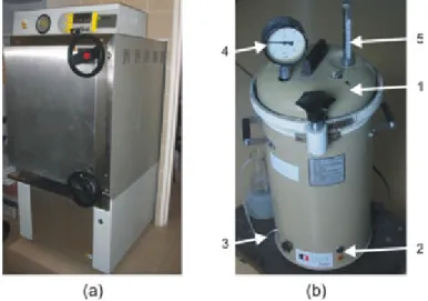
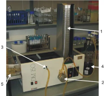
EXERCISE 1: OPERATION OF THE AUTOCLAVE
The use of saturated steam under high pressure is the most effective method to kill microorganisms. In the laborat- ories, a sealed heating device called autoclaveis used for this purpose (Fig. 2). From the inside of the carefully temperature-controlled autoclave, the air is expelled by the less dense steam and sterilisation takes place in a closed chamber at 121°C and overpressure. The household pressure cooker works on a similar principle but with lower
temperature. Autoclaves are widely used in microbiological practise mainly for sterilisation of culture media, glassware and heat-resistant plastic products before their use, and also for contaminated materials prior to disposal as municipal solid waste. To achieve sterilisation, generally 15 minutes of heat treatment at 121°C under 1.1 kg/cm2 pressure has to be applied. Most microbes are unable to tolerate this environment for more than 10 minutes.
However, the time used for sterilisation depends on the size and content of the load.
Fig. 2. Sterilisation by heat – the autoclave.(a) Bigger, automatic autoclave operated by external steam afflux.
(b) Smaller, manual autoclave: 1. lid, 2. power switch, 3. bleeder valve, 4. pressure gauge, 5. thermometer.
Object of study, test organisms:
culture medium in a flask Materials and equipment:
distilled water heat-proof gloves autoclave Practise:
1. Open the lid of the autoclave and check that there is sufficient amount of distilled or deionised water in it. If necessary, refill.
2. Place the correctly packaged materials (e.g. laboratory equipment, culture medium in a flask) into the chamber of the autoclave. Stick a piece of autoclave indicator tape onto the surface of materials!
3. Close the lid of the autoclave.
4. Make sure that the bleeder valve is open.
5. Turn on the heating of the autoclave (the indicator lamp is lit).
6. If an intense (a thick, milky white) steam outflow can be detected through the outlet tube of the bleeder valve (100°C on the built-in thermometer), wait for 4-5 minutes and close the bleeder valve (venting).
7. With the help of a built-in thermometer and manometer, check the temperature and pressure increase inside the chamber of the autoclave.
8. The sterilisation time (15 minutes or more) begins only when the temperature equalization (to 121°C) in the chamber has occurred. It is important that the operator stays with the device and controls the process of steril- isation from the time it is turned on until the end of the sterilisation period.
9. Turn off the power switch of the autoclave when the sterilisation cycle/period has ended.
10. Allow the device to cool down to at least 60-70°C.
11. For decompression, slowly open the bleeder valve. Thereafter, carefully open the lid of the autoclave and remove the sterilised materials, using heat-proof gloves. Check the colour of sterilisation indicator controls.
3.1.2. Sterilisation by radiation
Other forms of energy [e.g. ultraviolet (UV) and ionizing radiation] are also used for sterilisation especially for heat-sensitive materials. The full spectrum of UV radiation can damage microbes but only a small part is responsible for the so-called germicidal effect. Very strong "germicidal" effect can be achieved around 265 nm, because maximum UV absorption of DNA occurs at this wavelength. The main cause of cell death is the formation of pyrimidine dimers in nucleic acids. Bacteria are able to repair their nucleic acid after damage using different mechanisms; however, beyond a certain level of damage, the capacity of the enzyme system is not enough and the accumulation of mutations causes death. UV (germicidal) lamps are widely used in hospitals and laboratories (e.g.
in biological safety cabinets) for decontamination of air and any exposed surfaces. The disadvantage of the use of UV radiation is that it does not penetrate through glass, dirt films, water, and other substances.
Among the high-energy ionizing radiation, γ-rays from radioactive nuclides60Co are generally used for sterilisation of disposable needles, syringes, bandages, medicines and certain food (e.g. spices). The advantage of gamma radiation is its deep penetration through the packaging. Its disadvantage is the scattering in all directions, which requires special circumstances for application.
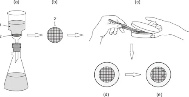
3.1.3. Filter sterilisation
The most commonly used mechanical method of sterilisation is filtration (Fig. 3). During filtration, liquids or gases are pressed through a filter, which (depending on its pore size) retains or adsorbs (e.g. asbestos filter pads) microbes, thereby the filtrate becomes sterile. The pore diameter of filters should be chosen carefully so that bacteria and other cellular components cannot penetrate.
Earlier Seitz-type asbestos or different glass filters were commonly used for the filtration of microorganisms. The modern membrane filters are usually composed of high tensile-strength polymers (cellulose acetate, cellulose nitrate or polysulfone, etc.). Their operation is based partly on the adsorption of microbes, partly on a mechanical sieve effect. The pure sieve-based filters can be beneficial because they do not change the composition of the filtered solution. To remove bacteria, membrane filters with pore size of 0.22 µm are the best choice.
Membrane filters are biologically neutral; do not hamper life activities of microorganisms remaining on the filter and do not inhibit their enzyme functions. Furthermore, nutrients can diffuse through the membranes, so bacteria can be cultured on a variety of media also by placing the filters onto their surface.
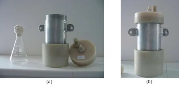
Fig. 3. Filter-sterilisation using syringe filter.Vitamin-solution (1) is added with filter-sterilisation (2) to the presterilised medium (3).
3.1.4. Sterilisation by chemicals
A wide range of chemicals is suitable to inhibit or kill microbes. Some of the antimicrobial agents only inhibit the growth of microorganisms (e.g. bacteriostatic, fungistatic, and virostatic compounds) while others kill them (e.g.
bacteriocidal, fungicidal, and virocidal agents). The -static or -cidal effect of a substance depends on the applied concentration and exposure time in addition to its quality. Only –cidal effect substances are used for chemical sterilisation. These substances have the following requirements: they should have a broad-spectrum effect, they should not be toxic to higher organisms, they should not enter detrimental reactions to the materials being treated with, they should not be biodegradable, they should be environmentally friendly, easy to apply and economical.
The materials used in chemical sterilisation are liquids or gases. Liquid agents are used especially for surface sterilisation. Among sterilising gases, those working at low temperature function by exposing the materials to be sterilised to high concentrations of very reactive gases (e.g. ethylene oxide, beta-propiolactone or formaldehide).
Due to their alkylating effect, these compounds cause the death of microbes by damaging their proteins and nucleic acids. The chemical agents used for sterilisation must be chemically compatible with the substances to be sterilised, therefore they have a great importance in sterilisation of pharmaceutical and thermoplastic materials. The chemicals used by the gas sterilisers are harmful to humans as well. Therefore, the application of gas sterilisers requires compliance with the precautions by the users.
3.2. Procedures of disinfection
Any process aimed at destroying or removing the infectious capability of pathogenic microbes that generally occur on inanimate objects, is called disinfection. The chemicals used for disinfection can be classified according to their chemical structure and their mode of action.
Among the alcohols, ethanol and isopropanol are widely used as disinfectants. 50-70% aqueous solution has excellent antiseptic properties. The action mechanism of alcohols depends on the applied concentration. Due to the solubility of lipids in 50-95% ethanol solutions, biological membranes are disintegrated. Alcohols pass through the cell membrane with altered permeability, denature the proteins inside the cell and have a dehydration effect as well.
Absolute alcohol (100% ethanol) provides the best dehydration effect but does not coagulate the intracellular proteins. 70% dilution of alcohols is the most effective way to kill the vegetative forms of bacteria and fungi, but less effective against spores and lipid-enveloped viruses.
Phenol called carbolic acid was first used as a disinfectant by Lister. Phenol denatures proteins, and irreversibly inactivates the membrane-bound oxidases and dehydrogenases. Due to the unfavourable physical, chemical and toxicological properties, phenol is no longer used. However, substituted (alkylated, halogenated) derivatives are often used in combination with surfactants or alcohols (e.g. cresol, hexachlorophene, chlorhexidine).
The halogens (F, Cl, I, Br) and their derivatives are very effective disinfectants and antiseptic agents; mainly their non-ionic forms have antimicrobial activity. Chlorine gas is used almost exclusively for the disinfection of drinking water or other waters. In addition, different compounds (e.g. chloride of lime, chloramine-B, sodium dichloroiso- cyanurate) are among the most widely used disinfectant agents. Sodium hypochlorite (“household bleach” is a mixture of 8% NaClO and 1% NaOH) is one of the oldest high-bleaching and deodorizing disinfectant. The basis of the effect of chlorine and its derivatives is that during decomposition in aqueous solution, a strong oxidant, nascent (atomic state) oxygen ('O'), is released. Nascent oxygen is very reactive and suitable to destroy bacteria, fungi and their spores as well as viruses.
Iodine is also a widely used disinfectant and antiseptic agent. There are two known preparations: tincture of iodine (alcoholic potassium iodide solution containing 5% iodine) and iodophors (aqueous solutions of iodine complexes with different natural detergents). It is applied in alcoholic solution to disinfect skin or in aquatic solution for washing prior surgery.
Aldehydes, such as formaldehyde and glutaraldehyde, are broad-spectrum disinfectants. They are used for decon- tamination of equipment and devices. Formalin is the 34-38% aqueous solution of formaldehyde gas. Its effect is based on the alkylation of proteins.
Heavy metals such as mercury, arsenic, silver, gold, copper, zinc and lead, and a variety of their compounds are highly efficient disinfectants but they are too damaging to living tissues to apply. They can be used as disinfectants
at very low concentrations. Inside the cell, they bind to the sulfhydryl groups of proteins. Primarily, organic and inorganic salts of silver and mercury-containing products are commercially available, which have bactericidal, fungicidal and virocidal effect.
Detergents or surfactants are amphiphilic organic molecules which have a hydrophilic "head" and a long hydrophobic
“tail” . Detergents can be non-ionic, anionic or cationic according to the charge of the carbon chain. Nonionic surfactants have no significant biocidal effect and anionic detergents are only of limited use because of their poor efficiency. The latter group includes soaps, which are long-chain carboxylic acids (fatty acids) of sodium or po- tassium salts. They are not disinfectants on their own, but are efficient cleaning agents due to their lipid-solubilising effect. Cationic detergents, such as quaternary ammonium salts, are the best disinfectants.
3.3. Control of the efficacy of sterilisation equipment
To monitor the efficacy of sterilisation equipment, several methods are available: instrumental monitoring, the use of chemical indicators and biological monitoring with spore preparations.
By instrumental monitoring, the vapour pressure, temperature and exposure time can be monitored inside the sterilizing equipment. In general, colour changes of chemical indicators on packaging show that temperature and duration are sufficient for effective sterilisation. The original Browne-type sterilisation control glass tubes contain a red indicator solution, which turns yellow during inadequate heat treatment, and turns green in the case of sufficient sterilisation. Another chemical indicator, the indicator tape, should be stuck onto the outer surfaces of the load (e.g. glassware or aluminium foil packaging). The strips change colour or a marking appears (e.g. "OK" or
"STERILE"), which indicates that sterilisation has taken place. The use of chemical indicators is recommended only in equipment previously qualified by biological tests.
The use of biological indicators is the most reliable method for the certification and periodic monitoring of steril- izing equipment. For this purpose, standardised bacterial spore products, the so-called “spore preparations” are required. Test organisms (e.g. spores ofGeobacillus stearothermophilus) are usually more resistant to heat steril- isation than most microorganisms. If the efficiency of sterilisation is inadequate, test microbes remain viable (spore germination is maintained). For the microbiological control of steam sterilisers, standardised bioindicators made of the spores of the type strain ofGeobacillusstearothermophilusATCC 7953, containing 1.2 x 10⁶CFU equivalent endospores are used. This amount of spores can be destroyed by an efficient autoclaving cycle. The destruction or survival of microorganisms can be detected by culturing in broth. The spore preparations should be placed, equally distributed, with the load within the chamber of the autoclave at the characteristic technical points.
EXERCISE 2: MICROBIOLOGICAL CONTROL OF AN AUTOCLAVE BY USING THE SPORE PREPARA- TION OFGEOBACILLUSSTEAROTHERMOPHILUSATCC 7953
Object of study, test organisms:
spore preparation ofGeobacillusstearothermophilusATCC 7953 Materials and equipment:
TSB medium (see Appendix) sterile scissors
sterile metal forceps autoclave
Bunsen burner
incubator (set at 55-60°C) Practise:
1. Place the spore preparation containing strips to the appropriate points of the autoclave chamber.
2. Conduct a sterilisation cycle in the autoclave. (See EXERCISE 1)
3. When the sterilisation cycle is completed, open the bags containing the spores with sterile scissors. Place the preparation of spores into sterile TSB medium aseptically using sterile forceps.
4. Incubate the broth containing the spores in an incubator at 55-60°C for a week.
5. After the incubation period, detect the presence of bacterial growth within the culture broth.
3.4. Determination of the microbiological effic- acy of disinfectants
Microbiological laboratories, especially those involved in epidemiological, medical or industrial processes (phar- maceutical companies, food industry), use disinfectant solutions for preventive, continuous or terminal disinfection.
In the case of an actual epidemiological event (epidemic, accumulation of infection), the effectiveness of these disinfectants is systematically inspected. The principle of efficacy testing is that the relevant disinfectant is incubated with a test bacterium for a defined time interval, and the treated bacteria are subsequently spread onto the surface of a suitable nutrient medium. Following the incubation period, based on the growth of the test microbe, conclusions can be drawn whether the disinfectant within the exposure time interval effectively killed the test microbe.
EXERCISE 3: DETERMINATION OF THE MICROBIOLOGICAL EFFICACY OF DISINFECTANTS Object of study, test organisms:
24-hour culture ofStaphylococcus aureusstrain in 50 mL TSB medium 24-hour culture ofPseudomonas aeruginosastrain in 50 mL TSB medium 24-hour culture ofBacillus subtilisstrain in 50 mL TSB medium
Materials and equipment:
TSB medium (see Appendix) pipettes, sterile pipette tips vortex mixer
water bath
TSA plates (see Appendix)
disinfectants: 1% and 2% sodium hypochlorite solution, other commercially available disinfectant (such as Domestos, Clorox)
control solution: 0.9% sodium chloride solution sterile plastic inoculating loops
9 mL sterile distilled water in test tubes sterile test tubes
Bunsen burner incubator Practise:
1. Measure 9-9 mL from each adequately diluted disinfectant solution and from the control (NaCl) solution into sterile test tubes and place the test tubes into 25°C water bath (Fig. 4).
2. Place sterile plastic inoculating loops into the liquid cultures of the test microbes for 10 minutes.
3. Following the 10-minute incubation, take out the plastic inoculating loops, drain the excess of the liquid culture by touching the inner side of the test tubes.
4. Place the “infected objects” in the appropriate disinfectant solution as well as into the control solution, and leave them there for predefined incubation periods (1, 5, 15, 30, 45 or 60 minutes).
5. Immerse the loops in sterile water for 1 minute (to remove the remaining disinfectant from the surface of the inoculating loop).
6. Inoculate the surface of the TSA plates with the plastic inoculating loops.
7. Place the infected agar plates into an incubator at 28°C for up to a week.
8. Evaluate bacterial growth compared with the controls (0.9% sodium chloride solution that has not been inoculated, and one that has been inoculated with the test microbes) on a scale of five (-, , +, ++, +++) (see Appendix for the evaluation table).
Fig. 4. Testing the efficacy of disinfectants.(a) Place sterile plastic inoculating loops into the suspension of the test microbes. (b) Place the “infected object” in the appropriate disinfectant solution. (c) Wash the loop in sterile
water. (d) Inoculate the surface of the TSA plates with the plastic inoculating loops.
MICROBIOLOGY
The way of sampling depends on the aim of the microbiological study, and must be done at the selected site with sterile equipment. Although sampling different environments (e.g. throat swab, blood, soil, sewage, water, etc.) require different methods and equipment, samples always have to be representative. Sampling must always be done in adequate number of parallels depending on the aim of the research. Not only sampling, but also the transfer of samples to the laboratory can be a critical point during the study. Recording details on site will help the inter- pretation of results later. Take extra care to avoid contaminating the sample container or the sample.
4.1. Sampling for diagnostic purposes
Sampling for diagnostic purposes and the successful diagnosis of pathogenic microbes needs professional sampling techniques and quick transfer of samples to the laboratory. Different methods are used for different samples (e.g.
tissue, urine, feces, blood, etc.), but the amount of contaminating microbes in the samples (which possibly mask the original pathogens) must be low. Samples are always put into sample containers that are adequate for the given sample. Samples must be precisely labelled and transferred to the laboratory with care.
4.2. Sampling from various environments
4.2.1. Collection of air samples
Air sampling in the context of microbiological assessment is the collection of airborne microorganisms. The atmo- sphere is not a living habitat for microbes, but they can spread through it, and therefore the atmosphere could act as a conveyor of pathogenic microbes. Studying microbes in the air has not only hygienic but also economic im- portance: microbes present in the atmosphere of a factory can be hazardous to crude materials and products, and also to the production processes. Consequences of using microbiologically contaminated materials can be serious, therefore checking the quality of the air is a critical factor in the cosmetics, pharmaceutical and food industries, etc.
There are different ways for sampling the air. The simplest way is the passive, “Koch-type” sedimentation method (using settle plates), which is adequate to detect well settling microbial particles. Active methods require impaction devices (Fig. 5). The volume of air for active sample collection depends on the device being used and on the anti- cipated concentration of the bioaerosol. With filtration (selecting adequate pore size filters) or gas washers not only microbes (e.g. fungi, bacteria) but also cell debris can be detected. The use of impingement (slit sampler) is also widespread. In this case, in a narrow canal, an air stream is generated and particles are caught by breaking the way of the air.
Where only low concentrations of microbial contaminants are expected, e.g. clean rooms, food production facilities and operating theatres, generally impaction methods are chosen. In highly contaminated environments, impaction techniques may 'oversample' even in short timescales and impingement or filter samples are more appropriate.
With the strict adherence to manufacturer's flow rates, sampling periods, culture media used and device placement, most techniques should yield comparable results, which are normally expressed in CFU/m³ of air (CFU= colony forming unit).