Contents lists available at ScienceDirect
Applied Surface Science
journal homepage: www.elsevier.com/locate/apsusc
Full Length Article
Preparation and characterization of noble metal modified titanium dioxide hollow spheres – new insights concerning the light trapping efficiency
Tamás Gyulavári
a,b, Kata Kovács
b, Zoltán Kovács
b,c, Enikő Bárdos
a,b, Gábor Kovács
a,b,c,d, Kornélia Baán
b, Klára Magyari
c, Gábor Veréb
a,e,⁎, Zsolt Pap
a,d,e,⁎, Klara Hernadi
a,ba Research Group of Environmental Chemistry, Institute of Chemistry, University of Szeged, Tisza Lajos krt. 103, H-6720 Szeged, Hungary
b Department of Applied and Environmental Chemistry, University of Szeged, Rerrich tér 1, H-6720 Szeged, Hungary
c Nanostructured Materials and Bio-Nano-Interfaces Center, Interdisciplinary Research Institute on Bio-Nano-Sciences, Babes-Bolyai University, Treboniu Laurian 42, RO- 400271 Cluj-Napoca, Romania
d Institute of Environmental Science and Technology, University of Szeged, Tisza Lajos krt. 103, H-6720 Szeged, Hungary
e Department of Process Engineering, Faculty of Engineering, University of Szeged, Moszkvai krt. 9, H-6725 Szeged, Hungary
A R T I C L E I N F O Keywords:
Titanium dioxide Hollow spheres Photocatalysis Noble metals Light trapping
A B S T R A C T
Titanium dioxide hollow spheres (TiO2-HSs) were fabricated by applying carbon spheres (CSs) as templates, which were eliminated by calcination. The most suitable TiO2 was selected and an attempt was made to further increase its photocatalytic activity via noble metal (Au and Pt, at 0.25 wt%) deposition. The photocatalytic efficiency was determined by the decomposition of phenol and oxalic acid under both UV-A and visible light irradiation. It was established, that both the unique morphology and the presence of noble metals contributed to the photocatalytic activity gain compared to the solid spherical reference. For the elucidation of the observed phenol degradation performance under UV-A light irradiation, new insights were proposed: within the TiO2
samples the ratio of HSs with enhanced light trapping properties were demonstrated, and the data was compared to the observed photocatalytic activities and a direct correlation was found.
1. Introduction
A large number of methods have been suggested to remedy the problem of wastewater treatment from which heterogeneous photo- catalysis is a suitable candidate to be used as an alternative technique.
Titanium dioxide remains to be one of the most appropriate semi- conductors for photocatalytic applications due to its apparent beneficial properties (it is photo- and chemically stable, cheap, accessible in considerable amounts); however, its photocatalytic efficiency is un- satisfactory in numerous cases, making the enhancement of it the main scope of numerous publications. There has been tremendous number of attempts to increase the photocatalytic activity and the excitability of TiO2, for example (i) preparing composites with other semiconductors [1–4], (ii) doping with various elements [5–9], (iii) sensitizing with dyes [10,11], (iv) modifying with noble metals [12–17] and (v) syn- thesizing TiO2 with various morphologies [17–21].
Recently, the preparation of hollow structural semiconductors has attracted considerable attention due to their unique chemical, thermal, optical, electrical and optoelectronic properties [22]. Semiconductors
possessing such morphology may have enhanced light harvesting cap- ability due to the multiple reflections of incident light within the hollow cavity which can lead to the improved utilization of light source [23,24], yielding more photogenerated charge carriers, resulting in enhanced photocatalytic efficiency of the semiconductor. Based on the publication of Xiang and Zhao, in which methyl blue was applied as model con- taminant, the (visible) light harvesting of the titania cannot be increased effectively only by controlling the morphology, but it is possible to en- hance it by combining morphology control with noble metal deposition [19]. Accordingly, it is probable that combining morphology control with other photocatalytic activity enhancement techniques (such as noble metal deposition) will be of particular interest.
It is well-known that noble metal deposition (using noble metals as co-catalysts) can increase the photocatalytic activity of titania because of the fast transfer of photogenerated electrons from TiO2 to noble metal nanoparticles resulting in decreased e-/h+ pair recombination and consequently, enhanced charge separation [25,26]. The deposition of noble metals can also enable the production of hydrogen, an alter- nate green energy source [12,27].
https://doi.org/10.1016/j.apsusc.2020.147327
Received 18 April 2020; Received in revised form 22 June 2020; Accepted 20 July 2020
⁎Corresponding authors at: Institute of Environmental Science and Technology, University of Szeged, H-6720 Szeged, Tisza Lajos krt. 103, Hungary (Z. Pap).
Institute of Process Engineering, Faculty of Engineering, University of Szeged, H-6725 Szeged, Moszkvai krt. 9, Hungary (G. Veréb).
E-mail addresses: verebg@mk.u-szeged.hu (G. Veréb), pzsolt@chem.u-szeged.hu (Z. Pap).
Available online 09 August 2020
0169-4332/ © 2020 The Author(s). Published by Elsevier B.V. This is an open access article under the CC BY license (http://creativecommons.org/licenses/by/4.0/).
T
Although there are some publications in the literature where noble metal-deposited (Au, Ag, Pt, Pd) hollow spherical TiO2-s were synthe- sized [17,19,28,29] their numbers are scarce. Thus, in this study these two techniques (morphology control and noble metal deposition) were combined to increase the photocatalytic activity of TiO2 photocatalysts.
The photocatalytic efficiencies were determined by either using the poorly adsorbing phenol, or oxalic acid with good adsorption properties as model contaminants under UV-A and visible light irradiation. The nature of photocatalytic activity gain was described in detail via in- depth morpho-structural characterization and photocatalytic degrada- tion tests using reference titania with or without noble metal co-cata- lysts and/or hollow spherical morphology. Even though there are nu- merous publications in the literature for the explanation of the photocatalytic enhancement caused by the deposition of noble metals, but in the case of photocatalytic activity gain caused by the unique hollow spherical morphology the enhanced light trapping properties are only implied in these publications to the best of our knowledge.
Thus, in this paper an attempt was made to establish connection be- tween the observed photocatalytic activity and the properties of the TiO2 hollow spheres based on calculations.
2. Experimental 2.1. Materials
For the synthesis of TiO2-HSs, Ti(IV) butoxide (Sigma-Aldrich; reagent grade; 97%), was applied as precursor. For the fabrication of CS templates ordinary table sugar (sucrose, Magyar Cukor Zrt., KoronásTM) – as carbon source –, NaOH (Molar Chemicals; a.r.; 50%), and ultrapure Millipore Milli-Q (MQ) water were used. For their purification either ethanol (Molar Chemicals; 96%) or acetone (Molar Chemicals; 99.96%) were used. For the determination of photocatalytic activities oxalic acid (Sharlau; analytical grade) and phenol (Spektrum 3D; analytical grade) were used as model pollutants. For the deposition of gold and platinum nanoparticles HAuCl4∙4 H2O (Sigma-Aldrich; 99.9%) and H2PtCl6 (Sigma- Aldrich; 99.9%) were applied, respectively. Trisodium citrate (Sigma- Aldrich; > 99%) was utilized to stabilize the growth of the noble metal particles and for their reduction NaBH4 (Alfa Aesar; 98%) was used.
2.2. Preparation of the carbon sphere templates
The CS templates were synthesized based on our recent publication [30] as follows. In a Teflon®-lined stainless-steel autoclave (Vtotal = 623 mL) 180.7 mL 0.15 M sucrose solution was prepared (Vfill/ Vtotal = 29%) and the pH was set to 12 using a 2 M NaOH solution. The as-prepared solution was subjected to hydrothermal treatment in a drying oven at 180 °C for 12 h. The CSs were purified from the residual organic contaminants – which form during the synthesis under the applied conditions – by centrifugation using either ethanol or acetone as solvent. During this procedure the CSs were washed in three cycles at the rate of 13,400 rpm for 3 min using 80 mL solvent per 1 g of CSs.
Then, the solid product was collected and dried in air at 40 °C, and finally, was ground in an agate mortar.
2.3. Preparation of TiO2 hollow spheres and the deposition of noble metals The preparation of the TiO2 coating was based on the publication of Ao et al. [23]. 0.1 g CS was added to 20 mL absolute ethanol in a beaker under vigorous magnetic stirring, to which 1 mL Ti(IV) butoxide was added dropwise with a constant rate of 1 mL·min−1. Then, the beaker was covered with parafilm to prevent evaporation, and the suspension was stirred for 4 h. After the formation of the coating, the samples were dried, and ultimately the CSs were eliminated by calcination in a Thermolyne 21,100 type tube furnace with constant air supply (30 L∙h−1) at either 500 or 800 °C for 3 h applying 5 °C∙min−1 heating rate, resulting in crystalline TiO2-HSs.
Subsequently, gold or platinum nanoparticles were deposited onto the surface of TiO2-HSs at 0.25 wt%. For this, 800 mg of the as-prepared sample was added to 87 mL MQ water, to which 12.5 mL trisodium citrate (c = 0.63∙10−4 M) was added. Then, either 1.6 mL HAuCl4 ∙ 4 H2O (c = 2.54∙10−2 M) or 1.3 mL H2PtCl6 (c = 3.13∙10−2 M) were added to the system. For the chemical reduction of noble metals 2 mL precooled (T ~ 0 °C) NaBH4 (0.15 M) was applied, and the suspension was stirred for 30 min then purified via centrifugation 3 times using MQ water. Finally, the samples were dried in air at 40 °C and ground in an agate mortar. The synthesis parameters – applied during the prepara- tion of the samples in this work – were summarized in Table 1. The as- prepared samples were named based on the following: TiO2_HSxyz, where ‘HS’ stands for hollow spheres, ‘x’ is the solvent which was used for the purification of the carbon spheres (‘Ac’ for acetone and ‘EtOH’
for ethanol), and ‘y’ is the calcination temperature which was used for both the elimination of carbon spheres and the crystallization of TiO2, and ‘z’ is the deposited noble metal.
For the evaluation of photocatalytic activity gain caused by the unique morphology multiple reference TiO2 samples were fabricated.
The synthesis procedure was carried out almost similarly; the only difference was the absence of CSs. In other words, the solid spherical counterparts of those TiO2-HSs were synthesized, which were used for the evaluation of photocatalytic activities. The naming of these samples was similar compared to the hollow spherical samples, with the addi- tion of ‘no’ (‘TiO2_no_HSxyz’), which refers to the absence of CSs, thus the solid spherical morphology of the samples.
2.4. Characterization methods and instrumentation
A Rigaku Miniflex II type diffractometer was used for the XRD measurements. The following parameters were applied: 30 mA, 40 kV, λCuKα = 0.15406 nm. The 20–40 (2θ°) interval was recorded using 1 (2θ°)∙min−1 scan speed. Mean primary crystallite sizes were calculated applying the Scherrer equation [31,32]. The rutile and anatase weight fractions were determined from their corresponding peak areas (27.5 (2θ°) for rutile and 25.3 (2θ°) for anatase, respectively).
The morphology of the samples was analyzed by a Hitachi S-4700 Type II scanning electron microscope and a FEI TECNAI G2 20 X-Twin type transmission electron microscope. The former was used to examine the general morphology, while the latter to observe the hollow struc- ture and the presence of noble metals on the surface of the TiO2
Table 1
Crystal phase compositions, mean diameters, circularities and specific surface areas of the TiO2-HSs samples.
Sample name CS purification
solvent Calcination temperature
(°C) Anatase
(wt%) Rutile (wt
%) DA (nm) DR (nm) Median (nm) Circularity Specific surface area (m2∙g−1)
TiO2_no_HSAc500 acetone 500 97 3 35 66.9 1313 0.99 ~6
TiO2_HSAc500 acetone 500 87 13 26.5 30.1 1198 0.99 ~6
TiO2_HSAc800 acetone 800 – 100 – 28.3 1129 0.94 ~6
TiO2_HSEtOH500 ethanol 500 96.6 3.4 18.1 20.9 948 0.98 ~6
TiO2_HSEtOH800 ethanol 800 12.5 87.5 35.1 32.4 1122 0.93 ~6
TiO2_HSAc500-Au acetone 500 90.01 9.99 22.2 20.5 1635 0.98 ~6
TiO2_HSAc500-Pt acetone 500 91.93 8.07 22.9 29 1333 0.98 ~6
samples. The recorded micrographs were utilized to determine the diameter and calculate the circularity degree [33] of the spheres using the ImageJ software. During SEM measurements the electron beam was produced using a cold field emission gun applying 10 kV acceleration voltage, while in the case of TEM 100 kV was set. The circularity (C) of the spheres was calculated based on the following equation:
=
C A
P 4
2
where A is the area and P is the perimeter of the projection of the spheres [33].
The diffuse reflectance (λ = 220–800 nm region) of the samples was studied by a Jasco-V650 spectrophotometer, equipped with an ILV- 724-type integration sphere. The band gap energies were evaluated from the derivative spectra of the samples.
The surface of the TiO2 samples was studied by FT-IR measurements using a Jasco 6000 spectrometer. The 400–4000 cm−1 range was re- corded applying 4 cm−1 spectral resolution.
The specific surface areas of the TiO2 samples were determined with a BELCAT-A device via N2 adsorption at 77 K using the BET method.
2.5. Evaluation of the photocatalytic efficiencies
The photocatalytic activities were evaluated by the photocatalytic decomposition of phenol (c0,phenol = 0.1 mM) and oxalic acid (c0,oxalic acid = 1 mM) both under UV-A (λmax = 365 nm) and visible light ir- radiation (λ > 400 nm). For the visible light experiments 4 conven- tional energy-saving lamps (Düwi 25920/R7S-24 W), while for the UV experiments 6 fluorescent tubes (Vilber-Lourmat T-6L UV-A, 6 W) were applied. The emission spectra of the applied lamps can be seen in Fig.
S1. The TiO2 suspensions were added to a glass vessel which was placed on a magnetic stirrer. During the visible light illumination 1 M NaNO2
solution was circulated in the thermostating jacket of the glass vessel, to cut off UV photons. Constant temperature (25 °C) was assured during the experiments via the NaNO2 solution (Vis) or water (UV) using an ultrathermostat. Constant dissolved oxygen level was maintained by supplying air into the photoreactor during the 4-hour-long measure- ments. The concentration of phenol and oxalic acid was measured with high performance liquid chromatography (HPLC) and the device con- sisted of a Merck Hitachi L-7100 low-pressure gradient pump equipped with a Merck-Hitachi L-4250 UV–Vis detector. For phenol as eluent 50–50% methanol/water mixture (λdetection = 210 nm), and for oxalic acid 19.3 mM H2SO4 eluent (λdetection = 206 nm) were applied.
3. Results and discussion 3.1. Characterization of TiO2-HSs
The CSs – which were used as templates for the shape-controlled synthesis – were characterized in detail in our previous publications [20,21,30]. For the purification of CSs various solvents (acetone or ethanol) were used, because – based on our previous results and the publication of Mahyar and Amani-Ghadim [34] – applying solvents of different polarity can result in the formation of titanium dioxides with different crystal phase compositions and characteristics. By the utili- zation of these CSs TiO2-HSs were prepared (Table 1) applying different calcination temperatures (500, 800 °C), which were characterized by XRD, SEM and TEM in order to determine which TiO2-HS possesses the best characteristics to be used as the base material for the deposition of noble metals.
The SEM and TEM micrographs of the TiO2-HSs are shown in Fig. 1.
Based on the SEM measurements it was found that the samples syn- thesized at similar conditions (Fig. 1a with Fig. 1b and Fig. 1c with Fig. 1d), i.e. same solvent and calcination temperature, similar char- acteristics could be observed (similar morphology and diameter/cir- cularity values) (Table 1). Based on our previous work [20] the most
critical step during the synthesis of HSs via template removal is the elimination of CSs by calcination. In the case of samples calcined at 500 °C (Figs. 1a and b) well-defined spheres were observed possessing near perfect morphology, which were not damaged during calcination.
The circularity values, medians and the diameter distribution histo- grams are summarized in Table 1 and Fig. S2, respectively. The TEM images of the samples calcined at 500 °C (Fig. 1e and f) confirmed that they had regular hollow cavities, as intended. The theoretical thickness of the TiO2 shell – evaluated from the quantities of the reactants – was calculated to be 70 nm from the relative weight fractions of CS and TiO2 coating (determined from the volume and density of these species). This value is in reasonably good accordance with the one obtained from the TEM micrographs, which was measured to be ~60 nm. By increasing the calcination temperature to 800 °C (Fig. 1c and d), it was found that hollow structures with raspberry-like surface were formed, in contrast with the smooth surfaces detected in the previous cases. It was also observed that the high temperature also caused the inner diameter of the hollow cavity to be smaller (Fig. 1g and h), which could be ex- plained as the result of the sintering of the nanoparticles [35].
Subsequently, XRD and nitrogen adsorption measurements were carried out and the results (crystal phase composition, primary crys- tallite sizes, specific surface areas) are summarized in Table 1. As ex- pected, the samples calcined at lower temperature (500 °C, TiO2_HSAc500
and TiO2_HSEtOH500 ) mainly consisted of anatase, while samples calcined at the higher temperature (800 °C, TiO2_HSAc800 and TiO2_HSEtOH800 ) con- tained predominantly rutile phase and larger crystallites. As for the N2 adsorption measurements, despite having relatively low crystallite sizes (18–35 nm), the corresponding specific surface areas were surprisingly low (~6 m2∙g−1 in all cases).
Summarizing these results, it was established, that sample TiO2_HSAc500 possessed the best characteristics to be used as base mate- rial for the determination of photocatalytic activity gain caused by the unique morphology (highest circularity value, regular hollow cavity).
Additionally, it was also the most appropriate from the economic point of view, since for the synthesis of this sample the CSs were purified by acetone, which is cheaper than ethanol, and for the elimination of the CSs only 500 °C temperature was applied making its production more cost-effective. Therefore, later on, for the deposition of noble metals this sample was selected as base material. Lastly, after the TiO2_HSAc500 sample was chosen as base material, a reference sample was also syn- thesized in the same way just without the application of CS templates (denoted as ‘TiO2_no_HSAc500′), to investigate the possible effects of the hollow structure. This sample was also examined via SEM and TEM measurements (Fig. 2). Based on the SEM micrographs (Fig. 2a) it was observed that it also contained well-defined spheres with relatively high diameter (~1300 nm), comparable to prior samples. Then, this sample was investigated by TEM measurements as well (Fig. 2b) and it was found that it contained solely solid spheres, which, most im- portantly, makes it most suitable for the investigation of the effects caused by the hollow morphology in the case of our other samples. For the formation of spherical morphology (in the absence of CS templates in the case of sample TiO2_no_HSAc500), a plausible explanation can be that – according to the Ostwald ripening phenomenon – the formation of larger structures is thermodynamically favored as there are less molecules on the surface in an energetically less stable position, making the development of the spherical shape (with the smallest specific surface area) favored [36]. The reason for the relatively high diameter (low specific surface area) could be – as it was already observed in our previous work – that applying a precursor containing relatively long alkyl chains can result in slower hydrolysis making the formation of larger structures favored [37].
3.2. Further characterization of reference TiO2-s and TiO2-HSs modified with noble metals
Following the deposition of gold and platinum noble metals at
0.25 wt% on the TiO2_HSAc500 sample TEM images were recorded (Fig. 3a and c, respectively). It was observed, that even after the deposition process the regular hollow spherical morphology remained intact, the
noble metals were evenly distributed on the surface and no aggregation occurred. Additionally, HRTEM images were utilized to measure the d- spacing of the TiO2 shell, the gold (Fig. 3b) and platinum (Fig. 3d) Fig. 1. SEM micrographs of samples TiO2_HSAc500 (a), TiO2_HSEtOH500 (b), TiO2_HSAc800 (c) TiO2_HSEtOH800 (d) and their corresponding TEM figures (e-h, respectively).
nanoparticles to confirm their presence. Accordingly, the measured 3.4 Å value was attributed to the TiO2 (101) crystallographic plane, 2.4 Å to the Au (111) plane and 2.0 Å to the Pt (200) plane.
Then, XRD measurements were carried out to determine the influ- ence of the chemical deposition process on the properties of the TiO2_HSAc500 base material (Fig. S3, Table 1). It was found that after the deposition process the noble metal-containing samples largely retained their crystal phase composition as expected. The rutile crystal phase content of ~8–13 wt% of the base material and noble metal-containing samples could be beneficial in terms of the photocatalytic activity of
these samples similarly to the well-known commercial reference pho- tocatalyst P25 [38]. The non-hollow TiO2_no_HSAc500 sample also con- tained rutile crystal phase in 3 wt%. Regarding the crystal phase compositions, it is well known that anatase – due to its large band gap (3.2 eV) – does not absorb visible light, whereas rutile has a narrower band gap (3.02 eV) making the absorption of visible light possible to a certain extent; a fact that can have a considerable influence on the resulting photocatalytic activities of the samples [39–41].
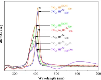
As the next step, the light absorption properties were investigated via DR measurements (Fig. 4). The band gap energies were determined Fig. 2. SEM (a) and TEM (b) images of reference non-hollow sample TiO2_no_HSAc500.
Fig. 3. TEM images of samples TiO2_HSAc500-Au (a, b) and TiO2_HSAc500-Pt (c, d).
from the first order derivatives of the DR spectra of the TiO2–s, because based on the publication of Flak et al. the excitability of the photo- catalysts can be determined more reliably by plotting the first-order derivatives of the DR spectra as a function of wavelength [42]. As ex- pected, rutile phase TiO2-s possessed lower band gaps (3.02 eV for TiO2_HSAc800 and TiO2_HSEtOH800 ) compared to the anatase phase TiO2-s (3.1 eV for TiO2_no_HSAc500, TiO2_HSEtOH500 and 3.14 eV for TiO2_HSAc500).
The band gap after noble metal deposition (3.13 eV for both TiO2_HSAc500-Au and TiO2_HSAc500-Pt) did not change significantly com- pared to the base material (TiO2_HSAc500, 3.14 eV). The inflection point at
~542 nm in the derivative spectrum can be attributed to the plasmon resonance of the gold nanoparticles in the case of sample TiO2-HSAc500- Au. The surface properties were examined by FT-IR measurements (Fig. 5). The bands at 424, 515 and 620 cm−1 can be attributed to the transverse optical vibrations of the Ti–O bonds [43–45]. The shape of these bands varied in accordance with the different crystal phases;
anatase has less defined Ti–O stretch band as it possesses less ordered structure (compared to rutile), consequently, in the case of mixed crystal phase compositions, the infrared absorption bands also re- presented this transmission between the two crystal phases [46]. The doublet at 2340, 2358 cm−1 is characteristic of adsorbed CO2 [47].
Additional bands which could be ascribed to leftover carbon from either the TiO2 precursor or the CS templates were not observed, from which
it was concluded that during the calcination process these were entirely eliminated from the system.
3.3. Evaluation of photocatalytic activity
The photocatalytic activities of the non-hollow, spherical reference sample (TiO2_no_HSAc500), the base material (TiO2_HSAc500) and the noble metal-containing samples (TiO2_HSAc500-Au and TiO2_HSAc500-Pt) were investigated by the photocatalytic degradation of phenol and oxalic acid under UV and visible light irradiation and the results were summarized in Fig. 6 (UV irradiation) and Fig. S4 (visible light irra- diation). Additionally, the photocatalytic activities of solid spherical TiO2_no_HSAc500-Au and TiO2_no_HSAc500-Pt samples were also investigated in the case of phenol degradation under UV light irradiation.
In the case of the spherical, but non-hollow TiO2_no_HSAc500 reference sample negligible photocatalytic activity was observed under visible light irradiation (~3–4% model pollutants were degraded) and by ap- plying UV irradiation 26% phenol and 36% oxalic acid were degraded after the 240-minute-long measurements.
As the next step, the hollow spherical base material (TiO2_HSAc500) was investigated, to determine the possible achievable effects of the unique morphology on the photocatalytic activity. In almost every case, the enhancement of photocatalytic activity was observed (except in the case of oxalic acid degradation under visible light irradiation). In the case of visible light irradiation only negligible differences were ob- served, however, under UV light irradiation notable 166% and 135%
enhancements were detected for phenol and oxalic acid degradations, respectively, compared to the non-hollow reference TiO2.
By the deposition of noble metals on the surface of the hollow spherical base catalyst TiO2_HSAc500, in almost every case the photo- catalytic activity increased (except in the case of TiO2_HSAc500-Au during the decomposition of phenol under UV light irradiation). Applying visible light irradiation 156% and 66% increases were observed in the degradation rate of phenol for sample TiO2-HSAc500_Au and TiO2_HSAc500- Pt, respectively, which values were 780% and 200% for oxalic acid, respectively. At the same time, in the case of UV irradiation these values were −20%, 30% and 18%, 18%, respectively.
Then, after comparing the photocatalytic activity difference be- tween (i) the bare solid spherical (TiO2_no_HSAc500) and bare hollow spherical (TiO2_HSAc500) samples (3rd paragraph), (ii) the bare hollow spherical (TiO2_HSAc500) and noble metal-containing hollow spherical (TiO2_HSAc500_Au and TiO2_HSAc500-Pt) samples (4th paragraph), finally, the photocatalytic activity difference between (iii) the noble metal- containing solid spherical (TiO2_no_HSAc500_Au and TiO2_no_HSAc500-Pt) and noble metal-containing hollow spherical samples (TiO2_HSAc500_Au and TiO2_HSAc500-Pt) were investigated additionally, for phenol de- gradation under UV light irradiation (Fig. 6 left side). It was found that the photocatalytic activities of hollow spherical samples were always higher compared to the solid spherical samples, further reinforcing the beneficial effect of the unique hollow spherical morphology on the photocatalytic performance. The TiO2_HSAc500_Au sample was more ef- ficient by 81% compared to TiO2_no_HSAc500_Au, while TiO2_HSAc500_Pt was more efficient by 18% compared to TiO2_no_HSAc500_Pt in terms of total degraded phenol amount.
For the explanation of the obtained results, numerous factors can be considered: (i) properties of the TiO2 (crystallite size, specific surface area, crystal phase composition, surface features, etc.) [16,48–50]; (ii) properties of the noble metal (size, shape, number) [14,15,51,52]; (iii) properties of the model pollutants (adsorption ability, charge carrier- scavenging attributes, features of intermediates, etc.) [14–16,48]; (iv) the emission spectrum of the light source and the temperature during the photocatalytic experiments [53–55].
Generally, it can be said that the investigated titania decomposed oxalic acid to a greater degree compared to phenol. It is well-known, that phenol is a poorly adsorbing compound and the degradation of pollutants takes place by the utilization of the generated ·OH radicals
300 400 500 600 700
TiO2_no_HSAc 500 TiO2_HSEtOH TiO2_HSAc 800
800
TiO2_HSEtOH 500 TiO2_HSAc
500 TiO2_HSAc
500-Pt
dλ/Rd).u.a(
Wavelength (nm)
TiO2_HSAc 500-Au
Fig. 4. First order derivative DR spectra of the as-prepared photocatalysts.
4000 3500 3000 2500 2000 1500 1000 500
Ti –O
TiO2_no_HSAc 500 TiO2_HSAc
500-Pt TiO2_HSAc
500-Au TiO2_HSEtOH
800
TiO2_HSEtOH 500 TiO2_HSAc
800
).u.a(ytisnetnI
Wavenumber (cm-1) TiO2_HSAc
500
ads. CO2
Fig. 5. FT-IR spectra of the investigated photocatalysts.
[56]; moreover, the forming degradation intermediates with different adsorption properties can also block the active sites of the photocatalyst hindering the further degradation process. Conversely, oxalic acid can adsorb to the surface of TiO2 extremely well facilitating its degradation by the generated charge carriers; furthermore, oxalic acid is a hole scavenger resulting in the efficient separation/increased lifespan of the electron-hole pairs [15].
In the case of oxalic acid degradation, the addition of Pt and Au nanoparticles resulted in the increase of photocatalytic activity in all cases (under both UV and visible light irradiation), reaching nearly 100% degradation efficiency in the former case. This result is in good accordance with literature data where the same results were observed in the case of commercial (predominantly) anatase phase TiO2-s (Aldrich anatase, Aeroxide P25) [15,16]. The photocatalytic activity enhancement of TiO2-HSAc500-Pt can be explained by a double charge trapping mechanism: oxalic acid acts as a hole scavenger, whereas the Pt separates the electron from the TiO2 nanoparticle [14]. In the case of TiO2_HSAc500-Au the photocatalytic activity enhancement can be asso- ciated with the results of our previous work, where the highest degree of photocatalytic activity gain was observed in the presence of spherical gold nanoparticles [52], similar to our present results (Fig. 3b).
As for the phenol degradation tests, only in the case of TiO2_HSAc500- Au sample under UV light irradiation was a photocatalytic activity decrease observed, which is in good accordance with the literature data: it is well-known, that in the case of commercial Aeroxide P25 (which has similar crystal phase composition compared to our samples) the deposition of gold nanoparticles results in decreased photocatalytic activity in the case of phenol under UV light irradiation [16,51,52,57].
However, in the case of the gold-deposited sample under visible light irradiation a photocatalytic activity enhancement was observed, which can be explained by the fact, that the electrons formed by surface plasmon resonance excitation can be efficiently injected into the con- duction band of TiO2 leading to enhanced charge separation [58]. The fact, that the surface resonance peak of gold is located in the visible light range, could explain the observed photocatalytic activity en- hancement under visible light irradiation. In the case of TiO2_HSAc500-Pt sample the photocatalytic activity increased after the deposition of platinum under both visible and UV light irradiation. The photo- catalytic activity gain – in the case of visible light irradiation – could be attributed to the rutile content of the sample, since electron trapping by platinum is much more significant on rutile than on anatase [59].
However, the photocatalytic activity enhancement observed in the case of our TiO2_HSAc500-Pt sample under UV irradiation could not be attrib- uted to literature data, as normally the deposition of platinum nano- particles on Aeroxide P25 (possessing similar crystal phase composi- tion) leads to decreased photocatalytic activity [15,60]. For the deeper
understanding of the as-mentioned results in the next paragraph the unique hollow spherical morphology of our samples was taken into account.
It is well-known that light waves (used for the excitation of the photocatalysts) cannot only interact with matter but with each other as well, which is called wave interference. Constructive interference oc- curs, if the crests of one wave overlap the crests of the other wave. For this to happen, the distance between two waves must be equal to an integer multiple of the wavelength and as a result, the crests combine to produce a wave with greater amplitude. If we excite our photocatalyst with such light waves with greater amplitude, then the excitation process can be more efficient resulting in higher photocatalytic activity.
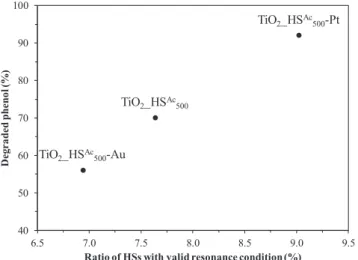
Based on this theory, the following steps were taken: the emission spectra of the applied lamps (Fig. S1) were compared to the diameter distribution histograms (Fig. S2) and it was ascertained, that what proportion of the hollow spheres happens to be precisely in the range where the travelled distance is the integer multiple of the wavelength of the emission maxima. For the calculations the most intense domain of the emission spectrum (360 ± 10 nm) was taken into account. Then, the percentage of the hollow spheres with valid resonance condition – where the constructive interference of the light waves can most prob- ably occur – was counted, by dividing the diameter of the hollow spheres by the wavelength of the light source. After these calculations were carried out, the as-calculated data were compared to the photo- catalytic activities (Fig. 6 left side) and these results are represented in Fig. 7. It can be seen in the figure, that the observed photocatalytic activity (TiO2_HSAc500-Au < TiO2_HSAc500 < TiO2_HSAc500-Pt) indeed cor- responds well to the increasing ratio of hollow spheres with valid re- sonance condition. Since it is well-known, that the excitability of a photocatalyst correlates with the intensity of the light source, and that the intensity of a wave is proportional to the square of its amplitude, then it can be expected, that after constructive interference occurs re- sulting in the increase of the amplitude, the light waves with higher intensity could indeed cause the enhancement of the photocatalytic activity. The validity of this phenomenon was further reinforced by the results mentioned in the 5th paragraph: if we compare the 3 hollow spherical samples appearing in Fig. 7 with their solid spherical coun- terparts, it is apparent that the hollow spherical samples were indeed more efficient in the degradation of phenol under UV light irradiation.
To sum up, based on the results of the photocatalytic activity ex- periments, it was observed that in each case reference solid spherical TiO2 samples proved to be less efficient compared to their hollow spherical counterparts. The unique morphology indeed increased the photocatalytic activity, as did the deposition of noble metals in most cases, thus the combination of the two photocatalytic activity enhan- cing methods was successful.
Fig. 6. Photocatalytic activity of the investigated photocatalysts under UV light irradiation.
4. Conclusions
Carbon spheres were used as templates to synthesize titanium di- oxide hollow spheres applying different synthesis parameters (CS pur- ification solvent, calcination temperature). The CS purification solvent (acetone or ethanol) did not influence the characteristics of the TiO2- HSs, whereas applying 500 °C calcination temperature resulted in anatase phase TiO2-HSs with perfect hollow spherical morphology, while applying 800 °C yielded rutile phase TiO2-HSs with less regular morphology.
The TiO2-HS possessing the best properties in terms of morphology was selected for the deposition of gold and platinum nanoparticles in 0.25 wt%. SEM and TEM measurements confirmed, that the mor- phology remained intact after the deposition process and that the noble metal nanoparticles were distributed evenly on the TiO2-HSs.
The photocatalytic activity enhancement caused by either the un- ique morphology and the presence of noble metals was investigated by the degradation of phenol and oxalic acid under both UV and visible light irradiation using the base hollow spherical, and solid (non-hollow) spherical TiO2-s as references. The hollow spherical morphology re- sulted in remarkable 166% and 135% increases in photocatalytic ac- tivity during UV light irradiation compared to the non-hollow solid reference sample in the case of phenol and oxalic acid, respectively.
Using the same reference, after combining the hollow spherical mor- phology with the deposition of noble metals in the case of the gold- deposited sample these values were 113% and 178% for phenol, along with 246% and 178% for oxalic acid in the case of platinum-deposited TiO2-s, respectively, under UV light irradiation. Summarizing, in the case of visible light irradiation, the gold-deposited hollow spherical TiO2 had the best photocatalytic activity, while in the case of UV ir- radiation the platinum containing hollow spherical TiO2 proved to be the most efficient for the degradation of both oxalic acid and phenol.
Unusual photocatalytic activity order was observed in the case of UV irradiation during the decomposition of phenol, and for its eluci- dation the solid spherical counterparts of each hollow spherical sample were applied to propose a plausible explanation. It was presumed that the hollow spherical TiO2-s have increased light harvesting properties, and the ratio of hollow spheres with this unique feature was calculated.
The photocatalytic activity indeed increased with increasing propor- tions of titanium dioxide hollow sphere diameters close to the integer multiple of the excitation light source’s wavelength, i.e. in which case the occurrence of constructive interference was more probable. The proposed explanation was in good agreement with the acquired results,
since the hollow spherical TiO2-s were always more efficient compared to their solid spherical counterparts.
CRediT authorship contribution statement
Tamás Gyulavári: Investigation, Writing - original draft, Conceptualization. Kata Kovács: Investigation. Zoltán Kovács:
Conceptualization. Enikő Bárdos: Investigation. Gábor Kovács:
Writing - review & editing. Kornélia Baán: Investigation. Klára Magyari: Investigation. Gábor Veréb: Writing - review & editing. Zsolt Pap: Supervision, Writing - original draft. Klara Hernadi: Funding acquisition, Writing - review & editing. : .
Declaration of Competing Interest
The authors declare that they have no known competing financial interests or personal relationships that could have appeared to influ- ence the work reported in this paper.
Acknowledgements
This study was financed by the NKFI-K-124212 project. T. Gyulavári is grateful for the financial support of the GINOP-2.3.2-15-2016-00013 and the NKFI-TNN-16-123631 projects. Zs. Pap acknowledges the Bolyai János scholarship provided by the Hungarian Academy of Sciences. Zs. Pap and G. Veréb were supported by the EFOP-3.6.2-16- 2017-00010 project of the Hungarian State and the European Union.
The financial support of project PN-III-P1-1.1-TE-2016-1588 is also greatly appreciated.
Appendix A. Supplementary data
Supplementary data to this article can be found online at https://
doi.org/10.1016/j.apsusc.2020.147327.
References
[1] Y. Lu, L. Hao, K. Matsuzaka, H. Yoshida, H. Asanuma, J. Chen, F. Pan, Titanium dioxide–nickel oxide composite coatings: preparation by mechanical coating/
thermal oxidation and photocatalytic activity, Mater. Sci. Semicond. Process. 24 (2014) 138–145.
[2] É. Karácsonyi, L. Baia, A. Dombi, V. Danciu, K. Mogyorósi, L.C. Pop, G. Kovács, V. Coşoveanu, A. Vulpoi, S. Simon, Z. Pap, The photocatalytic activity of TiO2/ WO3/noble metal (Au or Pt) nanoarchitectures obtained by selective photodeposi- tion, Catal. Today 208 (2013) 19–27.
[3] R.K. Mandal, S. Kundu, S. Sain, S.K. Pradhan, Enhanced photocatalytic performance of V2O5–TiO2 nanocomposites synthesized by mechanical alloying with morpho- logical hierarchy, New J. Chem. 43 (2019) 2804–2816.
[4] N.O. Balayeva, M. Fleisch, D.W. Bahnemann, Surface-grafted WO3/TiO2 photo- catalysts: Enhanced visible-light activity towards indoor air purification, Catal.
Today 313 (2018) 63–71.
[5] C. Pablos, J. Marugan, R. van Grieken, P.S.M. Dunlop, J.W.J. Hamilton, D.D. Dionysiou, J.A. Byrne, Electrochemical enhancement of photocatalytic disin- fection on aligned TiO2 and nitrogen doped TiO(2) nanotubes, Molecules 22 (2017).
[6] A. Gil, A.M. García, M. Fernández, M.A. Vicente, B. González-Rodríguez, V. Rives, S.A. Korili, Effect of dopants on the structure of titanium oxide used as a photo- catalyst for the removal of emergent contaminants, J. Ind. Eng. Chem. 53 (2017) 183–191.
[7] A. Samokhvalov, Hydrogen by photocatalysis with nitrogen codoped titanium di- oxide, Renew. Sust. Energ. Rev. 72 (2017) 981–1000.
[8] M.C. Wu, J. Hiltunen, A. Sapi, A. Avila, W. Larsson, H.C. Liao, M. Huuhtanen, G. Toth, A. Shchukarev, N. Laufer, A. Kukovecz, Z. Konya, J.P. Mikkola, R. Keiski, W.F. Su, Y.F. Chen, H. Jantunen, P.M. Ajayan, R. Vajtai, K. Kordas, Nitrogen-doped anatase nanofibers decorated with noble metal nanoparticles for photocatalytic production of hydrogen, ACS Nano 5 (2011) 5025–5030.
[9] R. Kun, S. Tarján, A. Oszkó, T. Seemann, V. Zöllmer, M. Busse, I. Dékány, Preparation and characterization of mesoporous N-doped and sulfuric acid treated anatase TiO2 catalysts and their photocatalytic activity under UV and Vis illumi- nation, J. Solid State Chem. 182 (2009) 3076–3084.
[10] M. Gratzel, Solar energy conversion by dye-sensitized photovoltaic cells, Inorg.
Chem. 44 (2005) 6841–6851.
[11] Z. Youssef, L. Colombeau, N. Yesmurzayeva, F. Baros, R. Vanderesse, T. Hamieh, J. Toufaily, C. Frochot, T. Roques-Carmes, S. Acherar, Dye-sensitized nanoparticles for heterogeneous photocatalysis: Cases studies with TiO2, ZnO, fullerene and 40
50 60 70 80 90 100
6.5 7.0 7.5 8.0 8.5 9.0 9.5
)%(lonehpdedargeD
Ratio of HSs with valid resonance condition (%) TiO2_HSAc500-Au
TiO2_HSAc500-Pt
TiO2_HSAc500
Fig. 7. Comparison of the ratio of TiO2-HSs with enhanced light trapping properties to the observed amount of degraded phenol model pollutant under UV light irradiation by the end of the 240-min-long experiments.
graphene for water purification, Dyes Pigm. 159 (2018) 49–71.
[12] Á. Kmetykó, Á. Szániel, C. Tsakiroglou, A. Dombi, K. Hernádi, Enhanced photo- catalytic H2 generation on noble metal modified TiO2 catalysts excited with visible light irradiation, React. Kinet. Mech. Catal. 117 (2015) 379–390.
[13] Z. Wei, M. Janczarek, M. Endo, K. Wang, A. Balčytis, A. Nitta, M.G. Méndez- Medrano, C. Colbeau-Justin, S. Juodkazis, B. Ohtani, E. Kowalska, Noble metal- modified faceted anatase titania photocatalysts: octahedron versus decahedron, Appl. Catal. B Environ. 237 (2018) 574–587.
[14] S. Fodor, G. Kovács, K. Hernádi, V. Danciu, L. Baia, Z. Pap, Shape tailored Pd na- noparticles’ effect on the photocatalytic activity of commercial TiO2, Catal. Today 284 (2017) 137–145.
[15] G. Kovács, S. Fodor, A. Vulpoi, K. Schrantz, A. Dombi, K. Hernádi, V. Danciu, Z. Pap, L. Baia, Polyhedral Pt vs. spherical Pt nanoparticles on commercial titanias: Is shape tailoring a guarantee of achieving high activity? J. Catal. 325 (2015) 156–167.
[16] Z.-R. Tóth, G. Kovács, K. Hernádi, L. Baia, Z. Pap, The investigation of the photo- catalytic efficiency of spherical gold nanocages/TiO2 and silver nanospheres/TiO2
composites, Sep. Purif. Technol. 183 (2017) 216–225.
[17] C.-C. Nguyen, N.-N. Vu, T.-O. Do, Efficient hollow double-shell photocatalysts for the degradation of organic pollutants under visible light and in darkness, J. Mater.
Chem. A 4 (2016) 4413–4419.
[18] Y. Zou, J.-W. Shi, D. Ma, Z. Fan, L. Lu, C. Niu, In situ synthesis of C-doped TiO2@g-C 3 N 4 core-shell hollow nanospheres with enhanced visible-light photocatalytic activity for H 2 evolution, Chem. Eng. J. 322 (2017) 435–444.
[19] L. Xiang, X. Zhao, Wet-Chemical Preparation of TiO2-Based Composites with Different Morphologies and Photocatalytic Properties, Nanomater. 7 (2017).
[20] T. Gyulavari, G. Vereb, Z. Pap, B. Reti, K. Baan, M. Todea, K. Magyari, I.M. Szilagyi, K. Hernadi, Utilization of carbon nanospheres in photocatalyst production: from composites to highly active hollow structures, Materials 12 (2019).
[21] N. Justh, L.P. Bakos, K. Hernadi, G. Kiss, B. Reti, Z. Erdelyi, B. Parditka, I.M. Szilagyi, Photocatalytic hollow TiO2 and ZnO nanospheres prepared by atomic layer deposition, Sci. Rep. 7 (2017) 4337.
[22] W. Raza, M.M. Haque, M. Muneer, D. Bahnemann, Synthesis of visible light driven TiO2 coated carbon nanospheres for degradation of dyes, Arab. J. Chem. (2015).
[23] Y. Ao, J. Xu, D. Fu, C. Yuan, A simple method for the preparation of titania hollow sphere, Catal. Commun. 9 (2008) 2574–2577.
[24] K. Lv, J. Li, X. Qing, W. Li, Q. Chen, Synthesis and photo-degradation application of WO3/TiO2 hollow spheres, J. Hazard. Mater. 189 (2011) 329–335.
[25] S. Semlali, T. Pigot, D. Flahaut, J. Allouche, S. Lacombe, L. Nicole, Mesoporous Pt- TiO2 thin films: photocatalytic efficiency under UV and visible light, Appl. Catal. B Environ. 150–151 (2014) 656–662.
[26] K. Mogyorósi, Á. Kmetykó, N. Czirbus, G. Veréb, P. Sipos, A. Dombi, Comparison of the substrate dependent performance of Pt-, Au- and Ag-doped TiO2 photocatalysts in H2-production and in decomposition of various organics, React. Kinet. Catal.
Lett. 98 (2009) 215–225.
[27] G.N. Nomikos, P. Panagiotopoulou, D.I. Kondarides, X.E. Verykios, Kinetic and mechanistic study of the photocatalytic reforming of methanol over Pt/TiO2 cata- lyst, Appl. Catal. B Environ. 146 (2014) 249–257.
[28] L. Liu, J. Yang, S. Liu, L. Bai, B. Liu, Q. Wang, G. Xu, P. Jing, S. Yu, J. Zhang, Hollow hybrid titanate/Au@TiO2 hierarchical architecture for highly efficient photo- catalytic application, Catal. Commun. 54 (2014) 66–71.
[29] E. Grabowska, M. Marchelek, T. Klimczuk, G. Trykowski, A. Zaleska-Medynska, Noble metal modified TiO2 microspheres: surface properties and photocatalytic activity under UV–vis and visible light, J. Mol. Catal. A: Chem. 423 (2016) 191–206.
[30] B. Réti, G.I. Kiss, T. Gyulavári, K. Baan, K. Magyari, K. Hernadi, Carbon sphere templates for TiO2 hollow structures: preparation, characterization and photo- catalytic activity, Catal. Today 284 (2017) 160–168.
[31] R.A. Spurr, H. Myers, Quantitative analysis of anatase-rutile mixtures with an X-ray diffractometer, Anal. Chem. 29 (1957) 760–762.
[32] H. Zhang, J.F. Banfield, Understanding polymorphic phase transformation behavior during growth of nanocrystalline aggregates: insights from TiO2, J. Phys. Chem. B 104 (2000) 3481–3487.
[33] E.P. Cox, A method of assigning numerical and percentage values to the degree of roundness of sand grains, J. Paleontol. 1 (1927) 179–183.
[34] A. Mahyar, A.R. Amani-Ghadim, Influence of solvent type on the characteristics and photocatalytic activity of TiO2 nanoparticles prepared by the sol–gel method, Micro Nano Lett. 6 (2011) 244.
[35] S.J. Kalita, S. Qiu, S. Verma, A quantitative study of the calcination and sintering of nanocrystalline titanium dioxide and its flexural strength properties, Mater. Chem.
Phys. 109 (2008) 392–398.
[36] P. Wang, L. Yang, L. Wang, J. Zhang, Template-free synthesis of hollow anatase TiO2 microspheres through stepwise water-releasing strategy, Mater. Lett. 164 (2016) 405–408.
[37] P. Berki, B. Reti, K. Terzi, I. Bountas, E. Horvath, D. Fejes, A. Magrez, C. Tsakiroglu, L. Forró, K. Hernadi, The effect of titania precursor on the morphology of prepared TiO2/MWCNT nanocomposite materials, Phys. Status Solidi B 251 (2014)
2384–2388.
[38] K.E. Rajashekhar, L.G. Devi, Polymorphic phase transformation of Degussa P25 TiO2 by the chelation of diaminopyridine on TiO62− octahedron: Correlation of anatase to rutile phase ratio on the photocatalytic activity, J. Mol. Catal. A: Chem.
374–375 (2013) 12–21.
[39] S. Yin, H. Hasegawa, D. Maeda, M. Ishitsuka, T. Sato, Synthesis of visible-light- active nanosize rutile titania photocatalyst by low temperature dis-
solution–reprecipitation process, J. Photochem. Photobiol. A Chem. 163 (2004) 1–8.
[40] J. Noh, M. Yi, S. Hwang, K.M. Im, T. Yu, J. Kim, A facile synthesis of rutile-rich titanium oxide nanoparticles using reverse micelle method and their photocatalytic applications, J. Ind. Eng. Chem. 33 (2016) 369–373.
[41] S. Banerjee, J. Gopal, P. Muraleedharan, A.K. Tyagi, B. Rai, Physics and chemistry of photocatalytic titanium dioxide: Visualization of bactericidal activity using atomic force microscopy, Curr. Sci. 90 (2006) 1378–1383.
[42] D. Flak, A. Braun, B.S. Mun, J.B. Park, M. Parlinska-Wojtan, T. Graule, M. Rekas, Spectroscopic assessment of the role of hydrogen in surface defects, in the electronic structure and transport properties of TiO2, ZnO and SnO2 nanoparticles, Phys.
Chem. Chem. Phys. 15 (2013) 1417–1430.
[43] T. Busani, R.A.B. Devine, Dielectric and infrared properties of TiO2 films containing anatase and rutile, Semicond. Sci. Technol. 20 (2005) 870–875.
[44] M.R. Ayers, A.J. Hunt, Titanium oxide aerogels prepared from titanium metal and hydrogen peroxide, Mater. Lett. 34 (1998) 290–293.
[45] V. Maria Vinosel, M. Asisi Janifer, S. Anand, S. Pauline, Structural and functional group characterization of nanocomposite Fe3O4/TiO2 and Its magnetic property, Mech. Mater. Sci. Eng. (2017).
[46] N.T. Nolan, M.K. Seery, S.C. Pillai, Spectroscopic investigation of the anatase-to- rutile transformation of Sol−Gel-Synthesized TiO2 photocatalysts, J. Phys. Chem. C 113 (2009) 16151–16157.
[47] J. Orlikowski, B. Tryba, J. Ziebro, A.W. Morawski, J. Przepiórski, A new method for preparation of rutile phase titania photoactive under visible light, Catal. Commun.
24 (2012) 5–10.
[48] L. Baia, A. Vulpoi, T. Radu, É. Karácsonyi, A. Dombi, K. Hernádi, V. Danciu, S. Simon, K. Norén, S.E. Canton, G. Kovács, Z. Pap, TiO2/WO3/Au nanoarchi- tectures’ photocatalytic activity “from degradation intermediates to catalysts’
structural peculiarities” Part II: Aerogel based composites – fine details by spec- troscopic means, Appl. Catal. B Environ. 148–149 (2014) 589–600.
[49] H.P. Boehm, Acidic and basic properties of hydroxylated metal oxide surfaces, Discuss. Faraday Soc. 52 (1971) 264.
[50] M.A. Fox, M.T. Dulay, Heterogeneous photocatalysis, Chem. Rev. 93 (1993) 341–357.
[51] G. Kovács, L. Baia, A. Vulpoi, T. Radu, É. Karácsonyi, A. Dombi, K. Hernádi, V. Danciu, S. Simon, Z. Pap, TiO2/WO3/Au nanoarchitectures’ photocatalytic ac- tivity, “from degradation intermediates to catalysts’ structural peculiarities”, Part I:
Aeroxide P25 based composites, Appl. Catal. B Environ. 147 (2014) 508–517.
[52] Z. Pap, Z.R. Toth, V. Danciu, L. Baia, G. Kovacs, Differently Shaped Au Nanoparticles: A Case Study on the Enhancement of the Photocatalytic Activity of Commercial TiO2, Materials 8 (2014) 162–180.
[53] Q. Hu, B. Liu, Z. Zhang, M. Song, X. Zhao, Temperature effect on the photocatalytic degradation of methyl orange under UV-vis light irradiation, Journal of Wuhan University of Technology-Mater. Sci. Ed., 25 (2010) 210-213.
[54] K. Nishijima, B. Ohtani, X. Yan, T.-A. Kamai, T. Chiyoya, T. Tsubota, N. Murakami, T. Ohno, Incident light dependence for photocatalytic degradation of acetaldehyde and acetic acid on S-doped and N-doped TiO2 photocatalysts, Chem. Phys. 339 (2007) 64–72.
[55] C.u. Gomes Silva, R. Juárez, T. Marino, R. Molinari, H. García, Influence of Excitation Wavelength (UV or Visible Light) on the Photocatalytic Activity of Titania Containing Gold Nanoparticles for the Generation of Hydrogen or Oxygen from Water, J. Am. Chem. Soc., 133 (2011) 595-602.
[56] G. Veréb, L. Manczinger, G. Bozsó, A. Sienkiewicz, L. Forró, K. Mogyorósi, K. Hernádi, A. Dombi, Comparison of the photocatalytic efficiencies of bare and doped rutile and anatase TiO2 photocatalysts under visible light for phenol de- gradation and E. coli inactivation, Appl. Catal. B Environ. 129 (2013) 566–574.
[57] G. Veréb, Z. Ambrus, Z. Pap, Á. Kmetykó, A. Dombi, V. Danciu, A. Cheesman, K. Mogyorósi, Comparative study on UV and visible light sensitive bare and doped titanium dioxide photocatalysts for the decomposition of environmental pollutants in water, Appl. Catal. A Gen. 417–418 (2012) 26–36.
[58] Z. Xu, M. Quintanilla, F. Vetrone, A.O. Govorov, M. Chaker, D. Ma, Harvesting lost photons: plasmon and upconversion enhanced broadband photocatalytic activity in Core@Shell microspheres based on lanthanide-doped NaYF4, TiO2, and Au, Adv.
Funct. Mater. 25 (2015) 2950–2960.
[59] T.A. Egerton, J.A. Mattinson, The influence of platinum on UV and ‘visible’ pho- tocatalysis by rutile and Degussa P25, J. Photochem. Photobiol. A Chem. 194 (2008) 283–289.
[60] B. Sun, A.V. Vorontsov, P.G. Smirniotis, Role of platinum deposited on TiO2 in phenol photocatalytic oxidation, Langmuir 19 (2003) 3151–3156.