1 Journal of Crohn's and Colitis, 2021, 1–6
https://doi.org/10.1093/ecco-jcc/jjab164 Advance Access publication September 11, 2021 Short Report
© The Author(s) 2021. Published by Oxford University Press on behalf of European Crohn’s and Colitis Organisation. All rights reserved.
For permissions, please email: journals.permissions@oup.com
Short Report
Diagnosis and Outcome of Extranodal Primary Intestinal Lymphoma in Inflammatory Bowel Disease: An ECCO CONFER Case Series
Frank Phillips,
a,Bram Verstockt,
b,Davide Giuseppe Ribaldone,
c,Ivan Guerra,
d,Niels Teich,
eKonstantinos Katsanos,
fRafal Filip,
gTamas Molner,
hKonstantinos Karmiris
i; ECCO CONFER Investigators
aNIHR Nottingham Digestive Diseases Biomedical Research Centre, Nottingham University Hospitals, Nottingham, UK bUniversity Hospitals Leuven, Gastroenterology and Hepatology, Leuven, Belgium; KU Leuven, Chronic Diseases, Metabolism and Ageing, TARGID-IBD Unit, Leuven, Belgium cUniversity of Turin, Department of Medical Sciences, Turin, Italy dHospital Universitario de Fuenlabrada and Instituto de Investigación de La Paz (IdiPaz), Madrid, Spain
eInternistische Gemeinschaftspraxis, Leipzig, Germany fDivision of Gastroenterology, Department of Internal Medicine, Faculty of Medicine, University of Ioannina School of Health Sciences, Ioannina, Greece gDepartment of Gastroenterology with IBD Unit of Clinical Hospital 2, University of Rzeszow, Poland hFirst Department of Medicine, University of Szeged, Szeged, Hungary iDepartment of Gastroenterology, Venizeleio General Hospital, Heraklion, Greece Corresponding author: Dr Frank Phillips MRCP, NIHR Nottingham Digestive Diseases Biomedical Research Centre, Nottingham University Hospitals, Nottingham, UK. Tel: 0115 969 1169; Fax: 0115 840 5812; Email: frankmphilllips@hotmail.com
Abstract
Background: There is a small but measurable increased risk of lymphoma in inflammatory bowel disease [IBD], with a suggestion that primary intestinal lymphoma [PIL] in IBD is associated with inflamed tissue and immunosuppressant use, mainly thiopurines.
Methods: This multicentre case series was supported by the European Crohn’s and Colitis Organisation [ECCO] and performed as part of the Collaborative Network of Exceptionally Rare case reports [CONFER] project. Clinical data were recorded in a standardized case report form.
Results: Fifteen patients with intestinal lymphoma from eight centres were included (12 males, 11 patients with Crohn’s disease [CD], mean age 47.8 [±16.4 SD, range 26–76] years at lymphoma diagnosis). Lymphoma type was diffuse large B-cell lymphoma [DLBCL] in eight, Hodgkin’s disease in two, mucosa-associated lymphoid tissue [MALT] lymphoma in three, and single cases of immunoblastic lymphoma and indolent T-cell lymphoma. Lymphoma was located within the IBD- affected area in ten patients. At lymphoma diagnosis, nine patients had a history of azathioprine or anti-tumour necrosis factor [TNF] use. Lymphoma was diagnosed at a mean time of 10.4 [±7.07, 1–24] years after IBD diagnosis in 11 patients, prior to IBD in two and concurrently in two. Sustained remission over a median follow-up time of 6.5 [1.5–20] years was achieved in ten patients after treatment; five of them had started biological therapy [including anti-TNFs, vedolizumab and ustekinumab] for active CD subsequent to their PIL treatment.
Conclusion: In this small case series, two-thirds of patients developed lymphoma in the IBD- affected area, and almost two-thirds had a history of thiopurine or anti-TNF use. Biologics were restarted without recurrence of lymphoma in half of the remitters.
Key Words: Lymphoma; lymphoproliferative disorder; inflammatory bowel disease
Downloaded from https://academic.oup.com/ecco-jcc/advance-article/doi/10.1093/ecco-jcc/jjab164/6368841 by University of Szeged user on 22 October 2021
1. Introduction
It has long been recognized that there is a slightly increased risk of malignancy in inflammatory bowel disease [IBD], which include Crohn’s disease [CD] and ulcerative colitis [UC], compared to the general population. The chronic inflammatory process can give rise to gastrointestinal carcinogenesis, with the risk of developing colo- rectal adenocarcinoma estimated to be 1% annually.1 There is also an increased risk of lymphoproliferative disease [LD] particularly in IBD patients exposed to thiopurines either as monotherapy or com- bined with anti-tumour necrosis factor [TNF] agents.2–4 A similar risk in large population studies of IBD patients not exposed to immunosuppressants has not been shown.5 The relationship be- tween immunosuppressants leading to LD is also recognized in the transplant literature, where chronic immunosuppression can lead to the development of post-transplant lymphoproliferative disease [PTLD].6,7
Primary intestinal lymphoma [PIL], also known as primary in- testinal lymphoproliferative disease, accounts for only 1–4% of all gastrointestinal malignancies. There are three case series and a few additional case reports in the literature of PIL associated with IBD.8–
19 Shephard et al. reported on ten cases of lymphoma [seven in UC and three in CD] in the colon and rectum.8 Holubar et al. reported on 15 cases from the Mayo Clinic in the pre-biologic era (93% male, median age of IBD diagnosis 30 [interquartile range, IQR 22–51]
years, median age of PIL diagnosis 47 [IQR 28–68] years, 66%
CD). Location was colorectal in 60%, small bowel in 27%, and one case each in stomach, duodenum and ileal pouch.9 Most cases of lymphoma arose in inflamed tissues and affected those exposed to immunosuppression, whilst Epstein–Barr virus [EBV] was frequently implicated. In an observational sub-study from the CESAME cohort, the intestines were involved in a quarter of cases [gastric and duo- denal cases were excluded]. Sokol et al. reported on 14 IBD patients identified with PIL, in nearly 20 000 patients with IBD in this co- hort.10 This gives a significantly higher risk of PIL in IBD compared with the general population, although the absolute risk was low [crude incidence of 0.12/1000 patient years]. All 14 cases were non- Hodgkin’s B-cell lymphoma, with 79% occurring in CD, 79% in males with a mean age of 55.1 [±5.6] years at lymphoma diagnosis and a median time from IBD diagnosis of 8.0 [IQR 3.0–15.8] years.
The risk was highest in those exposed to thiopurines; 86% were lo- cated within IBD lesions and 46% were found to be EBV-positive.10 We aimed to describe the clinical presentation, risk factors and outcome of a series of IBD patients with PIL.
2. Case Report
This was a retrospective multicentre case series supported by the European Crohn’s and Colitis Organisation [ECCO], and performed as part of the ECCO Collaborative Network for Exceptionally Rare case reports [CONFER] project. A call to all ECCO members was made to report on cases of ‘Extranodal intestinal lymphoma in in- flammatory bowel disease’. Clinical data were recorded in a stand- ardized data collection form including: demographics, Montreal classification, previous medications, PIL-related location and type, time to diagnosis, treatments and outcome.
2.1. Patients’ demographics
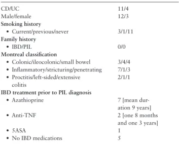
Our series included 15 IBD patients with PIL treated in eight dif- ferent centres (12 males [80.0%], mean [±SD, range] age 40.2 [±21.0, 13–76] years at IBD diagnosis and 46.4 [±17.0, 26–76] years at PIL diagnosis). Patients’ baseline characteristics are shown in Table 1. Six
patients had other significant comorbidities including Parkinson’s disease, myasthenia gravis, cardiovascular disease, Addison’s disease, diabetes and autoimmune polyglandular syndrome type II. An IBD- related surgery was performed in two patients prior to PIL diagnosis [ileocaecal resection in one, perianal drainage in one].
2.2. Primary intestinal lymphoma diagnosis
The diagnosis of PIL occurred at a mean time of 10.4 [±7.1, 1–24]
years after IBD diagnosis in 11 patients, prior to IBD in two and Table 1. Baseline characteristics [n = 15]
CD/UC 11/4
Male/female 12/3
Smoking history
• Current/previous/never 3/1/11
Family history
• IBD/PIL 0/0
Montreal classification
• Colonic/ileocolonic/small bowel 3/4/4
• Inflammatory/stricturing/penetrating 7/1/3
• Proctitis/left-sided/extensive colitis
2/1/1 IBD treatment prior to PIL diagnosis
• Azathioprine 7 [mean dur-
ation 9 years]
• Anti-TNF 2 [one 8 months
and one 3 years]
• 5ASA 1
• No IBD medications 5
PIL, primary intestinal lymphoma; IBD, inflammatory bowel disease; CD, Crohn’s disease; UC, ulcerative colitis; anti-TNF, anti-tumour necrosis factor;
5ASA, 5-aminosalicylic acid.
Table 2. PIL characteristics
N=
Time of PIL diagnosis in relation to IBD diagnosis
• PIL diagnosed after IBD 11 [mean time 10.4 years]
• PIL diagnosed concurrently with IBD 3
• PIL diagnosed before IBD 1 Lymphoma type
• DLBCL 8 [7 CD, 1 UC]
• Hodgkin’s lymphoma 2 [both CD]
• MALT lymphoma 3 [2 CD, 1 UC]
• Indolent T-cell lymphoma 1 [UC]
• Immunoblastic lymphoma 1 [UC]
Lymphoma location
• Rectum 3
• Colon 4 [including 1 at
anastomosis]
• Small bowel 4
• Stomach 1
• Multiple GI sites 3
Presentation
• Bowel obstruction 9
• Abdominal mass [non-obstructing] 2
• Others 4
PIL location in relation to IBD location
• In area affected by IBD 10
• In area distant to IBD 5
PIL, primary intestinal lymphoma; IBD, inflammatory bowel disease; CD, Crohn’s disease; UC, ulcerative colitis; DLBCL, diffuse large B-cell lymphoma, MALT, mucosa-associated lymphoid tissue.
Downloaded from https://academic.oup.com/ecco-jcc/advance-article/doi/10.1093/ecco-jcc/jjab164/6368841 by University of Szeged user on 22 October 2021
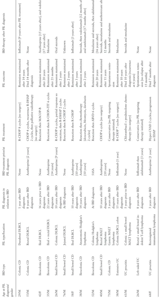
Table 3.Characteristics, treatment and outcome of primary intestinal lymphoma for each case Age at PIL diagnosis/ gender
IBD typePIL type/locationPIL diagnosis in relation to IBDIBD treatment prior/at PIL diagnosisPIL treatmentPIL outcomeIBD therapy after PIL diagnosis 29/MColonic CDDuodenal DLBCL 1 year after IBD diagnosisNoneR-CHOP 8 cycles [no surgery]Remission maintained after 14 yearsInfliximab [9 years after PIL treatment] 53/MColonic CDRectal DLBCL 8 years after IBD diagnosisAzathioprine [2 years]CHOP 4 cycles, recurrence R-DHAP 2 cycles, then palliative radiotherapy [no surgery]
Died 14 months after diagnosisNone 42/FIleocolonic CDIleal DLBCL 16 years prior to IBD diagnosisNoneResection then MACOP-BRemission maintained after 20 yearsAzathioprine [15 years after], and vedolizumab [17 years after] 35/MIleocolonic CDIleal + rectal DLBCL 22 years after IBD diagnosisAzathioprine [14 years]Resection then R-CHOP+TIT 6 cyclesRemission maintained after 21 monthsMesalazine 26/MSmall bowel CDColonic DLBCL 10 years after IBD diagnosisAzathioprine [9 years]R-CHOP 6 cycles, partial response so resection, then R-ESHAP 2 cyclesRemission maintained after 2 yearsOral steroids 76/MSmall bowel CDJejunal DLBCL At IBD diagnosisNoneResection then R-CHOP 2 cyclesRemission maintained after 2 yearsUnknown 58/FSmall bowel CDIleal DLBCL 10 years after IBD diagnosisAzathioprine [10 years]Resection then R-CHOP Remission maintained after 3 yearsInfliximab [3 years after] 38/MIleocolonic CDAnastomotic Hodgkin’s lymphoma 14 years after IBD diagnosisAzathioprine [10 years]Resection then chemotherapy [doxorubicin, bleomycin, darabazin 2 cycles]
Remission maintained after 2 yearsSteroids, then vedolizumab [12 months after] and ustekinumab [17 months after] 52/MIleocolonic CDColonic Hodgkin’s lymphoma At IBD diagnosis5ASA Resection then ABVD 6 cyclesRemission maintained after 20 yearsMesalazine and steroids, then adalimumab [16 years after] 40/MSmall bowel CDRectal MALT lymphoma10 years after IBD diagnosisAzathioprine [10 years]CHOP 6 cycles [no surgery]Remission maintained after 11 yearsSteroids, mesalazine and methotrexate after 12 months 75/MColonic CDGastric MALT lymphoma 3 years after IBD diagnosisNoneConservative [no PIL-targeting therapy initiated]Spontaneous remis- sionSteroids and mesalazine 38/MExtensive UCColonic DLBCL colon15 years after IBD diagnosisInfliximab [1 year]R-CHOP 7 cycles [no surgery]Remission maintained after 10 yearsMesalazine 61/MUC proctitisColonic + small bowel MALT lymphoma 5 years prior to IBD diagnosisNoneChlorambucil + prednisolone chemo- therapy [no surgery]Died 10 years after diagnosis [recurrence at 6 years]
Steroids and mesalazine 26/MLeft-sided UCRectal + small bowel in- dolent T-cell lymphoma 8 years after IBD diagnosisInfliximab then adalimumab [3 years]Conservative [no PIL-targeting therapy initiated]Spontaneous remis- sion [no relapse after 15 months]
None 68/FUC proctitisRectosigmoid immunoblast lymphoma 3 years after IBD diagnosisAzathioprine [1 year]Hyper-CVAD 2 cycles; progression so EDAP Died 7 months after diagnosisNone CD, Crohn’s disease; UC, ulcerative colitis; IBD, inflammatory bowel disease; PIL, primary intestinal lymphoma; DLBCL, diffuse large B-cell lymphoma; MALT, mucosa-associated lymphoid tissue; ABVD, doxorubicin, bleomycin, vinblastine, dacarbazine; CHOP, cyclophosphamide, doxorubicin, vincristine, prednisolone; R-CHOP, rituximab, cyclophosphamide, doxorubicin, vincristine, prednisolone; MA-COB, methotrexate with leucovorin rescue, doxorubicin, cyclophosphamide, vincristine, prednisone, bleo- mycin; R-ESHAP, rituximab, etoposide, methylprednisolone, cytarabine, cisplatin; R-DHAP, rituximab, dexamethasone, cisplatin, cytarabine; Hyper-CVAD, cyclophosphamide, vincristine, doxorubicin, dexamethasone, methotrexate, cytarabine; EDAP, etoposide, dexamethasone, cytarabine, cisplatin. Downloaded from https://academic.oup.com/ecco-jcc/advance-article/doi/10.1093/ecco-jcc/jjab164/6368841 by University of Szeged user on 22 October 2021
A B
C D
E F
Figure 1. Endoscopic and histological images of a Hodgkin’s lymphoma at the ileo-transverse colonic anastomosis. [A] Hodkin’s lymphoma. [B] Ulceration of mucous membranes due to lymphoma. [C] Lymphoma aggregate [magnification ×20]. [D] Ki-67 [magnification ×5]. [E] CD15-positive blasts. [F] CD30-positive blasts. Images courtesy of Niels Teich and Professors Arved Weimann and Volker Wiechmann, Leipzig, Germany.
Downloaded from https://academic.oup.com/ecco-jcc/advance-article/doi/10.1093/ecco-jcc/jjab164/6368841 by University of Szeged user on 22 October 2021
concurrently with IBD in two. The mean age of the four patients with a pre- or concurrent diagnosis was 52.5 [±17.8, 26–76] years, with no personal or family history of haematological disease. PIL characteristics for all 15 patients included in this series are shown in Table 2, whilst characteristics, treatment and outcome for each patient are given in Table 3. Diagnosis was made with the use of cross-sectional imaging and endoscopy, and histological confirm- ation was obtained in all cases. Selective endoscopic and histological images are shown in Figure 1.
PIL was located within the IBD-affected area in ten patients [six in the large bowel, three in the small bowel, and one in both ileum and rectum concurrently, with four of these having active intestinal inflam- mation at the time of diagnosis]. There was no extraintestinal or bone marrow involvement in any patient, except for the case of immunoblastic lymphoma who had non-contiguous extraintestinal spread.
IBD treatment was discontinued in the seven patients on azathioprine and the single patient on infliximab. Thirteen patients received PIL-related therapy, with seven receiving chemotherapy and surgery and six receiving only chemotherapy. The regimens are de- tailed in Table 3. Remission was achieved in ten patients after treat- ment with no signs of relapse at last follow-up. Three died from their PIL despite treatment: one with colonic CD and diffuse large B-cell lymphoma [DLBCL], one with UC proctitis and immunoblastic lymphoma with plasma cell differentiation, and one with UC proc- titis and mucosa-associated lymphoid tissue [MALT] lymphoma throughout the large and small bowel. Two are being monitored without therapy: one with an indolent T-cell lymphoma and the other with a gastric MALT lymphoma. The latter was not associated with Helicobacter pylori, and no further signs of lymphoma were found after 2 years of endoscopic follow-up.
IBD-related treatment was resumed in 11/15 patients [Table 3].
One of these patients was treated with 5-aminosalicylic acid [5ASA]
and oral steroids, later developing lymphoma recurrence and dying from this. No signs of PIL relapse were reported in the rest at last follow-up, at a median 6.5 years after PIL diagnosis [range 1.5–
20 years]. This included five patients [three with DLBCL in remission and two with Hodgkin’s lymphoma in remission] who have started biologics [infliximab in two cases, adalimumab in one, vedolizumab in one, and vedolizumab followed by ustekinumab in one], all for active luminal CD.
3. Discussion
This is a retrospective, international study reporting on a series of PIL in IBD patients, and the course of both PIL and subsequent IBD therapy. This study was not designed to assess the prevalence of PIL in IBD. Our series has shown some similarities with the previous case series, but also some important differences.
Similar to the Mayo and CESAME cohort series,9,10 we also ob- served that PIL mainly occurred in middle-aged men [80.0%], with an average age of 47.8 years at diagnosis. PIL was more frequently diagnosed in CD [11/15] than UC patients, and the most common histological type was B-cell lymphoma, including an immunoblastic lymphoma with plasma cell differentiation [9/15].
The diagnosis of PIL occurred at a mean of 10.4 [±7.1, 1–24]
years after IBD diagnosis, which is less than the 17 years reported in the Mayo series,9 and more compatible with the 8 years reported in the CESAME series.10 However, two patients were diagnosed with PIL simultaneously with IBD, highlighting the possibility that both may be present at the same time. Diagnostic work-up includes a combination of clinical presentation of major abdominal symptoms
not always attributed to IBD, cross-sectional imaging and endos- copy [if accessible], with compatible histological features, the last being a prerequisite for diagnosis. In some cases, diagnosis is made only after surgical resection of an obstructing lesion and subsequent histological examination of the resection specimen. Bowel obstruc- tion was the most common clinical presentation, whereas bloody diarrhoea has been previously reported as equally common in other series.9,10 Endoscopic features are not always pathognomonic.
The Mayo clinic and CESAME cohort series identified immuno- suppressants and active intestinal inflammation as potential risk fac- tors for the development of PIL.9,10 Sixty per cent of the cases in our series [9/15] may be related to these medications. Two-thirds developed PIL in the area affected by IBD inflammation, but that means a third of patients developed PIL in an area not involved in intestinal inflammation. Clinicians should therefore be mindful of this possibility. Two-thirds [10/15] achieved remission after receiving the appropriate PIL-related treatment.
A potential clinical dilemma is whether immunosuppressants can be safely restarted after lymphoma diagnosis. Our series shows IBD therapy was resumed in 11 patients, with relapse of PIL occurring in one case only that was probably unrelated to treat- ment. This includes five patients who restarted biologics for active luminal CD, including anti-TNFs, vedolizumab and ustekinumab, and none of them reported recurrence of lymphoma at last follow-up.
In conclusion, this case series of PIL in IBD illustrates a strong male predilection and a wide histological type range, with the majority being DLBCL. It is notable that two-thirds of patients had a history of thiopurine or anti-TNF use, and two-thirds devel- oped PIL in the luminal site affected by IBD. The majority of cases successfully recovered after appropriate treatment and, in half of them, IBD therapy was resumed uneventfully. However, these re- sults are based on a small case series and should be interpreted with caution.
Funding
No financial support was received for this work.
Conflict of Interest
The contributing authors declare the following: F.P., D.R.G., K.Kat.: none.
B.V.: financial support for research from Pfizer; lecture fees from Abbvie, Ferring, Takeda Pharmaceuticals, Janssen and R Biopharm; consultancy fees from Janssen, Guidepoint and Sandoz. I.G.: served as speaker, a con- sultant and advisory member for or has received research funding from Kern Pharma, Takeda and Janssen. N.T.: served as a speaker, a consultant and/or an advisory board member for AbbVie, Biogen, Falk Foundation, Janssen, MSD, Norgine, Shield Therapeutics, Takeda, Tillotts and Vifor and has re- ceived research funding from Ferring Arzneimittel GmbH. R.F.: support for research from EGIS, lecture fees from Takeda, Abbvie and Ferring. T.M.:
speaker’s honoraria from MSD, AbbVie, Egis, Goodwill Pharma, Takeda, Pfizer, Richter, Fresenius Kabi and Teva. K.Kar.: personal fees from Abbvie, MSD, Janssen, Pfizer and Takeda. All conflicts of interests stated here are outside the submitted work.
Author Contributions
F.P.: conceived the study, collected, analysed and interpreted the data and drafted the manuscript. B.V., D.G.R., I.G., N.T., K.Kat., R.F. and T.M.: con- tributed the cases and revised the manuscript. K.Kar.: supervised the project, interpreted the data and critically revised the manuscript. All authors ap- proved the final version.
Downloaded from https://academic.oup.com/ecco-jcc/advance-article/doi/10.1093/ecco-jcc/jjab164/6368841 by University of Szeged user on 22 October 2021
Data Availability
The data underlying this article are available within the article. Any additional information will be shared on reasonable request to the corresponding author.
Acknowledgments
We thank the ECCO CONFER Steering Committee members: Uri Kopylov [Israel, also member of the ECCO Clinical Research Committee], Pierre Ellul [Malta], Maria Chaparro Sanchez [Spain] and Idan Goren [Israel] and the ECCO Clinical Research Committee members: Krisztina Gecse [The Netherlands], Laurent Beaugerie [France], Peter Bossuyt [Belgium] and Shaji Sebastian [UK]
and all collaborators participating in the Feasibility Network for selecting this topic as a CONFER project and providing their insightful comments, as well as the ECCO Office personnel for their valuable contribution to the success of the project. We also thank Professors Arved Weimann and Volker Wiechmann for kindly providing the histology images presented in this paper.
References
1. Munkholm P. Review article: The incidence and prevalence of colorectal cancer in inflammatory bowel disease. Aliment Pharmacol Ther 2003;18 Suppl 2:1–5.
2. Beaugerie L, Brousse N, Bouvier AM, et al.; CESAME Study Group.
Lymphoproliferative disorders in patients receiving thiopurines for inflam- matory bowel disease: a prospective observational cohort study. Lancet 2009;374:1617–25.
3. Herrinton LJ, Liu L, Weng X, Lewis JD, Hutfless S, Allison JE. Role of thiopurine and anti-TNF therapy in lymphoma in inflammatory bowel disease. Am J Gastroenterol 2011;106:2146–53.
4. Kandiel A, Fraser AG, Korelitz BI, Brensinger C, Lewis JD. Increased risk of lymphoma among inflammatory bowel disease patients treated with azathioprine and 6-mercaptopurine. Gut 2005;54:1121–5.
5. Lewis JD, Bilker WB, Brensinger C, Deren JJ, Vaughn DJ, Strom BL.
Inflammatory bowel disease is not associated with an increased risk of lymphoma. Gastroenterology 2001;121:1080–7.
6. Taylor AL, Marcus R, Bradley JA. Post-transplant lymphoproliferative dis- orders (PTLD) after solid organ transplantation. Crit Rev Oncol Hematol 2005;56:155–67.
7. Ribaldone DG, Imperatore N, Le Grazie M, et al.; Italian Group for the study of Inflammatory Bowel Disease IG-IBD. Inflammatory bowel dis- ease course in liver transplant versus non-liver transplant patients for primary sclerosing cholangitis: LIVIBD, an IG-IBD study. Dig Liver Dis 2021;53:712–6.
8. Shepherd NA, Hall PA, Williams GT, et al. Primary malignant lymphoma of the large intestine complicating chronic inflammatory bowel disease.
Histopathology 1989;15:325–37.
9. Holubar SD, Dozois EJ, Loftus EV Jr, et al. Primary intestinal lymphoma in patients with inflammatory bowel disease: a descriptive series from the prebiologic therapy era. Inflamm Bowel Dis 2011;17:1557–63.
10. Sokol H, Beaugerie L, Maynadié M, et al.; CESAME Study Group. Excess primary intestinal lymphoproliferative disorders in patients with inflam- matory bowel disease. Inflamm Bowel Dis 2012;18:2063–71.
11. Salgueiro P, Lago P, Farrajota P, Santos M, Pedroto I. Primary intestinal Hodgkin’s lymphoma associated with Crohn’s disease. Rev Esp Enferm Dig 2013;105:293–5.
12. Kelly MD, Stuart M, Tschuchnigg M, Turner J, Tydd T. Primary intestinal Hodgkin’s disease complicating ileal Crohn’s disease. Aust N Z J Surg 1997;67:485–9.
13. Hecker R, Sheers R, Thomas D. Hodgkin’s disease as a complication of Crohn’s disease. Med J Aust 1978;2:603.
14. Hall CH Jr, Shamma M. Primary intestinal lymphoma complicating Crohn’s disease. J Clin Gastroenterol 2003;36:332–6.
15. Perosio PM, Brooks JJ, Saul SH, Haller DG. Primary intestinal lymphoma in Crohn’s disease: minute tumor with a fatal outcome. Am J Gastroenterol 1992;87:894–8.
16. Teare JP, Greenfield SM, Slater S. Rectal lymphoma after colectomy for ulcerative colitis. Gut 1992;33:138–9.
17. Kumar S, Fend F, Quintanilla-Martinez L, et al. Epstein-Barr virus-positive primary gastrointestinal Hodgkin’s disease: association with inflammatory bowel disease and immunosuppression. Am J Surg Pathol 2000;24:66–73.
18. Nyam DC, Pemberton JH, Sandborn WJ, Savcenko M. Lymphoma of the pouch after ileal pouch-anal anastomosis: report of a case. Dis Colon Rectum 1997;40:971–2.
19. Wong NA, Herbst H, Herrmann K, et al. Epstein-Barr virus infection in colorectal neoplasms associated with inflammatory bowel disease: de- tection of the virus in lymphomas but not in adenocarcinomas. J Pathol 2003;201:312–8.
Downloaded from https://academic.oup.com/ecco-jcc/advance-article/doi/10.1093/ecco-jcc/jjab164/6368841 by University of Szeged user on 22 October 2021