Relics from the Past: Molecular Biology and Genetic Applications of Resurrected
DNA Transposons in Vertebrates
Thesis for the Degree of
Doctor of the Hungarian Academy of Sciences
Zoltán Ivics
Max Delbrück Center for Molecular Medicine
Berlin 2010
Table of Contents
ACKNOWLEDGEMENTS... 5
LIST OF PUBLICATIONS THAT FORM THE BASIS OF THIS THESIS... 6
ABBREVIATIONS... 9
1 INTRODUCTION... 12
1.1 Discovery of transposable elements ... 12
1.2 Classification of transposable elements ... 13
1.2.1 RNA elements ... 14
1.2.2 DNA elements ... 16
1.2.2.1 The Tc1/mariner superfamily of transposons... 18
1.2.2.1.1 Structural and functional components of Tc1/mariner transposons ... 18
1.2.2.1.1.1 The transposase ... 18
The DNA-binding domain... 19
The catalytic domain... 21
1.2.2.1.1.2 The transposon inverted repeats ... 23
1.3 Modes of transposition ... 25
1.3.1 The biochemistry of cut-and-paste DNA transposition ... 26
1.3.1.1 Transposon excision... 27
1.3.1.2 Transposon integration and target site selection... 28
1.4 Regulation of transposition ... 30
1.4.1 Transcriptional control of transposition ... 32
1.4.2 Control of synaptic complex assembly during transposition ... 33
1.4.3 Regulation of transposition by chromatin... 35
1.4.4 Regulation by cell-cycle and DNA repair processes ... 36
1.4.5 Avoiding insertional damage to host cell genes by site-specific transposition... 38
1.5 DNA elements in natural hosts ... 40
1.5.1 The evolutionary life-cycle of DNA transposons ... 40
1.5.2 Impact of transposons on host genomes: Mutations, genome size and the evolution of novel gene functions ... 42
1.5.2.1 Transposons as a creative evolutionary force... 44
1.5.2.1.1 "Domesticated", transposase-derived cellular genes ... 45
1.6 Transposons as genetic tools ... 48
1.6.1 Insertional mutagenesis... 50
1.6.2 Transgenesis... 54
1.6.3 Transposons as vectors for gene therapy ... 56
1.6.3.1 The genotoxic risk of integrating gene therapy vector systems ... 58
2 AIMS... 61
2.1 Relics from the past: molecular biology of resurrected transposons and transposase-derived cellular genes in vertebrates... 61
2.2 DNA transposons as a gene delivery platform for genetic manipulations in vertebrates ... 61
3 RESULTS and DISCUSSION... 62
3.1 Relics from the past: molecular biology of resurrected transposons and transposase-derived cellular genes in vertebrates... 62
3.1.1 Molecular reconstruction of Sleeping Beauty, a Tc1-like transposon in fish, and its transposition in human cells (Papers I and II) ... 62
3.1.1.1 The molecular mechanism of Sleeping Beauty transposition ... 64
3.1.1.1.1 Transcriptional activities of the Sleeping Beauty transposon (Paper III) ... 64
3.1.1.1.2 Specific DNA-binding by the Sleeping Beauty transposase... 68
3.1.1.1.3 Synaptic complex assembly and the role of multiple binding sites for the transposase... 69
3.1.1.1.4 The role of HMGB1 in Sleeping Beauty transposition: Ordered assembly of synaptic complexes (Paper IV) ... 70
3.1.2 Sleeping Beauty transposase modulates cell-cycle progression through interaction with Miz-1 (Paper V) ... 71
3.1.3 Regulation of Sleeping Beauty transposition by DNA CpG methylation (Paper VI) ... 76
3.1.4 Common physical properties of DNA affecting target site selection of Sleeping Beauty and other Tc1/mariner transposable elements (Paper VII) ... 79
3.1.5 The Frog Prince: a reconstructed transposon from Rana pipiens with high activity in vertebrates (Paper VIII) ... 84
3.1.6 The ancient mariner sails again: Transposition of the human Hsmar1 element by a reconstructed transposase and activities of the SETMAR protein on
transposon ends (Paper IX) ... 91 3.1.7 Transposition of a reconstructed Harbinger element in human cells and
functional homology with two transposon-derived cellular genes (Paper X) ... 101 3.2 DNA transposons as a gene delivery platform for genetic manipulations in
vertebrates ... 111 3.2.1 Development of hyperactive Sleeping Beauty transposon vectors by mutational
analysis (Paper XI) ... 111 3.2.2 Frog Prince transposon-based RNAi vectors mediate efficient gene knockdown
in human cells (Paper XII)... 119 3.2.3 Comparative analysis of transposable element vector systems in human cells
(Paper XIII)... 123 3.2.4 Towards safer vectors for gene therapy I: Transcriptional shielding of Sleeping
Beauty's genetic cargo with insulators (Paper III) ... 134 3.2.5 Towards safer vectors for gene therapy II: Targeted Sleeping Beauty
transposition in human cells (Paper XIV)... 137 4 SUMMARY: Major discoveries and conclusions... 144 5 REFERENCES... 152
ACKNOWLEDGEMENTS
First and foremost I am grateful to my wife and long-term colleague Dr. Zsuzsanna Izsvak with whom I have been working side-by-side for the past nineteen years. This time has been an adventure to “boldly go where no man has gone before”.
I am grateful to all members of the “Transposition” and “Mobile DNA” groups at the Max Delbrück Center for Molecular Medicine for their dedicated work. Their spirit, innovative ideas as well as hard benchwork provide the basis of past and future discoveries.
Lastly, I thank my parents for their unconditional love and support.
Work in the author’s laboratory has been supported by EU FP5, FP6 and FP7 research grants, as well as financial support from the Deutsche Forschungsgemeinschaft, the Bundesministerium für Forschung und Bildung and the Volkswagen Stiftung.
LIST OF PUBLICATIONS THAT FORM THE BASIS OF THIS THESIS
I. Ivics, Z., Izsvák, Zs., Minter, A. and Hackett, P.B. (1996). Identification of functional domains and evolution of Tc1-like transposable elements. Proc. Natl. Acad. Sci. USA 93:5008-5013.
II. Ivics, Z., Hackett, P.B., Plasterk, R.H. and Izsvák, Zs. (1997). Molecular reconstruction of Sleeping Beauty, a Tc1-like transposon in fish, and its transposition in human cells.
Cell 91:501-510.
III. Walisko, O., Schorn, A., Rolfs, F., Devaraj, A., Miskey, C., Izsvák, Z. and Ivics, Z.
(2008). Transcriptional activities of the Sleeping Beauty transposon and shielding its genetic cargo with insulators. Mol. Ther. 16:359-69.
IV. Zayed, H., Izsvák, Zs., Khare, D., Heinemann, U. and Ivics, Z. (2003). The DNA- bending protein HMGB1 is a cellular cofactor of Sleeping Beauty transposition. Nucleic Acids Res. 31:2313-2322.
V. Walisko, O., Izsvák, Z., Szabó, K., Kaufman, C.D., Herold, S., and Ivics, Z. (2006).
Sleeping Beauty transposase modulates cell-cycle progression through interaction with Miz-1. Proc. Natl. Acad. Sci. USA 103:4062-4067.
VI. Jursch, T., Izsvák, Z. and Ivics, Z. (2010). Regulation of DNA transposition by CpG methylation and chromatin structure in human cells. Mobile DNA. (to be submitted) VII. Vigdal, T., Kaufman, C., Izsvak, Z., Voytas, D. and Ivics, Z. (2002). Common physical
properties of DNA affecting target site selection of Sleeping Beauty and other Tc1/mariner transposable elements. J. Mol. Biol. 323:441452.
VIII. Miskey, Cs., Izsvák, Zs., Plasterk, R.H. and Ivics, Z. (2003). The Frog Prince: a reconstructed transposon from Rana pipiens with high activity in vertebrates. Nucleic Acids Res. 31:6873-6881.
IX. Miskey, C., Papp, B., Mátés, L., Sinzelle, L., Keller, H., Izsvák, Z. and Ivics, Z. (2007).
The ancient mariner sails again: Transposition of the human Hsmar1 element by a
reconstructed transposase and activities of the SETMAR protein on transposon ends.
Mol. Cell. Biol. 27: 4589-600.
X. Sinzelle, L., Kapitonov, V.V., Grzela, D.P., Jursch, T., Jurka, J., Izsvák, Z. and Ivics, Z.
(2008). Transposition of a reconstructed Harbinger element in human cells and functional homology with two transposon-derived cellular genes. Proc. Natl. Acad. Sci.
USA 105:4715-20.
XI. Zayed, H., Izsvák, Zs., Walisko, O. and Ivics, Z. (2004). Development of hyperactive Sleeping Beauty transposon vectors by mutational analysis. Mol. Ther. 9:292-304.
XII. Kaufman, C.D., Izsvák, Z., Katzer, A. and Ivics, Z. (2005). Frog Prince transposon- based RNAi vectors mediate efficient gene knockdown in human cells. Journal of RNAi and Gene Silencing 1:97-104.
XIII. Grabundzija, I., Irgang, M., Mátés, L., Belay, E., Matrai, J., Gogol-Döring, A., Kawakami, K., Chen, W., Ruiz, P., Chuah, M.K., VandenDriessche, T., Izsvák, Z. and Ivics, Z. (2010). Comparative analysis of transposable element vector systems in human cells. Mol. Ther. (in press)
XIV. Ivics, Z., Katzer, A., Stüwe, E.E., Fiedler, D., Knespel, S. and Izsvák, Z. (2007).
Targeted Sleeping Beauty transposition in human cells. Mol. Ther. 15:1137–1144.
Other publications that significantly contributed to this thesis
Izsvák, Zs., Ivics, Z. and Hackett, P.B. (1995). Characterization of a Tc1-like transposable element in zebrafish (Danio rerio). Mol. Gen. Genet. 247:312-322.
Plasterk, R.H., Izsvák, Zs. and Ivics, Z. (1999). Resident Aliens: The Tc1/mariner superfamily of transposable elements. Trends Genet. 15:326-332.
Izsvák, Zs., Khare, D., Behlke, J., Heinemann, U., Plasterk, R.H. and Ivics, Z. (2002).
Involvement of a bifunctional, paired-like DNA-binding domain and a transpositional enhancer in Sleeping Beauty transposition. J. Biol. Chem. 277:34581-34588.
Walisko, O. and Ivics, Z. (2006). Interference with cell cycle progression by parasitic genetic elements: Sleeping Beauty joins the club. Cell Cycle. 5:1275-80.
Walisko, O., Jursch, T., Izsvák, Z. and Ivics. Z. (2008). Transposon-host cell interactions in the regulation of Sleeping Beauty transposition. IN (Volff, J.-N. and Lankenau, D.-H.
eds) Genome Dynamics and Stability vol. 4: Transposons and the Dynamic Genome.
Springer-Verlag Berlin Heidelberg, Germany, pp 109-132.
Voigt, K., Izsvák, Z. and Ivics, Z. (2008). Targeted gene insertion for molecular medicine. J.
Mol. Med. 86:1205-19.
Sinzelle, L., Izsvák, Z. and Ivics, Z. (2009). Molecular domestication of transposable elements: From detrimental parasites to useful host genes. Cell. Mol. Life Sci. 66:1073- 93.
Mátés, L., Chuah, M.K., Belay, E., Jerchow, B., Manoj, N., Acosta-Sanchez, A., Grzela, D.P., Schmitt, A., Becker, K., Matrai, J., Ma, L., Samara-Kuko, E., Gysemans, C., Pryputniewicz, D., Miskey, C., Fletcher, B., VandenDriessche, T., Ivics, Z. and Izsvák, Z. (2009). Molecular evolution of a novel hyperactive Sleeping Beauty transposase enables robust stable gene transfer in vertebrates. Nat Genet. 41:753-61.
ABBREVIATIONS
AAV Adeno Associated Virus
Ac Activator
ASLV Avian Sarcoma Leukosis Virus
ASV Avian Sarcoma Vurus
ATP adenosine 5´-triphosphate
Cdk cyclin-dependent kinase
cDNA complementary DNA
CHO Chinese hamster ovary
CMV cytomegalovirus
DBD DNA-binding domain
DDD amino acid sequence containing three aspartic acids DDE amino acid sequence containing two aspartic acids and
one glutamic acid
DNA deoxyribonucleic acid
DR direct repeat
E. coli Escherichia coli
EN endonuclease
ENU ethylnitrosourea
env envelope
ERV endogenous retrovirus
FACS fluorescence-activated cell sorting
FP Frog Prince
gag group-specific antigen
GFP green fluorescent protein
HA hemagglutinin
HDR homology-dependent repair
HIV human immunodeficiency virus
HMG high-mobility group protein
HSC hematopoietic stem cell
HTH helix-turn-helix
Hsmar1 Homo sapiens mariner type 1
IAP intracisternal A particle
IHF integration host factor
IN integrase
IR inverted repeat
IRES internal ribosome entry site
IR/DR inverted repeats containing direct repeats
IS insertion sequence
kbp kilo base pairs
kDa kilo Daltons
L1 LINE1
LINE long interspersed nuclear element
LTR long terminal repeat
MBP maltose binding protein
MLV murine leukemia virus
Myr million years
mRNA messenger ribonucleic acid
NHEJ nonhomologous end joining
NLS nuclear localization signal
OPI overproduction inhibition
ORF open reading frame
PCR polymerase chain reaction
PEC paired end complex
pol polymerase
polyA poly-adenylation signal
Pol II RNA polymerase II
Pol III RNA polymerase III
PR protease
RACE rapid amplification of cDNA ends
RAG recombination-activating gene
RNA ribonucleic acid
RNAi RNA interference
RSS recombination signal sequence
RT reverse transcriptase
SA splice acceptor
SB Sleeping Beauty
SD splice donor
sem standard error of the mean
shRNA short hairpin RNA
SINE short interspersed nuclear element
SV40 simian virus 40
Tc1 transposon of Caenorhabditis elegans 1
TE transposable element
Tn transposon
TSD target site duplication
Ty yeast retrotransposon
UTR untranslated region
V(D)J variable (diversity) joining
vs. versus
X-Gal 4-Cl-5-Br-3-indolyl-β-galactosidase
ZF zinc finger
ZFN zinc finger nuclease
Figure 1. Barbara McClintock – Nobel Prize 1983
1 INTRODUCTION
1.1 Discovery of transposable elements
Transposable genetic elements (“jumping genes”) were first discovered by Barbara McClintock in the 1940s. She found that certain spontaneous mutations in enzymes required for the productions of the purple anthocyanin pigment in maize are due to “controlling”
elements that could apparently move from site to site in different chromosomes. This idea of jumping genes ran contrarily to the traditional view of the age that genomes are stable and static entities. The possibility that pieces of DNA can
“jump around” in a genome was viewed by biologists with much skepticism. Therefore, McClintock’s observations were thought to be rare phenomena and not of general interest. In fact, at a historic Cold Spring Harbor meeting 1951. McClintock's work greeted with "stony silence." It took nearly 30 years until McClintock’s conclusions from the 1940s were confirmed by findings in bacteria (insertion sequences) and Drosophila melanogaster (hybrid dysgenesis), and another ten years until she was rewarded for the discovery of transposable elements with the Nobel prize in 1983 (Fig. 1).
With the great advances of the molecular biology in the 1970s, it turned out that McClintock’s discovery was just the tip of an iceberg. Mobile element were found to be widespread not only in maize but in all kingdoms of living organisms from bacteria to humans. It turned out that transposable elements are indeed so abundant that they form a major fraction of the eukaryotic genome [1]. However, most researchers still assumed that repetitive DNA elements do not have any function: they are useless, selfish DNA sequences [2]. The term “junk DNA” coined by Sozumu Ohno repelled mainstream research from studying repetitive elements for many years [3]. As Doolittle and Sapienza termed in Nature in 1980: transposons’ “only «function» is survival within genomes”…“thus no phenotypic or evolutionary function need to be assigned to them”.
This view started to change in the 1990s, when it became evident that transposons are important integral components of eukaryotic genomes with deep impacts on the host evolution. It turned out that they interact with the surrounding genomic environment, and increase the ability of the organism to evolve [4].
Since their discovery, transposable elements have been broadening the scope of many fields of modern biology ranging from evolutionary genetics to gene therapy. There are numerous aspects of viewing transposable elements as subjects of scientific investigation.
Transposons are of interest for genome annotators, for structural and evolutional geneticists who investigate the role of mobile elements in chromosome/genome dynamics and their different contributions to host evolution. The ongoing studies of molecular biologists are continuously increasing our understanding of the mechanism transposition. Moreover, experimental geneticists use transposons routinely for insertional mutagenesis, gene tagging, germline transformations, gene trapping, and gene therapy. Their experimental model organisms range from bacteria to mammals. Due to the discovery of a variety of different prokaryotic and eukaryotic transposons, they are now routinely used as genetic tools in functional biology. Thus, repetitive elements are relevant to a wide scale of genetic studies, and transposons begin to be viewed as genomic treasure [5, 6].
1.2 Classification of transposable elements
Discrete DNA sequences that possess an intrinsic capability to change their genomic locations are called transposable elements (TEs). TEs are distinguished whether their movement relies exclusively on DNA intermediates or includes an RNA stage. Transposons that move exclusively through a DNA intermediate are referred to as DNA elements. Mobile elements that move through an RNA intermediate (RNA elements or retroelements) are transcribed, reverse transcribed and integrate as double stranded cDNA. These elements include retroviruses and the retrotransposons. DNA elements can be found in both prokaryotic and eukaryotic organisms, whereas RNA elements are restricted to eukaryotes.
1.2.1 RNA elements
Based on their structural properties and evolutionary relationships those transposable elements that can mobilize themselves through an element-derived RNA intermediate are grouped to those with long terminal repeats (LTR-retrotransposons and retroviruses) and those without (non-LTR retrotransposons).
A common feature of LTR-retrotransposons and retroviruses is that their coding region is flanked by LTRs (Fig. 2C). These sequences contain important control sequences e.g. promoter, enhancer and polyadenylation (polyA) signals. The coding sequences are divided into at least two open reading frames (ORFs). The first ORF encodes the group- specific antigen (gag) protein, required for the assembly of the RNA transcript into cytoplasmic particles. The second ORF constitutes the pol gene, encoding a polyprotein, which consists of a protease (PR), a reverse transcriptase (RT) and an integrase (IN). The difference between retroviruses and LTR-retrotransposons is that retroviruses not only possess the capability to move between DNA molecules like other transposons, but they can leave their host cells too and integrate into new genomes. Nevertheless, retroviruses and LTR-retrotransposons are derived from a common progenitor [7].
LTR-retroelements can be subdivided into three families based on homologies within the RT gene. The first two groups are named after their founding members found in yeast and Drosophila, Ty1/copia and Ty3/gypsy [8]. The Ty3/gypsy elements form two subfamilies based on the presence or absence of a third ORF, env, encoding for envelope-like proteins.
Retroviruses cluster into the third family of LTR-elements; they always possess a completely functional env gene for their viral life cycle. Many retroviruses, for example human immunodeficiency virus type 1 (HIV-1), contain additional proteins [9]. Endogenous retroviruses (ERVs) appear to have been recently active in the mammalian genome. LTR- retrotransposons are widely destributed in eukaryotes, and make up about 8% of the human genome. Retroviruses were for long thought to be restricted to vertebrate genomes until it was shown that the gypsy retrotranspsoson is indeed an infectious retrovirus of Drosophila
A
B
IR IR
Transposase
IS-Right IS-Left
Antibiotic resistance genes polyA
EN RT
ORF1 ORF2 polyA 3’-UTR 5’-UTR
ORF1 ORF2
5’ -LTR 3’-LTR
gag PR IN RT-RH
E D C
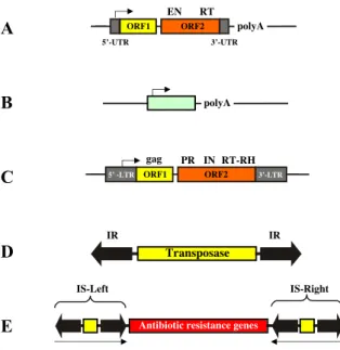
Figure 2. Structures and organization of the main types of transposable elements. (A) Non-LTR retrotransposon.
The element consists of a 5’ untranslated region that has promoter activity (arrow pointing towards the downstream genes), which is required to drive transcription of the element-encoded genes. ORF1 encodes a nucleic acid binding protein. ORF2 encodes an endonuclease (EN) and a reverse transcriptase (RT). The element has a polyA tail.
(B) A typical SINE. The element is a small, RNA-derived pseudogene, which is transcribed from an RNA polymerase III promoter within the element (arrow). The element has a polyA tail. (C) LTR-retrotransposon. The element consists of long terminal repeats (LTRs) similar to those of retroviruses.
The LTRs flank two open reading frames. ORF1 encodes the group specific antigen (gag), ORF2 encodes a protease (PR), an integrase (IN), and a reverse transcriptase- RNaseH (RT-RH) function. (D) DNA transposon. The central transposase gene (yellow box) is flanked by terminal inverted repeats (IRs, shown as black arrows). The IRs contain the binding sites for the transposase and sequences that are required for transposase-mediated cleavage. (E) Composite bacterial transposon. The element consists of antibiotic resistance genes (red box) flanked by two copies of an insertion sequence (IS) element that contains the transposase gene (yellow boxes). The arrows underneath indicate the inverted orientation of the IS elements.
melanogaster [10]. Transposition occurs through reverse transcription of the retrotransposon RNA, and integration of the resultant cDNA into a new location by the integrase protein.
The most abundant transposable elements in mammalians are non-LTR retrotransposons represented by the long interspersed nuclear elements (LINEs) and the short interspersed nuclear elements (SINEs). Although LINEs are especially abundant in mammals (they make up 26% of the human X chromosome alone) [11], they have also been found in protozoan, insects, reptiles and plants [12]. The major LINEs in humans (LINE1 or L1) are 6 kbp long and contain two ORFs (Fig. 2A). These encode for a nucleic acid binding protein and an enzyme with endonuclease (EN) and RT activity, respectively [13]. EN generates a single-stranded nick in the target DNA, and RT uses the nicked DNA to prime reverse transcription from the 3'-end of the L1 RNA [14]. Because reverse transcription is frequently incomplete, the majority of L1s is truncated, and thus nonfunctional.
Consequently, even though L1 has about 5 x 10⁵ copies in the human genome, thereby making up about 17% of human genomic DNA [11], the vast majority of these elements are inactive: in humans there are only 30-100 potentially active copies of L1 [15].
SINEs are short (about 100–400 bp) retrotransposable elements that encode no proteins; therefore, all of them are non- autonomous (Fig. 2B), and thought to use the enzymatic machinery of LINEs for transposition [16, 17]. The vast majority of known SINEs are derived from tRNA
sequences, with the exception of the human Alu element, which is derived from the 7SL component of the signal recognition particle [18]. Alu elements were originally identified as repetitive DNA elements in human DNA renaturation curves, and contain a recognition site for the restriction enzyme AluI. Alu elements are represented in the human genome with >1 x 10⁶ copies which make up about 11% of the total genome. Alu is the only active SINE in humans. Full-length Alus are 280bp long, contain promoter sequences for RNA polymerase III (Pol III) [19] and a polyA tail (Fig. 2B). The transcripts of Pol III-transcribed Alus terminate at Pol III termination signals fortuitously present in the 3’ flanking DNA. Rarely, RNA polymerase II (Pol II)-derived host gene transcripts can also be trans-mobilized by functional LINE proteins. These transposition products are named processed pseudogenes. They lack promoters, introns and end in a polyA tail. Only short target site duplications flanking these sequences provide evidence that these integrants are in fact transposition products.
1.2.2 DNA elements
These TEs can loosely be defined as sequences of DNA that can excise and insert into a variety of sites of a target DNA without the need to be reverse transcribed to cDNA. The simplest DNA elements are the insertion sequences (ISs) that were first characterized from bacteria in the late 1960s. Since then, approximately 800 ISs were identified (http://www- is.biotoul.fr). ISs are short (<2.5 kbps) and carry no genetic information except that necessary for their mobility. Thus, they are composed of a single gene coding for the transposase enzyme responsible for moving the element and of terminal inverted repeats (IRs) flanking it at both ends (Fig. 2D). The IRs are often called terminal inverted repeats (TIRs) or inverted terminal repeats (ITRs). The IRs contain the recombinationally active nucleotides at the very tips and specific recognition sequences for the transposase enzyme within.
Though most of the ISs are prokaryotic, a significant number of eukaryotic IS has also been documented. The largest and best-known group of these is the Tc1/mariner like elements that are structurally the closest to bacterial ISs (see more detail in the next section)
[20]. Another well-characterized member of the eukaryotic ISs is P element from Drosophila melanogaster.
Recently, new families of DNA elements have been identified from eukaryotes. The Helitron elements lack IRs and move by the rolling circle mechanism, similarly to the replication of plasmids, and together with their descendants they represent 2% of the Arabidopsis thaliana and the Caenorhabditis elegans genomes [21]. Polintons are a newly discovered, self-synthesizing, complex family of DNA transposons that possibly derived from ancient LTR retroelements [22]. The elements are very large (~20 kb), with IRs of several hundred bp in length and encode several proteins: i) a protein-primed DNA-dependent polymerase, ii) an ATPase, iii) a protease (not always), iv) an integrase, and 4-6 additional ORFs for proteins with unknown function. The 300,000 DNA transposon fossils in human add up to around 3% of the genome [11].
It became evident in the 1960s that genes responsible for antibiotic resistance in bacteria can move between DNA molecules in a process analogous to the movement of ISs [23]. It was suggested that mobile elements that carry one or more genes that encode other functions in addition to those related to transposition should be called transposons (today, the term „transposon“ is used in a wider sense, however: authors call all DNA elements, including ISs, transposons). Since these elements carry additional DNA they are usually larger than ISs (approximately 2.5-7 kbp). In some of these elements, called composite transposons, there are two complete ISs flanking a functional gene (Fig. 2E). This element can move as one functional unit, but also one or both of the bordering ISs can mobilize itself independently. There is a characteristic feature that distinguishes eukaryotic TEs from ISs and transposons in bacteria: the presence of a large number of inactive transposon copies that can in many cases be mobilized in trans by a limited number of active transposases [24].
Impala*
Bari1 Minos
Quetzal Fot1 Tc4
Pogo*
Tigger
Tc1*
Tc3 SB*
Tc1 Mariner
Pogo
Txr S Himar1
D.erecta p19 C. elegans MLE
H. cecropia MLE Mos1*
IS630*
IS911 IS3*
Copia Gypsy RSV
IS30 HIV-1*
D.tigrina Mar1 LTR- retrotranposons Retroviruses
Figure 3. Phylogeny of the Tc1/mariner superfamily.
DDE-containing recombinases are grouped into two major clusters: a DNA-transposon group and a retroelement group. Bacterial IS elements are DNA- transposons, but certain elements such as IS3, IS911 and IS30 are grouped together with the retroelement group, whereas the position of IS630 is close to the Tc1/mariner superfamily (green box) in the phylogenetic tree. The Tc1, mariner and pogo transposon families are probably monophyletic.
1.2.2.1 The Tc1/mariner superfamily of transposons
When David Hirsch and Scott Emmons discovered the Tc1 transposable element in 1983 as a repeat sequence in the genome of Caenorhabditis elegans [25], they probably did not realize how large the iceberg was of which they had found the tip. We now know that homologs of Tc1 and those of the related mariner transposon found in Drosophila mauritiana [26], are probably the most widespread DNA-transposons in nature, and can be found in fungi, plants, ciliates and animals, including nematodes, arthropods, fish, frogs and humans.
Together with related pogo transposons [27, 28], Tc1 and mariner elements are members of a large superfamily of transposable elements, the Tc1/mariner superfamily [29-31], so named after its two best studied members. Tc1/mariner elements are about 1300-2400 bp in length and contain a single gene encoding a transposase enzyme which is flanked by IRs. Although quite divergent in primary sequence (about 15% amino acid identity between the transposases of the different families [30]), members of the Tc1/mariner superfamily are probably monophyletic in origin (Fig. 3) [30, 32], and have similar structures and molecular mechanisms of transposition. As shown in Fig. 3, a more remote similarity exists between the above mentioned transposons and several bacterial IS elements, LTR-retrotransposons and retroviruses [33]. The recombinase proteins encoded by these diverse genetic elements are all related and contain a signature of three acidic amino acids (DDE or DDD, Fig. 2) with a characteristic spacing [32, 33].
1.2.2.1.1 Structural and functional components of Tc1/mariner transposons 1.2.2.1.1.1 The transposase
As discussed above, transposons are very diverse genetic entities; however, their enzymes carry out similar chemical reactions e.g. hydrolysis for strand cleavage and transesterification
for strand transfer. The similar activities of TEs are manifested in the remarkable overall structural similarity of the transposition proteins.
Both the transposases of ISs and transposons and INs of retroelements show structural similarities for their functional organization. Most of them can be divided into topological distinct functional domains. Partial proteolysis experiments revealed that the transposon-specific DNA-binding domains are generally localized in the N-terminal part, whereas the catalytic domain responsible for the strand cleavage and transfer is located in the C-terminal of the transposase protein (Fig. 4) [34, 35]. One possible explanation for this characteristic arrangement in prokaryotic elements is that during translation the N-terminal part of the premature transposase protein can fold independently of the C-terminal catalytic domain, and interact with its specific transposon binding sites close to the point of synthesis.
This hypothesis is reinforced by the observation that the presence of the C-terminal part of some bacterial transposases decreases the affinity of IR binding [36]. This arrangement can facilitate that the transposase is going to act on the transposon that produced it (a phenomenon called cis-preference) [37].
The DNA-binding domain
It is a key feature of all transposases that they recognize their specific transposon ends. TEs that move by transposon-specific transposases possess recognition sequences in their IRs.
The majority of ISs has simple, 10-40 bp long IRs, while others exhibit long and complex IRs.
Most transposon ends are composed of two functional parts. The 2-3 terminal base pairs of the ends are the recombinationally active sequences involved in the cleavage and the strand transfer reactions. The other functional part is situated within the IRs and it ensures the sequence-specific positioning of the transposase on the transposon ends [38, 39]. ISs have single transposase binding sites whereas for example M u and T n 7 have complex, asymmetric recognition sites [40, 41]. The bi-functionality of the transposon ends is reflected in the arrangement of the transposase on its cognate transposon. Due to the flexibility of the transposase, the N-terminal region of the enzyme attaches to the inner segment of IRs while
the C-terminal contacts the external ends. The sequence-specific DNA-binding of both eukaryotic and prokaryotic transposases is often carried out by a helix-turn-helix (HTH) motif.
This domain can be simple as it is the case of IS transposases [42], or can be complex and bipartite as found in Ac, Mu or in Tc transposases [29, 43]. The catalytic C-terminal domains of transposases are also involved in DNA-binding, however, this activity is not sequence specific and contributes to the correct positioning of the transposon end into the catalytic pocket [44].
The overall domain structure of the transposase is conserved in the entire Tc1/mariner superfamily [20]. Specific substrate recognition is mediated by an N-terminal, bipartite DNA-binding domain of the transposase (Fig. 4) [45-47]. This DNA-binding domain has been proposed to consist of two HTH motifs, similar to the paired domain of some transcription factors in both amino acid sequence and structure [47-49]. The modular paired domain has evolved versatility in binding to a range of different DNA sequences through various combinations of its subdomains (PAI+RED) [50]. The nucleotide sequences recognized by the composite paired domain are degenerate, the DNA-binding specificity is relaxed [51]. The origin of the paired domain is not clear, but phylogenetic analyses indicate that it might have been derived from an ancestral transposase [52].
The first of these HTH motifs, similar to the paired domain of some transcription factors (Fig. 4) [48, 49], has been crystallized in complex with double-stranded DNA corresponding to the termini of Tc3 transposons in C. elegans [53]. The crystal structure indeed showed a HTH fold, and a dimer of transposase subunits bringing together the two DNA ends. The paired-like domain is followed by a second HTH motif embedded in a homeo-like DNA-binding domain (Fig. 4). Secondary structure predictions indicate that mariner transposases might also contain such a bipartite DNA-binding domain consisting of two HTH motifs (Fig. 4). Pogo and certain bacterial transposases [54] contain "solo" HTH motifs (Fig. 4). We found that a GRPR-like sequence between the two HTH motifs is conserved in Tc1/mariner transposases (Fig. 4). The GRPR motif is characteristic to homeodomain proteins [55], and mediates interactions with DNA in the Hin invertase of Salmonella [56] and in the recombination activating gene (RAG1) recombinase that evolved
HHHHtttHHHH
HHHHtttHHHH
D 35
D E
IS630 IS3 HIV1-IN
Rag-1
~
~
~
~
~
~
~
HHHHtttHHHH GRPR
HHHHtttHHHH GKP HHHHtttHHHH
HHHHtttHHHH
HHHHtttHHHH
HHHHtttHHHH
D 34 E D
D
D 34 D
D
D 30/35 D
D
D 35 E
HHHHtttHHHH Zn2+
D D 35 E
GGRPR 1
Hin
GGRPR
RPR
Engrailed
?
1
D 254 E
1
1
1
1
124
145
~75
~45
~45
64
446
190
513 454
138 389
152
962 297
231
~300 282
285
55
600 181
140
~170 157
156
HHHHtttHHHH
~
~
~
~
~
~
~
~
~
D Tc1
Mariner Pogo/Fot1
HHHHtttHHHH
- Homeo-like domain - DDE domain
- Bipartite NLS
HHHHtttHHHH- Helix-turn-helix motif(HTH)
HHHHtttHHHH
- Paired-like domain
HHHHtttHHHH
- Zn-binding HTH
HHHHtttHHHH Zn2+
DNA-binding/NLS Catalysis
C343
D D E
N
C345
C343 C380 C288 C1053 N
N N N
N
N N N
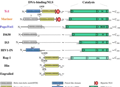
C~500
Figure 4. Functional domains of Tc1/mariner transposases. Modular structure of Tc1/mariner transposases and topology of their major functional domains in comparison with other DDE recombinases and DNA-binding proteins. Tc1/mariner transposases have an N-terminal DNA-binding domain followed by a nuclear localization signal and a C-terminal catalytic domain.
DDE recombinases have a DDE signature-containing core catalyzing polynucleotidyl transfer reactions. The catalytic core acquired different DNA- binding modules during evolution to give rise to a diverse family of recombinases.
from a DNA transposable element [57-59], and has been “domesticated” (as discussed in section 1.5.2.1.1) [60] to carry out recombination reactions to generate immunoglobulin and T-cell receptor gene diversity in jawed vertebrates [61-63]. The relatedness of DNA-binding by Tc1 transposase and RAG1 recombinase is further supported by DNA sequence similarities between their binding sites [64]. Members of the retroviral integrase family carry a combined motif of a zinc-binding domain [65] and an HTH motif (Fig. 4) that resembles the Tc3 paired-like structure [66].
The catalytic domain
The second major domain of the transposase has been referred to as the catalytic domain, because it is responsible for the DNA cleavage and joining reactions of transposition. The majority of known transposases and INs possess a well-conserved triad of amino acids, known as the aspartat-aspartat-glutamat, in short the DDE motif (actually, more of a signature than a “motif” in a usual sense) in their C-terminal catalytic domain (Fig. 4) [67].
The DDE motif is found in a large group of recombinases, including retrotransposon and retrovirus integrases, bacterial IS element transposases [33] and RAG1 [33, 68, 69].
Structural analyses of HIV-1 INs and mutational studies revealed that the DDE triad lies in the heart of the catalytic domain of transposases and INs [47, 70].
These amino acids play essential role in catalysis by coordinating, in general, two divalent cations necessary for activity. Retroviral INs were shown to be able to coordinate Ca++, Zn++ and Mn++ ions, but
Figure 5. Architecture of the Mos1 paired end complex. (A and B) Orthogonal views of the PEC crystal structure. Transposase monomer A is colored orange and monomer B blue. The two major- groove DNA-binding motifs contain HTH1 (residues 24–55) and HTH2 (residues 89–110). The minor-groove binding motif comprises residues 63–71. The two DNA duplexes bound by the DNA-binding domains are labeled IR DNA and the two extra DNA duplexes are labeled FL DNA. (C) Schematic diagram of the structure. An arrow indicates the 3’ end of each DNA strand and a black dot indicates the 5’ phosphate of the NTS. The purple sphere indicates the metal ion in active site A.
the biologically relevant cation is thought to be Mg++ [71, 72]. One metal ion acts as a Lewis acid, and stabilizes the transition state of the penta-coordinated phosphate, the other one acts as a general base and deprotonizes the incoming nucleophil during transesterification and strand transfer [44].
The C-terminal half of Tc1/mariner transposases was initially proposed to be the catalytic domain based on the presence of the characteristic DDE (or DDD in the case of mariner and pogo) motif (Fig. 4). Site-directed mutagenesis of these positions in the Tc3 transposase confirmed that these three amino acids are essential for all catalytic activities [73]. Interestingly, a change of the exceptional third D of mariner, turning the DDD into the canonical DDE, inactivates the transposase [74]. This is most easily explained by assuming that the catalytic role of either aspartic or glutamic acid is similar, but that the precise spatial position within the transposase fold requires the presence of the correct residue.
The crystal structure of the Mos1 mariner transposase from D. melanogaster has recently been solved [75]. The structure contains a dimer of transposase and four DNA duplexes. Two of these duplexes are recognized by the N-terminal DNA-binding domain of the transposase and are held in position in the catalytic domains as if they have just been cleaved (Fig. 5). In striking contrast to the anti-parallel orientation of transposon ends in the Tn5 synaptic complex, these duplexes are approximately parallel. The N- terminal domain of the transposase (residues 1–112) comprises two HTH motifs linked by a minor groove binding motif. Residues 113–125 form a linker between the DNA-binding domain and the catalytic domain (residues 126–161 and 190–345). Residues 162–189 form a clamp loop extending out from the catalytic domain making key interactions
with the linker of the other transposase monomer in the complex (Fig. 5). Two additional DNA duplexes are bound by the catalytic domains in positions that could represent binding sites for DNA flanking the transposon. The catalytic domain has an RNaseH-like fold. In addition to M o s 1 and other DDE-containing transposases and integrases [70], crystallographic analyses of the catalytic domains of proteins whose functions are not obviously related to transposition, such as RNAaseH [76] or RuvC [77] have revealed a remarkably similar overall fold.
The emerging picture reinforces the notion of a common structural motif that catalyses polynucleotidyl transfer reactions in diverse biological contexts [65, 70], and that the different specificities in binding to DNA might have evolved by the apparent acquisition of different DNA-binding domains, and combinations thereof, in the evolution of DDE recombinases [33].
1.2.2.1.1.2 The transposon inverted repeats
Tc1/mariner elements have a roughly uniform size of approximately 1.6-1.7 kb, indicating a natural selection in genomes for this particular size. The transposons are bracketed by IRs that contain binding sites for the transposase. IRs vary in length and contain transposase- binding sites in different numbers and patterns in the Tc1/mariner family (Fig. 6). Tc1 and mariner elements are the simplest and have repeats of less than 100 bp and a single binding site per repeat (Fig. 6) [47, 78]. Tc3 elements have IRs of more than 400 bp in length, each of which contains two binding sites, but the internal pair is not required for transposition (Fig.
6) [79]. A third subgroup of the Tc1/mariner superfamily is named IR/DR, and has a pair of transposase-binding sites at the ends of the 200-250 bp long IRs (Fig. 6) [80]. The binding sites contain short, 15-20 bp direct repeats (DRs). This structure can be found in several elements whose inverted repeats are not significantly similar at the DNA sequence level, such as Minos and S elements in flies [81, 82], Quetzal elements in mosquitos [83], Txr elements in frogs [84] and at least three Tc1-like transposon subfamilies in fish [49], including Sleeping Beauty, a reconstructed transposon of the salmonid subfamily (Fig. 6) [85]. There
Figure 6. Structure of Tc1/mariner transposons. The central transposase genes (tnpase) are flanked by terminal inverted repeats (TIR) that contain binding sites for the transposase. TIRs come in different lengths and contain binding sites in different numbers and patterns in the Tc1/mariner superfamily.
Dotted lines in Bari elements indicate that certain versions of these transposons have long inverted repeats. Actual or putative transposase binding sites are indicated as yellow arrows near the ends of the elements.
are two types of Bari elements in Drosophila; those that have short IRs similar to Tc1, and those that have IR/DR structure [86]. However, both types of Bari element have two putative transposase-binding sites flanking their transposase genes (Fig. 6). This suggests that it is not the long IRs per se, but the multiple binding sites for the transposase that are essential for the mobility of these elements.
Similar to the DNA-binding domains, the approximately 30 bp binding sites for Tc1- like transposases have a bipartite structure in which the 5'-part of the binding site is recognised by the homeo-like domain, whereas 3'-sequences interact with the paired-like domain of the transposase [47]. The binding sites for mariner transposase are also around 30 bp in length, supporting the hypothesis that these transposases also have bipartite DNA- binding domains. In contrast, pogo elements have binding sites of 12 bp within their short inverted repeats [87], consistent with the predicted single HTH motif in their DNA-binding domains. These binding sites are repeated either in direct or in inverted orientation at the ends of the element (Fig. 6), but it has not been determined whether they are required for the mobility of pogo elements. Taken together, the Tc1/mariner superfamily contains some simply structured elements in which the transposase gene is flanked by a pair of transposase binding sites, and more sophisticated ones with multiple binding sites that might impose some control over the timing and specificity of the transposition reaction.
Conservative transposition (cut-and-paste)
Replicative transposition (copy-and-paste)
Excision Replication
Integration into target DNA
Figure 7. Schematic representation of the two major mechanisms of transposition.
During conservative transposition, the element is excised from the donor DNA (red line), and integrates into a new target DNA (green line). The broken donor DNA has to be repaired by host factors, and this process can result in a small “footprint” (black dot) that marks the former presence of the element in that site. Replicative transposition requires amplification of the element either by replication or by copying of the element through transcription followed by reverse transcription. The amplified element gets inserted elsewhere in the genome.
1.3 Modes of transposition
The sum of molecular events involved in the movement of a transposable element from one chromosomal location to another is defined as transposition. There are two types of transposition reaction distinguished by whether the TE is replicated during the process or not (Fig. 7).
During the vast majority of replicative (copy-and-paste) transposition events, the transposon does not get excised from its donor locus, but instead a copy of it is produced that subsequently inserts elsewhere in the genome (Fig. 7).
Thus, replicative transposition leads to an increase in the copy number of the transposon within a genome. If the new copy is produced by transcription and subsequent reverse transcription of transposon sequences, the process is referred to as retrotransposition. The movement of retroviruses and retrotransposons is always of the replicative type, because it is the cDNA copy, not the original transposon, which is transposed. However, replicative transposition is not restricted to retroelements. For example, the IS6 family and Tn3 [42], and the complex DNA transposon, bacteriophage Mu [35], can also follow the replicative mode of transposition.
In non-replicative (also called conservative) transposition, the element is excised from a genomic locus and integrates to another through a so-called “cut-and-paste”
mechanism (Figs. 7 and 8). In non-replicative transposition, the genetic information of the element is carried by DNA. The bacterial IS10 [88], Tn7 [89] and eukaryotic transposons including the P element [90], members of the Tc1/mariner family and the maize transposon Ac/Ds discovered by McClintock all use the cut–and-paste mechanism for their transposition [20].
In cut-and-paste transposition, amplification is not inherent to the transposition process itself; nevertheless, the copy numbers of DNA transposons also increase over time.
Transposon amplification can occur when transposition takes place in the S-phase of the cell cycle. If a transposon is excised from an already replicated segment of the DNA, and reintegrates into a chromosome that has not been replicated, the process results in an increase by one copy of the transposon. If this event is followed by meiosis, two of the four germ cells have one more transposon copies compared to its parental cell [91]. Another way of increasing in copy number of non-replicative transposons was described for the P element [92] and the Mos1 mariner transposon [93]. After the excision of these elements the resulting gap in the donor chromosome can be sealed by a process called template-directed gap repair. This host repair mechanism uses the sister chromatid, the homologous chromosome or an ectopic site for refilling the gap created by the excised element.
1.3.1 The biochemistry of cut-and-paste transposition
Central to all transposition reactions are the excision and integration of a polynucleotide, therefore transposons execute polynucleotide transfer reactions. The transposase protein and the inverted repeats together engage in a series of molecular events that lead to the excision of the element from its DNA context and reintegration into a different locus, a process termed cut-and-paste transposition (Fig. 8). The transposition process can arbitrarily be divided into at least four major steps: 1) binding of the transposase to its sites within the transposon IRs; 2) formation of a synaptic complex in which the two ends of the elements are paired and held together by transposase subunits; 3) excision from the donor site by single-, or double-strand DNA cleavage; 4) reintegration at a target site and processing of the transposition product by host-encoded enzymes (Fig. 8) [44]. All transposition reactions involve DNA breakage and joining; the nature of the emerging transposition products depends on which strand of the DNA is cleaved and joined.
The transposase binds to the inverted repeats The transposase binds to the inverted repeats
TA TA
Transposase
The excised transposon integrates into the target DNA The excised transposon integrates into the target DNA Synaptic complex formation Synaptic complex formation
The transposase is expressed The transposase is expressed Transcriptional
control Transcriptional control
Chromatin control, architectural factors Chromatin control, architectural factors
Double-strand DNA break repair Cell-cycle control Double-strand DNA break repair Cell-cycle control
Primary DNA sequence DNA structure DNA accessibility Tethering factors Primary DNA sequence DNA structure DNA accessibility Tethering factors DNA accessibility
DNA accessibility
TA
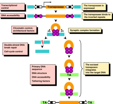
Figure 8. General mechanism and regulation of DNA transposition.
The transposable element consists of a gene encoding a transposase (orange box) bracketed by terminal inverted repeats (solid black arrows) that contain binding sites of the transposase (white arrows) and flanking donor DNA (blue boxes). Transcriptional control elements in the 5’-UTR of the transposon drive transcription (arrow) of the transposase gene.
The transposase (purple spheres) binds to its sites within the transposon inverted repeats. Excision takes place in a synaptic complex, and separates the transposon from the donor DNA. The excised element integrates into a new site (TA for Tc1/mariner transposons) in the target DNA (green box) that will be duplicated and will be flanking the newly integrated transposon. On the right, the various steps of transposition are shown. On the left, mechanisms and host factors regulating each step of the transposition reaction are indicated.
1.3.1.1 Transposon excision
The key process of all transposon excision is the exposure of the 3´-OH groups of the transposon ends, which will later be used at the strand transfer reaction for integration (Fig.
9) [94]. In the case of phage Mu and retroviral transposition the DNA cleavage involves only a single strand cut at each transposon ends. The vast majority of transposases, however, cleave both DNA strands of the corresponding transposon. During the excision of bacterial cut-and-paste elements, it is the first nick that generates the 3’-OH groups at the transposon ends. On the contrary, transposases of eukaryotic cut-and-paste transposons first generate a 5’-P on the transposon ends and the 3’-OH groups are exposed only as a result of the second strand cut [95]. In case of retroviruses, this process operates on the double-stranded cDNA of the element, and results in the cleavage of only two bases from the 3’-end of the cDNA [96].
Every DNA strand cleavage in all transposition reactions is a transposase- or i n t e g r a s e - c a t a l y z e d , M g++- dependent hydrolysis of the phosphodiester bonds of the DNA backbone, executed by a nucleophilic molecule. All the DDE recombinases catalyze similar chemical reactions [97], which begin with a single-strand nick that generates a free 3'-OH group. In the case of the first strand cleavage the nucleophilic molecule is H2O [94].
During cut-and-paste transposition, nicking of the element is followed by the cleavage of the complementary DNA strand too. To catalyze second
TA AT
TA AT
OH
OH
TA AT
TA AT TA
AT
AT
TA
TA AT
TA AT
TA AT
TA AT
Integration site
Excision site
DNA repair
Transposon
Reintegrated transposon Transposon footprint
TA AT TA
AT
Figure 9. Cut-and-paste transposition of Tc1/m a r i n e r transposons. The element (black box) is removed from its original site with staggered cuts, which leaves some transposon nucleotides at the site of excision. The excised element reintegrates elsewhere in the genome at a TA target dinucleotide. Repair of the single stranded gaps of the integration site results in the duplication of the target TA.
The excision site is predominantly repaired by non-homologous end joining, which leaves behind a transposon footprint.
strand cleavage, DDE recombinases developed versatile strategies [98]. This cleavage can occur at different positions relative to the transposon ends. The position of 5’-cleavege of the second strand required for the liberation of the element occurs directly opposite to the 3’- cleavage site in V(D)J recombination [99] and for the bacterial Tn10 element [100] (thereby generating blunt ended products). For Tn7 the cleavage occurs three nucleotides toward the 5’-end of the element [44]. In case of the Tc1/mariner elements the non-transferred strand is cleaved a few nucleotides within the transposon (Fig. 9) (two nucleotides for the Tc1 and Tc3 elements [73, 78], and three nucleotides inwards the element in case of mariner [78, 101]).
The double-strand DNA breaks (DSBs) generated by transposon excision are repaired either by the non-homologous end joining pathway (NHEJ), or by template-dependent gap repair [92, 93]. NHEJ generates transposon "footprints" (Fig. 9) that are therefore identical to the first or last 2-4 nucleotides of the transposon in Tc1/mariner transposition [101, 102]. In V(D)J recombination, the single-strand nick is converted into a DSB by a transesterification reaction in which the free 3’-OH attacks the opposite strand, thereby creating a hairpin intermediate [99, 103]. Tn5 and Tn10 transposons also transpose v i a a hairpin intermediate, with the difference that the hairpin is on the transposon and not on flanking DNA [100, 104].
1.3.1.2 Transposon integration and target site selection
The second step of the transposition reaction is the transfer of the exposed 3’-OH transposon tip to the target DNA molecule by transesterification (Fig. 9). Similarly to the initial DNA cut, the strand transfer is done by a nucleophilic attack. In this case, the 3’-OH groups of the already liberated transposon ends serve as a nucleophil that couples the element to
the target, without previous target DNA cleavage. As a result, the transposon ends are covalently attached to staggered positions: one of the transposon ends joining to one of the target strand, the other end joining to a displaced position of the target strand. Similarly to the initial strand cleavage, the strand transfer reaction does not need an external energy source, which suggest that it is the energy of the target phosphodiester bond that is used for the new transposon-target joint [94]. Although the initial excision and the strand transfer reactions are isoenergetic, many transposons such as Tn7 and the P element, need molecules with high- energy bonds (ATP and GTP, respectively) for transposition in vitro. However, these molecules do not serve as an energy source, rather they only play regulatory roles [90, 105].
The final steps of transposition reaction are performed by host proteins. Due to the staggered way of insertion during the strand transfer step, there are short, single stranded gaps flanking the new integrant (Fig. 9). Host DNA repair factors then repair these gaps generating characteristic short direct repeats, also called target site duplications (TSDs), the hallmarks of transposition.
Most TEs do not integrate randomly into target DNA, and display some degree of specificity in target site utilization [106]. There is a wide spectrum of specificity in target site selection, hereby defined as the mechanism by which the specific DNA sequences of target sites are chosen. For example, the bacterial Tn7 element is highly specialized to insert into a single sequence motif in the E. coli genome (discussed in more detail later in section 1.4.5) [106], whereas several other transposons, such as Tn5, can integrate at several locations even within a single gene [107]. Target selection may depend on primary DNA sequence and chromatin structure, which can influence target site utilization by modulating the accessibility of DNA. For some elements, such as Tn7 and the Ty1, Ty3 and Ty5 retrotransposons in yeast, either element- or host-encoded accessory proteins play a role to locate a potential target area (discussed in more detail later in section 1.4.5) [108-111]. In other systems, including the bacterial transposon Tn10 and the Tc1 and Tc3 transposons in Caenorhabditis elegans, target site selection is primarily determined by the transposase itself [112, 113].
Sequences responsible for target site selection of Tn10 and retroviruses have been mapped
to the core catalytic domain of the transposase (or integrase) [112, 114], containing an evolutionarily conserved catalytic domain, the DDE domain.
The DDE domain is shared by a large group of recombinase proteins, including the Tc1/mariner superfamily, some bacterial IS/Tn elements, retroviruses, and the RAG1 immunoglobulin gene recombinase (see section 1.2.2.1.1.1) [20]. Several members of this family integrate fairly randomly, yet not all possible sites are utilized within a genome with equal frequencies. Despite the implication that the conserved catalytic domain is responsible for locating the target site, no common pattern of integration can be recognized on the sequence level. Therefore, assuming that there might be common features of target selection in the DDE family, it is an attractive hypothesis that structural properties of the target DNA will be among them.
The secondary structure of DNA is likely an important factor in the transposable element’s insertional bias [106]. Indeed, secondary structural features influence integration of certain DNA transposons, including the bacterial elements Tn3 [115], Tn5 [107, 116, 117], Tn7 [118] and Tn10 [119, 120], P elements in Drosophila [121], retroviruses [122-125] and other retroelements [126, 127]. The insertional specificity for all of these groups is believed to exist because DNA at the site of integration forms an unusual or perturbed structure that allows better recognition by the transposition complex [118, 124, 126]. However, statistical significance of the structural features of transposon integration sites has not been considered in any of the previous analyses. Tc1/mariner elements insert into TA dinucleotides [20], and a transposon within this family, the Himar1 element from Haematobia irritans, has already been implicated as having a structural preference for sequences in addition to the canonical TA [128]. However, the nature of these structural determinants and their relationship to the insertion site preferences of other Tc1/mariner transposons is unknown.
1.4 Regulation of transposition
De novo transposition events only become evolutionarily manifested, i. e. they only survive, if they can be stably transmitted to the next generation. Hence, restriction of transposition