Electrophoresis2019,0,1–5 1
Boglarka Donczo1∗ Gabor Kiraly2 Andras Guttman1,3
1Horváth Csaba Laboratory of Bioseparation Sciences, Research Center for Molecular Medicine, Faculty of Medicine, University of Debrecen, Debrecen, Hungary
2Department of Biotechnology and Microbiology, University of Debrecen, Hungary
3MTA-PE Translational Glycomics Research Group, Research Institute for Biomolecular and Chemical Engineering, University of Pannonia, Veszprem, Hungary
Received February 26, 2019 Revised July 22, 2019 Accepted September 13, 2019
Research Article
Effect of the elapsed time between sampling and formalin fixation on the N -glycosylation profile of mouse tissue specimens
Formalin-fixed, paraffin-embedded (FFPE) samples are generally used for histology-study, however, they also possess important molecular diagnostics information. While it has been reported that theN-glycan moieties of glycoproteins is not affected by the FFPE process, no information is available about the effect of the elapsed time between sampling and fixa- tion on the resultingN-glycosylation profile. In this study, lung, brain, heart, spleen, liver, kidney, and intestine mouse tissue specimens were used forN-glycan profiling analysis and the elapsed sampling time effect was investigated with the lung tissue.N-glycan ex- traction from the tissue samples was performed by glycoprotein retrieval from the FFPE specimens using radioimmunoprecipitation assay (RIPA) buffer followed PNGase F di- gestion. The released oligosaccharides were fluorophore labeled and analyzed by capillary electrophoresis-laser induced fluorescent detection (CE-LIF).N-glycosylation profiles of freshly collected lung-tissue samples (zero time point), as well as 1 and 2 h after sam- pling were compared by carbohydrate profiling and exoglycosidase treatment based deep glycomic analysis. It was found that up to two hours of room temperature storage of tis- sue specimens apparently did not cause changes in theN-glycosylation profiles of com- plex carbohydrates, but resulted in considerable decrease in the amount of linear glucose oligomers and high mannose type glycans present in the samples.
Keywords:
capillary electrophoresis / FFPE sampling /N-glycans
DOI 10.1002/elps.201900109
Additional supporting information may be found online in the Supporting Infor- mation section at the end of the article.1 Introduction
Formalin-fixed, paraffin-embedded (FFPE) samples are routinely prepared in hospitals and pathology laboratories for histology, and kept for decades to archive tissue speci- mens [1], representing an unexploited clinical information source about biological processes and diseases. Albeit these specimens were traditionally used for histology-study based diagnostics, the advent of highly sensitive bioanalytical methods enabled the utilization of FFPE samples for molec- ular level investigation including genomics, proteomics,
Correspondence: Prof. Andras Guttman, Research Center for Molecular Medicine, Faculty of Medicine, University of Debrecen, Debrecen, Nagyerdei krt. 98., 4032, Hungary
E-mail: a.guttman@northeastern.edu
Abbreviations: APTS,8-aminopyrene-1,3,6-trisulfonic acid;
CE-LIF,CE-laser induced fluorescent detection;ER,endoplas- mic reticulum;FFPE,formalin-fixed, paraffin-embedded;RIPA, radioimmunoprecipitation assay
metabolomics, and glycomics [2]. However, for this latter one, no information is available about the effect of elapsed time be- tween sampling and fixation as glycomolecular level changes can occur during that time. It is generally assumed that tissue specimens are immediately fixed after sampling to prevent any post-sampling biochemical processes, which might change their global molecular profile. In the instance ofN- glycosylation type post-translational protein modifications, this elapsed time might affect some labile sugar residues such as sialylation and fucosylation (for the latter one, antennary and/or core), which play important structural and modula- tory roles in many normal and pathological processes [3–5].
While postmortem degradation was extensively exam- ined in proteomics and genomics studies, changes in N- glycan profiles have been investigated only in living organ- isms. It was found that protein kinase and phosphatase inhibitors preserved actin and myosin heavy chains from
∗Current address: Laboratory of Ion Beam Physics, Institute for Nuclear Research, Hungarian Academy of Sciences, Debrecen, Bem Square 18/c, 4026, Hungary
© The Authors.ElectrophoresisPublished by Wiley-VCH Verlag GmbH & Co. KGaA www.electrophoresis-journal.com
N-glycan degradation increased in the presence of glucosi- dase inhibitors, however, addition of proteasome and man- nosidase inhibitor stopped the degradation [8]. ER asso- ciated degradation in Arabidopsis revealed that during a chilling stress, smaller N-glycan intermediates were syn- thetized (Man5-7GlcNAc2instead of Man8-9GlcNAc2) [9]. Robb et al. analyzed N-glycosylation degradation in Streptococ- cus pneumonia using ribonuclease B as model glycopro- tein and showed that various mannosidases were essential for trimming high mannose typeN-glycans [10]. Cytosolic oligosaccharide chains degrade in the proteasome by au- tophagy, a lysosomal degradation process of cytoplasmic ma- terials. Proteasomal degradation is controlled byN-glycans attached to substrates of ER associated degradation [11].
Deglycosylation of unfolded proteins catalyzed by enzymes such as cytosolic endo-β-N-acetylglucosaminidase [12] and cytoplasmic PNGase cuts the glycans from the misfolded proteins [13].
In complex typeN-glycans, sialic acid residues are con- sidered as the most sensitive elements for environmental degradation effects. Kiyohara et al. found that the degrada- tion of sialylated human milk oligosaccharides is regulated by the expression of the exo-α-sialidase gene. Interestingly, it kept 80% of its activity even after 30 min incubation at 80°C, but the linkage preference of the enzyme changed fromα(2,6) andα(2,8) toα(2,3) [14]. It was reported that lyase epimerase and kinase enzymes controlled the catabolism of sialyl-mucin glycoproteins [15]. Sam et al. investigated the glycosylation of Chinese hamster ovary cells where the decrease in temper- ature reduced the level of sialylation. However, the increase of the amount of dissolved oxygen enhanced the activity of sialyltransferases, thus the sialic acid content of these glyco- proteins were increased [16].
Our earlier results revealed that the N-linked carbohy- drates of glycoproteins with neutral, sialylated and high man- nose structures were not affected by the formalin fixation and paraffin embedding process [17, 18]. Thus, it was considered that the fixation process archived the N-glycan structures and compositions of the sample at the very moment it was pre- pared. However, the elapsed time between sampling and fix- ation was not evaluated in respect to its effect on glycomolec- ular level changes. To the best of our knowledge, this is the first report examining possible time dependent degradation of asparagine-linked glycans in tissue specimens prior to for- malin fixation.
2 Materials and methods
2.1 Chemicals
Formaldehyde for the buffered formalin (3.7% formalde- hyde in 10 mM phosphate buffer, pH 7.4), the paraffin for
zyme (EC 3.2.1.20) were from Merck and (Kenilworth, NJ, USA). All other enzymes including PNGase F (EC 3.5.1.52), Arthrobacter ureafaciens sialidase (ABS) (EC 3.2.1.18), and α(1-2,3,6) mannosidase (EC 3.2.1.24) were from Prozyme (Hayward, CA, USA). The 8-aminopyrene-1,3,6-trisulfonate (APTS), the magnetic cleaning beads and the bracketing stan- dards of APTS-labeled maltose and maltopentadecaose were from the Fast Glycan Sample Preparation and Analysis kit of SCIEX (Brea, CA, USA).
2.2 Sample preparation
The SCID male mice were from the National Institute of On- cology (Budapest, Hungary) and treated in accordance with the Guidelines for Animal Experiments by the Institutional Ethics Committee (permission # 22.1/722/3/2010). For the N-glycan time-degradation study, lung-tissue samples from three SCID male mice were used. The freshly removed spec- imens were volume aliquoted into three equal portions (par- allel samples), followed by sample preparation forN-glycan profile analysis. The first aliquots were immediately pro- cessed. The second and third aliquots were stored at room temperature and processed 1 and 2 h after the tissue removal, respectively. In clinical practice, tissue samples are usually fixed within 2 hours after removal, so the experiments were planned accordingly.
The FFPE samples were washed twice by xylene and ethanol for 20 min, respectively, followed by centrifugation at 5000 ×gfor 10 min. For glycoprotein release (also re- ferred to as antigen retrieval), 40 mg of specimens were first homogenized in a Tapered Tissue Grinder (VWR, Budapest, Hungary), followed by the addition of 100 µL of RIPA buffer and 10 µL of 50 mM DTT to the homogenized samples and kept at 100˚C for 20 min, then at 65˚C for 120 min. After the protein extraction step, 10 µL of 50 mM iodoacetamide was added to the reaction mixture and incubated at 37˚C for 30 min in dark to alkylate the SH groups. This was followed by buffer exchange using 10 kDa spinfilters (VWR, Budapest, Hungary) and theN-glycan release took place on the filter us- ing 49 µL of 20 mM NaHCO3 buffer (pH 7.0) and 1 µL PN- Gase F (2.5 mU). The reaction mixture was incubated at 50˚C for 60 min. To collect the released sugars, the filters were cen- trifuged at 5000×gfor 10 min. The spinned content were dried in a centrifugal vacuum evaporator (Thermo Fisher Scientific, Waltham, MA, USA) followed by fluorophore la- beling with the addition of 6 µL of 20 mM APTS and 2 µL sodium-cyanoborohydride (in 1M THF) and incubated overnight at 37˚C. The fluorescently labeled samples were pu- rified by magnetic bead-based technology [19] and analyzed by CE with laser induced fluorescence detection (CE-LIF).
All experiments were done in triplicates and the RSDs were calculated.
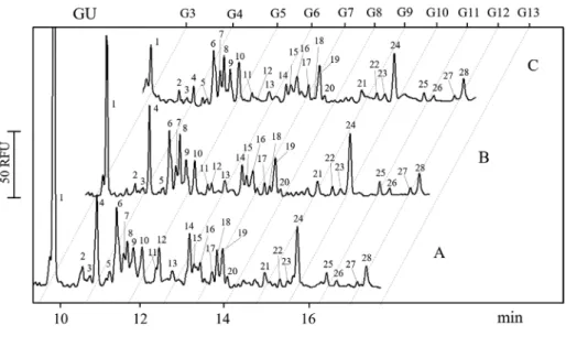
Figure 1. CE separation traces of APTS labeled N-glycan pools of mouse lung-tissue specimens pro- cessed at different times after sam- pling. (A) immediately prepared, (B) kept for 1 h at room temper- ature before processing, (C) kept for 2 h at room temperature before processing. Separation conditions:
bare fused silica capillary (50 μm i.d., 60 cm total length, 50 cm effec- tive length) filled with NCHO sepa- ration buffer, injection: 5 psi for 5 s, Separation voltage: 30 kV at 25˚C.
2.3 CE
All CE separations were performed using a PA800 Plus (SCIEX) Pharmaceutical Analysis system with laser induced fluorescence detection (488 nm excitation wavelength and a 520 nm emission filter) providing a low fmol detection limit.
The bare fused silica capillary (effective length: 50 cm, total length: 60 cm, internal diameter: 50 µm) was filled with N- CHO separation gel buffer (SCIEX) and the separations were performed in reversed polarity mode (cathode at the injection side) by the application of 30 kV electric potential. Samples were injected by 1 psi pressure for 5 s. APTS-labeled brack- eting standards (maltose and maltopentadecaose) were co- injected (10 nL) with each sample to assure migration time re- producibility of<0.1% RSD. GU value calculation and struc- turalN-glycan assignment were accomplished by using the GUcal software and database (www.GUcal.hu).
2.4 Exoglycosidase digestion based structural elucidation
Deep structural determination of the oligosaccharides of in- terest found in the releasedN-glycan pools from the mouse lung samples were accomplished by exoglycosidase-based studies [20]. Arthrobacter ureafaciens sialidase (ABS) was used to remove theα(2-3,6,8,9) bound sialic acids, Jack bean (Canavalia ensiformis) mannosidase (JBM) to cutα(1-2,3,6) mannoses (each 0.5 U, Prozyme,) andα-glucosidase (100 U from Merck and Co. Kenilworth, NJ, USA) to hydrolyze terminal, non-reducingα1-4-linked D-glucose residues. The aliquoted APTS-labeled samples were subject to exoglycosi- dase digestions separately with the application of one en- zyme at a time. The exoglycosidase reaction mixtures were incubated overnight at 37°C in 50 mM ammonium ac- etate buffer (pH 5.5) and analyzed by CE-LIF as described above.
3 Results
For mouse lung tissue, the effect of the elapsed time between the actual sampling and the formalin fixation, paraffin em- bedding process has been investigated to understand its in- fluence onN-glycosylation degradation in zero time point as well as after 1 and 2 h of room temperature storage. All other mouse tissue sample specimens were profiled for theirN- glycosylation since different tissue sample types have unique N-glycan profiles, specific to that particular organ [17] con- taining diverse proportions of high and low sialylated, high mannose as well as neutral carbohydrate structures in differ- ent size ranges, and interestingly a few low degree of poly- merization glucose oligomers.
Figure 1 compares the CE-LIF separation patterns of the APTS labeledN-glycan pool of mouse lung-tissue samples processed at different time points after sampling (0, 1 and 2 h). All measurements were done in triplicates and the re- sulting peak area reproducibility was 8.17% RSD. Trace A de- picts the resulting profile of the immediately prepared sam- ples after tissue specimen removal. Traces B and C show the glycoanalysis results of the corresponding aliquots of the samples kept at room temperature for one and two hours prior to processing, respectively. The numbers above the peaks correspond to the structures listed in Table 1. As one can observe, peaks 1, 4, 12, 14, and 18 were significantly de- creased after 1 and 2 h of room temperature storage prior to processing, but with different kinetics. For example, Peaks 1 and 14 decreased gradually between 0 and 2 h, Peak 4 practi- cally did not change up to 1 hour of room temperature stor- age but significantly decreased between 1 and 2 h. The size of peak 18 dropped between 0 and 1 h of storage. Quantitative data for each of the 28 peaks at the three different times are provided in Supporting Information Table 1.
GU values were calculated for all the peaks (zero hour time point) (Table 1) and their corresponding structures were suggested using the GlycoStore database (www.glycostore.
org). To verify the suggested GU value based structures and
+ + + + + + +
2 A4G(4)S(6,6,6,6)4 3.650 + − − − − − −
3 A4G(4)S(3,3,3,3)4 3.850 + − - − − − −
4 Maltotetraose 4 + + + + + + +
5 A3G(4)3S(6,6,6)3 4.250 + − − + + + +
6 A2G(4)2S(6,6)2 4.549 + + + + + + +
7 A2G(4)2S(3,6)2 4.721 + + + − + + +
8 F(6)A2G(4)2S(6,6)2 4.819 + + + + + + +
9 F(6)A2G(4)2S(3,6)2 4.996 + + + + − − −
10 A1[3]G(4)2S(3)1 5.240 + − + + + + −
11 A2[6]BG(4)1S(6)1 5.490 + − + + − + −
12 M4 5.580 + + + + + + +
13 F(6)A2[3]BG(4)1S(6)1 6.200 + − + + − + −
14 M5 6.607 + − + + + + +
15 F(6)A2[6]G(4)2S(6)1 6.713 + + + + + + +
16 F(6)A2G1Ga1S1 6.936 + − + + + + +
17 F(6)A2 7.326 + + + + + + +
18 M6 7.595 + + + + + + +
19 F(6)A2[3]G(4)2S(3)1 7.691 + − − − − − −
20 A2[6]BG(4)1 8.020 + + − − − − −
21 A2G(4)2 9.039 + + + − − + +
22 A2BG(4)2 9.530 + + + + + + +
23 F(6)A2BG(4)1 9.950 + + − − − + +
24 F(6)A2G(4)2 10.106 + + + + + + +
25 F(6)A2BG(4)2 10.860 + + − − − − +
26 A(6)3G3 11.250 + − − − − − +
27 A3G(4)3 11.970 + + + − − + +
28 FA2G2Ga2 12.366 + + − − − + −
29 A1[3]G(4)2 9.039 + − − − − − −
30 F(6)A2G2Ga1 11.096 + − − − − − −
*Abbreviated structure names followed the nomenclature suggested in the GlycoStore site (www.glycostore.org). Ga:α(1-3) galactose residue; (+) the structure was present in the tissue; (−) the structure was not present in the tissue
identify the oligosaccharides susceptible for delayed speci- men processing, exoglycosidase digestion based structural analysis was also accomplished for the lung specimen. Fig- ure 2 shows the results of the specific enzymatic releases of monosaccharide units in case of intact mouse lung-tissue sample after glucosidase (trace A), mannosidase (trace B), and sialidase (trace C) treatments. Structures were deter- mined for all peaks>0.1% (peak area). Glucosidase cut off allα-D-glucose units, apparently eliminating peaks one and four from trace A. The mannosidase treatment released all α(1-2,3,6) mannose residues eliminating peaks 12, 14, and 18 from trace B. Finally, the sialidase removed allα(2-3,6,8,9) sialic acid residues, shifting most of the peaks from the early migration time regime to the later migrating neutral species region. The Lung column in Table 1 lists all structures found during this exercise
All other mouse tissue samples of brain, heart, spleen, liver, kidney, and intestine were profiled by CE-LIF forN- glycosylation characterization only (no storage time effect) and the results are summarized in Table 1 in the correspond- ing columns.
4 Discussion
Elongated room temperature storage (up to 2 h) of mouse lung-tissue specimens resulted in the decrease of peak heights for some glucose oligomers and high mannose type N-glycan features. GU values from public databases and ex- oglycosidase digestion-based studies were implemented us- ing glucosidase, mannosidase and sialidase enzymes to iden- tify all carbohydrate structures in the lung samples. The glu- cosidase treatment resulted in disappearance of some of the low DP glucose oligomers of maltotriose and maltotetraose present in the samples. On the other hand, the formalin fixa- tion, paraffin embedding process apparently prevented their decomposition, so these structures were readily identifiable in the samples immediately fixed after removal. Mannosidase treatment eliminated all high mannose type structures but also affected the sizes of the maltotriose and maltotetraose peaks, assumably due to its apparent nonspecific glucosidase activity. As a first approximation, we suggest that the cause of the time based decrease in the size of these low DP glucose oligomers and high mannose N-linked structures was the
Figure 2. Exoglycosidase digestion based structural analysis of the N-glycans and maltooligosaccha- rides found in mouse lung-tissue specimens. (A)α(1-4) glucosidase, (B)α(1-2,3,6) mannosidase, and (C) α(2-3,6,8,9) sialidase treated sam- ples. Separation conditions were the same as in Fig. 1.
result of non-ATP mediated enzymatic reactions, which might still keep on working under postmortem conditions (personal communications with Prof Tamas Freund). Siali- dase treatment removed allα(2-3,6,8,9) bound sialic acids, re- sulting in shifts of all sialylated structures (see list in Table 1).
Tissue specimens are supposedly formalin fixed imme- diately after sampling at clinical and hospital facilities, but its exact time is usually unknown, and may significantly vary.
In this paper, we investigated the effect of elapsed time be- tween sampling and formalin fixation, paraffin embedding on the N-linked glycan profiles of mouse tissue specimens.
In all tissue types studied, two major peaks significantly de- creased with delayed processing, identified as maltotriose and maltotetraose, based on their GU value and glucosidase treatment results. It is important to note that no significant changes were observed in the profile of complex N-linked gly- cans including the sialylated structures up to two hours of storage of the removed specimens at room temperature.
The authors gratefully acknowledge the support of the NK- FIH (K 116263) grant of the Hungarian Government, and the BIONANO_GINOP-2.3.2-15-2016-00017 Project. The stimu- lating discussion with Dr Tamas Freund is also greatly appreci- ated. This is contribution number 148 from the Horvath Csaba Memorial Laboratory of Bioseparation Sciences.
The authors have declared no conflict of interest.
5 References
[1] Gnanapragasam, V. J.,BJU Int.2010,105, 274–278.
[2] Donczo, B., Guttman, A.,J. Pharm. Biomed. Anal.2018, 155, 125–134.
[3] Varki, A.,Trends Mol. Med.2008,14, 351–360.
[4] Schauer, R.,Glycoconj. J.2000,17, 485–499.
[5] Varki, A.,Nature2007,446, 1023–1029.
[6] Li, Z., Li, M., Du, M., Shen, Q. W., Zhang, D.,Food Chem.
2018,245, 233–239.
[7] Varki, A., in: Varki, A., Cummings, R. D., Esko, J. D., Freeze, H. H., Stanley, P., Bertozzi, C. R., Hart, G. W., Et- zler, M. E. (Eds.),Essentials of Glycobiology, Cold Spring Harbor, New York 2009.
[8] Chillaron, J., Adan, C., Haas, I. G.,Biol. Chem.2000,381, 1155–1164.
[9] Ma, J., Wang, D., She, J., Li, J., Zhu, J. K., She, Y. M.,New Phytol. 2016,212, 282–296.
[10] Robb, M., Hobbs, J. K., Woodiga, S. A., Shapiro-Ward, S., Suits, M. D., McGregor, N., Brumer, H., Yesilkaya, H., King, S. J., Boraston, A. B.,PLoS Pathog.2017,13, e1006090.
[11] Yoshida, Y., Tanaka, K., BioEssays 2018, 10.1002/bies.
201700215
[12] Huang, C., Harada, Y., Hosomi, A., Masahara-Negishi, Y., Seino, J., Fujihira, H., Funakoshi, Y., Suzuki, T., Dohmae, N., Suzuki, T.,Proc. Natl. Acad. Sci. U. S. A.2015,112, 1398–1403.
[13] Tanabe, K., Lennarz, W. J., Suzuki, T.,Meth. Enzymol.
2006,415, 46–55.
[14] Kiyohara, M., Tanigawa, K., Chaiwangsri, T., Katayama, T., Ashida, H., Yamamoto, K.,Glycobiology2011,21, 437–
447.
[15] Nishiyama, K., Nagai, A., Uribayashi, K., Yamamoto, Y., Mukai, T., Okada, N.,Anaerobe2018,52, 22-28.
[16] Thi Sam, N., Misaki, R., Ohashi, T., Fujiyama, K.,J. Biosci.
Bioeng.2018.
[17] Donczo, B., Szarka, M., Tovari, J., Ostoros, G., Csanky, E., Guttman, A.,Electrophoresis2017,38, 1602–1608.
[18] Donczo, B., Szigeti, M., Ostoros, G., Gacs, A., Tovari, J., Guttman, A.,Electrophoresis2016,37, 2292–2296.
[19] Varadi, C., Lew, C., Guttman, A.,Anal. Chem.2014,86, 5682–5687.
[20] Guttman, A., Ulfelder, K. W.,J. Chromatogr. A1997,781, 547–554.