Retrospective Research of Removing Dental Implants
Dóra Iványi
*, Béla Czinkóczky, Péter Kivovics
Semmelweis University, Department of Community Dentistry (Hungary, Budapest, 1088 Szentkirályi street 40.), Hungary
Research Open Access
Journal of
Dentistry and Oral Health
Received Date: April 02, 2019 Accepted Date: May 03, 2019 Published Date: May 06, 2019
Citation: Dóra Iványi (2019) Retrospective Research of Removing Dental Implants. J Dent Oral Health 6: 1-9.
*Corresponding author: Dóra Iványi, Semmelweis University, Department of Community Dentistry (Hungary, Budapest, 1088 Szentkirályi street 40.), Hungary; E-mail: divanyi132@gmail.com
Abstract
Aim: The purpose of this research is to make a comparative analysis of dental implant removals in the last five years in the Department of Community Dentistry.
Materials and methods: In the Department of Community Dentistry 74 implants of 39 patients were removed between 2014-2019. The relevant data were obtained by x-rays, medical charts and patient management program, called FOGÁSZ.
Data were evaluated with Microsoft Excel software.
Results: The average age was 63.2. 63.8% of the concerned individuals’ inserted implants were removed. There is nearly equal share of the location of removed implants between the maxilla and the mandible. 20.0% of the patients lost their implants within six months from surgery. The removed implants were possessed 5.4 years long on average. 43.6% of the patients commanded fixed prosthesis supported implant and teeth, this was the most common prosthesis type. The prevalence of peri-implantitis around removed implants was 79.7%. Out of the partly edentulous patients, horizontal bone resorption was discernible in 46.9%.
Conclusion: Teeth and implant supported fixed prostheses may cause implant loss, because of the biomechanical aspects of anchoring behave differently in the bone. Lack of peri-implantitis is a key factor in the success of implants. Periodontitis could also encourage the development of peri-implantitis.
Clinical significance:Avoid planning prostheses anchored at the same time to tooth and implant. Sufficient oral hygiene is essential for the prevention of inflammation. Patients with periodontitis should be cured of inflammation before implanta- tion.
Keywords: Endosseous dental implantation, Implant removal, Implants, Peri-implantitis, Research, retrospective study.
©2019 The Authors. Published by the JScholar under the terms of the Crea- tive Commons Attribution License http://creativecommons.org/licenses/
by/3.0/, which permits unrestricted use, provided the original author and source are credited.
2
Introduction
Nowadays, the use of implantation supported resto- rations have become a widely considered tooth replacement procedure in dentistry. With appropriate clinical and anatomi- cal factors, each types of edentulous patients can undergo pros- thetic treatments. Several types of dental implants are devel- oped and used in dentistry however, this article will focus on endosseous root-form implants which are the most frequently applied in dentistry today. According to statistics the success rate of implantation is high nevertheless there are still consid- erable implant failures that might require implant removals.
The aim of this retrospective study is to aid practising dentists by giving a comparative assessment of implant removals.
Literature shows that the success rate of implantation is within broad limits. According to certain authors, the sur- vival proportion is about 90-99% [1-4]. Different implantation related complications can occur. We may differentiate early and late complications which are related to implants. Com- plications can be cured by non-surgical or surgical therapy. In case the elected approach is ineffective the implant should be removed [5,6]. Indications of implant removal can be divided into two main groups: early and late indications. In the first case the implant removal happens before the osseointegration, and when the implant removal happens after the osseointegra- tion we define it as late indication [7] (Table 1.)
Early indications Late indications Tissue injury
• nerve injury
• tooth injury
Biological indication
• peri-implantitis
Malposition Mechanical indications
• implant fracture
• abutment fracture
• abutment screw fracture Inadequate primary
stability Concerning medical status Inflammation Focal infection
Table1: Indications of removing dental implants Early indications
Early indications involve tissue injury caused by im- plant placement. Temporary or permanent sensory impairment may derive from injuries to nerve trunks during implant sur- gery. Injuries may be treated medically or surgically depend- ing on the extent of the pathological alterations and the neu- rological symptoms reported by the patient [8,9]. Along with nerve trunk injuries, tooth near the implant could be damaged.
Various therapies are available, tooth might be endodontically treated, extracted, otherwise implant may be removed [10]. In case of inadequate planning or procedure the implant might be malpositioned, which may also effect implant removal [3,11].
Appropriate imaging process, e.g. CBCT, can aid the most ideal implant placement. Implant removal could be suggested if the primary stability is inadequate. Too hard primary stability in- duces bone resorption around the implant, however implants’
major amplitude micromovement caused by too low primary stability could inhibit osseointegration [12]. Mention must be made of the inflammatory processes before the osseointegra- tion. Excessive temperature generation during surgical drilling and inefficiently controlled wound healing could result inflam- mation in the surrounding bone [13].
Late indications
Biological indication includes peri-implantitis.
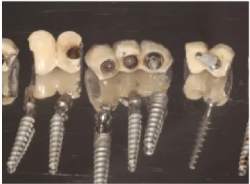
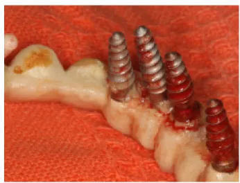
Peri-implantitis has been defined as an inflammatory pro- cess around an osseointegrated implant with progressive bone loss [14]. A study made in 2012 claims that the prevalence of peri-implantitis has been reported to be in the order of 10% of implants and 20% of patients [15]. Predisposing factors could be limited oral hygiene, smoking, systemic disease, poorly cleanable and overloaded prosthesis, history of peri-implanti- tis, soft tissue defects or poor quality soft tissue at the area of implants [16]. (Figure. 1.)
Figure 1: Implants were removed due to peri-implantitis.
The diagnosis of peri‐implantitis requires: 1.) Evi- dence of visual inflammatory changes in the peri-implant soft tissues combined with bleeding on probing and/or suppu- ration; 2.) Increasing probing pocket depths as compared to measurements obtained at placement of the supra‐structure;
3.) Progressive bone loss in relation to the radiographic bone level assessment at 1 year following the delivery of the implant‐
3 supported prosthetics reconstruction; 4.) In the absence of ini-
tial radiographs and probing depths, radiographic evidence of bone level ≥3 mm and/or probing depths ≥6 mm in conjunc- tion with profuse bleeding represents peri-implantitis [17].
The treatment of peri-implant infections comprises conserva- tive (non-surgical) and surgical approaches. Depending on the seriousness of peri-implant disease implant removal might be required [18].
Mechanical indications contain injury and fracture of the implant or the implants abutment. Most commonly, these conditions arise from implant overloading (Figure. 2.) [19].
Neither mechanical indications are absolute indications of im- plant removal [20].
Figure 2. Implant fracture.
Indications of implant removal concerning medical status are divided into several groups. Implant removal might be suggested surrounding maxillofacial tumours, according to the oncologic treatment [21,22]. Removal should be measured about implants are qualified as focal infections.
On account of the actuality of the present argument the Department of Community Dentistry’s workgroup pro- posed the investigation of implant removals. The aim of this research is to make a comparative interpretation of implant re- movals in the last three and a half years in the department. Our research implies the distribution of age and gender of the ex- amined population, location of inserted and removed implant in the jaws, elapsed time between implantation and removal, the types of prostheses anchored by removed dental implants and complications occurring with removed implants.
Material and methods
The last five years, 39 patients’ (23 women and 16 men) 74 implants were removed in the Department of Community Dentistry. 88.9% of removed implants were not inserted in the
Department. The applied data were obtained by x-rays, medi- cal charts and patient management program, called FOGÁSZ, found in the Department of Community Dentistry. Data were evaluated with Microsoft Excel software.
Results
Age distribution
Average age of the examined population was 63.2 years (deviation is 9.9 years). Female’s average age was 60.6 years (deviation is 14.0 years), whereas males was 64.2 years (deviation is 13.6 years). 94.9% of the patients were aged 50 or over. 30.8% were 51-60 years old, 48.7% were between 61-70 years, 15.4% were aged 71-80 and only 5.1% were in age group 31-40.
Implant position in the jaws
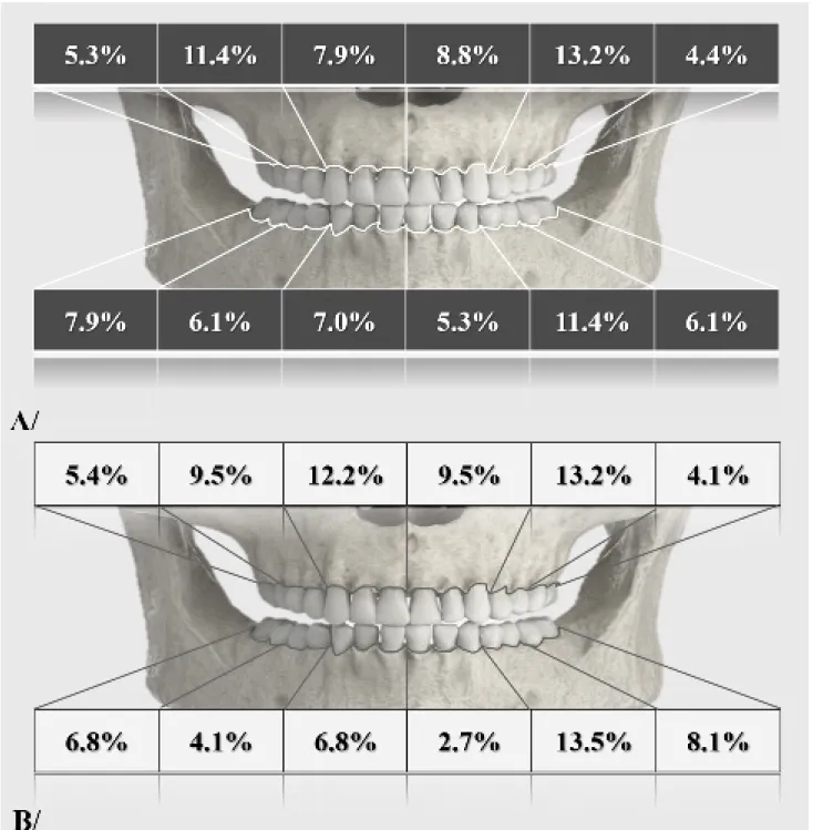
An analysis of the inserted and removed implants lo- cations within the jaws were made, considering laterality. 114 implants were placed to patients who got through implant re- moval, on average 2.9 implants (deviation is 2.7) per partici- pant. Inserted implants’ percental repartition is shown in Table 2. A (Table 2. A). 64.9% of the concerned individuals’ inserted implants were removed, counts 74 implants. 1.9 implants (de- viation is 1.6) were removed per individual. Removed implants’
percental repartition is shown in Table 2. B (Table 2. B). 58.1%
of removed implants located in the maxilla.
Implants survival time
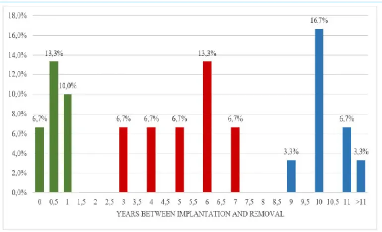
Data about removed implant lifetime were available in 30 cases. 20.0% of the patients lost their implants within six months from surgery, mainly because of early indications.
Out of 20.0% 6.7% had to be removed immediately afterward implantation. Percentage of the survival time of removed im- plants is given in Table 3. (Table 3) Removed implants were possessed 5.4 years (deviation is 4.1 years) long on average.
Types of prostheses anchored by removed dental implants
This research extends to observe the distribution of the prostheses types anchored by removed implants. 43.6 % of the patients commanded fixed prosthesis supported implant and teeth, 25.6% wore bridges anchored by dental implants, 15.4% wore full denture, 12.8% wore dental crown and merely 2.6% had all on four prosthesis type.
Table 2.A/ Inserted implants’ percental repartition. B/ Removed implants’ percental repartition.
4
Table 3: Removed implants survival time.
Table 4: Incidence of complications led to implant removal.
5
6
Complications concerning removed implants
The prevalence of peri-implantitis around removed implants was 79.7%. 20.5% of the population was fully eden- tulous. Out of the remaining patients, horizontal bone resorp- tion was discernible in 48.4%, over and above vertical bone defect was detectible in 12.9%. 9.5% of the removals were rec- ommended because of inflammation before osseointegration would have occurred. Only in tree cases were found malpo- sitioned implants. 6 of 74 removed implants were explanted through mechanical indications (Table 4.).
Discussion
Over the age of 50, the prevalence of diabetes melli- tus type two, cardiovascular diseases and tumorous diseases is rising [23,24,25]. Such morbi may decrease the resistance of the human body against bacteria and reduce the blood sup- ply in different tissues, thus they might influence the undis- turbed wound healing and the implant ossification. Accord- ingly, these common illnesses could be etiological factors in implant loss. According to a Hungarian study, the incidence of tooth loss is significantly growing over the age of 45 [26].
In parallel, the incidence of tooth loss, the rate of prosthesis is also in a tendency to increase [27]. As implantation supported prostheses are gaining ground in everyday dental practice, we can conclude that with traditional dentures, the prevalence of dentures on implants is also increasing in the older age group.
Consequently, inference might be deducted, that the frequent implantation in elder population may also heighten the im- plant removals over the age of 50. In our opinion, the 94.9%
incidence of people over 50 in the examined population can- not be explained with only the higher prevalence of systemat- ic diseases.
Observation can be made into the location of in- serted and removed implants. Notable difference is perceived between the right side in maxillary premolar, in the left side mandibular premolar and molar regions about removed im- plant’s location. Further investigations and the number of cases need to be extended to determine the exact cause of the significant deviation measured in the different side regions.
Removals in maxilla and in mandible were of nearly equal proportions. It should be noted that in large-scale studies, the proportion of placement of inserted implants does not match with sudden results [1]. In our case conclusion cannot be drawn, that the blood supply and structural differences in the maxillary and mandible affect the survival of the implants [28,29]. Further studies are needed to determine this infer- ence.
Concerning the lifetime of removed implants, we can observe that there are three maxima in the time axis. The first spike is located in the first year from implantation. In these cases, implants were removed due to early indications. The second spike could be detected for 6 years. Among patients belonged to this group, hard-to-clean prosthesis had been frequent, and except of three patients, everybody had a fixed denture with anchored teeth and implants. It is assumed that peri-implantitis due to inappropriate design and construction of the prosthesis has increased the number of implant remov- als. The last spike appears in the time axis around 10 years. In these cases, almost all types of prosthetics have occurred. The additive effects of low risk etiologic factors involved in the for- mation of peri-implantitis might reach inflammation and bone resorption at 10 years that may indicate implant removal.
In the examined population, fixed prostheses an- chored at the same time on implant and natural teeth was the most common type of prostheses anchored removed implants.
Along with designing an implant supported prosthesis, the bio- mechanical properties of natural teeth, implants and anchored dentures must be taken into account. Natural tooth provides a flexible connection with bone by periodontal ligaments so fixed prosthesis on the tooth may have micromovements [30].
The connection among implants and bone is rigid and anky- lotic. In the case of implant supported prosthesis, no or only very slight micromovements are observed. Micromovements generated by natural tooth are also transmitted to implants, as- suming that the two types of anchoring are connected. Micro- movement forces can weaken implant-bone relationship over time, helping penetration and adhesion of pathogens around implants, thereby promoting the formation of peri-implanti- tis [31,32]. The second most common type of prosthesis was fixed prosthesis anchored on implants. In many cases, the in- appropriate design of prosthesis led to increased accumulation of dental plaque, which can provoke the formation of peri-im- plant inflammation, thus contributing to the loss of the implant [33,14]. (Figure. 3.)
7
Figure 3: Dental plaque accumulated around implants due to inadequate dental prosthesis construction.
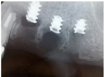
Peri-implantitis was observed nearly 80% of removed implants. High prevalence rates point to the fact that periim- plantal inflammation is one of the most important factors for losing implants (Figure. 4.). The presence of certain etiologic factors contributes to the development of peri-implantitis, such as insufficient oral hygiene, smoking, inadequate loading of the implant, various systemic diseases, and inadequately designed or completed dentures [16]. Periodontitis could also promote the development of peri-implantitis. In patients suffering peri- odontitis, the incidence of peri-implantitis is six times greater, due to the fact that the anaerobic bacterial flora around sore implants and periodontally affected tooth are largely identical [34,35]. Horizontal bone resorption can be observed in 48.46%
of patients. This confirms the assumption that there may be correlation between periodontal status and survival of im- plants. More than 10% of the removed implants were explant- ed due to inflammatory reactions before osseointegration. In these cases, the healing process might have been affected by traumatic surgical care, disturbed implant healing, dehiscence due to inadequate wound care and inadequate oral hygiene [36].
Figure 4: Seriously advanced peri-implantitis.
Conclusion
Based on our results, our conclusions were deducted:
• Avoid planning fixed prostheses supported at the same time by tooth and implant, because biomechanical as- pects.
• In cases of implant supported fixed prostheses the aim is to give properly cleanable prostheses and to help create the patient’s correct oral hygiene habits.
• Prevention of peri-implantitis is a key factor in the success of implants. We should try to minimize the presence of factors promoting the development of peri-implantitis. Suf- ficient oral hygiene is essential for the prevention of inflamma- tion.
• Patients with periodontitis should be cured of inflam- mation before implantation. Thus, we can reduce the chance of periodontal anaerobic flora adhesion around the implant caus- ing inflammation.
• An essential factor for osseointegration is the in- flammation-free healing. We can choose our best-controlled wound healing technique for our patients, as we can best re- duce the progression of inflammatory processes.
References
8
1. Romeo E, Lops D, Margutti E, Ghisolfi M, Chiapasco M, Vogel G (2004) Long-term Survival and Success of Oral Implants in the Treatment of Full and Partial Arches: A 7-year Prospective Study with the ITI Dental Implant System. Int J Oral Maxillofac Implants 19:247-259.
2. Compton SM, Clark D, Chan S, Kuc I, Wubie BA, et al. (2017) Dental Implants in the Elderly Population: A Long- Term Follow-up. Int J Oral Maxillofac Implants 32:164-170.
3. Divinyi T (2007) Orális Implantológia. Budapest : Semmelweis Kiadó.
4. Grisar K, Sinha D, Schoenaers J, Dormaar T, Politis C (2017) Retrospective Analysis of Dental Implants Placed Be- tween 2012 and 2014: Indications, Risk Factors, and Early Sur- vival. Int J Oral Maxillofac Implants 32:649–654, 32:649–654 ed.
5. ITI Treatment Guide Volume 8. Bragger U, Heitz- Mayfield LJ (2015) Berlin, Germany: Quintessence Publisch- ing Co.
6. Machtei, EE (2013) What do we do after an implant fails? A review of treatment alternatives for failed implants. Int J Periodontics Restorative Dent 33:111-119.
7. Iványi D and Kivovics P (2017) Possible causes and methods of removing dental implants 86.
8. Alhassani AA, AlGhamdi AS (2010) Inferior Alveo- lar Nerve Injury in Implant Dentistry: Diagnosis, Causes, Pre- vention, and Management. J of Oral Implantology 36:401-407.
9. Auyong TG, Le A (2011) Dentoalveolar nerve injury.
Oral Maxillofac Surg Clin North Am. 23:395-400.
10. Wook-Jae Y, Su-Gwan K, Mi-Ae J, Ji-Su O, Jae-Seek Y (2013) Prognosis and evaluation of tooth damage caused by implant fixtures. J Korean Assoc Oral Maxillofac Surg 39:
144–147.
11. Kahraman S, Bal BT, Asar NV, Turkyilmaz I, Tözüm TF (2009) Clinical study on the insertion torque and wireless resonance frequency analysis in the assessment of torque ca- pacity and stability of self-tapping dental implants. J Oral Re- habil 36:755-761.
12. Javed F, Ahmed HB, Crespi R, Romanos GE (2013) Role of primary stability for successful osseointegration of dental implants: Factors of influence and evaluation. Interv Med Appl Sci 5: 162–167.
13. Scharf DR, Tarnow DP (1993) The Swedish system of osseointegrated implants: problems and complications en- countered during a 4-year trial period. J Periodontol 64:954- 956.
14. Mombelli A, van Oosten M A, Schurch E Jr, Land N P (1987) The microbiota associated with successful or failing osseointegrated titanium implants. Oral Microbiol Immunol 2:
145–151.
15. Klinge B, Meyle J (2012) Peri-implant tissue destruc- tion. The Third EAO Consensus Conference 2012. Clin Oral Implants Res 6:108-110.
16. Heitz-Mayfield, LJ (2008) Peri-implant diseases: diag- nosis and risk indicators. J Clin Periodontol. 292-304.
17. S Renvert, G R Persson, F Q. Pirih, P M Camargo (2018) Peri‐implant health, peri‐implant mucositis, and peri‐
implantitis: Case definitions and diagnostic considerations.
Journal of Clinical Periodontology 20.
18. Heitz-Mayfield LJ, Needleman I, Salvi GE (2014)Con- sensus Statements and Clinical Recommendations for Preven- tion and Management of Biologic and Technical Implant Com- plications. The International Journal of Oral & Maxillofacial Implants 29: 346-350.
19. Schwarz MS (2000) Mechanical complications of den- tal implants. Clin Oral Implants Res 1:156-158.
20. Ramsey AA (2010) Unusual Broken Dental Implant Corrected. Dr. Ramsey Amin's Page.
21. Yerit KC, Posch M, Seemann M, , Hainich S, Dört- budak O, Turhani D, et al. (2006) Implant survival in mandi- bles of irradiated oral cancer patients. Clinical Oral Implants Research. 17:337-344.
22. Harrison JS, Strateman HS, Redding SW (2003) Den- tal implants for patients who have had radiation treatment for head and neck cancer. Special Care in Dentistry 23:223-229.
23. Jermendy Gy, Gaál Zs, Gerő L, Hídvégi T, et al. (2014) A diabetes mellitus kórismézése, a cukorbetegek kezelése és gondozása a felnőttkorban. Budapest : A Belgyógyászati Szak- mai Kollégium és a Magyar Diabetes Társaság.
24. Széles Gy, Vokó Z, Jenei T, Kardos L, Ádány R, Lun K, et al. (2002) Kardiovaszkuláris betegségek morbiditási viszon- yai Magyarországon.
25. Tompa, A (2011) A daganatos betegségek előfordulása, a hazai és a nemzetközi helyzet ismertetése. Magyar Tudomány 11: 1333-1344.
26. Madléna M, Hermann P, Jáhn M, Fejérdy P (2008) Caries prevalence and tooth loss in Hungarian adult popula- tion: results of a national survey. BMC Public Health. 8:364.
Submit your manuscript at
http://www.jscholaronline.org/submit-manuscript.php
Submit your manuscript to a JScholar journal and benefit from:
¶ Convenient online submission
¶ Rigorous peer review
¶ Immediate publication on acceptance
¶ Open access: articles freely available online
¶ High visibility within the field
¶ Better discount for your subsequent articles
9 27. Fábián T, Fejérdy P, Somogyi E (1998) Evaluation
of the dentition of the adult population in Hungary from the point of view of prothesis. Dental Reivew 91,383-389, 1998, 91:
383-389.
28. Chanavaz M (1995) Anatomy and histophysiology of the periosteum: quantification of the periosteal blood supply to the adjacent bone with 85Sr and gamma spectrometry. J Oral Implantol. 21:214-219.
29. Tolstunov L (2007) Implant zones of the jaws: implant location and related success rate. Journal of Oral Implantology 211-220.
30. Skalak R (2006) Biomedical Considerations in Osse- ointegrated Prostheses. The Journal of Prosthetic Dentistry 46:
843-848.
31. Winter W, Klein D, Karl, M (2013) Micromotion of Dental Implants: Basic Mechanical Considerations. Journal of Medical Engineering.
32. Charalampakis G, Leonhardt A, Rabe P, Dahlen G (2012) Clinical and microbiological characteristics of peri- implantitis cases: a retrospective multicentre study. Clin Oral Implants Res 23:1045-1054.
33. Ericsson I, Persson LG, Berglundh T, et al. (1995) Dif- ferent types of inflammatory reactions in peri-implant soft tis- sues. J Clin Periodontol 22: 255-261.
34. Zitzmann, NU, Walter, C és Berglundh, T (2006) Ätiologie, Diagnostik und Therapie der Periimplantitis – eine Übersicht. Deutsche Zahnärztliche Zeitschrift 61:642–649.
35. Rams TE, Degener JE, van Winkelhoff AJ (2013) An- tibiotic resistance in human peri-implantitis microbiota. Clin Oral Implants Res 25:82–90.
36. Salonen MA, Oikarinen K, Virtanen K, Pernu H (1993) Failures in the Osseointegration of Endosseous. Int J Oral Maxillofac Implants 8:92-97.