Two Nimrod receptors, NimC1 and Eater, synergistically contribute to bacterial phagocytosis in
Drosophila melanogaster
Claudia Melcarne1, Elodie Ramond1, Jan Dudzic1, Andrew J. Bretscher1,Eva Kurucz 2, Istvan Ando2 and Bruno Lemaitre1
1 Global Health Institute, School of Life Sciences,Ecole Polytechnique Federale de Lausanne (EPFL), Switzerland 2 Institute of Genetics, Biological Research Centre of the Hungarian Academy of Sciences, Szeged, Hungary
Keywords
Drosophila; haemocytes; innate immunity;
Nimrod; phagocytosis Correspondence
C. Melcarne, Global Health Institute, School of Life Science, Station 19 CH-1015, Lausanne, Switzerland
E-mail:claudia.melcarne@epfl.ch
B. Lemaitre, Global Health Institute, School of Life Sciences,Ecole Polytechnique Federale de Lausanne (EPFL), Lausanne, Switzerland
Tel: +41 21 693 18 31 E-mail:bruno.lemaitre@epfl.ch
(Received 29 November 2018, revised 19 March 2019, accepted 15 April 2019) doi:10.1111/febs.14857
Eater and NimC1 are transmembrane receptors of theDrosophila Nimrod family, specifically expressed in haemocytes, the insect blood cells. Previous ex vivoandin vivoRNAi studies have pointed to their role in the phagocyto- sis of bacteria. Here, we have created a novel NimC1 null mutant to re-evaluate the role of NimC1, alone or in combination with Eater, in the cellular immune response. We show that NimC1 functions as an adhesion moleculeex vivo, but in contrast to Eater it is not required for haemocyte sessility in vivo. Ex vivo phagocytosis assays and electron microscopy experiments confirmed that Eater is the main phagocytic receptor for Gram-positive, but not Gram-negative bacteria, and contributes to microbe tethering to haemocytes. Surprisingly,NimC1deletion did not impair phago- cytosis of bacteria, nor their adhesion to the haemocytes. However, phagocy- tosis of both types of bacteria was almost abolished in NimC11;eater1 haemocytes. This indicates that both receptors contribute synergistically to the phagocytosis of bacteria, but that Eater can bypass the requirement for NimC1. Finally, we uncovered that NimC1, but not Eater, is essential for uptake of latex beads and zymosan particles. We conclude that Eater and NimC1 are the two main receptors for phagocytosis of bacteria inDroso- phila,and that each receptor likely plays distinct roles in microbial uptake.
Introduction
Phagocytosis is an ancient and evolutionarily con- served process, generally defined as the cellular uptake of particles bigger than 0.5lm. Phagocytosis is an important feeding mechanism in primitive and unicel- lular organisms, such as amoeba [1]. In higher organ- isms, phagocytosis is performed by dedicated cells (phagocytes) and is used as a powerful process to internalize and eliminate pathogens, as well as to trig- ger host inflammation [2]. Moreover, phagocytosis contributes to tissue homeostasis and embryonic
development, mainly via the removal of apoptotic corpses [3]. Phagocytosis is a complex membrane-dri- ven process guided by the actin cytoskeleton of the host phagocytic cell. It involves the recognition and subsequent binding of the microbe by surface recep- tors. These interactions are essential to activate intra- cellular signalling pathways that finally culminate in the formation of the phagosome [4]. Several studies have highlighted similarities between the phagocytic machinery of Drosophila and mammals, such as the
Abbreviations
DAPI, 40,6- diamidino-2-phenylindole; EdU, 5-ethynyl-20-deoxyuridine; Hml, Hemolectin; Nim, Nimrod; PTU, phenylthiourea; SEM, scanning electron microscopy; TEM, transmission electron microscopy.
2670 The FEBS Journal286(2019) 2670–2691ª2019 The Authors.The FEBS Journalpublished by John Wiley & Sons Ltd on behalf of
involvement of actin and actin-related proteins [5–7].
Drosophila melanogaster harbours highly efficient phagocytes, called plasmatocytes, which originate from multipotent progenitors (prohaemocytes). In healthy larvae, prohaemocytes can differentiate into two mature haemocyte types: plasmatocytes and crystal cells. While the later are involved in the melanization response [8], plasmatocytes are professional phagocytes sharing functional features with mammalian macro- phages, and represent the most abundant haemocyte class at all developmental stages. They play a key role in bacterial clearance during infection, as well as in the removal of apoptotic corpses [9,10]. The ability of Drosophilahaemocytes to perform efficient phagocyto- sis relies on the expression of specific cell surface receptors that can bind particles and induce their engulfment. While many receptors have been impli- cated in bacterial phagocytosis, their specific involve- ment or individual contribution is less clear [11,12]. In this paper, we have characterized the phagocytic role of NimC1 and Eater, two EGF-like repeat Nimrod surface receptors specifically expressed in haemocytes [13,14]. The Nimrod family of proteins is characterized by the presence of epidermal growth factor (EGF)-like domains, also called ‘NIM repeats’. This family com- prises a cluster of 10 genes (NimA, NimB1-5 and NimC1-4) encoded by genes clustered on the chromo- some II, and two related haemocyte surface receptors, Eater and Draper, encoded by genes on chromosome 3 [14,15]. Early studies have shown the implication of some Nimrod C-type proteins in bacterial phagocytosis (Eater and NimC1) [13,14,16] or engulfment of apop- totic bodies (Draper and NimC4/SIMU) [17,18]. More recently, the Eater transmembrane receptor has also been involved in haemocyte adhesion and sessility [16].
Nimrod C1 (NimC1) is a 90-kDa transmembrane pro- tein characterized by 10 NIM repeats in its extracellu- lar region, a single transmembrane domain and a short cytosolic tail with unknown function [14]. NimC1 has been initially identified as the antigen of a haemocyte- specific antibody (P1), being involved in phagocytosis of bacteria [14]. Kuruczet al. [14] showed thatNimC1 silencing by RNAi decreases Staphylococcus aureus uptake by plasmatocytes, whereas its overexpression in S2 cells enhances phagocytosis of both S. aureus and Escherichia coli bacteria and makes the cells highly adherent. Here, we generated a null mutation in NimC1by homologous recombination (calledNimC11) and revisited its function in haemocyte-mediated immunity. Moreover, we recombined theNimC1muta- tion with the previously described eater1 mutant [16], generating a NimC11;eater1 double mutant. Using these genetic tools, we first show the involvement of
NimC1 in ex vivo cell adhesion and in the regulation of haemocyte proliferation. Contrasting with previous RNAi studies [14], our ex vivo phagocytosis assays demonstrate that NimC1 is not required for phagocy- tosis of Gram-positive or Gram-negative bacteria.
Nevertheless, we show that this Nimrod receptor con- tributes to the uptake of latex beads and zymosan yeast particles. The use of the NimC11;eater1 double mutant not only reconfirmed Eater as the main Gram- positive engulfing receptor, but, more importantly, revealed a synergistic action of NimC1 and Eater in microbe phagocytosis.NimC11;eater1haemocytes from third instar larvae, failed indeed to phagocytose any type of bacteria. Collectively, our study points to a major role of NimC1 and Eater in the phagocytosis of bacteria, and suggests that those proteins likely play distinct roles in microbial uptake, as tethering and docking receptors.
Results
Generation of aNimC1null mutant by homologous recombination
In order to characterize NimC1 functions, we gener- ated a null mutant by deleting the corresponding NimC1 gene region. The deletion removes the ATG translation start site and the following 852-bp sequence. The knockout was performed in the w1118 background, using homologous recombination [19], which also leads to the insertion of a 7.9-kb cassette carrying thewhite+gene (Fig. 1A,B). Functional dele- tion of NimC1 was confirmed by RT-PCR performed on total RNA and by P1 (anti-NimC1 antibody [14]) immunostaining (Fig. 1C,D). As NimC1 is specifically expressed in haemocytes and has been implicated in phagocytosis, we combined theNimC1 mutation with the previously describedeater1null mutant [16], gener- ating a double mutant NimC11;eater1 (Fig. 1). Both NimC11 and NimC11;eater1 flies were viable and did not show any developmental defect. For overexpres- sion studies, we also generated flies containing the NimC1gene downstream of theUASpromoter. Using these tools, we characterized the function of NimC1 focussing on haemocytes of third instar larvae.
NimC1-deficient haemocytes show adhesion defectsin vitro
Eater has been involved in haemocyte adhesion and ses- sility [16]. Given the structural similarities between NimC1 and Eater [14], we first investigated the role of NimC1 in cell adhesion. We observed that the cell area
of NimC11- and eater1-adherent haemocytes was decreased compared to that of w1118 wild-type control (Fig. 2A) [16]. Notably, the cell area ofNimC11;eater1- adherent haemocytes was significantly smaller than that of single mutants. Quantification analysis revealed that
wild-type haemocytes have a mean cell area of 237 lm2, while NimC11, eater1 and NimC11;eater1 mutants have 120, 114 and 99.7 lm2 respectively (Fig. 2B). Image-based cytometry analysis of free-float- ing haemocytes revealed that the spreading defects
13974 k 13976 k
2L NimC1-RC
NimC1-RB
NimC1-RA
Δ = 852 bp white+
3’ arm, 3.7 kb 5’ arm, 4.8 kb
A B
C D
eater eater
w NimC NimC w NimC
;eater
NimC
;eater eater_F/R NimC1_F/R
eater
w NimC NimC
;eater
NimC
;eater eater
w NimC
NimC1cDNA_F/R Rp49cDNA_F/R
w eater
NimC1 NimC1 ;eater
P1DAPI
Fig. 1.Gene targeting and deletion ofNimC1. (A)NimC1gene deletion by homologous recombination. TheNimC1gene is located on the left (L) arm of chromosome 2 and it encodes three isoforms. White and grey boxes represent exons and UTR regions respectively. Eye colour was transformed from white to red by thewhite+marker. Yellow and green arrows represent, respectively, the location of primers used in (B) and (C). (B) PCR genotyping confirming the targeted deletion of theNimC1gene, whereas theeaterlocus was not affected. (C) RT-PCRs confirming functional deletion ofNimC1. (D) NimC1 (P1) staining (green) of third instar larval haemocytes from the indicated genotypes. Cell nuclei are shown in DAPI (blue). The immunostaining was performed as previously described in [59]. Scale bar: 20lm.
Fig. 2.NimC11haemocytes show spreading defects in vitro. (A) Representative images of fixed haemocytes fromw1118,eater1,NimC11 andNimC11;eater1L3 wandering larvae combined withHmlDdsred.nlsmarker (red). Haemocytes of the indicated genotypes were extracted by larval bleeding, allowed to spread for 30 min on a glass slide, and stained with AlexaFluorTM488 phalloidin (green). Scale bar: 10lm. (B) Mean cell area quantification of fixedHmlDdsred.nlshaemocytes spread for 30 min on slides and stained with AlexaFluor488 phalloidin. Cell area of 750 cells was quantified using theCELLPROFILERsoftware (www.cellprofiler.org). (C) Size distribution of free-floating haemocytes from w1118,eater1,NimC11andNimC11;eater1L3 wandering larvae. Haemocyte size was measured with TALI imaged-based cytometer directly after larval bleeding of more than 7000 cells per genotype. (D) Representative SEM images of spread haemocytes from the indicated genotypes of L3 wandering larvae. Scale bar: 1lm. (E) Whole larva images ofw1118,eater1,NimC11andNimC11;eater1third instar larvae specifically expressingUAS-GFPin plasmatocytes driven byHmlΔ-GAL4. The dorsal side of the animal is shown. (F) Cross sections of the indicated genotypes from L3 wandering larvae combined with HmlDGal4>UAS-GFP (green). Rhodamine phalloidin staining (red) was performed after larva cross sectioning. Cell nuclei are shown in DAPI (blue). White arrows and asterisk indicate dorsal and lateral side of the animal respectively. (G) Representative images of fixed haemocytes from the indicated genotypes of L3 wandering larvae, stained with rhodamine phalloidin (red). Cell nuclei were stained with DAPI (blue). Arrows indicate the presence of filopodia in the corresponding genotype, found also in the eater deletion mutants. Scale bar: 10lm. (H) Whole larva images of third instar larvae of the indicated genotypes, specifically expressing GFP in plasmatocytes driven byHmlΔ-GAL4. The dorsal side of the animal is shown. ***P<0.001 by Mann–Whitney tests.
A B D
C
E
F
G
H
observed in our mutants were not due to an inherently smaller cell size (Fig. 2C). In order to get a deeper insight into these adhesion defects, we investigated haemocyte morphology by scanning electron micro- scopy (SEM). Lamellipodia are a key feature of highly motile cells, playing a central role in cell movement and migration [20]. They represent flat cellular protrusion, characterized by an enriched network of branched actin filaments. Filopodia, instead, are rather used by the cell to sense the surrounding microenvironment, and consist of parallel actin filaments that emerge from the lamel- lipodium. Spread plasmatocytes from wild-type larvae appeared as round adherent cells with a central bulge within the cell body, from which lamellipodia and filopodia extended (Fig. 2D). NimC1 and eater null haemocytes were still able to form narrow filopodia projections. However, both single and double mutants showed an obvious lamellipodium decreased region compared to wild-type control (Fig. 2D). Collectively, our results point to a role of NimC1 in haemocyte spreading and lamellipodia extension.
In the Drosophila larva, circulating haemocytes can attach to the inner layer of the cuticle, forming striped patterns along the dorsal vessel, and lateral patches in association with the endings of peripheral neurons [8,21–23]. These subepidermal sessile compartments are known as haematopoietic pockets [21,23–26]. Pre- vious work has shown thateaterlarvae lack the sessile haemocyte compartment and have all peripheral haemocytes in circulation [16]. To further investigate whether the NimC1 deletion affects sessility, we explored haemocyte localization using the haemocyte marker HmlΔGal4>UAS-GFP by whole larva imaging and cross-section visualization. In NimC11,HmlΔGal4, UAS-GFPthird instar (L3) wandering larvae, haemo- cytes were still able to enter the sessile state, forming dorsal and lateral patches (Fig. 2E,F). In contrast, both eater1 and NimC11;eater1 larvae lacked sessile haemocytes, all plasmatocytes being in circulation (Fig. 2E,F).In vivo RNAi targetingNimC1 confirmed
the haemocyte adhesion defect observed with the null mutant (Fig. 2G,H). This indicates that the observed phenotypes were indeed caused by the deletion of NimC1 and not the genetic background. Altogether, our data indicate that NimC1 contributes to haemo- cyte adhesion ex vivo, but in contrast to Eater, it is not directly required for haemocyte sessilityin vivo.
NimC1null larvae have an increased number of haemocytes
Drosophila haematopoiesis occurs in two successive waves. A first set of haemocytes is produced during embryogenesis, giving rise to a defined number of plasmatocytes and crystal cells. This embryonic haemocyte population expands in number during the following larval stages. The second haemocyte lineage derives from the lymph gland, a specialized organ that develops along all larval stages. The lymph gland acts as a reservoir of both prohaemocytes and mature haemocytes, which are released at the onset of meta- morphosis or upon parasitization [8,27–30]. Finally, accumulating evidence suggests that the sessile haematopoietic pockets also function as an active peripheral haematopoietic niche [21,23,26]. In order to further investigate the role of NimC1, and its poten- tial interaction with Eater in peripheral haematopoi- esis, we counted by flow cytometry the number of all the peripheral haemocyte populations (i.e. both sessile and circulating). Larvae containing the haemocyte marker HmlΔdsred.nls, combined with the NimC1and eater null mutants, were used (Fig. 3A–C). Our study confirmed that eater L3 wandering mutant larvae have more haemocytes than the wild-type [16]
(Fig. 3A). Similarly, NimC11 third instar larvae have 3.2 times more circulating haemocytes compared to the wild-type (Fig. 3A). As NimC11 L2 larvae have a wild-type like number of haemocytes, the increase in haemocyte counts in this mutant takes place at the end of larval development (Fig. 3C). Surprisingly,
Fig. 3.NimC11andNimC11;eater1larvae give rise to a higher number of haemocytes. Number of singlet peripheral haemocytes per 5 L3 wandering (A), 5 middle L3 (B) and 15 L2 (C) larvae ofw1118,eater1,NimC11,NimC11;eater1combined withHmlDdsred.nls. In (A–C) data are represented as meanSD from five independent experiments. (D) Representative confocal images of dissected lymph glands from w1118,eater1,NimC11,NimC11;eater1third instar larvae combined withHmlDdsred.nlshaemocyte marker (red). Lymph gland primary lobes are shown and boundaries are delimited by a white dashed line. Cell nuclei were stained with DAPI (blue) after paraformaldehyde fixation.
Scale bar: 50lm. Images were acquired with Zeiss LSM700 confocal microscope. (E) Absolute number ofHmlDdsred.nlscells in primary lymph gland lobes of the indicated genotypes. (F) Lymph gland area quantification in the indicated genotypes, performed by usingIMAGEJ
software tool (www.imagej.nih.gov). A.U., arbitrary units. (G) Percentage ofHmlDdsred.nlscells upon DAPI-positive cells in primary lymph gland lobes of the indicated genotypes. Five primary lymph gland primary lobes per genotypes were analysed in (E–G). Percentage of EdU- positive cells uponHmlDdsred.nlscells in middle L3 (H) and L2 larval (I) stage ofw1118,eater1,NimC11,NimC11;eater1. A number of at least six animals was used for each genotype. (J) Number of singlet peripheral haemocytes per five L3 wandering larvae of the indicated genotypes. Data are represented as meanSD from five independent experiments. *P<0.05, **P<0.01, ***P<0.001 by Mann– Whitney test.
w111
8
eater
1
Nim C1
1
NimC1
1;eater
1 40
50 60 70 80
EdU positive cells (%)
middle L3 larvae
**
w111
8
eater
1
NimC1
1
NimC1
1;eater
1 0
20 000 40 000 60 000 80 000 100 000
Number ofsinglet hemocytes per 5 larvae
L3 wandering larvae
*
**
w111
8
eater
1
NimC1
1
NimC1
1;eater
1 0
1000 2000 3000
Number ofsinglet hemocytes per 15 larvae
L2 larvae
*
w11
18
eater
1
NimC1
1
NimC1
1;eater
1 0
100 200 300 400 500
Number ofHmldsred cells per lymph gland ns
w111
8
eater
1
NimC1
1
NimC1
1;eater
1 0
20 40 60 80 100
% ofHmldsred cells
ns A
w111
8
eater
1
NimC1
1
NimC1
1;eater
1 0
10 000 20 000 30 000 40 000
Number ofsinglet hemocytes per 5 larvae
middle L3 larvae
**
B C
D E F
G H I
w11
18
eater
1
NimC1
1
NimC1
1;eater
1 0
20 40 60 80 100
EdU positive cells (%)
L2 larvae ns ***
w eater
NimC1 NimC1 eater
HmlΔdsred.nlsDAPI
w111 8
eater 1
NimC1 1
NimC1
1;eater 1 0
1 104 2 104 3 104 4 104
Lymph gland area (A.U.)
ns
Hml Gal4,UAS-GFP>
w11 18
Hml Gal4,UAS-
GFP>
eater IR
Hml Gal4,UAS-
GFP
>NimC1 IR
NimC1 1,Hml
Gal4,UAS -GFP>
NimC1 1;eater IR
Hml Gal4,UAS-
GFP
;eater1>NimC1 IR;ea ter1 0
20 000 40 000 60 000 80 000
L3 wandering larvae
** * **
**
Number ofsinglet hemocytes per 5 larvae
J
haemocyte number was six times higher in NimC11; eater1 double mutant L3 larvae (Fig. 3A), suggesting that eater and NimC1 additively regulate haemocyte counts. A higher haemocyte number was already observed in second instar larvae in the double mutant (Fig. 3C). We next investigated whether lymph glands from third instar mutant larvae had an increased number of mature haemocytes compared to wild-type.
Visual count ofHmlΔdsred.nls-positive cells from fixed primary lymph gland lobes revealed no major differ- ences between mutants and wild-type, although a decreased trend in haemocyte number in single and
double mutants could be observed (Fig. 3D,E). In agreement with this observation, primary lymph gland lobes of eater and NimC1 mutants showed a modest reduced area compared to control, which was not sta- tistically significant (Fig. 3F). Nevertheless, the ratio of HmlΔdsred.nls-positive cells to the all primary lymph gland cell population (i.e. DAPI positive), was not significantly altered between wild-type and mutants (Fig. 3G).
We then decided to explore whether the increase in peripheral haemocytes count observed in our mutants (Fig. 3A–C) was caused by a higher
HmlΔ>4laG-
w UAS-NimC1
PhalloidinDAPI PhalloidinDAPI
HmlΔGal4>UAS-GFP
>UAS-NimC1
>w
PhalloidinDAPI PhalloidinDAPI
HmlΔGal4,UAS-GFP
>w
HmlΔGal4,UAS-GFP;eater
>UAS-NimC1;eater
A B C
D E
Hml Gal4>w
1118
Hml
Gal4>UAS-NimC1
0 5000 10 000 15 000 20 000 25 000
Number ofsinglet hemocytes per 5 larvae
L3 wandering larva e ns
0 200 400 600 800 1000
Hemocytesarea(µm2) ns
Hml Gal4>w
1118
Hml
Gal4>UAS-Nim C1
Fig. 4.NimC1overexpression does not alter haemocyte number and adhesive properties. (A) Number of singlet peripheral haemocytes in third instar wandering larvae is not affected upon NimC1 overexpression. Results are represented as a sum of five animals with the indicated genotypes. Data are represented as meanSD from five independent experiments. (B) Upper panel: Representative images for fixed haemocytes from HmlDGal4>w1118 and HmlDGal4>UAS-NimC1L3 wandering larvae stained with rhodamine phalloidin (red). Cell nuclei are shown in DAPI (blue). Bottom panel: scanning electron micrographs on spread haemocytes from HmlDGal4>w1118 and HmlDGal4>UAS-NimC1of L3 wandering larvae. (C) Mean cell area quantification of fixed haemocytes of the indicated genotypes, spread for 30 min on slides and stained with AlexaFluor488 phalloidin. Cell area of 750 cells was quantified using theCELLPROFILERsoftware. (D) Whole larva imaging of HmlDGal4,UAS-GFP>w1118 and HmlDGal4,UAS-GFP>UAS-NimC1 shows no major difference in haemocyte localization pattern and adherence when NimC1 is specifically overexpressed in haemocytes. The dorsal side of the animal is shown. (E) Whole larva imaging and spreading assay showing the absence of rescue when overexpressingNimC1in aneatermutant background. Data in (A) and (C) were analysed by Mann–Whitney test. ns, not significant.
proliferation rate. EdU incorporation experiments revealed that NimC11 and eater1 single mutants have a higher frequency of peripheral proliferating haemo- cytes compared to wild-type in middle L3 but not L2 larvae (Fig. 3H,I). The higher proliferation rates might, therefore, explain the increased number of haemocyte counts in both L3 wandering (Fig. 3A) and middle L3 (Fig. 3B) larvae. Interestingly, we found that both haemocyte count and mitotic rate were higher in NimC11;eater1 in L2 and L3 larvae indicating that both receptors additively regulate
haemocyte proliferation levels (Fig. 3A–C,H,I). The higher haemocyte count in NimC1 mutant larvae was phenocopied when using an in vivo RNAi approach to silence NimC1 (Fig. 3J). Of note, over- expression of NimC1, using the HmlΔ-Gal4 plasma- tocyte driver, did not increase the peripheral haemocyte count (Fig. 4A), nor their adhesion prop- erties (Fig. 4B–D). Overexpression of NimC1 in haemocytes from eater-deficient larvae did not rescue the lack of sessility phenotype and the ex vivo adhe- sion defect caused by the absence of eater (Fig. 4E).
A B C
D E F
Fig. 5.Sessile and circulating crystal cell populations are mildly affected inNimC11larvae. (A) Heating larvae induces the spontaneous activation of the prophenoloxidase zymogen within crystal cells, leading to their blackening [60]. Consequently, the population of crystal cells attached under the cuticle becomes visible as black puncta. Sessile crystal cell numbers were mildly reduced inNimC11larvae, while being almost completely absent ineater1andNimC11;eater1larvae. Shown are representative images ofw1118, eater1, NimC11andNimC11; eater1L3 wandering larvae after heat treatment at 67°C for 20 min. (B) Black puncta count from the three posterior-most segments of heatedw1118, eater1, NimC11and NimC11;eater1third instar larvae. A number of at least 18 animals was used for each genotype. (C)In vivoimaging using the lzGal4>UAS-GFPcrystal cell marker confirmed the previous observations. Shown is the dorsal view of the five posterior-most segments inyw,eater1,NimC11andNimC11;eater1L3 wandering larvae, previously combined with the crystal cell lineage markerlzGal4>UAS-GFP. (D-E) Flow cytometry counting oflzGal4>UAS-GFP–positive cells revealed a wild-type number of crystal cells in NimC1andeater-deficient L3 wandering larvae (D), and a moderately decreased ratio of crystal cells over the total haemocyte population (E), pointing to a mild defect in sessility but not in the general ability to differentiate crystal cells. Data are represented as meanSD from five independent experiments. (F) Melanization response to epithelial wounding is wild-type like in eater and NimC1 mutants larvae.
Representative images of melanized larvae were acquired 20 min after pricking. In (A) and (F)PPO1,2,3mutant larvae were used as negative control. *P<0.05, ***P<0.001, by Mann–Whitney tests. ns: not significant.
A B C
D
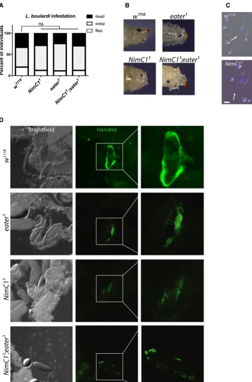
Fig. 6.NimC1 mutants do not show any major encapsulation defects after wasp infestation. (A) Quantification of emerging Drosophila melanogaster adult,Leptopilina boulardi wasp, and dead animals following parasitization with the parasitoid wasp L. boulardi.
Data are shown as a sum of three experiments, with a total of 90 animals for each genotype. Data were analysed using Chi-square statistical test (P-value>0.05). ns, not significant. (B) Shown are representative images of melanized wasp eggs in w1118control and mutants, 70 h afterL. boulardi infestation. (C) Lamellocyte differentiation is observed ineaterandNimC1mutants upon wasp infestation.
Representative images showing circulating haemocytes 70 h afterL. boulardiinfestation. Arrows and arrowheads indicate haemocytes with lamellocyte (La) and plasmatocyte (P) morphology respectively. Cell nuclei were stained with DAPI (blue) Scale bar: 20lm. (D) Representative images showing early wasp egg recognition by peripheral plasmatocytes (haemocytes stained with anti-Hemese antibody [61], green) 20 h after infestation. Haemocytes of both wild-type andeaterandNimC1mutants attached to the eggs. However, single and double-mutant plasmatocytes adhere with slightly decreased spreading ability compared to the wild-type.
We also investigated a possible role of NimC1 in crystal cell and lamellocyte differentiation.
Crystal cells are the second haemocyte type present in noninfected larvae, specifically involved in the melanization response and wound healing [31]. Crystal cells can be found in both the sessile and circulating state. Recent studies have shown that a fraction of those cells derive from sessile plasmatocyte by transdif- ferentiation [26]. Consequently, crystal cells need ses- sile plasmatocytes to be, themselves, sessile [16].
Lamellocytes are barely present in healthy larvae, but can differentiate from plasmatocytes [25,32] or pro- haemocytes [33] in response to specific stress signals, such as parasitization. They are thought to play an essential role in encapsulation of parasitoid wasp eggs.
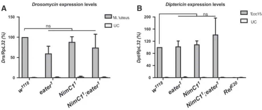
Our study indicates that NimC1 mutants retain the ability to differentiate fully mature crystal cells (Fig. 5). Moreover, our data also show that the NimC1deletion does not affect the ability to encapsu- late parasitoid wasp eggs (Fig. 6). Finally, we did not uncover any role of NimC1 in the systemic antimicro- bial response of larvae against Gram-positive (Micro- coccus luteus) or Gram-negative bacteria (Erwinia carotovora carotovora), as revealed by the wild-type–like induction of Diptericin and Drosomycin gene expression, two target genes of the Imd and Toll pathways respectively [34] (Fig. 7).
NimC1 contributes with Eater to phagocytosis of bacteria
A previous in vivo RNAi approach had revealed a role of NimC1 in the phagocytosis of Gram-positive bacteria [14]. We used the NimC1 deletion to further elucidate the requirement of this receptor in bacterial uptake by performing ex vivo phagocytosis assays at
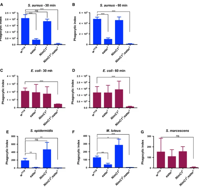
two different time points (early-30 min and late- 60 min). As previously reported [16], eater null mutant haemocytes were impaired in their capacity to phagocytose the Gram-positive bacterium S. au- reus (Fig. 8A,B), but not the Gram-negative bac- terium E. coli (Fig. 8C,D). In contrast to the previous RNAi experiments [14], loss of NimC1 affected neither the phagocytosis of S. aureus nor that of E. coli (Fig. 8A–D). However, haemocytes derived from NimC1;eater mutant larvae were not only severely impaired in the phagocytosis of S. au- reus (Fig. 8A,B), as expected, but also of E. coli (Fig. 8C,D). This indicates that Eater and NimC1 contribute redundantly to the phagocytosis of Gram- negative bacteria, as the presence of Eater or NimC1 is able to compensate for the absence of the other.
The use of a double mutant also revealed a contri- bution of NimC1 to the phagocytosis of S. aureus, although Eater plays the predominant role. To fur- ther confirm these phagocytosis defects, we extended the analysis to two additional Gram-positive (Sta- phylococcus epidermidis, M. luteus) (Fig. 8E,F) and one Gram-negative (Serratia marcescens) (Fig. 8G) bacteria. Phagocytosis of all those microbes was not impaired in NimC11 null haemocytes. However, NimC11;eater1 double mutant haemocytes showed a strongly reduced phagocytosis for both the Gram- positive bacteria S. epidermidis and M. luteus (Fig. 8E,F), and the Gram-negative bacterium S. marcescens (although statistically nonsignificant due to the high variability of the wild-type) (Fig. 8G). Those data further confirmed our initial findings (Fig. 8A–D). Interestingly, NimC1 null haemocytes showed a higher phagocytic index, when compared to wild-type, for S. epidermidis and M. lu- teus bacteria. We hypothesized that the absence of
w111
8
eater
1
NimC1
1
NimC1
1;eater
1 0
50 100 150
Drs/RpL32 (%)
Drosomycin expression levels
M. luteus ns UC
w11
18
eate r1
NimC1
1
NimC1
1;eater1 Rel
E20 0
40 80 120 160 200
Dpt/RpL32 (%)
Diptericin expression levels
Ecc15 UC
A B ns
Fig. 7.NimC1 is not involved in humoral immunity. Expression levels ofDiptericin(A) andDrosomycin(B) relative toRpL32inw1118,eater1, NimC11andNimC11;eater1third instar larvae. Total RNA from infected animals was extracted 4 h afterEcc15orMicrococcus luteusseptic injury. UC, unchallenged controls. The Imd pathway mutant Relish (RelE20) was used as an immune-deficient control in (A). Data are represented as meanSD from 3 independent experiments ns: not significant, by Mann–Whitney test.
NimC1 could trigger a compensatory pathway in plasmatocytes, specific for certain bacteria, in order to fulfil NimC1 phagocytic functions. The signalling of this putative compensatory pathway, that would eventually finally lead to a higher bacteria uptake by plasmatocytes, might be dependent on Eater, given the dramatically reduced phagocytic ability of the double mutant.
Eater and NimC1 receptors play a critical role in adhesion to bacteria
To better understand the cause of eater1 andNimC11; eater1 phagocytosis defects, and thereby to elucidate the unique role of these receptors in bacterial uptake, we performed scanning and transmission (TEM) elec- tron microscopy experiments. Both these techniques
w11
18
eater
1
NimC1
1
NimC1
1;eater
1 0
2 105 4 105 6 105
Phagocytic index
S. aureus - 60 min
*** ***
w111
8
eater
1
NimC1
1
NimC1
1;eater
1 0.0
5.0 104 1.0 105 1.5 105 2.0 105 2.5 105
Phagocytic index
S. aureus - 30 min
*** ns ***
w111
8
eater
1
NimC1
1
NimC1
1;eater
1 0
1 105 2 105 3 105 4 105
Phagocytic index
E. coli- 30 min
***
w11
18
eater
1
NimC1
1
NimC1
1;eater
1 0.0
5.0 105 1.0 106 1.5 106 2.0 106 2.5 106
Phagocytic index
E. coli- 60 min
***
w111
8
eater
1
NimC1
1
NimC1
1;eater
1 0
200 400 600 800
S. epidermidis
Phagocytic index
**
ns **
w11
18
eater
1
NimC1
1
NimC1
1;eater
1 0
100 200 300 400
M. luteus
Phagocytic index
**
* ***
w11
18
eater
1
NimC1
1
NimC1
1;eater
1 0
100 200 300
S. marcescens
Phagocytic index
ns
A B
C D
E F G
Fig. 8.NimC1 contributes with Eater to phagocytosis of Gram-positive and Gram-negative bacteria. Ex vivo phagocytosis assay using Staphylococcus aureus(A, B) andEscherichia coli(C, D) AlexaFluorTM488 BioParticlesTM(Invitrogen).HmlDdsred.nlshaemocytes from third instar wandering larvae were incubated with the particles for 60 (A, C) or 30 (B, D) min at room temperature. In (A–D) data are represented as meanSD from four independent experiments.Ex vivo phagocytosis assay using the Gram-positiveStaphylococcus epidermidis (E), Micrococcus luteus(F) and Gram-negativeSerratia marcescens(G) bacteria. Bacteria were first heat inactivated and subsequently labelled with fluorescein isothiocynate (FITC). Haemocytes from third instar wandering larvae of the indicated genotypes were incubated with the bacteria for 40 min at room temperature. In (E, F) data are represented as meanSD from three independent experiments. In (A–G), phagocytosis was quantified by flow cytometry, and the fluorescence of extracellular particles quenched by adding trypan blue. *P<0.05,
**P<0.01, ***P<0.001, by Studentttests. ns: not significant.
allow following the different membrane-driven events during the phagocytosis process. Haemocytes from the corresponding genotypes were incubated with either E. coliorS. aureuslive bacteria for 30 min to evaluate bacterial adhesion by SEM, and to follow bacterial uptake at 60 min by TEM. In wild-type and NimC11 haemocytes incubated with S. aureus, we observed plasma membrane remodelling, with the formation of a phagocytic cup and pseudopod protrusions that pro- gressively surrounded bacteria, finally leading to their engulfment (Fig. 9A,B white arrowheads). Similar observations were made for wild-type, NimC11 and eater1 haemocytes incubated with E. coli bacteria (Fig. 9C,D). Surprisingly, upon incubation of eater1 andNimC11;eater1 haemocytes withS. aureus, no bac- teria were present on the cell surface (Fig. 9A). A decreased level of bacteria adherence was also observed inNimC11;eater1 haemocytes incubated with E. coli (Fig. 9C). In accordance with SEM experi- ments, transmitted electron micrographs showed numerous engulfment events in wild-type and NimC11 haemocytes with S. aureus bacteria (Fig. 9B, arrows), as well as for E. coli in wild-type, NimC11 and eater1 haemocytes (Fig. 9D). Altogether, these experiments point to the importance of Eater in binding Gram-pos- itive bacteria, which is consistent with a previous report [35], but also to a redundant role of NimC1 and Eater in binding Gram-negative bacteria. Further- more, they suggest that these two receptors do not play any critical role in bacteria internalization, as NimC1;eater mutant showed (rare) engulfment events (Fig. 9B,D arrows).
To further confirm the bacteria adhesion defects, we incubated haemocytes and live fluorescent bacteria either on ice or with Cytochalasin D. Both treatments inhibit the engulfment process, without altering the binding of the bacteria to the phagocytic cell [6]. In both conditions (Fig. 9E,F), we observed less bacteria binding to plasmatocytes in the same genotypes that were defective for phagocytosis in our ex vivo assays (eater1 for S. aureus, andNimC11;eater1 for S. aureus andE. coli, Fig. 8A–D).
Phagocytosis of latex beads and zymosan yeast particles is impaired inNimC1null mutants To further understand the role of Eater and NimC1 in the phagocytosis process, we proceeded to analyse the uptake of ‘neutral’ latex beads particles. We also tested their role in the phagocytosis of zymosan, a compound found on the cell wall of yeast. While bacteria present at their surface-specific targets for the engulfing receptors, latex beads can be seen as nonimmunogenic particles,
that do not bear any ligands for the phagocyte. We observed that the phagocytic index of latex beads was wild-type like ineaternull plasmatocytes. Interestingly, plasmatocytes lacking the NimC1 receptor showed a sig- nificantly reduced ability to engulf latex beads, as well as zymosan yeast particles (Fig. 10A,B). Thus, we could uncover a phagocytic defect in theNimC1 single mutant only when using particles that do not display bacterial motifs, suggesting that bacteria can bypass NimC1, probably by recruiting other phagocytic receptors, such as Eater.
Bacteria adhesion and latex beads engulfment are not impaired incroquemortanddraper mutant haemocytes
The drastic effect observed with the NimC11;eater1 double mutant on phagocytosis and bacteria adhesion led us to explore the contribution of other previously characterized phagocytic receptors using the same assays. Draper and Croquemort are two transmem- brane receptors expressed by plasmatocytes, and belong to the Nimrod and CD36 family respectively [14,36]. With SIMU (NimC4), they both play a key role in the engulfment of apoptotic bodies [18,36–39].
Moreover, a role in S. aureus phagocytosis has also been described for Draper and Croquemort, as well as in E. coli phagocytosis for Draper [17,40–42].
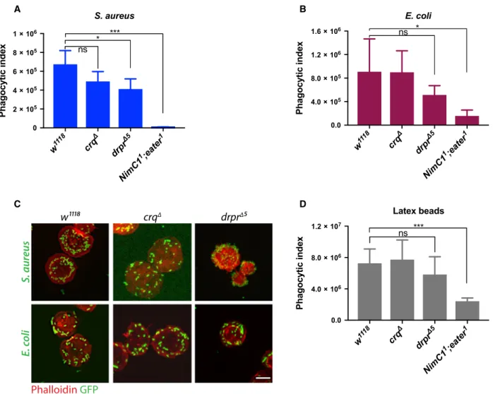
Although we did observe a modest decrease in S. au- reus phagocytosis in croquemort and draper mutants (called crqΔ and drprΔ5 respectively), and E. coli in drprΔ5 (Fig. 11A,B), bacteria adhesion to the haemo- cytes was not impaired in these mutants (Fig. 11C).
This further supports a specific role of Eater as the main tethering receptor in Gram-positive bacteria phagocytosis. Moreover, drprΔ5 and crqΔ haemocytes showed a wild-type like engulfment of latex beads (Fig. 11D), further indicating a specific role of Eater in microbe uptake, likely via the recognition of a key bacterial surface determinant.
Discussion and Conclusions
NimC1 was initially identified as an antigen for the plasmatocyte-specific monoclonal antibody P1. It belongs to the Nimrod gene family that has been implicated in the cellular innate immune response in Drosophila [43,44]. A previous study pointed to the importance of NimC1 in the phagocytosis of bacteria, since RNAi-mediated silencing of this gene resulted in decreased S. aureus uptake by plasmatocytes [14]. In the present work, we further re-evaluated the function
w eater
NimC1 NimC1 ;eater S. aureus
w eater
NimC1 NimC1 ;eater E. coli
eater
NimC1 ;eater
eater
NimC1 ;eater Ice incubation w
NimC1
w
NimC1
suerua.Siloc.E
Phalloidin GFP
Phalloidin GFP
w eater
NimC1 NimC1 ;eater E. coli
eater
NimC1 ;eater
eater
NimC1 ;eater Cytochalasin D w
NimC1
w
NimC1
suerua.Siloc.E
Phalloidin GFP Phalloidin GFP
A B
C D
E F
w eater
NimC1 NimC1 ;eater S. aureus
of the NimC1 protein by using a novel null mutant, revealing its precise role in haemocyte adhesion, prolif- eration and phagocytic ability.
By performingex vivospreading assays, we observed that the cell area of adherent NimC1 null haemocytes was reduced compared to wild-type control, suggesting that NimC1 works as an adhesion molecule. Consis- tent with this observation, SEM on spread haemocytes of NimC11 mutants revealed a defect in lamellipodia extension. Spreading defects were also observed in eater1 [16] and NimC11;eater1 haemocytes. Thus, two structurally related Nimrod receptors, NimC1 and Eater, are involved in lamellipodia extension and haemocyte adhesion. It will be interesting to analyse, in future work, the implications of NimC1 and Eater in haemocyte migration during metamorphosis or wound healing. Our results also indicate that Eater and NimC1 additively regulate haemocyte adherence.
In contrast to eater-deficient larvae, NimC1 is, how- ever, not directly required for plasmatocyte sessility
in vivo. Whether NimC1 contributes to haemocyte ses- sility through additional scaffold proteins has to be further investigated, even though the present evidence might favour a model where Eater is the only essential protein required for haemocyte sessility [16].
During larval development, the peripheral haemo- cyte population undergoes a significant proliferation, expanding by self-renewal [8,21]. Moreover, during these developmental stages, plasmatocytes are charac- terized by a dynamic behaviour, continuously exchang- ing between the sessile and circulating state. In 2011, Makhijaniet al. [21] provided evidence that plasmato- cyte proliferation rate is higher in the haematopoietic pockets, where haemocytes cluster on the lateral side of the larval body. At this location, sessile plasmato- cytes are in contact with the endings of peripheral neu- rons, which are thought to provide a trophic environment to the blood cells. More recently, it has been shown that sensory neurons of the peripheral ner- vous system produce Activin-b, which turned out to be
w111 8
eater 1
NimC1 1
NimC1 1;eater
1 0
2 106 4 106 6 106 8 106 1 107
Phagocytic index
ns ***
Latex beads
w111 8
eater 1
NimC1 1
NimC1 1;eater
1 0.0
5.0 104 1.0 105 1.5 105
Phagocytic index
Zymosan
ns **
A B
Fig. 10.NimC11haemocytes show impaired phagocytosis of latex beads and zymosan particles. (A) Phagocytic index quantification of latex beads (Sigma-Aldrich, St. Louis, MO, USA) engulfment in haemocytes fromw1118,eater1,NimC11andNimC11;eater1L3 wandering larvae after 30 min incubation. Data are represented as meanSD from five independent experiments (B) Phagocytic index quantification of AlexaFluorTM488 Zymosan (S. cerevisiae) BioParticlesTMengulfment. Zymosan BioParticles were incubated withw1118or mutant haemocytes from L3 wandering larvae for 90 min. Data are represented as meanSD from four independent experiments. In (A) and (B), phagocytosis was quantified by flow cytometry, and the fluorescence of extracellular particles quenched by adding trypan blue. **P<0.01,
***P<0.001, by Studentttests. ns, not significant.
Fig. 9.eater1andNimC11;eater1haemocytes show bacteria adhesion defects. (A) Representative SEM images of haemocytes from the indicated genotypes of L3 wandering larvae after 30 min incubation withStaphylococcus aureuslive bacteria, at room temperature. Scale bar: 2lm. (B) Representative transmission electron micrographs of haemocytes from the indicated genotypes of L3 wandering larvae after 60 min incubation withS. aureuslive bacteria, at room temperature. (C) Representative SEM images of haemocytes from the indicated genotypes of L3 wandering larvae after 30 min incubation withEscherichia colilive bacteria, at room temperature. Scale bar: 1lm. (D) Representative transmission electron micrographs of haemocytes from the indicated genotypes of L3 wandering larvae after 60 min incubation with E. coli live bacteria, at room temperature. Arrowheads and arrows indicate haemocyte membrane protrusions and internalized bacteria respectively. (E, F) Haemocytes from the corresponding genotypes were incubated withS. aureus GFP (green) or E. coliGFP (green) live bacteria on ice (E), or with Cytochalasin D (F), for 1 h (see Material and Methods section for further details). After fixation with 4% paraformaldehyde, haemocytes were stained with rhodamine phalloidin (red). Scale bar: 10lm.