Accepted Manuscript
Title: Biochemical and pharmacological characterization of three opioid-nociceptin hybrid peptide ligands reveals substantially differing modes of their actions
Authors: Anna I. Erdei, Adina Borb´ely, Anna Magyar, N´ora Taricska, Andr´as Perczel, Ott´o Zs´ıros, Gyoz˝ o Garab, Edina˝ Sz˝ucs, Ferenc ¨Otv¨os, Ferenc Z´ador, Mih´aly Balogh,
Mahmoud Al-Khrasani, S´andor Benyhe
PII: S0196-9781(17)30318-2
DOI: https://doi.org/10.1016/j.peptides.2017.10.005
Reference: PEP 69842
To appear in: Peptides Received date: 24-4-2017 Revised date: 10-10-2017 Accepted date: 11-10-2017
Please cite this article as: Erdei Anna I, Borb´ely Adina, Magyar Anna, Taricska N´ora, Perczel Andr´as, Zs´ıros Ott´o, Garab Gyoz˝˝ o, Szucs Edina,˝
¨Otv¨os Ferenc, Z´ador Ferenc, Balogh Mih´aly, Al-Khrasani Mahmoud, Benyhe S´andor.Biochemical and pharmacological characterization of three opioid-nociceptin hybrid peptide ligands reveals substantially differing modes of their actions.Peptides https://doi.org/10.1016/j.peptides.2017.10.005
This is a PDF file of an unedited manuscript that has been accepted for publication.
As a service to our customers we are providing this early version of the manuscript.
The manuscript will undergo copyediting, typesetting, and review of the resulting proof before it is published in itsfinal form. Please note that during the production process errors may be discovered which could affect the content, and all legal disclaimers that apply to the journal pertain.
Biochemical and pharmacological characterization of three opioid-nociceptin hybrid peptide ligands reveals substantially differing modes of their actions
Running title: Opioid-like receptor labelling by bivalent peptides
Anna I Erdei1, Adina Borbély2, Anna Magyar2, Nóra Taricska3, András Perczel3,4, Ottó Zsíros5, Győző Garab5, Edina Szűcs1, Ferenc Ötvös1, Ferenc Zádor1, Mihály Balogh6, Mahmoud Al-Khrasani6, Sándor Benyhe1*
1 Institute of Biochemistry, Biological Research Center, Hungarian Academy of Sciences, H- 6726 Szeged, Temesvári krt. 62., Hungary
2 MTA-ELTE Research Group of Peptide Chemistry, Hungarian Academy of Sciences, Eötvös Loránd University, H-1117 Budapest, Pázmány Péter sétány 1/A Budapest, Hungary
3 Laboratory of Structural Chemistry and Biology, Institute of Chemistry, Eötvös Loránd University, Pázmány P. sétány 1/A, Budapest, H-1117 Hungary;
4 MTA-ELTE Protein Modelling Research Group, Institute of Chemistry, Hungarian Academy of Sciences, Eötvös Loránd University, H-1117 Budapest, Pázmány Péter sétány 1/A Budapest, Hungary
5 Institute of Plant Biology, Biological Research Center, Hungarian Academy of Sciences, H-6726 Szeged, Temesvári krt. 62., Hungary
6 Department of Pharmacology and Pharmacotherapy, Semmelweis University, H-1445 Budapest, Nagyvárad tér 4., Hungary
* Corresponding author:
Sándor Benyhe
Institute of Biochemistry, Biological Research Center, Hungarian Academy of Sciences,
H-6701 Szeged, P.O. Box 521, Hungary.
Tel.: +36 62 432 099;
Fax: +36 62 433 506.
E-mail address: benyhe@brc.hu
Highlights
Bivalent ligands are research tools for GPCRs and their interacting complexes
Three opioid-nociceptin hybrid peptides were studied by biochemical pharmacology
Receptor binding and activation patterns of the peptides were sequence specific
Abstract
In an attempt to design opioid-nociceptin hybrid peptides, three novel bivalent ligands, H-YGGFGGGRYYRIK-NH2, H-YGGFRYYRIK-NH2 and Ac-RYYRIKGGGYGGFL-OH were synthesized and studied by biochemical, pharmacological, biophysical and molecular modelling tools. These chimeric molecules consist of YGGF sequence, a crucial motif in the N-terminus of natural opioid peptides, and Ac-RYYRIK-NH2, which was isolated from a combinatorial peptide library as an antagonist or partial agonist that inhibits the biological activity of the endogenously occurring heptadecapeptide nociceptin. Solution structures for the peptides were studied by analysing their circular dichroism spectra. Receptor binding affinities were measured by equilibrium competition experiments using four highly selective radioligands. G-protein activating properties of the multitarget peptides were estimated in [35S]GTPS binding tests. The three compounds were also measured in electrically stimulated mouse vas deferens (MVD) bioassay. H-YGGFGGGRYYRIK-NH2 (BA55), carrying N- terminal opioid and C-terminal nociceptin-like sequences interconnected with GGG tripeptide spacer displayed a tendency of having either unordered or -sheet structures, was moderately potent in MVD and possessed a NOP/KOP receptor preference. A similar peptide without spacer H-YGGFRYYRIK-NH2 (BA62) exhibited the weakest effect in MVD, more -helical periodicity was present in its structure and it exhibited the most efficacious agonist actions in the G-protein stimulation assays. The third hybrid peptide Ac-RYYRIKGGGYGGFL-OH (BA61) unexpectedly displayed opioid receptor affinities, because the opioid message motif is hidden within the C-terminus. The designed chimeric peptide ligands presented in this study accommodate well into a group of multitarget opioid compounds that include opioid-non-opioid peptide dimer analogues, dual non-peptide dimers and mixed peptide- non-peptide bifunctional ligands.
Keywords: radioligand binding, mouse vas deferens, opioid receptors, NOP receptor, nociception, bivalent ligands
1. INTRODUCTION
G protein-coupled receptors (GPCRs) are ancestrally related membrane proteins on various cells that mediate the physiological and pharmacological effect of most drugs, hormones and neurotransmitters. GPCRs are the largest family of proteins encoded in the human genome. One of the most important types of GPCRs is the opioid receptors [1–6]. Opioid family receptors consist of four closely related cell surface proteins expressed in all vertebrate animals examined to date. The three classical types of opioid receptors shown unequivocally to mediate analgesia in animal models and in humans are the mu- (MOP), delta- (DOP), and kappa- (KOP) opioid receptor proteins. The fourth and most recent member of the opioid receptor family described is the nociceptin or orphanin FQ receptor also called as NOP receptor or ORL-1 (opioid receptor like) receptor [7,8]. The role of NOP receptor and its ligands in mediating analgesia is not as clear, with both analgesic and hyperalgesic effects reported.
Natural ligands for the opioid receptor subfamily are endogenous opioid peptides. They include Met- and Leu-enkephalin [9], β-endorphin [10], dynorphin A [11] and nociceptin or orphanin FQ [7,8]. Opioid peptides are easy and frequent targets for chemical modifications, so it is not surprising that a huge amount of synthetic opioid peptides have been reported with various structural modifications for reviews see [12–14] and [15–18]. Hybrid or bifunctional peptide or non-peptide ligands developed recently, bearing two pharmacophores, represent novel biochemical tools in investigating GPCRs and their interacting complexes. The pioneer synthetic bifunctional opioid peptide was a double enkephalin later named biphalin [19].
Biphalin (Tyr-D-Ala-Gly-Phe-NH-NH<-Phe<-Gly<-D-Ala<-Tyr) is an opioid octapeptide with a dimeric structure based on two identical pharmacophore portions, derived from enkephalins, joined "tail to tail" by a hydrazide bridge. Numerous structure-activity relationship studies (SAR) were performed in order to understand the elements responsible for the high activity of biphalin [20]. Beside biphalin, a number of other chimeric opioid peptides linked by spacer have been studied so far. Opioid peptides have often been combined with other bioactive neurotransmitters and peptide hormones that are involved in pain perception, e.g. substance P, neurotensin, cholecystokinin, cannabinoids, neuromedin ligands, etc. [21]. Such novel peptide chimeras (also called designed multiple ligands or twin or hybrid drugs), may interact independently with their respective receptor proteins.
Here we describe and characterize three novel hybrid opioid peptides using in vitro displacement binding and functional [35S]GTPS binding assay as well as mouse vas deferens bioassay . These bivalent ligands (BA55 and BA62) are composed either of the minimum opioid
tetrapeptide structure (Tyr-Gly-Gly-Phe; YGGF) or (Tyr-Gly-Gly-Phe-Leu; YGGFL) targeting the opioid receptors, and a NOP receptor recognizing synthetic sequence Ac-Arg-Tyr-Tyr-Arg- Ile-Lys-NH2 (Ac-RYYRIK-NH2) isolated originally from combinatorial chemical libraries [22]. Ac-RYYRIK-NH2 and its related hexapeptides were reported to behave as partial agonists [22], therefore they are capable of antagonizing NOP receptor selective pure agonist ligands [23–27]. The related peptide fragments were either fused directly or connected via an arbitrary given short tripeptide spacer composed of three glycine residues (GGG). The structures of the three hybrid peptides are H-YGGFGGGRYYRIK-NH2, H-YGGFRYYRIK-NH2, Ac- RYYRIKGGGYGGFL-OH. Combining the MOP/DOP/KOP receptor agonist structure with the NOP receptor partial agonist/antagonist sequence Ac-RYYRIK-NH2 would result in effective bivalent compounds targeting the individual opioid receptors and perhaps their interacting complexes, e.g., MOP/NOP or DOP/NOP receptor heterodimers.
2. MATERIALS AND METHODS 2.1. Chemicals
All amino acid derivatives, resins (Rink-amide MBHA, 2-chlorotrityl resin) and coupling agents were purchased from IRIS Biotech GmbH (Marktredwitz, Germany), Reanal (Budapest, Hungary) or Fluka (Buchs, Switzerland). Solvents for synthesis and HPLC were from Molar Chemicals (Budapest, Hungary) or Merck Kft (Budapest, Hungary). Tris-HCl, MgCl2 x 6H2O, EGTA, NaCl, GDP, the GTP analogue GTPS, Bovine serum albumin (BSA), the [d-Ala2, d-Leu5] enkephalin (DADLE) and U-69,593 were purchased from Sigma-Aldrich (Budapest, Hungary). YGGF, Leu-enkephalin and nociceptin was obtained from Bachem Holding AG (Bubendorf, Switzerland). The Tyr-d-Ala-Gly-(NMe)Phe-Gly-ol (DAMGO), the Ile5,6-deltorphin II (IleDelt II) were synthesized in the Laboratory of Chemical Biology group of the Biological Research Center (BRC, Szeged, Hungary) and the Ac-RYYRIK-NH2 was synthesized in the Research Group of Peptide Chemistry of MTA-ELTE, Budapest. The U50,488H was obtained from the Upjohn Company, (Kalamazoo, MI, USA) and the ethylketocyclazocine (EKC) was purchased from Sterling Winthrop (Rensselaer, NY, USA).
The naltrindole, nor-BNI, and JTC-801 were purchased from Tocris Bioscience (Bristol, UK).
The naloxone was kindly provided by the company Endo Laboratories DuPont de Nemours (Wilmington, DE, USA) and the cyprodime was a gift from Prof. Helmut Schmidhammer, Innsbruck University, Innsbuck, Austria. Ligands were dissolved in water and were stored in 1
mM stock solution at −20 C. The radiolabelled GTP analogue, [35S]GTPS (specific activity:
1000 Ci/mmol) was purchased from Hartmann Analytic (Braunschweig, Germany).
[3H]DAMGO (specific activity: 38.8 Ci/mmol), [3H]IleDelt II (specific activity: 19.6 Ci/mmol) and [3H]HS665 (specific activity: 13.1 Ci/mmol) were radiolabelled by the Laboratory of Chemical Biology group in BRC (Szeged, Hungary) and were characterized previously. [3H]U- 69,593 (specific activity: 43.6 Ci/mmol) and [3H]Nociceptin (specific activity: 115.5 Ci/mmol) were purchased from PerkinElmer (Boston, USA). The UltimaGoldTM MV aqueous scintillation cocktail was purchased from PerkinElmer (Boston, USA).
2.2. Circular dichroism (CD) spectroscopy
Far-UV CD spectra were recorded at 25 C temperature on Jasco J-810 spectropolarimeter on the peptide. The CD spectra were measured at between 260 and 185 nm with an optical pathlength of 1 mm, the peptide concentration was ~ 0.1 mg/ml in Milli-Q water.
The bandwidth was 2 nm and data pitch 1.0 nm, the scan speed was set to 100 nm/min and the integration time was 1 sec. 10 spectra were accumulated and plotted. Far UV spectra were analized by CDSSTR method (dichroweb.cryst.bbk.ac.uk/html/home.shtml).
Near-UV range: Typical spectral accumulation parameters were the scanning speed 50 nm/min with 1 nm bandwidth and the 0.2 nm step resolution over wavelength range 240-325 nm with five scans averaged for each spectrum. The temperature at the cuvette was controlled by Peltier-type heating system. The raw ellipticity data were converted into molar ellipticity ([θ]/ deg*cm 2*dmol - 1) for the near-UV region.
2.3. Molecular dynamics calculations
The conformers were generated by simulated annealing and energy minimization method. An initial unrestrained molecular dynamics simulation was performed at 1000 K to override the conformational barriers. The trajectory was sampled every picoseconds and the resulting snapshots were annealed to 50 K for two picoseconds and then energy minimized by RMS gradient convergence criterion of 0.001. Molecular modelling was performed by the Tinker program package v. 6.3.3 (Software Tools for Molecular Design Washington University Medical School, USA). All molecular dynamics steps were performed using the amber99 force field and GBSA implicit solvent environment. Investigation of the secondary structure of the conformers was performed by the Stride program [28].
2.4. Animals
In experiments designed for receptor binding assay: male and female Wistar rats (250–
300 g body weight) and male guinea pigs (~700 g body weight, LAL/HA/BR strain) were used.
Rats were housed in the local animal house of BRC (Szeged, Hungary), while guinea pigs were housed in LAB-ÁLL Bt. (Budapest, Hungary). For MVD experiments NMRI mice (35-45 g) were used. Mice were purchased from Toxicoop (Budapest, Hungary) and they were housed in the local animal house of the Department of Pharmacology and Pharmacotherapy, Semmelweis University (Budapest, Hungary) in group of 5 animals /cage.
Animals were kept in a temperature controlled room (21-24 oC) under a 12:12 light and dark cycle, allowed free access to tap water and standard rodent food until the time of sacrifice. The animals were handled humanely, in complete accordance with the European directive 2010/63/EU on the protection of animals used for scientific purposes and the Hungarian Act for the Protection of Animals in Research (XXVIII.tv. 32.§). Both the number of rats and their suffering were minimized throughout our experiments.
2.5. Peptide synthesis
The peptides were synthesized by solid phase peptide synthesis method using Fmoc strategy on Rink-amide MBHA (H-YGGFGGGRYYRIK-NH2, H-YGGFRYYRIK-NH2) or on 2-chlorotrityl resin (Ac-RYYRIKGGGYGGFL-OH). Amino acids were coupled as Fmoc derivatives by the DIC/HOBt coupling method in DMF. After removal of the last Fmoc group, the N-terminus was acetylated by acetic anhydride and DIEA. The crude products were purified by semipreparative RP-HPLC and the purified compounds were characterized by analytical RP- HPLC and ESI-ion trap mass spectrometry.
2.6. Rat and guinea pig brain membrane homogenate preparation for binding assays Animals were decapitated and rat or guinea pig brains were quickly removed. The brains were prepared according to a method previously described [29] and partly used for binding experiments and partly were further prepared for the [35S]GTPS binding experiments according to [30]. Briefly, the full brain (without cerebellum) were homogenized, centrifuged in ice-cold 50 mM Tris-HCl (pH 7.4) buffer and incubated at 37 oC for 30 min in a shaking water-bath for details see [31]. After incubation the centrifugation was repeated as described before and the final pellet was suspended in 50 mM Tris-HCl pH 7.4 buffer containing 0.32 M sucrose. For the [35S]GTPS binding experiments the final pellet of rat or guinea pig brain membrane homogenate was suspended in ice-cold TEM (50 mM Tris-HCl, 1 mM EGTA, 3 mM MgCl2) buffer so that the desired protein concentration for the assay (~10 μg/ml) could be achieved. The proteins were stored at −80 oC until future use.
2.7. Displacement binding assay
In competition binding experiments the affinity of an unlabelled compound is analysed by measuring radioligand specific binding in the presence of increasing concentrations of the unlabelled compound in question. Aliquots of frozen rat brain membrane homogenates were
suspended in 50 mM Tris-HCl buffer (pH 7.4). Membrane fractions containing 0.3–0.5 mg/ml of protein were incubated with crescent concentrations (10-10–10-5 M) of the unlabelled tested ligands and 1 nM of the radioligands. Incubation conditions for rat brain homogenates were as follows: for [3H]DAMGO and [3H] IleDelt II 35 °C for 45 min, for [3H]U-69,593 24 °C for 45 min, for [3H]Nociceptin 30 °C for 30 min. In case of [3H]U-69,593 in guinea pig brain membrane homogenates (guinea pig brain has significantly more kappa receptors than rat brain). Additionally, unlabelled IleDelt II, U-69,593, DAMGO and nociceptin were also incubated together with their labelled counterparts in increasing concentrations (10-10–10-5 M) for control. For experiments performed with [3H]Nociceptin the incubation mixture also contained 50 mM Tris/HCl, 2.5 mM EGTA, 5 mM MgCl2 and 0.5 mg/ml fatty acid-free BSA (pH 7.4). The level of non-specific binding was determined in the presence of 10 mM unlabelled naloxone, U-69,593, nociceptin or naltrindole, while total binding was determined in the absence of cold compounds. The reaction was terminated by rapid filtration under vacuum (Brandel M24R Cell Harvester; Brandel Harvesters, Gaithersburg, MD), and washed three times with 5 ml ice-cold 50 mM Tris-HCl or 50 mM Tris/ HCl, 2.5 mM EGTA, 5 mM MgCl2, 0.5% BSA (pH 7.4) in case of [3H]Nociceptin. The filtration was accomplished through Whatman GF/C ([3H]DAMGO, [3H]IleDelt II) or GF/B ([3H]U-69593 and [3H]Nociceptin) glass fibre filters (GE Healthcare, Little Chalfont, UK). The radioactivity was detected in UltimaGold MV aqueous scintillation cocktail (Perkin Elmer, Waltham, MA) with Packard Tricarb 2300TR LSC spectrometer. The competition binding assays were performed in duplicates and repeated at least three times.
2.8. Functional [35S]GTPS binding experiments
The functional [35S]GTPS binding experiments were performed as previously described by [32], with slight modifications. Briefly the rat brain membrane fractions (~10 μg protein per sample) were incubated at 30 C for 60 min in Tris-EGTA buffer (pH 7.4). The buffer was composed of 50 mM Tris-HCl, 1 mM EGTA, 3 mM MgCl2, 100 mM NaCl, 30 μM GDP, containing 20 MBq/0.05 cm3 [35S]GTPS (0.05 nM) and increasing concentrations (10-10 – 10-5 M) of the tested compounds in the presence or absence of 10 µM receptor specific antagonist (cyprodime, naltrindole or norbinaltorphimine). The final volume was 1 ml. Total binding (T) was measured in the absence of the test compounds, while non-specific binding (NS) was determined in the presence of 10 μM unlabelled GTPS and subtracted from the total binding value, to determine the specific binding. Throughout this paper, G-protein activation is
given as percentage over the specific [35S]GTPS binding obtained in the absence of receptor ligands (basal activity). The difference of total binding (T) and non-specific binding (NS) represents the basal activity and was defined as 100%. After incubation, bound and unbound [35S]GTPS were separated by vacuum filtration through Whatman GF/B glass fiber filters with a Brandel M24R Cell harvester. Filters were washed three times with 5 ml ice-cold buffer (pH 7.4), and the radioactivity of the filters was measured in UltimaGOLD™ (Perkin Elmer) scintillation cocktail with a Packard TriCarb 2300TR counter. The experiments were performed in triplicates and repeated at least three times.
2.9. Mouse Vas Deferens (MVD) bioassay
Vasa deferentia from NMRI mice (35-45 g) were prepared as described previously [33]).
Briefly, vasa deferentia (a single organ/bath) were mounted between two electrodes under an initial tension 0.1g in Mg2+ free Krebs solution aerated with carbogen (O2:CO2=95:5) at 31 C.
Field electrical stimulation (upper ring, lower straight wire electrode arrangement) was used.
The stimulation parameters were as follows: field stimulation, pairs (100 ms pulse distance) of rectangular impulses (1 ms pulse width, 9 V/cm i.e. supramaximal intensity) were repeated by 10 s. The muscle contractions were monitored by computer, using a data recording and analysis system (LabChart 5, ADInstruments Pty LTD, Australia).
2.10. Experimental Paradigms of MVD
In the mouse vas deferens experiments, before adding the first dose of agonists, 30–40 min equilibration was used for tissues under stimulation. For all experiments, only one concentration-effect curve for the agonists was constructed per tissue [34] and each of these was constructed in cumulative manner. The drug exposure was less than 2 min, and the administration cycle 12–18 min. Preparations were then washed out and allowed to regain their pre-drug twitch height. Then they were equilibrated for 20 min with antagonists (naloxone, JTC-801, naltrindole, nor-BNI), and without washing a single concentration of agonist was added. To determine dissociation constants of the antagonist, dose ratio (DR) values were obtained by the single-dose method described [35].
2.11. Data analysis
Radioligand binding experiments were performed in duplicate and the [35S]GTPS binding assays were carried out in triplicate. Experimental data were analysed and graphically
processed as means ± S.E.M in the function of the applied ligand concentration range in logarithm form. Points were fitted with the professional curve fitting program, GraphPad Prism 6.0 (GraphPad Prism Software Inc., San Diego, CA, USA, www.graphpad.com), using non- linear regression. In the radioligand competition binding assays the ‘One-site competition’, while in [35S]GTPS binding assays the ‘Sigmoid dose-response’ equation was applied to determine IC50 and Ki (unlabelled ligand affinity) and ligand potency (EC50) and the maximum G-protein efficacy (Emax), respectively. For IC50 and EC50 values standard error is only given in their logarithm form by the curve fitting program due to the data representation. The specific binding of either radiolabelled compound was calculated by the subtraction of non-specific binding from total binding and was given in percentage. The data was normalized to total specific binding, which was settled 100%, which in case of [35S]GTPS also represents the basal activity of the G-protein. In the MVD bioassay, the 50% effective concentration (EC50) and maximal effect (Emax) was determined from the non-linear regression (Hill 4 parameters equation) of individual logarithmic concentration-response curves. The equilibrium dissociation constant of antagonists (Ke) was calculated with the single-dose method [35].
Antagonist affinities (Ke values) were calculated as: Ke= [antagonist concentration]/dose ratio
−1. Experimental data were analysed and graphically represented as means ± S.E.M with curve fitting program, SigmaPlot 11.0. (Systat Software, San Jose, CA). Differences between two data sets unpaired t-test with two-tailed P-value statistical analysis was used, while for three or more data sets one-way ANOVA with Tukey's post hoc test was performed to determine statistical significance. Significance was accepted at the P < 0.05 level.
3. RESULTS
3.1. Circular dichroism (CD) spectroscopy
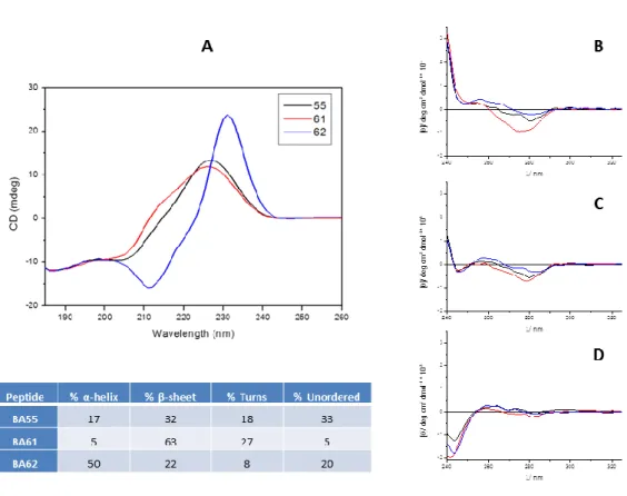
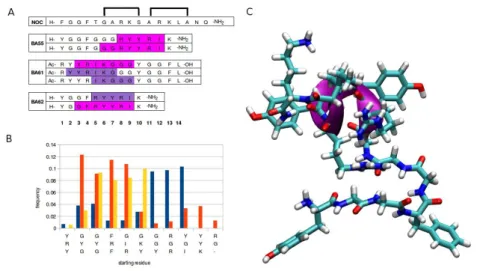
Three novel hybrid peptides composed of opioid (YGGF) and nociceptin-like (RYYRIK) building blocks were synthesized and studied. A detailed analysis of the different ECD (electron-capture dissociation) spectra of the bivalent peptides provided the following structural information: Table within the Fig. 1. shows the relative distributions of secondary structures observed. Spectral data collected at 25 °C were analysed by CDSSTR method using reference dataset: 3 [36]. Peptide BA55 has been associated with the unordered motive and the turns, while BA61 and BA62 the β-sheet and α-helix motives are dominant, respectively. Due to the aromatic amino acid content of these peptides the near-UV ECD region can also be informative (Fig. 1. B, C and D). All three peptides have similar spectral properties at all 3 temperatures recorded: i) a negative band at around 280 nm (Lb of Tyr) disappearing at 85°C a bit more pronounced for BA61, ii) a positive band at about 260 nm (most probably Lb of Phe) and iii) the shoulder of a larger band (248 nm) positive at 5°C reversed in sign at 85°C. In summary, the atypical far-UV ECD spectra, with bands strongly influenced by electronic transitions arising from the aromatic residues suggest rather an unstructured polypeptide main chain, which otherwise could have some partial order unrevealed at lower T, completely vanishing at elevated temperature.
3.2. Molecular dynamics calculations
The effect of the structure of the peptides on the biological activity on the NOP receptor was related to their secondary structure in solution analysed by both experimental and theoretical methods. Due to the conformational flexibility of peptides, a widely used experimental method to investigate their secondary structure is the electronic circular dichroism (CD) spectroscopy. In silico investigation of flexible peptides can be performed by the conformational analysis of conformer ensembles. Here we analysed ensembles of 50000 energy minimized conformers generated by molecular dynamics (MD) simulation (Fig. 2.). The binding activities shown for the NOP receptor can be correlated to the secondary structure patterns of the peptides. The amount of the turns present in the peptides seems to be proportional to the affinity, as in the most active BA61 turns were found in nearly 50%. Contrary to the turns, the presence of helices in the peptides showed a reverse effect, as the most helical BA62 proved to be inactive on NOP receptor. The percentile ratios and the location of the different secondary structure elements distributed within the conformer ensembles were compared to the
experimental results obtained by CD. The percentage of a specific secondary structure was calculated as follows:
100 x (sum of the affected resides in the ensemble) / (sum of all residues in the ensemble) The negligible amount of beta strands in the conformers was in accordance of their absence in the CD spectra. The rank order of the overall amount of turns was the same as obtained by CD.
In the case of helices, the helix content of the inactive BA62 was the highest, as expected.
Besides the overall amount of the secondary structure elements their location was also investigated. The most abundant beta turn type IV distributed almost uniformly along the peptide chains. beta turn type I, the second highest amount turn type, mostly abounded close to the N-terminal in the most active BA61 but farther from the N-terminal in other two peptides (Figure turns). The distribution of helices in the peptides seems to be related to the RYYRIK segment (Figure helices). However, besides the core RYYRIK segment, the helical character seems to be prune to extend toward the nearby glycine rich regions too.
3.3. Receptor binding assays and functional [35S]GTPS binding experiments
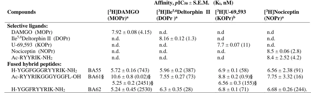
Heterologous equilibrium competition curves are shown in Fig. 3. and these studies are summarized in Table 1. Each of the three peptides displaced the receptor type-selective radioligands with various affinities. The highest NOP receptor affinity was obtained with BA61 (Ki 16 nM, Table 1.) confirming that the Ac-RYYRIK-NH2 N-terminal sequence carries NOP receptor interacting motifs. Most of the curves were accurately fitted by the ‘one binding site’
model. However, the curve for [3H]DAMGO / BA61 the ’two site fit’ was really perfect with Ki values of 0.02 nM (high affinity site) and 2.4 μM (low affinity site), respectively. The Ki
values of BA61 were 0.9 and 155 nM measured with [3H]U-69,593 suggesting again biphasic interaction in the case of KOP receptor (‘two site fit’ calculation). Moreover, the displacement curve of this competition was not complete (Fig. 3. C panel) with substantial residual binding activity (around 50%). The apparent high affinities of H-YGGF-OH and BA61 in the KOP receptor assay (Fig. 3. C panel) were also accompanied by higher residual binding levels.
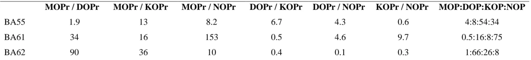
Binding selectivity ratios were calculated as quotients of the corresponding Ki values (Table 2.), while % relative affinity values (Table 2. last column) were calculated according to [37]. BA55 is characterized by higher KOP/NOP receptor preference, although sequence predictions suggested rather a MOP/DOP receptor selectivity for BA55. BA61 peptide exhibits NOP receptor preference (75%), while the characteristic target for the peptide BA62 is the DOP receptor (66%).
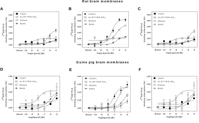
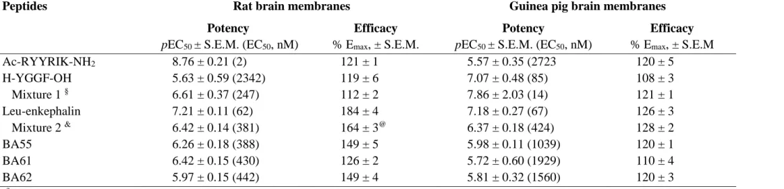
Agonist induced and receptor mediated G-protein activation was studied by [35S]GTPS binding assays performed on rat or guinea pig brain membranes (Fig. 4.). All three bivalent peptide ligands effectively stimulated the activity of regulatory G-proteins in rat brain membranes with maximal stimulation levels of 149:126:149 % for BA55:BA61:BA62, respectively (Table 3.). On the basis of these Emax values and those of Leu-enkephalin (H- YGGFL-OH), BA55 and BA62 are full agonists, while peptide BA61 seems to be a partial agonist ligand. In guinea pig brain membrane fractions, containing mainly KOP receptors, the stimulation order was BA55 BA62 > BA61, indicating again the partial agonist nature of BA61. The effects of the three hybrid peptides were also compared with those of the simple mixtures of their peptide building components. Thus, curves for the mixtures were substantially different from the curves depicting the real hybrid compounds (see Table 3. for significant differences).
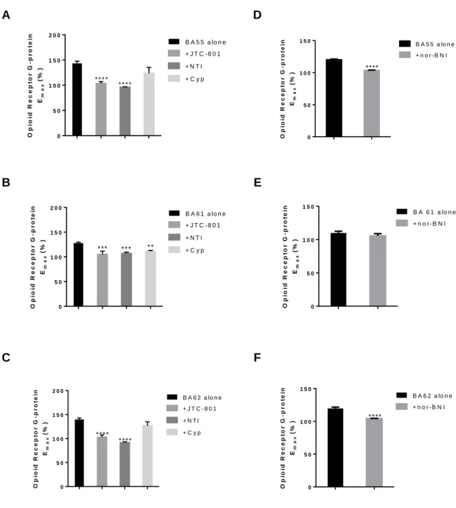
Antagonism by receptor type-selective ligands was also studied in [35S]GTPS binding experiments (Fig 5.). Stimulating effect of BA55 was significantly antagonised by DOP receptor, KOP receptor and NOP receptor antagonists, but not by cyprodime, a MOP receptor selective antagonist. G-protein activation by BA61 was substantially inhibited (significance level was ** or ***) in the presence of MOP receptor , DOP receptor and NOP receptor antagonists, while not with nor-BNI, the most selective KOP receptor antagonist, indicating that interaction of BA61 with the KOP receptor is weaker. Activating effect of the third hybrid peptide, BA62 was effectively antagonised by NOP receptor, DOP receptor and KOP receptor antagonist, but similarly to BA55, it was not inhibited by cyprodime. This suggests again that the MOP receptor is weakly involved in mediating the effects of these two peptides.
3.4 Mouse vas deferens bioassay
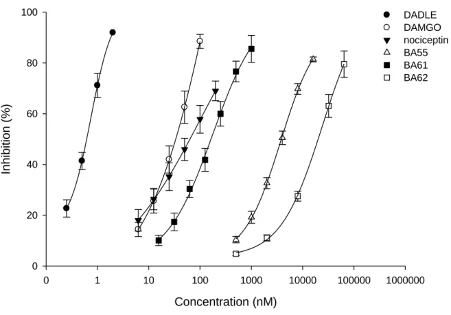
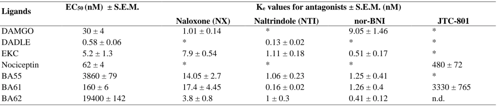
Fig. 6. depicts that the BA61, BA55, BA62 similarly to reference compounds inhibited the electrically-evoked MVD muscle contractions in concentration dependent manner. In MVD bioassay the EC50 (nM) values were 160, 3860, 19400 for BA61, BA55, BA62, respectively (Table 4.). BA61 was lesser potent than any of the reference compounds but about 24- and 121- fold more potent than BA55 or BA62. The average Emax was 100 % for each compounds tested in the present work (Table 4.). The DOP receptor antagonist naltrindole (NTI) Ke values against BA61, BA55, BA62 and [D-Ala2,D-Leu5] enkephalin (DADLE, DOPr agonist) were all within the range of 0.14−1.06 nM. The Ke (nM) value of norbinaltorphimine (nor-BNI, KOPr
antagonist) against BA55, BA62 and EKC (KOPr agonist) were 1.25 ± 0.41 (n=3), 0.41 ± 0.12 (n=4) and 0.51 ± 0.17 (n=3), respectively (Table 4.). On the other side the naloxone (NX) Ke
values against BA55 and BA62 were 14.05 ± 2.7 (n=5) and 3.8 ± 0.8 (n=5) nM, respectively.
The calculated Ke value of NX against DAMGO (typical MOPr agonist) was 1.01 ± 0.14 (n=3) and of JTC-801 against nociceptin (NOPr agonist) was 480 ± 72 (n=3) nM.
4 DISCUSSION
Different types of pain of moderate to severe intensity is often treated with opioid compounds with variable success. However, central and peripheral origin unwanted effects such as respiratory depression, constipation, tolerance and dependence are among main factors that limit the use of opiate therapy. One strategy to overcome the major side effects and to prolong the analgesic efficiency of the applied drugs involves the creation of bi- or multifunctional compounds which contain hybridized structures [21,38]. Combination of opioid agonist and antagonist pharmacophores in a single molecule has been considered and extensively investigated e.g. [39], but opioids have also been combined with other bioactive neurotransmitters and peptide hormones that are involved in pain perception (e.g. neurotensin, neurokinins, such as substance P, cholecystokinin, cannabinoids, etc. [40–42].
Three chimeric peptides were synthesized and studied in the current work. Amino acid sequence of the opioid peptide building blocks (YGGF) was selected according to the message- address model [43]), while the composition of the other pharmacophore (RYYRIK) based on artificial hexapeptide sequences isolated from a combinatorial peptide library [22]. Ac- RYYRIK-NH2 and Ac-RYYRIK-ol have been reported to display NOP receptor partial agonists with full antagonist properties in the presence of the pure agonist N/FQ [24–26,44].
Combination of NOP receptor antagonists with MOP receptor agonists would be perspective as analgesics, although for that reason non-peptide structures could be optimal. Transmembrane helix II of the NOP receptor was shown to be involved in the recognition of the Ac-RYYRIK- NH2 hexapeptide by photoaffinity labelling [45] while another molecular modelling and docking study described that both nociceptin and Ac-RYYRIK-NH2 can able to activate NOP receptor and their receptor-bound conformations have similar 3D structures [46].
Our novel peptide hybrids represent designed structures targeting at least two different GPCRs and perhaps their interacting complexes. Two peptides contain the YGGF opioid message motif at their N-terminus, and this region is followed by the C-terminally located Ac- RYYRIK-NH2 hexapeptide sequence. The two pharmacophores were connected directly in BA62, or interconnected with a triglycine (GGG) spacer sequence in BA55. The third chimeric peptide ligand bears a Ac-RYYRIK-NH2 sequence at the N-terminus, that was combined with the YGGF opioid receptor recognising motif or message sequence [43] via a triglycine (GGG) spacer. It is worth noting that, our peptide sequences are different from those reported by [47].
These authors described MOP/NOP receptor hybrid peptides consisting of full or partial dermorphin sequences found originally in frog skin [48]), fused with the Dooley’s Ac- RYYRIK-NH2 hexapeptide [47].
Based on the different abundance in the peptides, the presence of turns close to the N- terminal may be important for the activity at NOP receptor. The presence of helices close to the N-terminal is contradictory. Due to its low percentage in BA61, the effect of helix on the affinity is presumably negligible and the RYYRIK segment is capable of exerting its effect. Comparing BA55 and BA62, despite they uniform N-terminal segment, the latter is inactive. This may be the consequence of helical segments close to N-termini, starting at Gly3. In addition, BA62 is the most abundant in helical segments according to CD spectra. In BA55, the helical segments are shifted from 3 residue away from the N-terminal, by the amount of the Gly-Gly-Gly spacer between the nociceptin fragment YGGF and RYYRIK. It seems that the helicity induced by Ac-RYYRIK-NH2 less disturbed the structure of the N-terminal fragment in BA55 than did in BA62. Additionally, BA55 is less helical than BA62, according to CD.
In terms of binding affinity, the rank order of potency of the three peptides was BA61 >
BA55 > BA62 at the KOP receptor. The good KOP receptor activity of BA55 and BA62 is not surprising, because the accumulation of positively charged amino acid side chains (two arginines and one lysine, R..R.K) at the opioid address domain is present in various prodynorphin- (PDYN) derived endogenous opioid peptides [49], including dynorphin A 1-17 (RR...K.K), dynorphin B or rimorphin (RR..K...) and -neo-endorphin (RK..K). These data are consistent with our hypothesis that appropriate modifications in the address domain of dynorphin analogues may affect efficacy. Our results confirm also the importance of the opioid
"message" displayed by many opioid ligands but also suggest a potential role of receptor recognition and activation that may be mediated by EL2 through interactions with the "address"
component of dynorphin-like peptides [50]. The good MOP and KOP receptor affinities of BA61 (Ac-RYYRIKGGGYGGF-OH) were not expected, because this peptide has no N-
terminal tyrosine (Tyr, Y) at all, which is common in all endogenous opioid peptides, - endorphin, enkephalins, dynorphins and neo-endorphins. Moreover, the original opioid message tetrapeptide is hidden at the C-terminus of BA61, so the C-terminal address region of this synthetic oligopeptide ligand has no positively charged amino acids important for the KOP- receptor recognition. The high KOP receptor affinity was not observed among the Dooley’s hexapeptide analogues described earlier [24–27,51].
Peptides having the opioid message sequence YGGF at the N-terminus remained comparable full agonists with Leu-enkephalin (YGGFL) when their G-protein activating properties were studied in [35S]GTPS binding experiments (Fig. 3.). The Emax (efficacy) values went up to 149, 149 and 184% for BA55, BA62 and Leu-enkephalin, respectively. The third peptide starting with Ac-RYYRIK-NH2 sequence possesses partial agonist properties reaching an Emax value of 126% (Table 3.). Ac-RYYRIK-NH2 hexapeptides were consistently found to be partial agonists and/or antagonist ligands in a variety of biochemical and pharmacological assays [24–27,52]. It is also worthy to mention here that in the G-protein stimulation assays, simple mixtures composed from the parent peptide components of the hybridised ligands behaved differently when their effects were compared to the multitarget fused sequences bearing real chemical connections, i.e., covalent bonding. Ac-RYYRIK-NH2 was able to inhibit the stimulation by Leu-enkephalin (Fig. 3. B panel), when it was present in the mixture, indicating that the Dooley’s peptide has opioid antagonist activity in addition to the well-known antagonism observed at the NOP receptors. In guinea pig brain membranes, where KOP receptors are predominant, BA55 (Fig. 3. D panel) and BA62 (Fig. 3. F panel) mediated effects were additive by mixing them with the opioid tetrapeptide YGGF, the latter represents a minimum structure for opioid receptor activation [53].
BA55 and BA62 were significantly antagonized by the KOP receptor specific nor- binaltorphimine (nor-BNI) in the [35S]GTPS binding experiments (Fig. 4. D and F panels), while BA61 was not affected by the addition of nor-BNI (Fig. 4. E panel). This phenomenon supports the KOP receptor preference of those hybrid peptides that contain the N-terminal opioid message sequence YGGF in the correct, N-terminally position. Interestingly enough, when the opioid tetrapeptide is located at the C terminus of the hybrid compound BA61, only nor-BNI was not able to antagonize the stimulation, while JTC-801, cyprodime and naltrindole produced significant inhibitions.
For a comparison, we extended our study to examine the pharmacological properties of the test peptides in MVD, which hosting MOP, DOP, KOP and NOP receptors. The order of
potency in this tissue was BA61 BA55 BA62. Applying different opioid receptor subtype antagonists revealed that, BA55 and BA62 showed non selective opioid receptor subtype mediated action (MOP, DOP and KOP receptor), whereas BA61 displayed affinity for DOP and NOP but not for KOP receptor. Based on the above mentioned data, the test peptides displayed potency order that matches what was determined in the binding assay. This similarity was also found in the action of BA55 and BA62 for KOP. In addition, the two peptides showed DOP receptor-mediated effect. However, the selectivity of these two peptides for DOP receptor was less that of DADLE, indicated by dissociation constants (Table 4). The involvement of DOP and KOP receptor in the action of BA55 and BA62 is not surprising, since the two peptides have the same address and message domains. In contrast, BA61 has different N-terminal domain (Ac-R), and yet different C-terminal (carboxyl group). This observation is in good agreement with previous results, according to which the free carboxyl group containing enkephalins preferred DOP receptors [54,55]. Moreover, keeping in mind that BA61 has N- terminal preferred by NOP receptor [24–26,44]. Consequently, the observed pharmacological effect of this peptide might be stand on these changes. Indeed, we could not parallel measure the contribution of NOP and DOP receptor mediated by BA61, because MVD hosting all opioid receptor subtypes but the DOPr reserve is high [56,57]. This tendency makes MVD very sensitive for DOP receptor mediated effect of BA61. Therefore, we measured the effect of JTC- 801 against BA61 in the presence of NTI, a selective DOP receptor antagonist. Applying this strategy we could show the action of BA61 on NOP receptor.
Based on the different abundance in the peptides, the presence of turns close to the N- terminal may be important for the activity at NOP receptor. The presence of helices close to the N-terminal is contradictory. Due to its low percentage in BA61, the effect of helix on the affinity is presumably negligible and the Ac-RYYRIK segment is capable of exerting its effect.
Comparing BA55 and BA62, despite their uniform N-terminal segment, the latter is inactive.
This may be the consequence of helical segments close to N-termini, starting at Gly3. In addition, BA62 is the most abundant in helical segments according to CD spectra. In BA55, the helical segments are shifted from 3 residue away from the N-terminal, by the amount of the Gly-Gly-Gly spacer between the nociceptin fragment YGGF and RYYRIK. It seems that the helicity induced by RYYRIK disturbed less the structure of the N-terminal fragment in BA55 than did in BA62. Additionally, the location of the helices in BA55 is very similar to that in helix-constrained nociceptin analogues [58] (Fig. 2) allowing a nociceptin-like activation mechanism. A typical conformer of peptide BA55 having helical C-terminal tail is shown in Fig 2C.
Summarizing the results we can tell that each of the three novel hybrid peptides carries either opioid- or NOP receptor interacting properties or both, as expected during the design.
One of the original goal that was the combination of the natural opioid agonist tetrapeptide
“message” region with the artificial NOP-receptor selective antagonist (RYYRIK) peptide sequence is fulfilled, although some of the data did not yield the desired specific result. On the other hand, fine selectivity for an individual receptor of the novel peptides was not expected, because these are targeting at least two receptor proteins. The resulting hybrid structures can at least partly served as bivalent compounds for receptor labelling. Our most important and surprising observation is that BA61 behaved as partial agonist at the opioid receptors notwithstanding there is no N-terminal opioid “message” sequence in its structure. Taken together, the tridecapeptide H-YGGFGGGRYYRIK-NH2 (BA55) is moderately potent in the MVD bioassay, it exhibits a KOP/NOP receptor preference, and it has mainly unordered conformations in solution. The shorter hybrid decapeptide composed of opioid message and NOP receptor / nociceptin address domains, H-YGGFRYYRIK-NH2 (BA62), exhibited the weakest inhibition on the mouse vas deferens, it has more -helix in its structure, and this chimeric peptide has been the most efficacious agonist in the G-protein stimulation assays. The third bivalent compound, the tetradecapeptide Ac-RYYRIKGGGYGGFL-OH (BA61) seems to be a quaint KOP receptor ligand, although its amino acid sequence is completely differing from those of the kappa-receptor selective endogenous opioid peptides. Bearing a NOP receptor recognising hexapeptide sequence, the high affinity of BA61 toward the NOP receptor has also been restored. Experiments on receptor co-expressing cell lines are in progress to examine the effects of these hybrid peptides on MOP-NOP receptor heterodimers.
ACKNOWLEDGEMENTS
This research was supported by an OTKA-108518 grant provided by National Research, Development and Innovation Office (NKFIH), Budapest, Hungary.
REFERENCES
[1] M. Waldhoer, S.E. Bartlett, J.L. Whistler, Opioid receptors., Annu. Rev. Biochem. 73 (2004) 953–90. doi:10.1146/annurev.biochem.73.011303.073940.
[2] B.L. Kieffer, C.J. Evans, Opioid receptors: from binding sites to visible molecules in vivo., Neuropharmacology. 56 Suppl 1 (2009) 205–12.
doi:10.1016/j.neuropharm.2008.07.033.
[3] Y. Feng, X. He, Y. Yang, D. Chao, L.H. Lazarus, Y. Xia, Current research on opioid receptor function., Curr. Drug Targets. 13 (2012) 230–46.
http://www.pubmedcentral.nih.gov/articlerender.fcgi?artid=3371376&tool=pmcentrez
&rendertype=abstract (accessed September 14, 2016).
[4] B.M. Cox, M.J. Christie, L. Devi, L. Toll, J.R. Traynor, Challenges for opioid receptor nomenclature: IUPHAR Review 9., Br. J. Pharmacol. 172 (2015) 317–23.
doi:10.1111/bph.12612.
[5] C.W. Stevens, Bioinformatics and evolution of vertebrate nociceptin and opioid receptors., Vitam. Horm. 97 (2015) 57–94. doi:10.1016/bs.vh.2014.10.002.
[6] S. Benyhe, F. Z??dor, F. ??tv??s, Biochemistry of opioid (morphine) receptors: Binding, structure and molecular modelling, Acta Biol. Szeged. 59 (2015) 17–37.
[7] J.C. Meunier, C. Mollereau, L. Toll, C. Suaudeau, C. Moisand, P. Alvinerie, J.L. Butour, J.C. Guillemot, P. Ferrara, B. Monsarrat, Isolation and structure of the endogenous agonist of opioid receptor-like ORL1 receptor., Nature. 377 (1995) 532–5.
doi:10.1038/377532a0.
[8] R.K. Reinscheid, H.P. Nothacker, A. Bourson, A. Ardati, R.A. Henningsen, J.R.
Bunzow, D.K. Grandy, H. Langen, F.J. Monsma, O. Civelli, Orphanin FQ: a neuropeptide that activates an opioidlike G protein-coupled receptor., Science. 270 (1995) 792–4. http://www.ncbi.nlm.nih.gov/pubmed/7481766 (accessed September 14, 2016).
[9] J. Hughes, T.W. Smith, H.W. Kosterlitz, L.A. Fothergill, B.A. Morgan, H.R. Morris, Identification of two related pentapeptides from the brain with potent opiate agonist activity., Nature. 258 (1975) 577–80. http://www.ncbi.nlm.nih.gov/pubmed/1207728 (accessed September 14, 2016).
[10] C.H. Li, D. Chung, Isolation and structure of an untriakontapeptide with opiate activity from camel pituitary glands., Proc. Natl. Acad. Sci. U. S. A. 73 (1976) 1145–8.
http://www.pubmedcentral.nih.gov/articlerender.fcgi?artid=430217&tool=pmcentrez&
rendertype=abstract (accessed September 14, 2016).
[11] A. Goldstein, W. Fischli, L.I. Lowney, M. Hunkapiller, L. Hood, Porcine pituitary dynorphin: complete amino acid sequence of the biologically active heptadecapeptide., Proc. Natl. Acad. Sci. U. S. A. 78 (1981) 7219–23.
http://www.pubmedcentral.nih.gov/articlerender.fcgi?artid=349228&tool=pmcentrez&
rendertype=abstract (accessed September 14, 2016).
[12] S.D. Bryant, Y. Jinsmaa, S. Salvadori, Y. Okada, L.H. Lazarus, Dmt and opioid peptides:
a potent alliance., Biopolymers. 71 (2003) 86–102. doi:10.1002/bip.10399.
[13] L. Gentilucci, New trends in the development of opioid peptide analogues as advanced remedies for pain relief., Curr. Top. Med. Chem. 4 (2004) 19–38.
http://www.ncbi.nlm.nih.gov/pubmed/14754374 (accessed September 14, 2016).
[14] A. Janecka, J. Fichna, T. Janecki, Opioid receptors and their ligands., Curr. Top. Med.
Chem. 4 (2004) 1–17. http://www.ncbi.nlm.nih.gov/pubmed/14754373 (accessed September 14, 2016).
[15] A. Janecka, R. Staniszewska, J. Fichna, Endomorphin analogs., Curr. Med. Chem. 14 (2007) 3201–8. http://www.ncbi.nlm.nih.gov/pubmed/18220754 (accessed September 14, 2016).
[16] P.W. Schiller, Opioid peptide-derived analgesics., AAPS J. 7 (2005) E560-5.
doi:10.1208/aapsj070356.
[17] L. Gentilucci, A. Tolomelli, F. Squassabia, Peptides and peptidomimetics in medicine, surgery and biotechnology., Curr. Med. Chem. 13 (2006) 2449–66.
http://www.ncbi.nlm.nih.gov/pubmed/16918365 (accessed September 14, 2016).
[18] A.K. Giri, V.J. Hruby, Investigational peptide and peptidomimetic μ and δ opioid receptor agonists in the relief of pain., Expert Opin. Investig. Drugs. 23 (2014) 227–41.
doi:10.1517/13543784.2014.856879.
[19] A.W. Lipkowski, A.M. Konecka, I. Sroczyńska, Double-enkephalins--synthesis, activity on guinea-pig ileum, and analgesic effect., Peptides. 3 (1982) 697–700.
http://www.ncbi.nlm.nih.gov/pubmed/7134034 (accessed September 14, 2016).
[20] F. Feliciani, F. Pinnen, A. Stefanucci, R. Costante, I. Cacciatore, G. Lucente, A. Mollica, Structure-activity relationships of biphalin analogs and their biological evaluation on opioid receptors., Mini Rev. Med. Chem. 13 (2013) 11–33.
http://www.ncbi.nlm.nih.gov/pubmed/22512573 (accessed September 14, 2016).
[21] P. Kleczkowska, A.W. Lipkowski, D. Tourwé, S. Ballet, Hybrid opioid/non-opioid ligands in pain research., Curr. Pharm. Des. 19 (2013) 7435–50.
http://www.ncbi.nlm.nih.gov/pubmed/23448481 (accessed November 2, 2016).
[22] C.T. Dooley, C.G. Spaeth, I.P. Berzetei-Gurske, K. Craymer, I.D. Adapa, S.R. Brandt, R.A. Houghten, L. Toll, Binding and in vitro activities of peptides with high affinity for the nociceptin/orphanin FQ receptor, ORL1., J. Pharmacol. Exp. Ther. 283 (1997) 735–
41. http://www.ncbi.nlm.nih.gov/pubmed/9353393.
[23] H. Berger, E. Albrecht, G. Wallukat, M. Bienert, Antagonism by acetyl-RYYRIK-NH2 of G protein activation in rat brain preparations and of chronotropic effect on rat cardiomyocytes evoked by nociceptin/orphanin FQ., Br. J. Pharmacol. 126 (1999) 555–
8. doi:10.1038/sj.bjp.0702353.
[24] C. Kawano, K. Okada, T. Honda, T. Nose, K. Sakaguchi, T. Costa, Y. Shimohigashi, Structural requirements of nociceptin antagonist Ac-RYYRIK-NH2 for receptor binding., J. Pept. Sci. 8 (2002) 561–9. doi:10.1002/psc.415.
[25] L. Kocsis, G. Orosz, A. Magyar, M. Al-Khrasani, E. Kató, A.Z. Rónai, B. Bes, J.-C.
Meunier, O. Gündüz, G. Tóth, A. Borsodi, S. Benyhe, Nociceptin antagonism: probing the receptor by N-acetyl oligopeptides., Regul. Pept. 122 (2004) 199–207.
doi:10.1016/j.regpep.2004.06.019.
[26] Ö. Gündüz, F. Sipos, B. Spagnolo, L. Kocsis, A. Magyar, G. Orosz, A. Borsodi, G. Caló, S. Benyhe, In vitro binding and functional studies of Ac-RYYRIK-ol and its derivatives, novel partial agonists of the nociceptin/orphanin F/Q receptor, NeuroSignals. 15 (2006) 91–101. doi:10.1159/000094743.
[27] O. Gündüz, A. Rizzi, A. Baldisserotto, R. Guerrini, B. Spagnolo, E.C. Gavioli, L. Kocsis, A. Magyar, S. Benyhe, A. Borsodi, G. Calò, In vitro and in vivo pharmacological characterization of the nociceptin/orphanin FQ receptor ligand Ac-RYYRIK-ol., Eur. J.
Pharmacol. 539 (2006) 39–48. doi:10.1016/j.ejphar.2006.03.075.
[28] D. Frishman, P. Argos, Knowledge-based protein secondary structure assignment, Proteins Struct. Funct. Genet. 23 (1995) 566–579. doi:10.1002/prot.340230412.
[29] E. Bojnik, J. Farkas, A. Magyar, C. Tömböly, U. Güçlü, O. Gündüz, A. Borsodi, M.
Corbani, S. Benyhe, Selective and high affinity labeling of neuronal and recombinant nociceptin receptors with the hexapeptide radioprobe [(3)H]Ac-RYYRIK-ol., Neurochem. Int. 55 (2009) 458–66. doi:10.1016/j.neuint.2009.04.014.
[30] F. Zádor, D. Kocsis, A. Borsodi, S. Benyhe, Micromolar concentrations of rimonabant directly inhibits delta opioid receptor specific ligand binding and agonist-induced G- protein activity., Neurochem. Int. 67 (2014) 14–22. doi:10.1016/j.neuint.2013.12.005.
[31] S. Benyhe, J. Farkas, G. Tóth, M. Wollemann, Met5-enkephalin-Arg6-Phe7, an
endogenous neuropeptide, binds to multiple opioid and nonopioid sites in rat brain., J.
Neurosci. Res. 48 (1997) 249–58.
[32] J.R. Traynor, S.R. Nahorski, Modulation by mu-opioid agonists of guanosine-5’-O-(3- [35S]thio)triphosphate binding to membranes from human neuroblastoma SH-SY5Y
cells., Mol. Pharmacol. 47 (1995) 848–54.
http://www.ncbi.nlm.nih.gov/pubmed/7723747 (accessed March 13, 2017).
[33] A.Z. Rónai, L. Gráf, J.I. Székely, Z. Dunai-Kovács, S. Bajusz, Differential behaviour of LPH-(61-91)-peptide in different model systems: Comparison of the opioid activities of LPH-(61-91)-peptide and its fragments, FEBS Lett. 74 (1977) 182–184.
doi:10.1016/0014-5793(77)80842-9.
[34] R.G. Pertwee, S.R. Fernando, G. Griffin, W. Ryan, R.K. Razdan, D.R. Compton, B.R.
Martin, Agonist-antagonist characterization of 6′-cyanohex-2′-yne-Δ8- tetrahydrocannabinol in two isolated tissue preparations, Eur. J. Pharmacol. 315 (1996) 195–201. doi:10.1016/S0014-2999(96)00631-0.
[35] H.W. Kosterlitz, A.J. Watt, Kinetic parameters of narcotic agonists and antagonists, with particular reference to N-allylnoroxymorphone (naloxone)., Br. J. Pharmacol.
Chemother. 33 (1968) 266–76.
http://www.pubmedcentral.nih.gov/articlerender.fcgi?artid=1570231&tool=pmcentrez
&rendertype=abstract.
[36] N. Sreerama, R.W. Woody, Estimation of Protein Secondary Structure from Circular Dichroism Spectra: Comparison of CONTIN, SELCON, and CDSSTR Methods with an Expanded Reference Set, Anal. Biochem. 287 (2000) 252–260.
doi:10.1006/abio.2000.4880.
[37] H.W. Kosterlitz, S.J. Paterson, J.S. Morley, Characterization of Opioid Receptors in Nervous Tissue [and Discussion], Proc. R. Soc. B Biol. Sci. 210 (1980) 113–122.
doi:10.1098/rspb.1980.0122.
[38] S. Dvoracsko, A. Stefanucci, E. Novellino, A. Mollica, The design of multitarget ligands for chronic and neuropathic pain., Future Med. Chem. 7 (2015) 2469–83.
doi:10.4155/fmc.15.156.
[39] K. Guillemyn, J. Starnowska, C. Lagard, J. Dyniewicz, E. Rojewska, J. Mika, N.N.
Chung, V. Utard, P. Kosson, A.W. Lipkowski, L. Chevillard, P. Arranz-Gibert, M.
Teixidó, B. Megarbane, D. Tourwé, F. Simonin, B. Przewlocka, P.W. Schiller, S. Ballet, Bifunctional Peptide-Based Opioid Agonist-Nociceptin Antagonist Ligands for Dual Treatment of Acute and Neuropathic Pain., J. Med. Chem. 59 (2016) 3777–92.
doi:10.1021/acs.jmedchem.5b01976.
[40] A. Mollica, R. Costante, A. Stefanucci, F. Pinnen, G. Luisi, S. Pieretti, A. Borsodi, E.
Bojnik, S. Benyhe, Hybrid peptides endomorphin-2/DAMGO: design, synthesis and biological evaluation., Eur. J. Med. Chem. 68 (2013) 167–77.
doi:10.1016/j.ejmech.2013.07.044.
[41] P. Kleczkowska, E. Bojnik, A. Leśniak, P. Kosson, I. Van den Eynde, S. Ballet, S.
Benyhe, D. Tourwé, A.W. Lipkowski, Identification of Dmt-D-Lys-Phe-Phe-OH as a highly antinociceptive tetrapeptide metabolite of the opioid-neurotensin hybrid peptide
PK20., Pharmacol. Rep. 65 (2013) 836–46.
http://www.ncbi.nlm.nih.gov/pubmed/24145077 (accessed September 14, 2016).
[42] A. Kowalczyk, P. Kleczkowska, M. Rękawek, K. Kulik, A. Lesniak, A. Erdei, A. Borics, C. Martin, K. Pawlik, A.W. Lipkowski, S. Benyhe, H. Makulska-Nowak, S. Ballet, M.
Bujalska-Zadrozny, Biological evaluation and molecular docking studies of AA3052, a compound containing a μ-selective opioid peptide agonist DALDA and d-Phe-Phe-d- Phe-Leu-Leu-NH2, a substance P analogue., Eur. J. Pharm. Sci. 93 (2016) 11–20.
doi:10.1016/j.ejps.2016.07.009.
[43] R. Schwyzer, Molecular mechanism of opioid receptor selection, Biochemistry. 25 (1986) 6335–6342. doi:10.1021/bi00368a075.
[44] H. Berger, R. Bigoni, E. Albrecht, R.M. Richter, E. Krause, M. Bienert, G. Calo’, The nociceptin/orphanin FQ receptor ligand acetyl-RYYRIK-amide exhibits antagonistic and agonistic properties., Peptides. 21 (2000) 1131–9.
http://www.ncbi.nlm.nih.gov/pubmed/10998548 (accessed November 2, 2016).
[45] B. Bes, J.-C. Meunier, Identification of a hexapeptide binding region in the nociceptin (ORL1) receptor by photo-affinity labelling with Ac-Arg-Bpa-Tyr-Arg-Trp-Arg-NH2., Biochem. Biophys. Res. Commun. 310 (2003) 992–1001.
http://www.ncbi.nlm.nih.gov/pubmed/14550303 (accessed November 2, 2016).
[46] N. Akuzawa, S. Takeda, M. Ishiguro, Structural modelling and mutation analysis of a nociceptin receptor and its ligand complexes., J. Biochem. 141 (2007) 907–16.
doi:10.1093/jb/mvm100.
[47] S. Kawano, R. Ito, M. Nishiyama, M. Kubo, T. Matsushima, M. Minamisawa, A. Ambo, Y. Sasaki, Receptor binding properties and antinociceptive effects of chimeric peptides consisting of a micro-opioid receptor agonist and an ORL1 receptor antagonist., Biol.
Pharm. Bull. 30 (2007) 1260–4. http://www.ncbi.nlm.nih.gov/pubmed/17603164 (accessed September 14, 2016).
[48] V. Erspamer, P. Melchiorri, M. Broccardo, G.F. Erspamer, P. Falaschi, G. Improta, L.
Negri, T. Renda, The brain-gut-skin triangle: New peptides, Peptides. 2 (1981) 7–16.
doi:10.1016/0196-9781(81)90003-6.
[49] H. Kakidani, Y. Furutani, H. Takahashi, M. Noda, Y. Morimoto, T. Hirose, M. Asai, S.
Inayama, S. Nakanishi, S. Numa, Cloning and sequence analysis of cDNA for porcine beta-neo-endorphin/dynorphin precursor., Nature. 298 (1982) 245–9.
http://www.ncbi.nlm.nih.gov/pubmed/6123953 (accessed September 14, 2016).
[50] G. Paterlini, P.S. Portoghese, D.M. Ferguson, Molecular simulation of dynorphin A-(1- 10) binding to extracellular loop 2 of the kappa-opioid receptor. A model for receptor activation., J. Med. Chem. 40 (1997) 3254–62. doi:10.1021/jm970252j.
[51] J. Li, H. Nishimura, A. Matsushima, Y. Shimohigashi, N-methylthioacetylation of RYYRIK-NH2 with enhanced specific binding affinity and high antagonist activity for nociceptin ORL1 receptor., Bioorg. Med. Chem. 22 (2014) 5721–6.
doi:10.1016/j.bmc.2014.09.049.
[52] J. Li, K. Isozaki, K. Okada, A. Matsushima, T. Nose, T. Costa, Y. Shimohigashi, Designed modification of partial agonist of ORL1 nociceptin receptor for conversion into highly potent antagonist., Bioorg. Med. Chem. 16 (2008) 2635–44.
doi:10.1016/j.bmc.2007.11.043.
[53] a R. Jacobson, a R. Gintzler, L.M. Sayre, Minimum-structure enkephalin analogues incorporating L-tyrosine, D(or L)-phenylalanine, and a diamine spacer., J. Med. Chem.
32 (1989) 1708–17. doi:10.1021/jm00128a007.
[54] A.Z. Rónai, I. Berzétei, S. Bajusz, Differentiation between opioid peptides by naltrexone, Eur. J. Pharmacol. 45 (1977) 393–394. doi:10.1016/0014-2999(77)90281-3.
[55] A.Z. Rónai, J.I. Székely, I. Berzétei, E. Miglécz, S. Bajusz, Tetrapeptide-amide analogues of enkephalin: The role of C-terminus in determining the character of opioid activity, Biochem. Biophys. Res. Commun. 91 (1979) 1239–1249. doi:10.1016/0006- 291X(79)91200-2.
[56] J.A. Lord, A.A. Waterfield, J. Hughes, H.W. Kosterlitz, Endogenous opioid peptides:
multiple agonists and receptors., Nature. 267 (1977) 495–9.
http://www.ncbi.nlm.nih.gov/pubmed/195217 (accessed March 13, 2017).
[57] M. Al-Khrasani, G. Orosz, L. Kocsis, V. Farkas, A. Magyar, I. Lengyel, S. Benyhe, A.
Borsodi, A.Z. Rónai, Receptor constants for endomorphin-1 and endomorphin-1-ol indicate differences in efficacy and receptor occupancy., Eur. J. Pharmacol. 421 (2001) 61–7. http://www.ncbi.nlm.nih.gov/pubmed/11408050 (accessed March 13, 2017).
[58] R.-J. Lohman, R.S. Harrison, G. Ruiz-Gómez, H.N. Hoang, N.E. Shepherd, S. Chow, T.A. Hill, P.K. Madala, D.P. Fairlie, Helix-constrained nociceptin peptides are potent agonists and antagonists of ORL-1 and nociception., Vitam. Horm. 97 (2015) 1–55.
doi:10.1016/bs.vh.2014.10.001.
Figures and legends
Fig 1. Far (left) and near UV ECD spectra of the three hybrid peptides. A: Far UV spectra measured at 25°C. Temperature dependent near-UV spectra at B: 5°C, C: 35°C and D) 85°C of BA55 (black), BA61 (red) and BA62 (blue).
Fig. 2. Distribution of abundant secondary structures along the peptide sequences. Alpha- helices, more abundant highlighted in magenta (A); Starting positions of beta turns type I (B).
Amino acid numbering is shown at the bottom. (C) Conformer of BA55 possessing α-helix in the Arg8-Ile12 region. Backbone of the helical part is marked by magenta ribbon. Carbon, hydrogen, nitrogen and oxygen atoms are colored in cyan, white, blue and red, respectively.
- 1 0 - 9 - 8 - 7 - 6 - 5 0
2 0 4 0 6 0 8 0 1 0 0 1 2 0
D A M G O
B A 5 5 B A 6 1 B A 6 2
R a t b r a in m e m b r a n e
lo g [ L ig a n d ] , M [3H] DAMGO specific binding (%)
T o t a l Y G G F
- 1 0 - 9 - 8 - 7 - 6 - 5
0 2 0 4 0 6 0 8 0 1 0 0 1 2 0
T o t a l N o c ic e p tin
B A 5 5
B A 6 2 B A 6 1
R a t b r a in m e m b r a n e
lo g [ L ig a n d ] , M [3H] Nociceptin specific binding (%)
A c - R Y Y R IK - N H2
- 1 0 - 9 - 8 - 7 - 6 - 5
0 2 0 4 0 6 0 8 0 1 0 0 1 2 0
Ile D e lt II
B A 5 5 B A 6 1 B A 6 2
R a t b r a in m e m b r a n e
lo g [ L ig a n d ] , M [3H] DIDI specific binding (%)
T o t a l Y G G F
- 1 0 - 9 - 8 - 7 - 6 - 5
0 2 0 4 0 6 0 8 0 1 0 0 1 2 0
U - 6 9 5 9 3
B A 5 5 B A 6 1 B A 6 2
G u in e a p ig b r a i n m e m b r a n e
lo g [ L ig a n d ] , ( M ) [3H] U-69593 specific binding (%)
T o t a l Y G G F A c - R Y Y R IK - N H2
A B
C D
Fig. 3. Displacement of [3H]DAMGO, [3H]Ile5,6Deltorphin II, [3H]U69,593 and [3H]Nociceptin radioprobes by the hybrid peptides and reference ligands. A) MOP receptor, B) DOP receptor, C) KOP receptor, D) NOP receptor assays were done in duplicates and repeated at least three times. The equilibrium competition binding experiments were performed in rat (A, B and D) and in guinea pig (C) brain membrane homogenates.
![Fig. 3. Displacement of [ 3 H]DAMGO, [ 3 H]Ile5,6Deltorphin II, [ 3 H]U69,593 and [ 3 H]Nociceptin radioprobes by the hybrid peptides and reference ligands](https://thumb-eu.123doks.com/thumbv2/9dokorg/1393399.116031/29.892.120.746.125.710/displacement-damgo-deltorphin-nociceptin-radioprobes-peptides-reference-ligands.webp)