Left atrial rather than left ventricular impaired mechanics are associated with the pro-fibrotic ST2 marker and
outcomes in heart failure with preserved ejection fraction
A. I. Nagy
1, C. Hage
2,3, B. Merkely
1, E. Donal
4, J.-C. Daubert
4, C. Linde
2,3, L. H. Lund
2,3& A. Manouras
2,3From the1Heart and Vascular Center, Semmelweis University, Budapest, Hungary;2Theme of Heart and Vessels, Karolinska University Hospital;3Institution for Medicine, Karolinska Institutet, Stockholm, Sweden; and4Departement de Cardiologie & CIC-IT U 804, Centre Hospitalier Universitaire de Rennes, Rennes, France
Abstract.Nagy AI, Hage C, Merkely B, Donal E, Daubert J-C, Linde C, Lund LH, Manouras A (Semmelweis University, Budapest, Hungary; Karolinska University Hospital; Karolinska Institutet, Stockholm, Sweden; Centre Hospitalier Universitaire de Rennes, France). Left atrial rather than left ventricular impaired mechanics are associated with the pro-fibrotic ST2 marker and outcomes in heart failure with preserved ejection fraction.J Intern Med 2018;283: 380–391.
Aims. Left ventricular (LV) mechanics have been extensively investigated in heart failure with pre- served ejection fraction (HFpEF) overshadowing for a long time the potential role of left atrium (LA) in that setting. Soluble suppression of tumorigenic- ity-2 receptor (ST2) is a novel biomarker of pro- fibrotic burden in HF. We hypothesized that due to the thinner LA wall, the fibrotic myocardial changes in HFpEF as indicated by elevated ST2 levels might more readily be reflected by impair- ments in the LA rather than the LV performance.
Methods and Results. In 86 patients with HFpEF, enrolled in the Karolinska Rennes (KaRen)
biomarker prospective substudy, global LA strain (GL-LS) along with other echocardiographic as well as haemodynamic parameters and ST2 levels were measured.ST2 levels were inversely associated with LA-GS (r=0.30, P=0.009), but not with LA size, LV geometry, systolic or diastolic LV function (P>0.05 for all). Furthermore, symptom severity correlated with ST2 and LA-GS, but not with LV structural or functional indices. Finally, during a median 18-month follow-up, LA-GS independently predicted the composite endpoint of HF hospitalization and all-cause mortality, even after adjustment for potential clinical and cardiac mechanical confounders, including LV global longitudinal strain and filling pressures (odds ratio: 4.15; confidence interval: 1.2–14, P =0.023).
Conclusions. Reduced LA-GS but not LV functional systolic and diastolic parameters were associated with the pro-fibrotic ST2 marker, HF symptoms and outcome in HFpEF.
Keywords: ST2, heart failure, preserved ejection frac- tion, left atrial strain, prognosis.
Introduction
Heart failure with preserved ejection fraction (HFpEF) accounts for up to half of patients with heart failure (HF) [1, 2], with a prognosis compa- rable to that of HF with reduced ejection fraction (EF) [1] and an increasing prevalence.
For a long time, investigations aiming to uncover the pathophysiology underlying HFpEF have focused on left ventricular (LV) mechanics;
however, no pathognomonic LV functional or structural alterations have been identified. While LV hypertrophy is common in HFpEF, nearly half of the HFpEF patients show no signs of hypertrophy [2]. Similarly, although increased LV stiffness is typical in this patient population [3, 4], it may also occur in subjects without clinical signs and symp- toms of HF [5]. Furthermore, whereas LV diastolic dysfunction is considered as a hallmark of HFpEF, abnormal diastolic LV performance is an almost universal finding in elderly patients without HFpEF [5], which underscores the significance of other mechanisms in the pathophysiology of HFpEF.
ClinicalTrials.gov. NCT00774709
Recently, a subtle shift in the focus of interest in the HFpEF field has occurred with an increasing number of studies investigating the role of the left atrium (LA) rather than that of the LV in the HFpEF [6–9]. Elevated filling pressures and subsequent LA dilation are a characteristic finding in HFpEF. The degree LA enlargement reflects disease chronicity [10] and is prognostic in HFpEF [9]. Importantly, apart from the structural remodelling, increased LA stiffness has also been demonstrated in HFpEF, which further aggravates the elevated LA pres- sures, particularly during exertion.
Increasing evidence supports the concept that HFpEF ultimately develops on the basis of a pro- inflammatory state triggered by comorbidities [11].
The soluble suppression of tumorigenicity-2 recep- tor (ST2) is an established biomarker of inflamma- tion and fibrosis with an emerging role in the diagnosis and prognostication of HF. In the context of HF, ST2 expression is thought to be triggered by myocardial stretch and consequent fibrosis [12]. In patients with reduced EF, ST2 has superior prog- nostic power compared to conventional biomarkers such as N-terminal pro-brain natriuretic peptide (NT-proBNP) [13]. Patients with HFpEF also demonstrate elevated ST2 levels. Interestingly how- ever, in this condition no significant correlation between ST2 levels and LV structure or function has been found [14].
As HFpEF is postulated to develop as a result of a systemic inflammatory reaction, we hypothesized that the LA, given its much thinner wall, is likely to be more susceptible to fibrotic changes as com- pared to the LV. Thus, early signs of the disease, as indicated by elevated ST2 levels, might more promptly manifest in altered LA mechanics, rather than in impaired LV performance. Accordingly, employing information from the biomarker Karolin- ska Rennes (KaRen) prospective, multicentre sub- study, we tested the hypothesis that in HFpEF (1) plasma ST2 levels correlate with LA function; and (2) impaired LA function is associated with worse outcomes.
Materials and methods Study population
The study included 86 HFpEF patients enrolled in the prespecified KaRen Biomarker Study which comprised a substudy of the Karolinska Rennes (KaRen) prospective, observational, multicentre study [15]. Patients admitted for acute HF
symptoms, NT-proBNP >300 ng L1 and LVEF
≥45% were enrolled between May 2007 and Decem- ber 2011. Four to 8 weeks after enrolment, when patients were in a stable condition, blood samples were taken and clinical investigations, including echocardiography, were performed. The patients were followed until September 2012 when vital status was assessed by telephone contact or by the Swedish National Patient and Population Registers and then centrally adjudicated. All HF hospitaliza- tions were adjudicated and defined according to clinical judgement by the local specialist investi- gator and additionally centrally validated to con- firm the presence of HF at hospitalization. The primary outcome was the composite of time to death from any cause or first hospitalization for HF (Figure S1).
Biomarker assays
Fasting blood samples were taken from subjects in a stable condition and euvolaemic state, collected in chilled EDTA tubes, immediately centrifuged at 4°C and stored in aliquots at 70°C until anal- ysis. NT-proBNP was analysed by proBNPII (Roche Diagnostics, Bromma, Sweden). Plasma ST2 levels were measured by the Presage ST2 Assay (Critical Diagnostics, San Diego, California). Estimated glomerular filtration rate (eGFR) was calculated according to the MDRD study equation:
GFR½mL=min=1:73 m2 ¼175 ½creatinine1:154 ½Age0:203 ½0:742 if female;
Creatinine in mg/dL, age in years:
Echocardiographic data
All subjects underwent transthoracic echocardio- graphy using a Vivid-7 system (GE Ultrasound, Horten, Norway) equipped with a 2.5-MHz matrix array transducer. Images were analysed offline (EchoPAC PC, version 2.0 GE Ultrasound, Wauke- sha, Wisconsin) by a single echocardiographer, blinded to the patients’ clinical data. Stroke volume index (SVi) was measured by Doppler method. For LA volumetric analysis, the method of disc method was used. Myocardial deformation was analysed by two-dimensional speckle track- ing, using dedicated software designed for the LV and LA, respectively (TomTec Imaging Systems, Unterschleissheim, Germany). LV global longitu- dinal strain (LV-GLS) was calculated as the aver- age of longitudinal strain measured in 12
segments obtained from the apical four- and two- chamber views. LA global strain (LA-GS) was measured in the apical two-chamber view, accord- ing to the manufacturer’s recommendations. The LA endocardial border was traced so that the LA appendage and pulmonary veins were excluded.
LA reservoir function was estimated by peak LA- GS during ventricular systole. All measurements were averaged over 3 cardiac cycles. At the time of echocardiographic examination, 21 patients were in atrial fibrillation (AF). In these cases, measure- ments were averaged over 5 cycles. In case of significant foreshortening of the cavity or poor tracking quality, the measurements were consid- ered unreliable and excluded from the analysis (n=7).
Intra-observer variability for LA-GS measurements was assessed in 10 randomly selected patients.
The coefficient of variation was 8%, and the intr- aclass correlation coefficient was 0.94 (95%
CI=0.631–0.991).
Measurements of the atrioventricular coupling and vascular function Effective arterial elastance (Ea) constitutes a
‘lumped index’ of LV afterload in the time domain and was calculated as Ea=LVESP/SV, where LVESP is the LV end-systolic pressure. LVESP values were estimated as derived from the equa- tion: LVESP =0.99SBP, where SBP is the systolic systemic blood pressure [16]. LV end-systolic elas- tance (Ees) was calculated using the single-beat approach developed by Chen et al [17]. Total arterial compliance was estimated by the SV-to- pulse pressure ratio [18] and systemic vascular resistance index (SVRi) as: mean arterial pressure/
cardiac index 980.
Assessment of LV relaxation rate and filling pressures
The mean value of the lateral and septal mitral annular early diastolic velocity (e0) was deter- mined by spectral tissue Doppler imaging using standard methods. The e0 velocity is relatively preload independent and inversely related to the time constant of isovolumic relaxation (tau), which was derived from the previously validated formula:
tau=(14.70–1009e0)/0.15 [19]. Early transmi- tral flow velocity (E) was measured by pulsed-wave Doppler. LV end-diastolic pressure (LVEDP) was estimated as follows: LVEDP=11.96+0.5969E/e0,
as previously determined from Doppler and invasive EDP measurements [19].
Determination of LV diastolic stiffness
The validated single-beat approach [20] was used to characterize the LVEDP–end-diastolic volume (EDV) relationship (EDPVR) based on the equation:
EDP=a 9EDVb; where a is a curve-fitting con- stant and b is the diastolic stiffness constant describing the steepness of the EDPVR curve.
Measured EDP and EDV were used to derive a and b in each subject. Additionally, LV end- diastolic stiffness was assessed by the ratio between EDP and EDV.
Ethics
The study conformed to the Declaration of Helsinki had ethics approval by local ethics committees, and all participants provided written informed consent.
Statistics
IBM SPSS statistics version 23.0 (IBM Corp., Armonk, NY, USA) was used. Normality was tested using the Kolmogorov–Smirnov test. Continuous variables were expressed as meanSD or median and interquartile range (IQR) whereas categorical variables as absolute values and percentage.
Comparisons between groups were performed with Mann–Whitney rank-sum test. Correlations were tested by the Pearson’s two-tailed test. All tests were performed at 95% confidence intervals. AllP- values were two-sided, and statistical significance was, except for the Bonferroni-adjusted correla- tions, set at 0.05. HFpEF patients were categorized according to quartiles of LA-GS, and trend tests were applied across the groups to investigate the association between LA-GS and demographic char- acteristics and echocardiographic measures of cardiac structure and function.
The association of LA-GS with the combined out- come of death and/or hospitalization was tested with univariate and multivariable Cox proportional hazards models and Kaplan and Meier nonparamet- ric test and compared using a log-rank test, using a time-to-event analysis. Adjustment for demographic and clinical covariates (age, history of AF, logST2, eGFR, LV-GLS, LAVi,E/e0 andEa) was performed.
The proportional hazards assumption was tested for
all analyses. No violation of the proportional haz- ards assumption by LA-GS was found.
Because of the biomarker levels not being normally distributed, all biomarker data were natural-loga- rithmically (log)-transformed.
Analysis of inter- and intra-observer variability was performed for LA-GS in 10 randomly selected patients by two observers. Methodological error (Err) in a single measurement estimated from double measurements was calculated according to formula: Err=(SDdiff9100%)/(total mean9√2), where SDdiff is the SD of the difference between the measurements [21].
Results
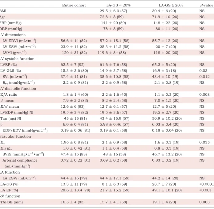
Demographic characteristics of the patient popu- lation are provided in Table 1. Median EF for the whole cohort was 63% (Q1:57%, Q3:68%). At the time of enrolment, all patients were highly symp- tomatic (88% in New York Heart Association func- tional class (NYHA) III-IV, 12% in NYHA II status);
however, when the echocardiographic examination and biochemical analyses were performed (in stable state 4–8 weeks after enrolment), the symp- toms were significantly alleviated (17% in NYHA III, 59% in NYHA II and 23% in NYHA I; 69% on diuretics).
Similar to prior studies in HFpEF, there was a slight over-representation of women (51% vs. 49%) and nearly half (41%) of the patients demonstrated severe obesity. Fifty-two patients (60%) had previ- ously been diagnosed with AF, of whom 21 were in AF at the time of the echocardiographic examina- tion.
ST2 levels and cardiac mechanics
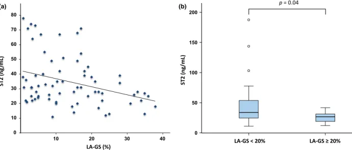
The median ST2 concentration in our cohort was 32 ng mL1 [Q1:24–Q3:48 ng mL1]. Serum ST2 levels inversely correlated with LA-GS (r=0.30, P=0.009, Fig. 1a). However, no association was found between ST2 and the degree of LA enlarge- ment (LA volume index, LAVi) or with indices of LV geometrical remodelling (LV mass index (LVMi), LV systolic and diastolic volumes), LV systolic func- tional parameters (LVEF, LV-GLS, Ees), measures of the LV relaxation and end-diastolic function (tau, b, EDP/EDV, E/e0) or indices of the AV- coupling and the systemic vascular function (Ea/Ees, arterial compliance, SVRi). Importantly,
Table 1 Demographic data General
Age years 7210
Gender male/female 42/44 (49/51) Medical history
Atrial fibrillation/flutter 52 (60)
Hypertension 68 (79)
Diabetes mellitus 28 (33)
COPD 17 (20)
Cancer 15 (17)
Coronary disease 13 (15)
NYHA I 19 (22)
NYHA II 46 (53)
NYHA III 19 (22)
NYHA IV 0
Clinical measurements
BMI kg m2 306
Obesity (BMI≥30) 34 (41)
SBP (mmHg) 14221
DBP (mmHg) 799
HR (beats/min) 7015
Treatment
ARB or ACE-I 65 (76)
Statin 37 (43)
Digoxin 10 (12)
Loop diuretic 59 (69)
Beta-blocker 67 (80)
Calcium channel blocker 26 (30) Laboratory findings
NT-proBNP (ng∗L1) 1000 (Q1:465;Q3:2335) ST2 (ng∗mL1) 32 (Q1:24;Q3:48) eGFR (mL∗min1/
1.73 m2)
70 (Q1:54;Q3:85)
Haemoglobin (g∗L1) 13.1 (Q1:12.2;Q3:14.2) White blood cell
count (109/L)
8.0 (Q1:7.1;Q3:9.9)
COPD, chronic obstructive pulmonary disease; NYHA, New York Heart Association functional class; BMI, body mass index; SBP, systolic blood pressure; DBP, diastolic blood pressure; HR, heart rate; ARB, angiotensin recep- tor blocker; ACE-I, ACE inhibitor; NT-proBNP, N-term- inal pro-brain natriuretic peptide; ST2, soluble suppression of tumorigenicity-2 receptor; eGFR, esti- mated glomerular filtration rate. Data are provided as absolute numbers followed by percentages in brackets;
or median values followed by 1st (Q1) and 3rd (Q3) quartiles in brackets
an inverse association of ST2 with RV function, as assessed by tricuspid annular plane systolic excur- sion (TAPSE), was demonstrated (r=0.28, P =0.01), whereas no correlation between ST2 and renal functional indices (s-Creatinine, eGFR) or CRP was observed.
Determinants of LA strain
The LA-GS did not show significant association with the LVMi or LV volumes. Although it was significantly related to the SV (r=0.23,P<0.05), no correlation with other indices of LV systolic performance (LVEF, LV-GLS; Ees) was evident.
Similarly, LA-GS was not associated with either the LV end-diastolic function as assessed by EDP/
EDV ratio, or thebvalue representing the slope of the EDPVR or the preload as estimated by E/e0. On the other hand, LA-GS was inversely related with LV afterload as described by Ea (r=0.28, P =0.01). Importantly, no significant relationship between LA-GS and LAVi was found. Similar to ST2, LA-GS was also significantly associated with TAPSE (r=0.41, P<0.001). In a multiple regres- sion analysis, including logST2, age, eGFR, LV-GLS, LAVi, E/e0 and Ea as potential predic- tors, only logST2 and eGFR were identified as inde- pendent predictors of the LA-GS (LA-GS=39.7–
5.4 ∗logST2-0.1139eGFR; P= 0.003). However, when the occurrence of AF was added in
the analysis, eGFR and AF only remained as predictors of LA-GS (LA-GS=29.3–7.8∗AF- 0.0929eGFR; P=0.001). When we restricted the above analysis to patients in sinus rhythm, similar to the entire population, only eGFR and logST2 acted as independent predictors of LA-GS (LA-GS=2.636–0.28∗logST2-0.1159eGFR;
P<0.001).
Association between HF symptoms and indices of LA and LV function In order to investigate the relationship between the functional status and cardiac performance, we dichotomized our study cohort into asymp- tomatic patients (NYHA I,n =19) and those with moderate to severe symptoms (NYHA II-III, n =65). LA-GS and ST2 were the only markers demonstrating significant difference between the two groups (LA-GS: 18.7 10.7 vs. 11.7 10.8%, P=0.01; ST2: 30.214.1 vs. 42.8 29.0 ng mL1, P=0.04, asymptomatic vs. symptomatic patients) (Fig. 1). On the other hand, neither the systolic (LV-GLS, SVi) nor the diastolic LV metrics (b, tau, EDP/EDV, E/e0) or AV-coupling and the vascular function indices (Ea, SVRi) differ signifi- cantly between the two groups (Fig. 2). Additionally, there was no difference in NYHA class between patients in sinus and those in AF during the examination (P=0.62).
0 10 20 30 40 50 60 70 80
10 20 30 40
p = 0.04
(a) (b)
Fig. 1 Left atrial strain and ST2 levels. (a) correlation between plasma soluble suppression of tumorigenicity-2 receptor (ST2) levels and left atrial strain (LA-GS) (r=0.3,P=0.009). (b) Comparison of ST2 levels between patients with LA- GS<20% and LA-GS≥20%.
LA strain and outcome
To investigate whether increased LA stiffness as assessed by LA-GS had a direct influence on patient prognosis, we dichotomized our patient cohort based on the LA-GS, using the third interquartile (LA-GS: 20%) as a cut-off value.
As shown in Table 2, patients with LA-GS<20%
displayed significantly lower SVi, LV-GLS and RV function along with higher Ea. Importantly, E/e0 values were similar in the two groups. ST2 and NT- proBNP levels were higher in patients with more reduced LA-GS, whereas eGFR did not significantly differ between the two groups.
Over a median follow-up of 572 days (IQR: 467–
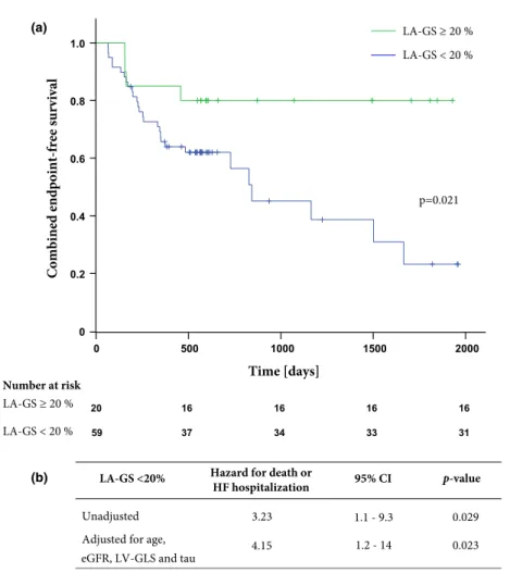
1369), 32 primary outcome events occurred (five deaths, 27 first HF hospitalizations). No patients were lost to follow-up. LA-GS<20% was associ- ated with an increased risk of the primary com- posite endpoint (P=0.02) in unadjusted analysis [odds ratio (OR) 3.23; confidence interval (CI) 1.1–
9.3, P=0.029]; Fig. 3a. After adjustment for age, eGFR, LV-GLS and tau, LA-GS remained an inde- pendent predictor of the outcome [OR: 4.15; CI:
1.2–14,P=0.023]; Fig. 3b.
As AF impacts on LA functional parameters, we proceed by further adjustment employing AF as covariate in the aforementioned regression model.
50
40
30
20
10
0
100
80
60
40
20
0
–25
–20
–15
–10
–5
0 200
150
100
50
0
NYHA I NYHA II – III NYHA I NYHA II – III
p = 0.224 p = 0.235
p = 0.01 p = 0.04
LA-GS [%] ST2 [ng/mL]
LAVi [mL/m2] LV-GLS [%]
NYHA II–III NYHA I NYHA II–III
NYHA I
(a) (b)
(c) (d)
Fig. 2 Association between heart failure symptoms, echocardiographic parameters and ST2 levels. Comparison of left atrial strain (LA-GS) (a), soluble suppression of tumorigenicity-2 receptor (ST2) levels (b), left atrial volume index (LAVi) (c) and left ventricular global longitudinal strain (LV-GLS) (d) between patient groups with (NYHA II-III) or without (NYHA I) heart failure symptoms. NYHA, New York Heart Failure Functional Classification.
LA-GS retained its significant predictive ability [OR: 4.56; CI: 1.3–15.7, P=0.016] although sig- nificant but weaker predictive capacity was even demonstrated for LV-GLS [P=0.042]; in contrast, neither the indices of AV-coupling (Ea,Ees,Ea/Ees) nor theE/e0demonstrated any predictive ability for death or hospitalization. In addition, selective analysis of the patients in sinus rhythm also showed that, after adjustment for age, eGFR, tau
and LV-GLS, LA-GS remained an independent predictor of outcome in this population [OR: 4.24;
CI: 1.2–14.9,P=0.019].
Discussion and conclusion
In the present prospective study, we demonstrate that in HFpEF: (i) pro-fibrotic changes as indicated by the ST2 biomarker are associated with
Table 2 Cardiac and vascular geometric and functional measures in the two groups stratified according to LA-GS.
Entire cohort LA-GS<20% LA-GS≥20% P-value
BMI 29.56.0 (57) 30.46 (20) NS
Age 72.88 (59) 71.910 (20) NS
SBP (mmHg) 14120 (59) 14822 (20) NS
DBP (mmHg) 788 (59) 8011 (20) NS
LV dimensions
LV EDVi (mL∗m2) 56.614 (82) 57.215.1 (58) 55.712 (20) NS
LV ESVi (mL∗m2) 23.911 (82) 25.311.2 (58) 207 (20) NS
LVMi (g∗m2) 12031 (82) 119.634 (58) 11820 (20) NS
LV systolic function
LVEF (%) 62.57 (82) 61.67.6 (58) 65.25 (20) NS
LV-GLS (%) 15.33.6 (80) 14.93.7 (58) 16.93 (18) 0.03
SVi (mL∗m2) 37.411 (81) 35.610.8 (58) 43.410 (19) 0.012
Ees(mmHg∗mL1) 2.20.9 (81) 2.20.9 (58) 2.10.8 (19) NS
LV diastolic function
E/A ratio 1.81.4 (60) 2.21.6 (40) 1.10.3 (20) 0.008
e0mean 7.92.2 (83) 8.22.4 (58) 7.01.5 (20) NS
E/e0mean 12.66 (83) 12.76.1 (57) 12.75 (20) NS
LVEDP (mmHg) NI 19.53.4 (82) 19.53.6 (57) 19.52.7 (20) NS
Tau (ms) NI 4515 (81) 43.415.9 (57) 50.910.2 (20) NS
b 6.00.4 (81) 5.980.46 (57) 6.030.4 (20) NS
EDP/EDV (mmHg∗mL1) 0.190.06 (81) 0.190.1 (58) 0.180.04 (20) NS Vascular function
Ea 1.960.8 (81) 2.10.9 (58) 1.60.3 (19) 0.035
Ea/Ees 1.00.42 (81) 1.10.4 (58) 0.80.3 (19) NS
SVRi (mmHg∗L1∗m2) 47.415 (83) 4816 (58) 46.713.2 (20) NS Arterial compliance
(mL∗mmHg1)
0.720.22 (81) 0.690.2 (58) 0.830.2 (19) NS
LA function
LA ESVi (mL∗m2) 44.416 (79) 44.417.1 (59) 44.214 (20) NS
LA-GS (%) 13.311 (79) 8.16.3 (59) 28.77 (20) <0.0001
LA EF (%) 28.618.4 (79) 21.715.2 (59) 49.110.1 (20) <0.001
RV function
TAPSE (mm) 16.54 (83) 15.74.1 (58) 19.14 (20) 0.003
mechanical alterations of the LA but not the LV;
and (ii) the LA strain comprised an independent predictor of death or hospitalization independently of the degree of LV remodelling or dysfunction.
The LA reservoir function is influenced by both LV systolic function and the intrinsic LA compliance and plays an important role in disease progression in various pathologies including AF, acute myocar- dial ischaemia and HF. LA strain is an emerging noninvasive method for the quantification of LA reservoir function [6].
Left atrium strain has been shown to reflect the extent of LA fibrosis in various pathological states.
Kuppahally et al. [22] assessed the degree of LA wall fibrosis by delayed-enhancement MRI in AF patients and found that LA strain inversely asso- ciated with the degree of LA fibrosis. In another report, in patients undergoing mitral valve surgery, preoperatively measured LA strain was the stron- gest independent predictor of the degree of histopathologically quantified LA wall fibrosis [23].
In our study, ST2, an established pro-fibrotic marker, significantly associated with LA-GS. Con- versely, measures of the systolic, early diastolic and late diastolic LV function or those of the AV- coupling and vascular function were not related to ST2 levels. In agreement with our results, previous studies in HFpEF failed to demonstrate any rela- tionship between ST2 levels and LV echocardio- graphic parameters [14, 24]. Even though ST2 has
repeatedly been shown to be a reliable marker of disease severity in HFpEF, the aforementioned observation has led to the misconception that elevated ST2 might barely indicate systemic inflammation, rather than reflect direct cardiac alterations [14, 25]. No studies, however, have specifically investigated the association between LA function and ST2 levels in HFpEF.
The LV contraction towards the apex is expected to act as a major determinant of the LA deformation during systole [26]. In our cohort, however, no association between the LV longitudinal deforma- tion and LA-GS was found. This might be explained by disparate responses of the LA and the LV to inflammation, as myocardial remodelling at the atrial level has been shown to involve differential pathophysiologic pathways from the LV. In a tachycardia-induced HF model, considerably dif- ferent cellular responses were observed in these two chambers, with more pronounced inflamma- tory and pro-fibrotic reaction detected in the LA as compared to the LV wall [27]. In another study, angiotensin II infusion resulted in progressive LA fibrosis that was independent of LV wall stress but directly related to circulating hormone levels [28].
Conceivably, due to its thinner wall, the LA might be more susceptible to myocardial fibrosis and exhibit more apparent mechanical changes as compared to LV. Our results advocate that LA-GS comprises a surrogate marker of LA mechanical changes partly ascribed to a pro-fibrotic reaction and imply that LA structural and functional Table 2 (Continued)
Entire cohort LA-GS<20% LA-GS≥20% P-value
Biochemical
NT-proBNP (ng∗L1) 1400 (Q1:556; Q3:2633) 495 (Q1:430; Q3:822) 0.003
ST2 (ng∗mL1) 35.0 (Q1:25; Q3:55) 26.7 (Q1:19; Q3:33) 0.003
eGFR (mL∗min1/1.73 m2) 70 (Q1:56; Q3:86) 59.5 (Q1:45; Q3:80) NS BMI, body mass index; SBP, systolic blood pressure; DBP, diastolic blood pressure; LV, left ventricle; EDVi, end-diastolic volume index; ESVi, end-systolic volume index; LVMi, LV mass index; EF, ejection fraction; LV-GLS, LV global longitudinal strain; SVi, stroke volume index;Ees, LV end-systolic elastance;E/A, ratio between the early diastolic inflow velocity (E) to the inflow velocity due to atrial contraction (A);e0mean, mean value of early myocardial velocity in LV basal septal and lateral wall;E/e0, ratio between theE and thee0; EDP, end-diastolic pressure; NI, noninvasive; tau, time constant of LV isovolumic relaxation;b, diastolic stiffness constant describing the steepness of the EDPVR curve; EDP/
EDV, end-diastolic pressure-to-end-diastolic volume ratio; Ea, effective arterial elastance; SVRi, systemic vascular resistance index; LA, left atrium; LA ESVi, left atrial end-systolic volume; LA-GS, left atrial global longitudinal strain; LA EF, left atrial ejection fraction; TAPSE, tricuspid annular plane systolic excursion; NT-proBNP, N-terminal pro-brain natriuretic peptide; ST2, soluble suppression of tumorigenicity-2 receptor; eGFR, estimated glomerular filtration rate; NS, nonsignificant (P≥0.05). Data are provided as meanSD followed by patient number in brackets; or median values followed by 1st and 3rd quartiles in brackets.
abnormalities might develop on the ground of intrinsic LA alterations, independently of LV dys- function.
Normally, the distensible LA accommodates blood from the pulmonary veins without a considerable rise in the LA pressure (LAP). In cases of reduced LA compliance, as in LA fibrosis, the LA pressure–
volume curve is shifted upwards resulting in dis- proportional rise of LAP for the same volume entering the chamber. Chronically elevated LAP leads to LA enlargement. Based on physical prin- ciples, increased LA volume would mitigate the elevated wall stress and accordingly the LAP.
Paradoxically however, the degree of LA remod- elling has been shown to be positively related to the
severity of pulmonary hypertension [29]. This observation can be physiologically explained by concomitantly occurring LA wall fibrosis, which counteracts the alleviating effect of LA volume increase. Of note, in this study, LAVi was not associated with either ST2 levels or the LA-GS.
Similar findings were also reported by others, suggesting that the degree of LA fibrosis is not solely or even primarily determined by the degree of LA enlargement [6, 10], which supports our hypothesis that LA fibrosis, as reflected by elevated ST2, might be a result of an inflammatory process rather than haemodynamic overload. In our study, LA enlargement was not associated with the patients’ functional class either, whereas a signif- icant relationship between LA-GS and symptom
2000 1500
1000 500
0 1.0
0.8
0.6
0.4
0.2
0
-GS -GS <
21
Number at risk LA-GS 20 % LA-GS < 20 %
16 16
16 16
20
31 33
34 37
59
Time [days]
Combined endpoint-free survival
LA-GS <20% Hazard for death or
HF hospitalization 95% CI p-value Unadjusted
Adjusted for age, eGFR, LV-GLS and tau
3.23 4.15
1.1 - 9.3 0.029
1.2 - 14 0.023
(a)
(b)
Fig. 3 Left atrial strain and patient outcome. (a) Kaplan–Meier analysis of patients stratified by left atrial strain (LA-GS).
Group I, LA-GS<20%; Group II, LA-GS≥20%. (b) Hazard ratio for death or heart failure (HF) hospitalization for patients with LA-GS<20% compared to LA-GS≥20%. eGFR, estimated glomerular filtration rate; LV-GLS, left ventricular global longitudinal strain; tau, time constant of LV isovolumic relaxation; CI, confidence interval.
severity was observed. Similar association was also found between ST2 levels and symptomatology, further supporting the notion of the potential impact of inflammatory activity on LA mechanics.
LV diastolic properties have been extensively stud- ied in regard to their association with symptoms in HFpEF. Employing measurements with conduc- tance catheters, Liu et al. [4] demonstrated that HFpEF patients display increased LV end-diastolic stiffness and impaired relaxation as compared to healthy controls. These findings were confirmed in a larger scale study in which noninvasive estimates of LV relaxation and stiffness were used [8]. In another report, however, early relaxation and LV end-diastolic stiffness were similar between HFpEF patients and healthy subjects at rest [30] and elevated EDP in HFpEF was not accompanied by increasedb values advocating for the influence of extracardiac forces rather than passive LV stiffness on the elevated filling pressures. In our study, indices of LV relaxation and diastolic stiffness did not correlate with ST2 levels. However, there was a weak association between tau and LA-GS suggest- ing that apart from fibrotic changes, mechanical alterations in the LV function during the relaxation phase may also influence the LA mechanics.
LA strain and prognosis
To date, few studies have attempted to assess the predictive value of LA function regarding outcome in HFpEF. Recently, Melenovsky et al.[7] demon- strated that LA EF was an independent predictor of mortality in HFpEF. In another study, Santos and colleagues showed that reduced LA strain implied an increased risk of HF hospitalization in HFpEF patients; however, it did not remain prognostic after adjustment for LV deformation and the E/e0 [31]. Accordingly, the authors conclude that the predictive ability of LA strain is to be attributed merely to its association with LV performance [31].
Importantly, our findings contrast this observation as we show that LA-GS independently predicted outcome even when adjusted for measurements of LV longitudinal systolic deformation and diastolic performance as well as for indices of atrioventric- ular coupling. These disparities might be explained by differences in patient profile. Our cohort con- sisted of older patients, in whom the structural and functional LA indices as well as the higher preva- lence of AF indicated more advanced disease. Also, in the present work, a dedicated software for LA strain analysis was employed, presumably yielding
more representative measurements, as compared to earlier studies that applied LV strain measuring algorithm for LA strain analysis.
Importantly, in our study other well-established indices such as those describing the AV-coupling and theE/e0did not demonstrate significant prog- nostic value. The later noninvasive marker of LA filling pressures has been shown to entail signifi- cant predictive value in HFpEF. Our findings indicate that LA strain, reflecting not merely the haemodynamic filling state but, as previously dis- cussed, also the degree of pro-fibrotic alterations, constitutes a more robust marker of disease sever- ity in this clinical condition.
AF is a common condition in HFpEF that impor- tantly influences LA-GS measurements and thus could be a concern for the reliability and utility of LA-GS measurement in these patients. In order to rule out the confounding effect of AF on LA-GS measurement, we tested the association of ST2 with LA-GS confining our analysis to patients in sinus rhythm. ST2 showed a significant correla- tion with LA-GS in these patients also. Similarly, the independent prognostic value of LA-GS for outcome was maintained in this subgroup of patients.
Limitations
LA strain is a more and more widely used nonin- vasive metric of LA reservoir function; however, the actual haemodynamic meaning of LA strain is rather ambiguous. Although the LA deformation during systole is expected to represent a surrogate of LA compliance, LA strain is also influenced by other components of the cardiac mechanics. A more accurate assessment of the LA reservoir function would require direct measurement of LA stiffness by an invasive approach, preferably with micromanometric catheters. On the other hand, the obvious correlations found with both ST2 levels and patient outcome speak for the utility of LA strain, as a readily obtainable metric providing clinically important information. The relatively limited size of our study, as well as the fact, that of the 32 outcome events that occurred during follow-up, 27 were heart failure hospital- izations, warrants larger scale investigations to confirm our results. At the same time, the prog- nostic information evident even at this patient number corroborates the clinical significance of our findings.
Conclusions
Our results indicate that ST2 is a sensitive marker of LA dysfunction in HFpEF, elevation of which may specifically reflect LA mechanical alterations, independently of LV performance. Impaired LA strain remained an independent predictor of HF hospitalization and mortality, even after adjust- ment for clinical variables and LV functional indices.
Considering the fact that currently no effective therapy for HFpEF is available, a reliable tool for monitoring the evolution of this disease is of major clinical significance for the timely recogni- tion and thus prevention of patients at increased risk, prior to the development of irreversible LA remodelling.
Sources of Funding
This project was supported by the Janos Bolyai Scholarship of the Hungarian Academy of Sciences.
Conflict of Interest None.
References
1 Owan TE, Hodge DO, Herges RMet al.Trends in prevalence and outcome of heart failure with preserved ejection fraction.
N Engl J Med2006;355:251–9.
2 Zile MR, Gottdiener JS, Hetzel SJ et al. Prevalence and significance of alterations in cardiac structure and function in patients with heart failure and a preserved ejection fraction.Circulation2011;124:2491–501.
3 Zile MR, Baicu CF, Gaasch WH. Diastolic heart failure–
abnormalities in active relaxation and passive stiffness of the left ventricle.N Engl J Med2004;350:1953–9.
4 Liu CP, Ting CT, Lawrence Wet al.Diminished contractile response to increased heart rate in intact human left ventricular hypertrophy. Systolic versus diastolic determi- nants.Circulation1993;88:1893–906.
5 Redfield MM, Jacobsen SJ, Burnett JC Jret al.Burden of systolic and diastolic ventricular dysfunction in the commu- nity: appreciating the scope of the heart failure epidemic.
JAMA2003;289:194–202.
6 Santos AB, Kraigher-Krainer E, Gupta DKet al.Impaired left atrial function in heart failure with preserved ejection frac- tion.Eur J Heart Fail2014;16:1096–103.
7 Melenovsky V, Hwang SJ, Redfield MM et al. Left atrial remodeling and function in advanced heart failure with preserved or reduced ejection fraction.Circ Heart Fail2015;
8:295–303.
8 Lam CS, Roger VL, Rodeheffer RJet al.Cardiac structure and ventricular-vascular function in persons with heart failure and preserved ejection fraction from Olmsted County, Min- nesota.Circulation2007;115:1982–90.
9 Rossi A, Gheorghiade M, Triposkiadis Fet al.Left atrium in heart failure with preserved ejection fraction: structure, function, and significance.Circ Heart Fail2014;7:1042–9.
10 Melenovsky V, Borlaug BA, Rosen B et al.Cardiovascular features of heart failure with preserved ejection fraction versus nonfailing hypertensive left ventricular hypertrophy in the urban Baltimore community: the role of atrial remod- eling/dysfunction.J Am Coll Cardiol2007;49:198–207.
11 Paulus WJ, Tschope C. A novel paradigm for heart failure with preserved ejection fraction: comorbidities drive myocardial dysfunction and remodeling through coronary microvascular endothelial inflammation.J Am Coll Cardiol2013;62:263– 71.
12 de Boer RA, Daniels LB, Maisel AS, Januzzi JL Jr. State of the Art: Newer biomarkers in heart failure.Eur J Heart Fail2015;
17:559–69.
13 Zhou H, Ni J, Yuan Yet al.Soluble ST2 may possess special superiority as a risk predictor in heart failure patients.Int J Cardiol2015;186:146–7.
14 AbouEzzeddine OF, McKie PM, Dunlay SMet al.Suppression of tumorigenicity 2 in heart failure with preserved ejection fraction.J Am Heart Assoc2017;6:e004382.
15 Donal E, Lund LH, Linde Cet al.Rationale and design of the Karolinska-Rennes (KaRen) prospective study of dyssyn- chrony in heart failure with preserved ejection fraction.Eur J Heart Fail2009;11:198–204.
16 Kelly RP, Ting CT, Yang TMet al.Effective arterial elastance as index of arterial vascular load in humans. Circulation 1992;86:513–21.
17 Chen CH, Fetics B, Nevo E et al. Noninvasive single-beat determination of left ventricular end-systolic elastance in humans.J Am Coll Cardiol2001;38:2028–34.
18 Chemla D, Hebert JL, Coirault Cet al.Total arterial compli- ance estimated by stroke volume-to-aortic pulse pressure ratio in humans.Am J Physiol1998;274:H500–5.
19 Ommen SR, Nishimura RA, Appleton CPet al.Clinical utility of Doppler echocardiography and tissue Doppler imaging in the estimation of left ventricular filling pressures: A compar- ative simultaneous Doppler-catheterization study.Circulation 2000;102:1788–94.
20 Klotz S, Hay I, Dickstein MLet al.Single-beat estimation of end-diastolic pressure-volume relationship: a novel method with potential for noninvasive application.Am J Physiol Heart Circ Physiol2006;291:H403–12.
21 Dahlberg G. Statistical Methods for Medical and Biological Students. London: G. Allen & Unwin Ltd. 1940.
22 Kuppahally SS, Akoum N, Burgon NSet al.Left atrial strain and strain rate in patients with paroxysmal and persistent atrial fibrillation: relationship to left atrial structural remod- eling detected by delayed-enhancement MRI.Circ Cardiovasc Imaging2010;3:231–9.
23 Cameli M, Lisi M, Righini FM et al. Usefulness of atrial deformation analysis to predict left atrial fibrosis and endo- cardial thickness in patients undergoing mitral valve opera- tions for severe mitral regurgitation secondary to mitral valve prolapse.Am J Cardiol2013;111:595–601.
24 Zile MR, Jhund PS, Baicu CF et al. Plasma biomarkers reflecting profibrotic processes in heart failure with a
preserved ejection fraction: data from the prospective com- parison of ARNI with ARB on management of heart failure with preserved ejection fraction study.Circ Heart Fail2016;
9:e002551.
25 Bartunek J, Delrue L, Van Durme Fet al. Nonmyocardial production of ST2 protein in human hypertrophy and failure is related to diastolic load.J Am Coll Cardiol2008;52:2166–
74.
26 Appleton CP, Kovacs SJ. The role of left atrial function in diastolic heart failure.Circ Cardiovasc Imaging2009;2:6–9.
27 Hanna N, Cardin S, Leung TK, Nattel S. Differences in atrial versus ventricular remodeling in dogs with ventricular tachy- pacing-induced congestive heart failure. Cardiovasc Res 2004;63:236–44.
28 Sun Y, Ramires FJ, Weber KT. Fibrosis of atria and great vessels in response to angiotensin II or aldosterone infusion.
Cardiovasc Res1997;35:138–47.
29 Le Tourneau T, Richardson M, Juthier Fet al.Echocardiog- raphy predictors and prognostic value of pulmonary artery systolic pressure in chronic organic mitral regurgitation.
Heart2010;96:1311–7.
30 Kawaguchi M, Hay I, Fetics B, Kass DA. Combined ventricular systolic and arterial stiffening in patients with heart failure and preserved ejection fraction: implications for systolic and diastolic reserve limitations.Circulation2003;107:714–20.
31 Santos AB, Roca GQ, Claggett Bet al.Prognostic relevance of left atrial dysfunction in heart failure with preserved ejection fraction.Circ Heart Fail2016;9:e002763.
Correspondence: Aniko Ilona Nagy, Semmelweis University, Heart and Vascular Center, 68. Varosmajor u., 1122 Budapest, Hungary.
(phone: +36-20-8259738; e-mail: anychophora@gmail.com)
Supporting Information
Additional Supporting Information may be found in the online version of this article:
Figure S1. Diagram illustrating the temporal sequence of study events.