Review Article
Molecular Mechanisms of Neurodegeneration Related to C9orf72 Hexanucleotide Repeat Expansion
Mirjana Babi ć Leko,
1Vera Ž upunski,
2Jason Kirincich ,
1Dinko Smilovi ć ,
1Tibor Hortobágyi ,
3,4Patrick R. Hof,
5and Goran Š imi ć
11Department of Neuroscience, Croatian Institute for Brain Research, University of Zagreb School of Medicine, Zagreb, Croatia
2Department of Biochemistry, Faculty of Chemistry and Chemical Technology, University of Ljubljana, Ljubljana, Slovenia
3Institute of Pathology, Faculty of Medicine, University of Szeged, Szeged, Hungary
4MTA-DE Cerebrovascular and Neurodegenerative Research Group, University of Debrecen, Debrecen, Hungary
5Fishberg Department of Neuroscience, Friedman Brain Institute, Ronald M. Loeb Center for Alzheimer’s Disease, Icahn School of Medicine at Mount Sinai, New York, USA
Correspondence should be addressed to GoranŠimić; gsimic@hiim.hr
Received 7 June 2018; Revised 28 August 2018; Accepted 18 September 2018; Published 15 January 2019 Guest Editor: Edoardo G. Spinelli
Copyright © 2019 Mirjana BabićLeko et al. This is an open access article distributed under the Creative Commons Attribution License, which permits unrestricted use, distribution, and reproduction in any medium, provided the original work is properly cited.
Two clinically distinct diseases, amyotrophic lateral sclerosis (ALS) and frontotemporal dementia (FTD), have recently been classified as two extremes of the FTD/ALS spectrum. The neuropathological correlate of FTD is frontotemporal lobar degeneration (FTLD), characterized by tau-, TDP-43-, and FUS-immunoreactive neuronal inclusions. An earlier discovery that a hexanucleotide repeat expansion mutation in chromosome 9 open reading frame 72 (C9orf72) gene causes ALS and FTD established a special subtype of ALS and FTLD with TDP-43 pathology (C9FTD/ALS). Normal individuals carry 2–10 hexanucleotide GGGGCC repeats in the C9orf72gene, while more than a few hundred repeats represent a risk for ALS and FTD. The proposed molecular mechanisms by whichC9orf72repeat expansions induce neurodegenerative changes are C9orf72 loss-of-function through haploinsufficiency, RNA toxic gain-of-function, and gain-of-function through the accumulation of toxic dipeptide repeat proteins. However, many more cellular processes are affected by pathological processes in C9FTD/ALS, including nucleocytoplasmic transport, RNA processing, normal function of nucleolus, formation of membraneless organelles, translation, ubiquitin proteasome system, Notch signalling pathway, granule transport, and normal function of TAR DNA- binding protein 43 (TDP-43). Although the exact molecular mechanisms through whichC9orf72repeat expansions account for neurodegeneration have not been elucidated, some potential therapeutics, such as antisense oligonucleotides targeting hexanucleotide GGGGCC repeats in mRNA, were successful in preclinical trials and are awaiting phase 1 clinical trials. In this review, we critically discuss each proposed mechanism and provide insight into the most recent studies aiming to elucidate the molecular underpinnings of C9FTD/ALS.
1. Introduction
Amyotrophic lateral sclerosis (ALS) and frontotemporal dementia (FTD) are two clinically distinct entities. ALS, also known as motor neuron disease (MND), Lou Gehrig
’s dis- ease, and Charcot disease, a
ffects both upper and lower motor neurons leading to hyperre
flexia, spasticity, fascicula- tions, and muscular atrophy [1]. The disease onset is mostly after 60 years of age with a prevalence of around 5 cases per
100,000 [2]. ALS patients can be divided into subgroups according to neuropsychological deficits: ALS with cognitive impairment (ALS-ci), ALS with behavioral impairment (ALS-bi), and ALS with combined cognitive and behavioral impairment (ALS-cbi) [3]. In FTD, neurodegeneration a
ffects the frontal and temporal lobes causing frontotemporal lobar degeneration (FTLD) and is associated with changes in behavior and personality, de
ficits in frontal executive func- tions, and language impairment [3]. FTD is considered to
Volume 2019, Article ID 2909168, 18 pages https://doi.org/10.1155/2019/2909168
be one of the most common forms of dementia in the popu- lation under 65 years of age with the prevalence around 10 cases per 100,000 [4]. FTD patients are divided in three clin- ical syndromes according to their symptomatology: two lan- guage variants (progressive non
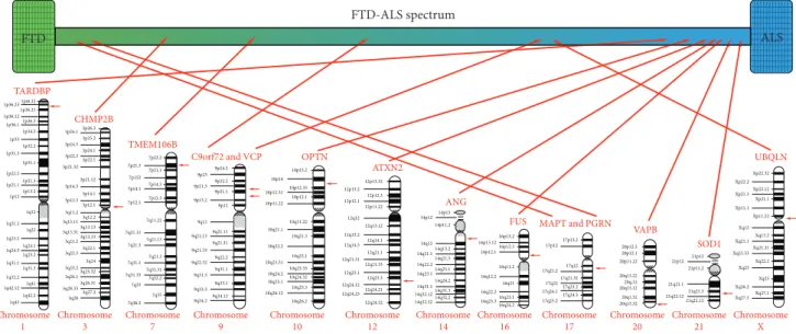
fluent aphasia (PNFA) and semantic dementia (SD)), and behavioral variant of fronto- temporal dementia (bvFTD) [3]. Based on the clinical, genetic, and neuropathological overlap between ALS and FTD, these two diseases are now considered as two extremes of the FTD/ALS spectrum (Figure 1) [1]. About 15% of FTD patients show symptoms of ALS, and up to 50% of ALS patients have symptoms of FTD [5].
2. Main Features of ALS and FTLD-TDP Neuropathology
The vast majority of ALS cases (ALS-TDP) and the most common FTD pathological subtype (FTLD-TDP) show TDP-43 (TAR DNA-binding protein 43) immunoreactive aggregates forming characteristic inclusions (for overview see [6, 7]). The hallmarks of ALS with TDP-43 pathology (ALS-TDP) are skein-like, granular, and compact inclusions in motor neurons, whereas in FTLD-TDP, there are four dis- tinct morphological types (A
–D) with characteristic distribu- tion and morphology of dystrophic neurites, cytoplasmic, and intranuclear inclusions. There is a good correlation with both clinical phenotype and genetic alterations [8]. For example, cases with hexanucleotide repeat expansions in C9orf72 are typically associated with type A and type B pathology, whereas mutations in the progranulin gene GRN and valosin-containing protein gene (VCP) are associated with type A and type D, respectively. Regarding the correla- tion with clinical phenotypes, for example, PNFA is associ- ated with type A, SD with type C, and inclusion body myopathy with frontotemporal dementia (IBMFD) with type D. In both ALS and FTLD, there is glial pathology mainly a
ffecting oligodendroglia, with TDP-43-immunoreactive cytoplasmic inclusions [6, 7]. While most of the ALS cases
are sporadic, about half of all FTD patients show a familial pattern of inheritance linked to mutations in several different genes [6, 9–11].
3. Hexanucleotide Repeat Expansion in C9orf72 Gene
A mutation in chromosome 9 open reading frame 72 (C9orf72) has been identified as a common genetic cause of both ALS and FTD. The link of ALS and FTD to chromo- some 9 was
first reported in 2006 in two independent studies[12, 13]. It was later shown that the mutation is an expansion of GGGGCC (G4C2) hexanucleotide repeat in a noncoding region of the C9orf72 gene [14
–16], which is the most com- mon genetic cause of ALS and FTD (so called C9FTD/ALS) in certain populations.
In humans, three transcript variants of C9orf72 have been identified. Transcript variants 2 and 3 give rise to identical protein isoforms of 481 amino acids, while the 3′ site of transcript variant 1 is truncated and encodes an isoform of 222 amino acids. The hexanucleotide repeat is located within the promoter region for transcript variant 2, which corresponds to the position of the
first intron in transcript variants 1 and 3 depending on the transcription start site used [17, 18].
While the number of G4C2 repeat units in the DNA of healthy individuals is up to 25, the number of repeats in the DNA of ALS and FTD patients is usually 400 to several thou- sand [14–16, 19–21]. A small percentage of patients have shorter expansions, from 45–80 repeats [19], and an even shorter expansion, around 30 repeats, has been associated with the disease [15]. Notably, there is an apparent gap between short pathogenic repeat sizes of 45 to 80 and long expansions from 400 to several thousand units. This is likely due to high genomic instability of the intermediate long repeats, which may have a tendency to either expand or contract [19]. Interestingly, longer expansions have been
FTD-ALS spectrum
FTD ALS
TARDBP
CHMP2B
TMEM106B
C9orf72 and VCP OPTN
ATXN2
ANG
FUS MAPT and PGRN VAPB
SOD1
UBQLN
Chromosome 1
Chromosome 3
Chromosome 7
Chromosome 9
Chromosome 10
Chromosome 12
Chromosome 14
Chromosome 16
Chromosome 17
Chromosome 20
Chromosome 21
Chromosome X
1pt6.32 1p36.23
1p36.21 1p36.12
1p36.3 1p36.1
1p34.2 1p33
1p32.2 1p31.3
1p31.1 1p22.2
1p21.3 1p21.1
1p13.2 1p12
1q12 1q21.2
1q22 1q23.2
1q24.1 1q24.3
1q25.2 1q31.1
1q31.3 1q32.2
1q41 1q42.12
1q42.2 1q43
3p26.3 3p26.1
3p25.2 3p24.3
3p24.1 3p22.3
3p22.1 3p21.32
3p21.12 3p14.3
3p14.1 3p12.3
3p12.1 3q11.2 3q13.11
3q12.2 3q13.13 3q13.31
3q13.33 3q21.2
3q22.1 3q22.3
3q24 3q25.2
3q25.32 3q26.1
3q26.31 3q28.33
3q28 3q27.2
7p22.2 7p21.3
7p21.1 7p152
7p14.3 7p14.1
7p12.3 7p12.1
7q11.22 7q21.11
7q21.13 7q21.3
7q21.3 7q31.1
7q31.31 7q31.33
7q32.2 7q33
7q35 7q36.2
9p24.2 9p23
9p22.2 9p21.3
9p21.1 9p13.2
9p12
9q12 9q21.11 9q21.13
9q21.31 9q21.33
9q22.2 9q22.32
9q31.1 9q31.3
9q33.1 9q33.3
9q34.12 9q34.2
10p15.2 10p14
10p12.33 10p12.31
10p12.1 10p11.22
10q11.22 10q21.1
10q21.3 10q22.2
10q23.1 10q23.31
10q23.33 10q24.2
10q24.32 10q25.1
10q25.3 10q26.12
10q26.2
12p13.32 12p13.2
12p12.3 12p12.1
12p11.22 12q12
12q13.12 12q13.2
12q14.1 12q14.3
12q21.1 12q21.31
12q21.33 12q23.1
12q23.3 12q24.12
12q24.21 12q24.23
12q24.32
14p13 14p12
14p11.2
14q12 14q13.2 14q21.1
14q21.3 14q22.2
14q23.1 14q23.1
14q24.2 14q31.1
14q31.3 14q32.12
14q32.2 14q32.32
16p13.2
17p13.2 17p12
17q12 17q21.2
17q21.32 17q22
17q23.2 17q24.1
17q24.3 17q25.2 16p13.12
16p12.3 16p12.1
16q11.2 16q12.2
16q21 16q22.2
16q23.1 16q23.3
16q24.2
20p12.3 20p12.1 20p11.22 20q11.22 20q.12 20q13.12 20q1.32 20q13.32
21p13 21p12
21p11.2
21q21.1 21q21.3 21q22.12
21q22.12
Xp22.32 Xp22.2
Xp22.12 Xp21.1 Xp21.3 Xp11.1
Xp11.22 Xq12
Xq13.2 Xq21.1 Xq21.33
Xq22.2 Xq23
Xq25 Xq26.2
Xq27.1 Xq27.3
Xq21.31
Figure1: Influence of different genes on FTD/ALS clinical spectrum.
recently correlated with an earlier onset of disease [19]. In contrast, other studies detected either positive correlation [22–25] or no association [22, 25] between disease severity and expansion size. Additionally, the results of these studies were variable depending on the tissue in which expansion size was measured [22, 25].
4. C9orf72 Protein
The function of C9orf72 protein is still unclear. Bioinformat- ics predictions suggest that C9orf72 belongs to the family of DENN (differentially expressed in normal and neoplastic cells) proteins, which function as RabGEF (guanine exchange factor) regulators of membrane tra
fficking by activating RabGTPases [26, 27]. RabGEF facilitates a release of GDP from Rab and exchanges it for GTP. To support the role of C9orf72 as RabGEF, a study conducted on neuronal cell lines and human spinal cord tissue revealed that it was colocalized and coprecipitated with Rab proteins [28]. Therefore, C9orf72 could be involved in Rab-mediated cellular traffick- ing and protein degradation. Additionally, C9orf72 protein was detected at presynaptic sites, and as it interacts with Rab proteins, it is believed that C9orf72 could regulate synap- tic vesicles as RabGEF for RAB3 proteins [29]. Xiao et al.
detected interactions of C9orf72 with di
fferent components of the nuclear pore complex (NPC) and nuclear receptors in human brain tissue, indicating a possible role of C9orf72 in nucleocytoplasmic tra
fficking [30]. Nuclear transport is compromised in di
fferent C9FTD/ALS models (such as Drosophila, induced pluripotent stem cells (iPSC) derived neurons, human brain tissue, yeast cells, mouse primary neu- rons, and primary human dermal
fibroblasts) [31–33]. Asknockdown of C9orf72 leads to disruption in endosomal trafficking and formation of autophagosome [28], it is pro- posed that C9orf72 may be also involved in the regulation of endosomal tra
fficking and autophagy. Additional studies established the involvement of C9orf72 in the regulation of autophagy [34
–36]. In studies conducted on cell lines, it was also shown that C9orf72 could be involved in the regula- tion of lysosomal function [37, 38] and in the formation of stress granules [39]. In mice, C9orf72 is required for the nor- mal macrophage and microglial function [40]. It was shown that decreased levels of C9orf72 cause dysfunctional microg- lia that are related to neurodegeneration [40].
5. Neuropathological Features of C9FTD/ALS Cases with hexanucleotide repeat expansion in C9orf72 are typically associated with type A and B FTLD-TDP pathology, as previously mentioned. The neuropathology of C9FTD/ALS shows pathognomonic ubiquitin- and p62- positive and, rarely, TDP-43-containing inclusions in the cerebellum (Purkinje cells and granular cells) and hippo- campus [41, 42]. A unique feature is the presence of aggre- gating dipeptide repeat proteins within a proportion of inclusion bodies, some of which may not show phosphory- lated TDP-43 (pTDP-43) immunoreactivity [43, 44]. Ultra- structurally, the characteristic inclusions in FTLD and ALS with C9orf72 mutation are granular and
filamentous [45].6. Potential Mechanisms of C9orf72 Hexanucleotide Repeat Expansion- Mediated Neurodegeneration
Three mechanisms have been proposed for G4C2 hexanu- cleotide repeat expansion (HRE) in C9orf72 to induce neuro- degenerative changes: (1) loss of C9orf72 function through haploinsufficiency, (2) toxic gain-of-function due to the generation of aberrant HRE-containing RNA, and (3) toxic gain-of-function through the accumulation of dipeptide repeat proteins (DPR) translated from hexanucleotide repeat RNA. Potential mechanisms by which C9orf72 repeat expan- sions result in neurodegeneration are summarized in Figure 2. Microphotograph with characteristic histopatho- logical changes is given in Figure 3. Studies investigating these mechanisms are summarized in Table 1.
6.1. Loss of C9orf72 Function through Haploinsu
fficiency.
Carriers of C9orf72 HRE have decreased levels of C9orf72 transcripts [14, 16], which are also reflected in the decreased C9orf72 protein levels in the frontal and temporal cortex [20], presumably due to the loss of transcription from the mutant allele carrying the HRE. A systematic study of C9orf72 levels in patients with HRE revealed decreased levels of C9orf72 transcripts in comparison to non-HRE patients and controls [46]. Interestingly, the levels of the long C9orf72 protein isoform were decreased in the brain, while levels of the short C9orf72 protein isoform were increased (as detected by Western blot). Immunohistochemical analy- sis of spinal motor neurons showed decreased expression of short C9orf72 protein isoform on nuclear membrane in C9ALS cases compared to controls, while subcellular locali- zation of long C9orf72 protein isoform was unchanged [30]. Another study detected 80% reduction of long C9orf72 protein isoform in the cerebellum of C9orf72 HRE carriers in comparison to controls [29]. C9orf72 hexanucleo- tide repeat expansion might induce DNA hypermethylation and consequently lead to decreased C9orf72 transcription [47]. HRE is methylated when the number of repeats is larger than 90 [48]. Moreover, the DNA from a fraction of C9orf72 HRE carriers is also methylated in the 5′ CpG island [47, 49, 50]. The repeat expansion-induced DNA methyla- tion in the C9orf72 promoter region, which results in down- regulated C9orf72 transcription, therefore provides a likely explanation for the association between the size of repeat expansion and the age of onset of the disease [19]. More pre- cisely, the authors tested C9orf72 promoter activity in human kidney and neuroblastoma cell lines and observed reduced C9orf72 transcription in cells with larger repeats and increased methylation. Thus, they proposed that higher methylation of C9orf72 promoter may be an explanation of how repeat expansion could lead to loss-of-function, without excluding the possibility that repeat expansion could also lead to toxic gain-of-function [19]. There is also a possibility that higher-order DNA structures formed on G4C2 repeats could lead to abortive transcription of C9orf72 and therefore be the cause for decreased C9orf72 transcription [51].
C9orf72 loss-of-function in C. elegans and zebra
fish
models results in motor neuron degeneration, indicating a
possible disease mechanism [52, 53]. However, human C9orf72 gene has only partial homology with its ortholog in C. elegans and zebra
fish. Thus, loss-of-function mechanisms were assessed in C9orf72 knockout mice as the mouse ortho- log of C9orf72 gene is more similar to the human C9orf72
gene [18]. Various knockout and knockdown models have been developed, yet none of the mouse models showed phe- notypes characteristic for ALS or FTD [35, 40, 54–57], indi- cating that C9orf72 loss-of-function is not the true cause of disease. The observation that patients homozygous for C9orf72 repeat expansion do not have more severe symptoms of disease compared to heterozygotes further supports these
findings [58, 59] (Table 2).6.2. Toxic Gain-of-Function due to Aberrant HRE-Containing RNA
6.2.1. Higher-Order Structures of DNA and RNA Formed by C9orf72 HRE Sequence. Due to the uniformity of the G4C2 sequence and the abundance of guanine nucleotides, expanded hexanucleotide repeats in the C9orf72 gene form higher-order DNA structures called G-quadruplexes [51, 60].
G-quadruplexes formed in the DNA can adopt both parallel- and antiparallel-stranded conformations, and increasing the length of the repeats creates a heterogeneous mixture of these structures. Both C-rich sense and antisense strands can assemble as i-motifs and hairpin structures [61]. G- quadruplex structures formed in the C9orf72 HRE region
Figure3: TDP-43-immunoreactive cytoplasmic inclusions, finelygranular aggregates, and lack of nuclear labelling in a spinal cord motoneuron of a patient with ALS caused by C9orf72 hexanucleotide repeat expansion.
G G
G G G G G
G G G G
G G G G
G G
G G G G
G G G G
G G G G
G G G G
G G G G
G G G G
G G G G
G G G
G G C
C C C C
C C G C G G G C C C C C C C C G
C C C
G G G
G C
G G
C C C C C
C C G C G G G C C C C C C C C G
C C C G G G
CG
Expanded G4C2 repeats in C9orf72 DNA
5′ 3′
1
2 DNA G-quadruplex
3 RNA G-quadruplex
4 RNA hairpin
5 RNA Foci
6 RAN translation (GGGGCC)n
1a 1b 2 10 11
Parallel Antiparallel
Transcription
Translation
Frame 1
Frame 2
Frame 3 Frame -3 Frame -1
Frame -2
Nucleolin
RNA-binding protein Glycine
Alanine Arginine Proline
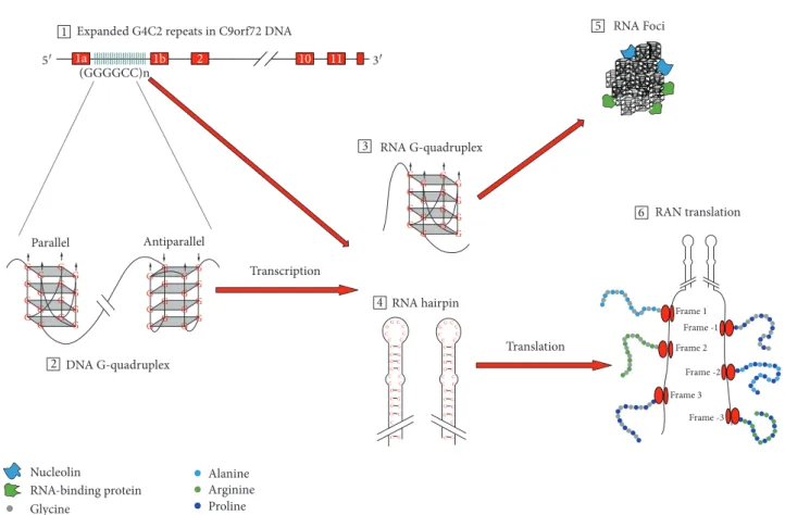
Figure2: Potential mechanisms ofC9orf72hexanucleotide repeat expansion (HRE)-mediated neurodegeneration. Pathology due to repeats inC9orf72gene may emerge from C9orf72 haploinsufficiency, RNA toxicity, and DPR accumulation. HRE in the noncoding region of the C9orf72 gene (1) form G-quadruplex structures (2). RNA transcribed from HRE DNA region can form different structures including G-quadruplexes (3) and RNA hairpins (4). HRE-containing RNA form RNA foci (5), which bind RNA-binding proteins. The last possible mechanism underlying pathology in C9FTD/ALS is through the repeat-associated non-ATG (RAN) translation, in which five different dipeptide repeat proteins can be formed—poly-GA, poly-GP, and poly-GR from the sense strand and poly-GP, poly- PA, and poly-PR from the antisense strand (6).
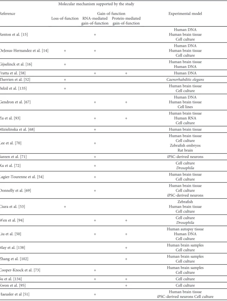
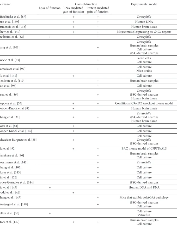
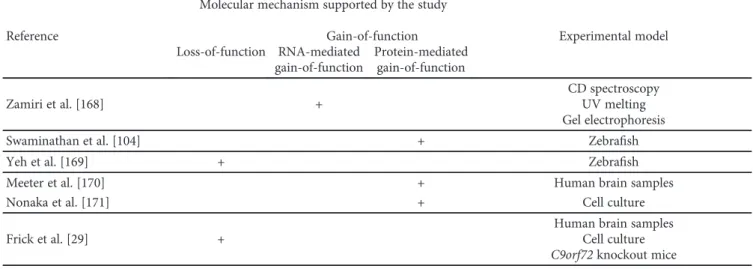
Table1: Summary of studies on the mechanisms by whichC9orf72repeat expansions cause neurodegeneration.
Reference
Molecular mechanism supported by the study
Experimental model Loss-of-function
Gain-of-function RNA-mediated
gain-of-function
Protein-mediated gain-of-function
Renton et al. [15] +
Human DNA Human brain tissue
Cell culture
DeJesus-Hernandez et al. [14] + +
Human DNA Human brain tissue
Cell culture
Gijselinck et al. [16] + Human brain tissue
Human DNA
Fratta et al. [58] + + Human DNA
Therrien et al. [52] + Caenorhabditis elegans
Belzil et al. [135] + Human brain tissue
Cell culture
Gendron et al. [67] + +
Human DNA Human brain tissue
Cell lines
Zu et al. [93] + +
Human brain tissue Human RNA
Cell culture
Mizielinska et al. [68] + Human brain tissue
Lee et al. [70] +
Human brain tissue Cell culture Zebrafish embryos
Rat brain
Sareen et al. [71] + iPSC-derived neurons
Xu et al. [72] + Cell culture
Drosophila
Lagier-Tourenne et al. [54] + Human brain tissue
Cell culture
Donnelly et al. [69] +
Human brain tissue Cell culture iPSC-derived neurons
Ciura et al. [53] +
Zebrafish Human brain tissue
Cell culture
Wen et al. [94] + + Cell culture
Drosophila
Liu et al. [50] + +
Human autopsy tissue Human DNA
Cell culture
May et al. [138] + Human brain samples
Cell culture
Zhang et al. [102] + Human brain samples
Cell culture
Cooper-Knock et al. [73] + Human brain samples
Cell culture
Su et al. [134] + + Cell culture
Kwon et al. [95] + Cell culture
Haeusler et al [51] + Human brain tissue
iPSC-derived neurons Cell culture
Table1: Continued.
Reference
Molecular mechanism supported by the study
Experimental model Loss-of-function
Gain-of-function RNA-mediated
gain-of-function
Protein-mediated gain-of-function
Mizielinska et al. [87] + + Drosophila
Russ et al. [139] + + Human DNA
Prudencio et al. [113] + + Human brain tissue
Chew et al. [140] + + Mouse model expressing 66 G4C2 repeats
Freibaum et al. [32] + + Drosophila
Yang et al. [101] +
Drosophila Human brain samples
Cell culture iPSC-derived neurons
Jovičićet al. [33] + Yeast cells
Cell culture
Yamakawa et al. [99] + Cell culture
Mice brains
Hu et al. [141] + Cell culture
Gendron et al. [110] + Human brain samples
Tao et al. [98] + Cell culture
Tran et al. [86] +
Drosophila iPSC-derived neurons
Human brain tissue
Koppers et al. [55] + + Conditional C9orf72 knockout mouse model
Cooper-Knock et al. [83] + Human brain tissue
Zhang et al. [31] + +
Drosophila iPSC-derived neurons
Human brain tissue
Rossi et al. [84] + Cell culture
Cooper-Knock et al. [116] + Cell culture
Schweizer Burguete et al. [85] +
Cell culture Drosophila iPSC-derived neurons
Liu et al. [92] + + BAC mouse model of C9FTD/ALS
Kanekura et al. [96] + Human brain samples
Cell culture
Boeynaems et al. [142] + Drosophila
Chang et al. [103] + Cell culture
Flores et al. [143] + Cell culture
Lin et al. [126] + Cell culture
Lopez-Gonzalez et al. [144] + iPSC-derived neurons
Liu et al. [145] + Human DNA and RNA
Dodd et al. [146] +
Zhang et al. [147] + Mice that exhibit poly(GA) pathology
Westergard et al. [148] + iPSC-derived neurons
Cell culture
Sellier et al. [36] + Cell culture
Zebrafish
Mori et al. [149] + + Human brain samples
Cell culture
Table1: Continued.
Reference
Molecular mechanism supported by the study
Experimental model Loss-of-function
Gain-of-function RNA-mediated
gain-of-function
Protein-mediated gain-of-function
Gijselinck et al. [19] + Human DNA
Ugolino et al. [150] + C9orf72 knockout mice
C9orf72 knockdown cell models
Lee et al. [125] + Drosophila
Cell culture
O’Rourke et al. [40] + + C9orf72 +/−and C9orf72−/−mice
Atanasio et al. [56] + + C9orf72-deficient mouse
Jiang et al. [91] + +
C9orf72 +/−and C9orf72−/−mice Mice expressing BAC with the human expanded
C9orf72gene
Webster et al. [34] + Cell culture
iNeurons (obtained by differentiation of iNPCs)
Ji et al. [151] + C9orf72 knockout mouse model
Cell culture
Ohki et al. [152] + Zebrafish
Liu et al. [153] + Cell culture
Hu et al. [154] + Cell culture
Green et al. [155] + Cell culture
Schludi et al. [156] + Transgenic mice expressing codon-modified
(GA)149
Gupta et al. [157] + Cell culture
Khosravi et al. [158] + Cell culture
Shi et al. [159] + Cell culture
Saberi et al. [160] + Human brain samples
Kramer et al. [105] + Cell culture
Herranz-Martin et al. [161] + + Mice that overexpress 10 or 102 interrupted
G4C2 repeats
Zhou et al. [162] + Cell culture
Human brain samples
Lehmer et al. [163] + Human CSF
Hautbergue et al. [82] + + Drosophila
Cell culture
Boeynaems et al. [100] + Cell culture
Maharjan et al. [39] + + + Cell culture
Moens et al. [88] + + Drosophila
Cheng et al. [164] + Cell culture
Swinnen et al. [165] + Zebrafish
Shi et al. [59] + +
Cell culture iPSC-derived neurons Human brain samples
Simone et al. [137] + + iPSC-derived neurons
Drosophila
Tabet et al. [166] + Human brain sections
Cell culture
Corrionero and Horvitz [167] + Caenorhabditis elegans
cause generation of truncated RNA transcripts that are aborted in the hexanucleotide repeat region [51]. These aberrant RNA transcripts containing repetitive hexanucleo- tide sequence can also form G-quadruplexes and RNA hair- pin structures. Additionally, HRE-containing RNA can form hybrids with HRE-containing DNA called R-loops [51, 62–64]. Together, these higher-order structures of DNA and RNA are thought to act as promoters and
regulatory elements affecting replication, transcription, and translation of the surrounding region [65, 66], and to exert deleterious effect on cells by causing nucleolar stress and impeding RNA processing [51], as discussed below.
6.2.2. Sequestration of RNA-Binding Proteins into Nuclear Aggregates by the C9orf72 HRE-RNA. HRE-containing RNA transcripts accumulate and form nuclear aggregates, or
Table1: Continued.Reference
Molecular mechanism supported by the study
Experimental model Loss-of-function
Gain-of-function RNA-mediated
gain-of-function
Protein-mediated gain-of-function
Zamiri et al. [168] +
CD spectroscopy UV melting Gel electrophoresis
Swaminathan et al. [104] + Zebrafish
Yeh et al. [169] + Zebrafish
Meeter et al. [170] + Human brain samples
Nonaka et al. [171] + Cell culture
Frick et al. [29] +
Human brain samples Cell culture C9orf72knockout mice
BAC: bacterial artificial chromosome; CSF: cerebrospinalfluid; iNPCs: induced neural progenitor cells; iPSC: induced pluripotent stem cells. Studies investigating pathological mechanisms ofC9orf72HRE but not clearly supporting any of the three proposed disease mechanisms are not included in this table. Search for these studies was completed on May 20, 2018. The literature search was performed in Google Scholar using the keywords:“C9orf72”,“mechanism”,“pathological”,
“loss-of-function”,“gain-of-function”,“haploinsufficiency”,“RNA foci”, and“DPR”. The literature search was conducted by two independent researchers.
Table2: Evaluation of potential mechanisms underlying pathology in C9FTD/ALS.
Molecular mechanism underlying
pathology in C9FTD/ALS Pros Cons
Loss-of-function
C9orf72loss-of-function models inC. elegansand
zebrafish result in motor neuron degeneration C9orf72loss-of-function mouse models do not show phenotype characteristic for ALS and FTD Carriers ofC9orf72HRE have decreased levels of
C9orf72 mRNA and proteins in the brain
Patients homozygous forC9orf72repeat expansion do not have more severe symptoms of disease
RNA-mediated gain-of-function
HRE-containing RNA transcripts accumulate and form nuclear aggregates, or RNA foci, in the brain
of patients with mutatedC9orf72
Drosophilamodels of RNA toxic gain-of-function fail to produce neurodegeneration Sequestration of RNA-binding proteins into RNA
foci can disrupt RNA processing, translation, nucleocytoplasmic transport, and granule
transport and lead to nucleolar stress
The results on RNA toxic gain-of-function mouse models are conflicting and need to be further
investigated Higher abundance of RNA foci in patients
carrying C9FTD/ALS HRE is associated with earlier disease onset
Protein-mediated gain-of-function
Drosophilamodel of protein-mediated gain-of- function develops neurodegeneration
Amounts of DPR in the brain do not correlate with clinical phenotype, severity of diseases, and
neurodegeneration DPR disrupt nucleocytoplasmic transport, RNA
processing, translation, ubiquitin proteasome system, formation of stress granule, and Notch signalling pathway and can lead to nucleolar stress
Abundance of DPR is low in the brain regions most affected by ALS and FTD
ALS: amyotrophic lateral sclerosis; C9FTD/ALS: hexanucleotide repeat expansion in C9orf72 causing ALS and FTD; DPR proteins: dipeptide repeat proteins;
FTD: frontotemporal dementia; HRE: hexanucleotide repeat expansion.
RNA foci, in the brain of patients with C9orf72 HRE [14]. As the transcription of C9orf72 HRE DNA region can also occur in the antisense direction, antisense RNA is also found within foci in the brains of patients with C9orf72 HRE [67, 68].
Several proteins speci
fically bind to C9orf72 HRE-containing RNA, including ADARB2, ALYREF, hnRNP H, hnRNP A1, nucleolin, Pur
α, and SRSF2 [51, 69
–73]. A number of RNA-binding proteins (RBP) are implicated in the HRE- induced neurodegeneration.
(1) Nucleolin. One of the main constituents of the nucleolus, nucleolin, has a high affinity for C9orf72 RNA containing G-quadruplex structures of G4C2 repeat [51]. Nucleolin bound to RNA G-quadruplexes dislocates from the nucleoli and disperses through the nucleus. This results in impaired rRNA processing, followed by decreased maturation of ribo- somes, and
finally, accumulation of untranslated mRNA in the neuronal cytoplasm [51].
(2) Pur
α. Another protein that has been identified as a HRE-RNA binding is Pur
α[72]. Involvement of Pur
αin the path- ogenesis of ALS/FTD is supported by experiments showing that Pur
αoverexpression alleviates HRE-mediated toxicity.
In other words, Pur
αoverexpression prevents cell death both in mammalian cells and in Drosophila [72]. Pur
αis a compo- nent of the RNA-transport granules
—particles that carry mRNAs to the nerve
fibers where translation of those mRNAs into proteins occurs [74, 75]
—and is involved in the regulation of the cell cycle [76] and in cell di
fferentiation [77, 78]. Sequestration of Pur
αby the C9orf72 hexanucleo- tide RNA repeats could impair neuronal mRNA transport thus leading to neurodegeneration [79].
(3) hnRNPs. The largest group of proteins sequestered by the C9orf72 HRE-containing RNA are heterogeneous nuclear ribonucleoproteins (hnRNPs) [44, 51]. It was shown that C9orf72 interacts with hnRNP-U, hnRNP-F, hnRNP-K [51], hnRNP-A2/B1, and hnRNP-A1 [28], while hnRNP A1 and hnRNP H colocalize with RNA foci [70, 71]. HnRNP H is involved in the regulation of RNA processing [80]; thus, hnRNP H sequestration in RNA cause aberrant RNA pro- cessing and may enhance neurodegeneration [70].
(4) ADARB2. An RNA-editing enzyme ADARB2 also inter- acts with the HRE-containing RNAs and colocalizes with RNA foci in C9ALS cases [69]. Although the mechanism by which ADARB2 sequestration in RNA foci could contribute to HRE-mediated toxicity is unclear, it was proposed that this protein may be important for RNA foci formation because ADARBP knockdown results in the reduction of neurons that contain RNA foci [69].
(5) SRSF1 and SRSF2. It was shown that serine-arginine- rich splicing factor 1 (SRSF1) and SRSF2 colocalize with RNA foci [70]. Given that SRSF2 is a marker of nuclear speckles, nuclear regions important for storage of splicing factors [81], its accumulation in RNA foci could disrupt the function of these speckles and lead to aberrant RNA processing [73]. Additionally, binding of the nuclear export
adaptor SRSF1 to C9orf72 repeats promotes export of C9orf72 repeats from the nucleus. As it was demonstrated that SRSF1 knockdown in C9FTD/ALS Drosophila model blocks neurodegeneration, SRSF1 could be a potential ther- apeutic target [82].
(6) ALYREF. Because aberrant nuclear transport is observed in di
fferent C9FTD/ALS models [31
–33], the observation that ALYREF (Aly/REF export factor) also colocalizes with RNA foci in C9FTD/ALS cases [70, 73, 83] is of interest.
However, although knockdown of nuclear export adaptor SRSF1 blocks neurodegeneration in Drosophila, only a modest decrease in neurodegeneration was observed after knockdown of ALYREF [82].
In conclusion, toxic gain-of-function due to aberrant HRE-containing RNA can lead to dysfunctional RNA pro- cessing [73], aberrant translation [84], nucleolar stress [51], disrupted nucleocytoplasmic transport [31], and dysfunction in granule transport [85]. Additionally, it was observed that higher abundance of RNA foci in the frontal cortex of C9FTD patients is associated with earlier disease onset [68].
However, several experimental studies failed to detect neuro- degeneration in in vivo C9FTD/ALS models displaying RNA foci (Table 2). Neurodegeneration was not observed in Dro- sophila models carrying 160 G4C2 repeats [86], 288 G4C2 repeats [87], and 1000 G4C2 repeats [88]. More precisely, Drosophila models carrying
“RNA-only
”repeats that formed RNA foci but not DPRs failed to produce neurodegeneration, while Drosophila models carrying
“pure repeats
”that could form both RNA foci and DPRs displayed neurodegeneration [87, 88]. However, two transgenic C9FTD/ALS mouse models with both RNA and protein toxic gain-of-function failed to produce neurodegeneration [89, 90], while two other transgenic mouse models showed signs of neurodegeneration [91, 92]. Regarding the latter two C9FTD/ALS mice models, whether neurodegeneration was caused by toxic HRE- containing RNAs or DPRs translated from hexanucleotide repeat RNA remains to be assessed [91, 92].
6.3. Toxic Gain-of-Function through Accumulation of Dipeptide Repeat Proteins Translated from C9orf72 HRE- Containing RNA. RNA transcripts of C9orf72 HRE region can undergo repeat-associated non-ATG (RAN) translation in which different DPR proteins are synthesized [43, 44, 93].
Poly-Gly-Ala, poly-Gly-Pro, and poly-Gly-Arg are translated
from di
fferent open reading fragments on the sense transcript
and poly-Gly-Pro, poly-Pro-Arg, and poly-Pro-Ala from the
antisense transcript. Toxicity of DPR proteins seems to be
mainly dependent on arginine-containing DPR proteins, par-
ticularly poly-Pro-Arg [33, 87, 94, 95]. Arginine-rich DPRs
appear to disrupt primarily nucleocytoplasmic transport
[33] and RNA processing [33, 95], which can cause dysregu-
lation of translation [96, 97], nucleolar stress [98], disturb
ubiquitin proteasome system [99], affect the formation of
stress granules [100], and influence the Notch signalling path-
way [101]. Additionally, it was shown that poly-Gly-Ala DPR
proteins can disturb the ubiquitin proteasome system and
cause endoplasmic reticulum stress [102], and enhance the
formation of toxic amyloid
fibrils [103].
Although there is molecular evidence of DPR proteins’
toxicity in in vitro and in vivo C9FTD/ALS models [104, 105], post mortem analyses of human brain revealed inconsistent results (Table 2). No correlation of DPR pro- teins with clinical phenotype, severity of disease, and neuro- degeneration was observed, and the abundance of DPR proteins was low in the brain regions most a
ffected in ALS and FTD [106
–109]. This lack of correlation may signal that neurons carrying toxic DPR proteins are possibly dead at the time of autopsy [18]. Additionally, there were dis- crepancies in the outcome of the studies comparing the distribution of DPR proteins between ALS and FTD cases [107, 110, 111].
7. Cellular Processes Affected in C9FTD/ALS Although many cellular processes are affected by C9orf72 repeat expansion, including translation [96], ubiquitin proteasome system [99], Notch signalling pathway [101], granule transport [85], and normal function of TDP-43 (for review see [17]), here we discuss the cellular processes affected by C9orf72 repeat expansion for which sufficient information is available.
7.1. E
ffects of DPR Proteins and HRE-Containing RNAs on Nucleocytoplasmic Transport. A hallmark morphological feature of TDP-43 proteinopathies is the cytoplasmic accu- mulation and aggregate formation of TDP-43. We have dem- onstrated earlier that impaired nucleocytoplasmic transport plays a major role in this process [112]. Importantly, C9orf72 repeat expansion compromises nucleocytoplasmic transport of proteins and RNA through the nuclear pores.
A study in yeast expressing arginine-rich DPR constructs demonstrated that one of the main targets of toxic DPR pro- teins is nucleocytoplasmic transport [33]. In support of these
findings, genetic screens in Drosophila designed to identify genes linked to HRE-induced toxicity also identi
fied compo- nents of the nuclear pore complex and nucleocytoplasmic transport machinery [31, 32]. Furthermore, RCC1, which is a human protein required for nucleocytoplasmic transport, is mislocalized in cells derived from patients with the C9orf72 HRE, providing preliminary evidence that C9FTD/
ALS HRE might affect nucleocytoplasmic transport in human cells as well [33]. Taken together, the studies in yeast and Drosophila provided the
first evidence that C9orf72 repeat expansion causes degeneration by impairing nucleo- cytoplasmic transport of proteins and RNA [31
–33]; how- ever, the e
ffect of C9FTD/ALS HRE on nucleocytoplasmic transport in human cells needs to be further investigated.
These
findings open a possibility of targeting nucleocytoplas-mic transport as a potential new therapeutic target in ALS and FTD [31, 32]. Additionally, as mRNA export factor ALYREF [70, 73, 83] and Ran GTPase-activating protein (RanGAP, regulator of nucleocytoplasmic transport) [31] colocalize with RNA foci (observed in C9FTD/ALS brains) and their function is disrupted, it seems that not only DPRs can lead to aberration of nucleocytoplasmic transport but also HRE- containing RNAs.
7.2. C9orf72 Hexanucleotide Repeat Expansion and RNA Processing. Several studies reported different transcrip- tional profiles between C9FTD/ALS patients and controls [54, 69, 71, 113–115]. Additionally, Cooper-Knock et al.
observed an enrichment in RNA splicing factors in C9FTD/ALS patients [116], further supporting the obser- vation of aberrant RNA processing caused by pathological processes in C9FTD/ALS. The main mechanism that leads to disturbed RNA processing in C9FTD/ALS patients is through sequestration of RBP in RNA foci [14, 73], but it could be also caused by DPR proteins as they can bind to RPB too [33, 95].
7.3. HRE-Containing RNAs and Dipeptide Repeat Proteins Can Lead to Nucleolar Stress. The way by which nucleolin binding to RNA foci disrupts the normal function of the nucleolus [51] was discussed above. It was also observed that DPR proteins can cause nucleolar stress in cell lines [98].
Enlargement of the nucleolus was observed in in vitro [94, 98] and in vivo [101] C9FTD/ALS models expressing arginine-rich DPR proteins. Enlargement of the nucleolus in these models leads to its fragmentation and decreased mat- uration of rRNA [98].
7.4. The Effect of DPRs on Formation and Function of Membraneless Organelles. Cells possess several RNA and protein-containing membraneless organelles, collectively referred to as ribonucleoprotein (RNP) granules, which sep- arate from the cytoplasm or nucleoplasm into a distinct liq- uid phase-like state that is typically slightly denser than the surrounding [117]. Examples of such organelles are nucleoli and Cajal bodies in the nucleus and processing bodies (P-bodies), stress granules, and transport granules in the cytoplasm [117–119]. Formation of such compartments is triggered by intrinsically disordered low complexity poly- peptide sequences present within RBP, which have the ability to undergo phase transitions [120–122]. Furthermore, it has been shown that nuclear membraneless suborganelles, such as Cajal bodies and nuclear speckles, can be nucleated by sev- eral coding and noncoding RNAs through the recruitment of proteins residing in these nuclear bodies [123]. Stress induces the formation of many membraneless compartments [124].
Lee et al. reported that arginine-rich DPRs bind low com- plexity polypeptide sequences of RBP that are components of membraneless organelles. Arginine-rich DPRs alter phase separation of those proteins and, in that way, disturb the function of membraneless organelles [125]. Liquid-liquid phase separation of RBP is very important for the normal for- mation of membraneless organelles. Other authors also showed that arginine-rich DPRs disturb the function and dynamics of membraneless organelles [126], more precisely, stress granules [98, 100]. Because it has been proposed that stress granules could be seeding points where aggregation of pathological proteins in FTD and ALS begins [127, 128], understanding the influence of DPR proteins on dynamics of membraneless organelles is highly relevant in this context.
7.5. C9orf72 HRE Affects Autophagy and Apoptosis. Adulter-
ations in the autophagic pathway can alter protein
homeostasis, causing detrimental effects on neurons that have been implicated in neurodegenerative diseases like Par- kinson’s disease and tauopathies. Autophagy prevents pro- teotoxic cell death by removing damaged, misfolded, and unwanted proteins [129]. Hindrance of C9orf72 function leads to a consequential decrease in Rab GTPase activity, a protein involved in membrane fusion, vesicle formation, and vesicle tra
fficking, which impairs the endocytosis process and autophagy. This causes an increased amount of p62/
SQSTM1 and TDP-43, known markers of ALS-FTD [36].
Using C9orf72-deficient mice and cell lines, Sullivan et al.
demonstrated that C9orf72 forms a binding complex with SMCR8 and WDR41, which allow it to interact with FIP200/Ulk1/ATG13/ATG101 complex and initiate autoph- agy [35]. Additionally, C9orf72 HRE have been implicated in increased endoplasmic reticulum stress by causing dysregula- tion of its calcium channels that leads to neuron apoptosis.
This has been quanti
fied by observing a decrease in antiapop- totic genes Bcl-2 and BcL-X
L, while causing an increased expression of proapoptotic gene BAK [130].
7.6. C9orf72 HRE and Neuroinflammation. Experimental studies investigating C9orf72 loss-of-function in C9orf72- deficient mice revealed an increase in proinflammatory cytokines, lymphadenopathy, splenomegaly, alterations in myeloid cells from spleen, and in some cases of autoimmu- nity [40, 56, 57]. Additionally, a human autopsy study showed that C9ALS cases had more severe microglial pathol- ogy in the medulla and motor cortex than ALS cases without C9orf72 HRE [131]. These
findings raised questions about increased neuroin
flammation in C9FTD/ALS patients and possible involvement of activated microglia in disease patho- genesis. It was proposed that microglia could represent a link between three potential pathological mechanisms of C9orf72 HRE, whereby microglia is being most affected by loss of C9orf72 function, while neurons are most affected by a gain-of-function mechanism. Altogether, increased neuroin-
flammation, accumulation of RNA foci, and DPRs could leadto neuronal death (reviewed in [132]).
8. Potential Therapeutic Approaches
Potential therapeutics most commonly target HRE- containing RNA, because its degradation abolishes RNA tox- icity and the formation of DPR proteins by RAN translation.
The following approaches have been considered so far.
8.1. Antisense Oligonucleotides. Antisense oligonucleotides (ASOs) targeting HRE-containing RNAs were tested in dif- ferent studies [54, 69, 91]. After treatment of C9FTD/ALS cells in in vitro [54, 69, 71] and in vivo models with ASOs (mice expressing bacterial artificial chromosome (BAC) with the human expanded C9orf72 gene [91] and adeno- associated virus (AAV) (G
4C
2)
66mice [133]), a reduction in RNA foci was observed. Additionally, treatment with ASOs also led to a decrease in DPRs [91, 133]. Although ASOs used in these studies did not a
ffect the levels of normal C9orf72 protein [54, 71, 91], the possibility of total silencing C9orf72 cannot be confirmed, so these approaches need to
be considered with caution. A clinical trial for ASO targeting HRE-containing RNAs (WVE-3972-01; https://adisinsight .springer.com/trials/700291284) is expected in the fourth quarter of 2018.
8.2. Small Molecules. The advantage of using small molecules as therapeutics for C9FTD/ALS is that these compounds could not only target HRE-containing RNAs or DPR pro- teins but also cellular processes affected by pathology under- lying C9FTD/ALS. Su et al. developed three small molecules that target hexanucleotide repeat region of RNAs and could stop RAN translation in (G4C2)
66-expressing COS7 cells [134]. As epigenetic alterations were observed in C9FTD/
ALS cases, epigenetic changes in the C9orf72 gene were also targeted. Usage of G-quadruplex-binding small molecules yielded increased expression of C9orf72 protein [135
–137].
9. The Mechanisms of C9orf72 HRE-Mediated Neurodegeneration Are Not
Mutually Exclusive
It is possible that the proposed mechanisms of C9FTD/ALS coexist and act in a combined manner. Maharjan et al. sug- gested that HRE in C9orf72 gene affects normal expression of C9orf72, diminishes levels of C9orf72 protein, and conse- quently impairs the formation of stress granules during the cellular stress (caused by formation of RNA foci and DPRs).
In other words, C9orf72 loss-of-function made cells more sensitive to toxicity caused by gain-of-function mechanisms [39]. Additionally, Lall and Baloh [132] proposed a model that uni
fies all three pathological mechanisms mentioned above. Other authors stressed the importance of generation of rodent experimental models in which all three mecha- nisms coexist, for example, by crossing C9orf72 knockout mouse and mouse carrying BAC with human expanded C9orf72 gene [18]. Although it was shown that ASO targeting HRE-containing RNAs do not affect the levels of normal C9orf72 protein [54, 71, 91], the possibility of silencing C9orf72 totally should not be overlooked. As ASO targeting HRE-containing RNAs moves into phase 1 of clinical trials, the development of novel cellular and animal experimental models that exhibit all three pathological mechanisms is highly relevant.
10. Conclusions
Seven years after the discovery of hexanucleotide repeat
expansion in C9orf72 gene, a lot of progress has been made
in the clarification of molecular mechanisms through which
C9orf72 repeat expansions cause neurodegeneration. Three
possible, not mutually exclusive, mechanisms could together
contribute to the pathogenesis of disease [39]. The majority
of the studies investigating the molecular mechanisms of
pathological processes in C9FTD/ALS support either toxic
HRE-RNA or DPR-dependent gain-of-function, with many
studies supporting both mechanisms (Table 1). Hence, to
better identify potential therapeutic targets, further studies
are needed to fully understand molecular events underlying
pathological processes in C9FTD/ALS.
Abbreviations
ALS: Amyotrophic lateral sclerosis ALS-bi: ALS with behavioral impairment ALS-ci: ALS with cognitive impairment
ALS-cbi: ALS with combined cognitive and behav- ioral impairment
ALYREF: Aly/REF export factor ASOs: Antisense oligonucleotides BAC: Bacterial arti
ficial chromosome bvFTD: Behavioral variant of frontotemporal
dementia
C9orf72: Chromosome 9 open reading frame 72 C9FTD/ALS: Hexanucleotide repeat expansion in
C9orf72 causing ALS and FTD CSF: Cerebrospinal
fluidDENN proteins: Di
fferentially expressed in normal and neoplasia proteins
DPR proteins: Dipeptide repeat proteins FTD: Frontotemporal dementia
FTLD: Frontotemporal lobar degeneration
FUS: Fused in sarcoma
G4C2: GGGGCC
GEF: Guanine exchange factor hnRNPs: Heterogeneous nuclear
ribonucleoproteins
HRE: Hexanucleotide repeat expansion IBMFD: Inclusion body myopathy with fronto-
temporal dementia
iNPCs: Induced neural progenitor cells iPSC: Induced pluripotent stem cells MND: Motor neuron disease
NCL: Nucleolin
NPC: Nuclear pore complex P-bodies: Processing bodies
PNFA: Progressive nonfluent aphasia pTDP-43: Phosphorylated TDP-43
RAN translation: Repeat-associated non-ATG translation RanGAP: Ran GTPase-activating protein
RBP: RNA binding proteins
RNP: Ribonucleoprotein
SD: Semantic dementia
SRSF: Serine-arginine-rich splicing factor TDP-43: TAR DNA-binding protein 43 VCP: Valosin-containing protein.
Conflicts of Interest
The authors declare no con
flict of interest.
Acknowledgments
We thank Mirta Boban for her help in improving the initial version of the manuscript. This work was supported by the Croatian Science Foundation grant IP-2014-09-9730 to G
Š, by the Scienti
fic Centre of Excellence for Basic, Clinical and Translational Neuroscience CoreNeuro of the Croatian Institute for Brain Research, University of
Zagreb Medical School, grant KK.01.1.1.01.0007, and in part by NIH grant P50 AG005138 to PRH.
References
[1] T. van Langenhove, J. van der Zee, and C. van Broeckhoven,
“The molecular basis of the frontotemporal lobar degenera- tion–amyotrophic lateral sclerosis spectrum,” Annals of Medicine, vol. 44, no. 8, pp. 817–828, 2012.
[2] P. M. Worms,“The epidemiology of motor neuron diseases: a review of recent studies,”Journal of the Neurological Sciences, vol. 191, no. 1-2, pp. 3–9, 2001.
[3] M. J. Strong, S. Abrahams, L. H. Goldstein et al.,“Amyotro- phic lateral sclerosis - frontotemporal spectrum disorder (ALS-FTSD): revised diagnostic criteria,” Amyotrophic Lateral Sclerosis and Frontotemporal Degeneration, vol. 18, no. 3-4, pp. 153–174, 2017.
[4] E. Ratnavalli, C. Brayne, K. Dawson, and J. R. Hodges,“The prevalence of frontotemporal dementia,”Neurology, vol. 58, no. 11, pp. 1615–1621, 2002.
[5] A. S. L. Ng, R. Rademakers, and B. L. Miller,“Frontotemporal dementia: a bridge between dementia and neuromuscular disease,” Annals of the New York Academy of Sciences, vol. 1338, no. 1, pp. 71–93, 2015.
[6] T. Hortobágyi and N. Cairns,“Amyotrophic lateral sclerosis and frontotemporal dementia,”inNeuropathology of Neuro- degenerative Diseases: A Practical Guide, G. Kovacs, Ed., pp. 209–248, Cambridge University Press, 2015.
[7] T. Hortobágyi and N. J. Cairns,“Chapter 26 - Amyotrophic lateral sclerosis and non-tau frontotemporal lobar degenera- tion,”Handbook of clinical neurology, vol. 145, pp. 369–381, 2017.
[8] I. R. A. Mackenzie, M. Neumann, A. Baborie et al.,“A harmo- nized classification system for FTLD-TDP pathology,”Acta Neuropathologica, vol. 122, no. 1, pp. 111–113, 2011.
[9] T. W. Chow, B. L. Miller, V. N. Hayashi, and D. H.
Geschwind, “Inheritance of frontotemporal dementia,” Archives of Neurology, vol. 56, no. 7, pp. 817–822, 1999.
[10] S. M. Rosso, L. D. Kaat, T. Baks et al., “Frontotemporal dementia in the Netherlands: patient characteristics and prevalence estimates from a population-based study,”Brain, vol. 126, no. 9, pp. 2016–2022, 2003.
[11] M. J. Strong, T. Hortobágyi, K. Okamoto, and S. Kato,“Amyo- trophic lateral sclerosis, primary lateral sclerosis and spinal muscular atrophy,” in Neurodegeneration: The Molecular Pathology of Dementia and Movement Disorders, D. Dickson and R. Weller, Eds., pp. 418–433, Wiley-Blackwell, 2011.
[12] C. Vance, A. al-Chalabi, D. Ruddy et al.,“Familial amyotro- phic lateral sclerosis with frontotemporal dementia is linked to a locus on chromosome 9p13.2–21.3,” Brain, vol. 129, no. 4, pp. 868–876, 2006.
[13] M. Morita, A. al-Chalabi, P. M. Andersen et al.,“A locus on chromosome 9p confers susceptibility to ALS and frontotem- poral dementia,”Neurology, vol. 66, no. 6, pp. 839–844, 2006.
[14] M. DeJesus-Hernandez, I. R. Mackenzie, B. F. Boeve et al.,
“Expanded GGGGCC hexanucleotide repeat in noncoding region of C9ORF72 causes chromosome 9p-linked FTD and ALS,”Neuron, vol. 72, no. 2, pp. 245–256, 2011.
[15] A. E. Renton, E. Majounie, A. Waite et al.,“A hexanucleotide repeat expansion in C9ORF72 is the cause of chromosome
9p21-linked ALS-FTD,”Neuron, vol. 72, no. 2, pp. 257–268, 2011.
[16] I. Gijselinck, T. van Langenhove, J. van der Zee et al., “A C9orf72 promoter repeat expansion in a Flanders-Belgian cohort with disorders of the frontotemporal lobar degeneration-amyotrophic lateral sclerosis spectrum: a gene identification study,” The Lancet Neurology, vol. 11, no. 1, pp. 54–65, 2012.
[17] T. W. Todd and L. Petrucelli,“Insights into the pathogenic mechanisms of chromosome 9 open reading frame 72 (C9orf72) repeat expansions,” Journal of Neurochemistry, vol. 138, pp. 145–162, 2016.
[18] X. Wen, T. Westergard, P. Pasinelli, and D. Trotti,“Patho- genic determinants and mechanisms of ALS/FTD linked to hexanucleotide repeat expansions in the C9orf72 gene,”
Neuroscience Letters, vol. 636, pp. 16–26, 2017.
[19] I. Gijselinck, on behalf of the BELNEU CONSORTIUM13, S. Van Mossevelde et al.,“TheC9orf72repeat size correlates with onset age of disease, DNA methylation and transcrip- tional downregulation of the promoter,”Molecular Psychia- try, vol. 21, no. 8, pp. 1112–1124, 2016.
[20] A. J. Waite, D. Bäumer, S. East et al.,“Reduced C9orf72 pro- tein levels in frontal cortex of amyotrophic lateral sclerosis and frontotemporal degeneration brain with the C9ORF72 hexanucleotide repeat expansion,” Neurobiology of Aging, vol. 35, no. 7, pp. 1779.e5–1779.e13, 2014.
[21] O. Dols-Icardo, A. Garcia-Redondo, R. Rojas-Garcia et al.,
“Characterization of the repeat expansion size inC9orf72in amyotrophic lateral sclerosis and frontotemporal dementia,” Human Molecular Genetics, vol. 23, no. 3, pp. 749–754, 2014.
[22] M. van Blitterswijk, M. DeJesus-Hernandez, E. Niemantsverdriet et al.,“Association between repeat sizes and clinical and pathological characteristics in carriers of C9ORF72 repeat expansions (Xpansize-72): a cross- sectional cohort study,” The Lancet Neurology, vol. 12, no. 10, pp. 978–988, 2013.
[23] J. Beck, M. Poulter, D. Hensman et al.,“LargeC9orf72hexa- nucleotide repeat expansions are seen in multiple neurode- generative syndromes and are more frequent than expected in the UK population,” The American Journal of Human Genetics, vol. 92, no. 3, pp. 345–353, 2013.
[24] A. Hübers, N. Marroquin, B. Schmoll et al.,“Polymerase chain reaction and Southern blot-based analysis of the C9orf72 hexanucleotide repeat in different motor neuron diseases,” Neurobiology of Aging, vol. 35, no. 5, pp. 1214.e1– 1214.e6, 2014.
[25] A. Nordin, C. Akimoto, A. Wuolikainen et al.,“Extensive size variability of the GGGGCC expansion in C9orf72 in both neuronal and non-neuronal tissues in 18 patients with ALS or FTD,” Human Molecular Genetics, vol. 24, no. 11, pp. 3133–3142, 2015.
[26] D. Zhang, L. M. Iyer, F. He, and L. Aravind,“Discovery of novel DENN proteins: implications for the evolution of eukaryotic intracellular membrane structures and human disease,”Frontiers in Genetics, vol. 3, p. 283, 2012.
[27] T. P. Levine, R. D. Daniels, A. T. Gatta, L. H. Wong, and M. J.
Hayes,“The product of C9orf72, a gene strongly implicated in neurodegeneration, is structurally related to DENN Rab- GEFs,”Bioinformatics, vol. 29, no. 4, pp. 499–503, 2013.
[28] M. A. Farg, V. Sundaramoorthy, J. M. Sultana et al.,
“C9ORF72, implicated in amytrophic lateral sclerosis and
frontotemporal dementia, regulates endosomal trafficking,” Human Molecular Genetics, vol. 23, no. 13, pp. 3579–3595, 2014.
[29] P. Frick, C. Sellier, I. R. A. Mackenzie et al.,“Novel antibodies reveal presynaptic localization of C9orf72 protein and reduced protein levels inC9orf72 mutation carriers,”Acta Neuropathologica Communications, vol. 6, no. 1, p. 72, 2018.
[30] S. Xiao, L. MacNair, P. McGoldrick et al.,“Isoform-specific antibodies reveal distinct subcellular localizations of C9orf72in amyotrophic lateral sclerosis,”Annals of Neurol- ogy, vol. 78, no. 4, pp. 568–583, 2015.
[31] K. Zhang, C. J. Donnelly, A. R. Haeusler et al.,“The C9orf72 repeat expansion disrupts nucleocytoplasmic transport,” Nature, vol. 525, no. 7567, pp. 56–61, 2015.
[32] B. D. Freibaum, Y. Lu, R. Lopez-Gonzalez et al.,“GGGGCC repeat expansion in C9orf72 compromises nucleocytoplas- mic transport,”Nature, vol. 525, no. 7567, pp. 129–133, 2015.
[33] A. Jovičić, J. Mertens, S. Boeynaems et al., “Modifiers of C9orf72 dipeptide repeat toxicity connect nucleocytoplasmic transport defects to FTD/ALS,”Nature Neuroscience, vol. 18, no. 9, pp. 1226–1229, 2015.
[34] C. P. Webster, E. F. Smith, C. S. Bauer et al.,“The C9orf72 protein interacts with Rab1a and the ULK1 complex to regu- late initiation of autophagy,” The EMBO Journal, vol. 35, no. 15, pp. 1656–1676, 2016.
[35] P. M. Sullivan, X. Zhou, A. M. Robins et al.,“The ALS/FTLD associated protein C9orf72 associates with SMCR8 and WDR41 to regulate the autophagy-lysosome pathway,”Acta Neuropathologica Communications, vol. 4, no. 1, p. 51, 2016.
[36] C. Sellier, M. L. Campanari, C. Julie Corbier et al.,“Loss of C9ORF72 impairs autophagy and synergizes with polyQ ataxin-2 to induce motor neuron dysfunction and cell death,” The EMBO Journal, vol. 35, no. 12, pp. 1276–1297, 2016.
[37] J. Amick, A. Roczniak-Ferguson, and S. M. Ferguson,
“C9orf72 binds SMCR8, localizes to lysosomes, and regulates mTORC1 signaling,”Molecular Biology of the Cell, vol. 27, no. 20, pp. 3040–3051, 2016.
[38] J. Amick and S. M. Ferguson,“C9orf72: at the intersection of lysosome cell biology and neurodegenerative disease,”Traffic, vol. 18, no. 5, pp. 267–276, 2017.
[39] N. Maharjan, C. Künzli, K. Buthey, and S. Saxena,“C9ORF72 regulates stress granule formation and its deficiency impairs stress granule assembly, hypersensitizing cells to stress,”
Molecular Neurobiology, vol. 54, no. 4, pp. 3062–3077, 2017.
[40] J. G. O'Rourke, L. Bogdanik, A. Yanez et al., “C9orf72 is required for proper macrophage and microglial function in mice,”Science, vol. 351, no. 6279, pp. 1324–1329, 2016.
[41] S. Al-Sarraj, A. King, C. Troakes et al.,“p62 positive, TDP-43 negative, neuronal cytoplasmic and intranuclear inclusions in the cerebellum and hippocampus define the pathology of C9orf72-linked FTLD and MND/ALS,”Acta Neuropatholo- gica, vol. 122, no. 6, pp. 691–702, 2011.
[42] M. E. Murray, M. DeJesus-Hernandez, N. J. Rutherford et al.,
“Clinical and neuropathologic heterogeneity of c9FTD/ALS associated with hexanucleotide repeat expansion in C9ORF72,”Acta Neuropathologica, vol. 122, no. 6, pp. 673– 690, 2011.
[43] P. E. A. Ash, K. F. Bieniek, T. F. Gendron et al.,“Unconven- tional translation of C9ORF72 GGGGCC expansion gener- ates insoluble polypeptides specific to c9FTD/ALS,”Neuron, vol. 77, no. 4, pp. 639–646, 2013.
[44] K. Mori, S. M. Weng, T. Arzberger et al., “The C9orf72 GGGGCC repeat is translated into aggregating dipeptide- repeat proteins in FTLD/ALS,”Science, vol. 339, no. 6125, pp. 1335–1338, 2013.
[45] C. Troakes, S. Maekawa, L. Wijesekera et al.,“An MND/ALS phenotype associated with C9orf72 repeat expansion: abun- dant p62-positive, TDP-43-negative inclusions in cerebral cortex, hippocampus and cerebellum but without associated cognitive decline,”Neuropathology, vol. 32, no. 5, pp. 505– 514, 2012.
[46] M. van Blitterswijk, T. F. Gendron, M. C. Baker et al.,“Novel clinical associations with specific C9ORF72 transcripts in patients with repeat expansions in C9ORF72,”Acta Neuro- pathologica, vol. 130, no. 6, pp. 863–876, 2015.
[47] Z. Xi, L. Zinman, D. Moreno et al.,“Hypermethylation of the CpG island near the G4C2 repeat in ALS with a C9orf72 expansion,” American Journal of Human Genetics, vol. 92, no. 6, pp. 981–989, 2013.
[48] Z. Xi, M. Zhang, A. C. Bruni et al., “The C9orf72 repeat expansion itself is methylated in ALS and FTLD patients,” Acta Neuropathologica, vol. 129, no. 5, pp. 715–727, 2015.
[49] Z. Xi, I. Rainero, E. Rubino et al.,“Hypermethylation of the CpG-island near the C9orf72 G4C2-repeat expansion in FTLD patients,”Human Molecular Genetics, vol. 23, no. 21, pp. 5630–5637, 2014.
[50] E. Y. Liu, J. Russ, K. Wu et al.,“C9orf72 hypermethylation protects against repeat expansion-associated pathology in ALS/FTD,”Acta Neuropathologica, vol. 128, no. 4, pp. 525– 541, 2014.
[51] A. R. Haeusler, C. J. Donnelly, G. Periz et al.,“C9orf72 nucle- otide repeat structures initiate molecular cascades of disease,”
Nature, vol. 507, no. 7491, pp. 195–200, 2014.
[52] M. Therrien, G. A. Rouleau, P. A. Dion, and J. A. Parker,
“Deletion of C9ORF72 results in motor neuron degeneration and stress sensitivity in C. elegans,”PLoS One, vol. 8, no. 12, article e83450, 2013.
[53] S. Ciura, S. Lattante, I. le Ber et al., “Loss of function of C9orf72 causes motor deficits in a zebrafish model of amyo- trophic lateral sclerosis,”Annals of Neurology, vol. 74, no. 2, pp. 180–187, 2013.
[54] C. Lagier-Tourenne, M. Baughn, F. Rigo et al., “Targeted degradation of sense and antisense C9orf72 RNA foci as therapy for ALS and frontotemporal degeneration,” Pro- ceedings of the National Academy of Sciences of the United States of America, vol. 110, no. 47, pp. E4530–E4539, 2013.
[55] M. Koppers, A. M. Blokhuis, H. J. Westeneng et al.,“C9orf72 ablation in mice does not cause motor neuron degeneration or motor deficits,” Annals of Neurology, vol. 78, no. 3, pp. 426–438, 2015.
[56] A. Atanasio, V. Decman, D. White et al.,“C9orf72 ablation causes immune dysregulation characterized by leukocyte expansion, autoantibody production and glomerulonephro- pathy in mice,” Scientific Reports, vol. 6, no. 1, p. 23204, 2016.
[57] E. Sudria-Lopez, M. Koppers, M. de Wit et al.,“Full ablation of C9orf72 in mice causes immune system-related pathology and neoplastic events but no motor neuron defects,” Acta Neuropathologica, vol. 132, no. 1, pp. 145–147, 2016.
[58] P. Fratta, M. Poulter, T. Lashley et al.,“Homozygosity for the C9orf72 GGGGCC repeat expansion in frontotemporal
dementia,”Acta Neuropathologica, vol. 126, no. 3, pp. 401– 409, 2013.
[59] Y. Shi, S. Lin, K. A. Staats et al.,“Haploinsufficiency leads to neurodegeneration in C9ORF72 ALS/FTD human induced motor neurons,” Nature Medicine, vol. 24, no. 3, pp. 313– 325, 2018.
[60] P.Šket, J. Pohleven, A. Kovanda et al.,“Characterization of DNA G-quadruplex species forming from C9ORF72 G4C2- expanded repeats associated with amyotrophic lateral sclero- sis and frontotemporal lobar degeneration,”Neurobiology of Aging, vol. 36, no. 2, pp. 1091–1096, 2015.
[61] A. Kovanda, M. Zalar, P. Šket, J. Plavec, and B. Rogelj,
“Anti-sense DNA d(GGCCCC)n expansions in C9ORF72 form i-motifs and protonated hairpins,” Scientific Reports, vol. 5, no. 1, article 17944, 2015.
[62] P. Fratta, S. Mizielinska, A. J. Nicoll et al.,“C9orf72 hexanu- cleotide repeat associated with amyotrophic lateral sclerosis and frontotemporal dementia forms RNA G-quadruplexes,” Scientific Reports, vol. 2, no. 1, p. 1016, 2012.
[63] K. Reddy, B. Zamiri, S. Y. R. Stanley, R. B. Macgregor Jr., and C. E. Pearson,“The disease-associated r(GGGGCC)nrepeat from the C9orf72 gene forms tract length-dependent uni- and multimolecular RNA G-quadruplex structures,”Journal of Biological Chemistry, vol. 288, no. 14, pp. 9860–9866, 2013.
[64] K. Reddy, M. H. M. Schmidt, J. M. Geist et al.,“Processing of double-R-loops in (CAG)·(CTG) and C9orf72 (GGGG CC)·(GGCCCC) repeats causes instability,” Nucleic Acids Research, vol. 42, no. 16, pp. 10473–10487, 2014.
[65] S. Kendrick and L. H. Hurley,“The role of G-quadruplex/i- motif secondary structures as cis-acting regulatory elements,” Pure and Applied Chemistry, vol. 82, no. 8, pp. 1609–1621, 2010.
[66] T. A. Brooks, S. Kendrick, and L. Hurley,“Making sense of G-quadruplex and i-motif functions in oncogene pro- moters,” The FEBS Journal, vol. 277, no. 17, pp. 3459– 3469, 2010.
[67] T. F. Gendron, K. F. Bieniek, Y. J. Zhang et al.,“Antisense transcripts of the expanded C9ORF72 hexanucleotide repeat form nuclear RNA foci and undergo repeat-associated non- ATG translation in c9FTD/ALS,” Acta Neuropathologica, vol. 126, no. 6, pp. 829–844, 2013.
[68] S. Mizielinska, T. Lashley, F. E. Norona et al.,“C9orf72 fron- totemporal lobar degeneration is characterised by frequent neuronal sense and antisense RNA foci,”Acta Neuropatholo- gica, vol. 126, no. 6, pp. 845–857, 2013.
[69] C. J. Donnelly, P. W. Zhang, J. T. Pham et al.,“RNA toxicity from the ALS/FTD C9ORF72 expansion is mitigated by anti- sense intervention,” Neuron, vol. 80, no. 2, pp. 415–428, 2013.
[70] Y.-B. Lee, H. J. Chen, J. N. Peres et al., “Hexanucleotide repeats in ALS/FTD form length-dependent RNA foci, sequester RNA binding proteins, and are neurotoxic,” Cell Reports, vol. 5, no. 5, pp. 1178–1186, 2013.
[71] D. Sareen, J. G. O'Rourke, P. Meera et al.,“Targeting RNA foci in iPSC-derived motor neurons from ALS patients with aC9ORF72repeat expansion,”Science Translational Medi- cine, vol. 5, no. 208, article 208ra149, 2013.
[72] Z. Xu, M. Poidevin, X. Li et al.,“Expanded GGGGCC repeat RNA associated with amyotrophic lateral sclerosis and fron- totemporal dementia causes neurodegeneration,”Proceedings