Salicylic Acid and Hydrogen Peroxide Accumulation in Relation to Hydrolyte Leakage in Barley Plants Challenged
with Cochliobolus sativus
A. Al-DAouDe*, e. Al-ShehADAh, A. ShoAib, M. JAwhAr and M.i.e. ArAbi Department of Molecular Biology and Biotechnology, AECS, P.O. Box 6091 Damascus, Syria
(Received 8 February 2018; Accepted 4 July 2018;
Communicated by N. Rostoks)
Spot blotch (SB) caused by the hemibiotrophic fungal pathogen Cochliobolus sativus is a destructive disease of barley worldwide. To better understand the mechanisms of resistance to this disease, the involvements of salicylic acid (SA), hydrogen peroxide (H2O2) and ion fluxes during the interaction between resistant and susceptible barley seedlings and C. sati- vus were investigated. Early SA accumulation in leaf tissues was accompanied with an increase in H2O2 concentration in both compatible and incompatible interactions. The resist- ant cultivar constitutively contained higher levels of H2O2 and SA, as well as during the 72 h as compared with the un-infected control (0 h). However, levels increased rapidly upon infection in both cultivars. Moreover, a markedly greater increase in ion fluxes from the compatible material compared with the incompatible one was observed. Results suggest that SA and H2O2 accumulation are important during both compatible and incompatible barley- C. sativus interactions.
Keywords: barley, Cochliobolus sativus, hydrogen peroxide, salicylic acid, electrolyte leakage, interaction
Introduction
Cochliobolus sativus (Ito & Kurib.) Drechsl. ex Dast. [anamorph: Bipolaris sorokiniana (Sacc. in Sorok.) Shoem.], the cause of spot blotch (SB), is an economically important disease of barley (Hordeum vulgare) worldwide (Mathre 1997). C. sativus infection on barley plants results in various host-specific biochemical responses which show strategies deployed by the plant to withstand diseases (Kumar et al. 2002; Jawhar et al. 2017). These responses are regulated by a concerted expression of different plant signaling pathways, and associated with an oxidative response detectable as the localized accumulation of reactive oxygen species (ROS) molecules in attacked and/or neighboring cells of the in- fection site (Leng and Zhong 2015; Rodríguez-Decuadro et al. 2014).
C. sativus has been classified as an hemibiotrophic fungus, and its pathogenesis in- cludes germination of conidia on the leaf surface and formation of an appressorium at the tip of the germ tube that supports direct penetration through the host cuticle (Kumar et al.
*Corresponding author; E-mail: ascientific@aec.org.sy
2002). The apparently biotrophic growth phase is primarily confined to a single epidermal cell invaded by infection hyphae, whereas the necrotrophic growth phase starts upon in- vasion of the mesophyll tissue followed by host cell death, which appears to be a conse- quence of toxin secretion (Apoga et al. 2002). However, ROS, especially H2O2 accumula- tion at the site of pathogen infection is one of the first cytologically detectable symptoms of plant pathogen interaction (Nanda et al. 2010).
SA, a plant hormone widely distributed in angiosperms, is known to be a key element in pathogen defense and its simple and accurate quantification in plants is therefore of prominent importance (Belt et al. 2017). Different resistance mechanisms related to SA signaling including hypersensitive response (HR), oxidative burst and pathogenesis-relat- ed (PR) gene expression have been reported (Häffner et al. 2014; Vásquez et al. 2015).
Additionally, changes in ion fluxes are considered as the first events during the initial establishment of the HR which can provide a robust method for quantifying cell collapse (Bestwick et al. 1998).
To complete the picture of barley biochemical responses drawn (Al-Daoude et al.
2013), we investigated in the current study the possible changes of endogenous H2O2 and SA and electrolyte leakage during barley interaction with C. sativus at different time points.
Materials and Methods Host genotypes
After an extensive screening of barley (Hordeum vulgare L.) genotypes for more than 15 years in the greenhouse and laboratory experiments, the German cv. Banteng has proved to be the most resistant genotype to all C. sativus isolates available so far (Arabi and Jawhar 2003; 2004), therefore, it was used as a plant material in this study. The universal susceptible control genotype (cv. WI2291) from Australia was also included in the ex- periments. Barley seedlings were grown in 20-cm pots filled with sterilized peat moss.
Pots were placed in a greenhouse and arranged in a randomized block design with three replicates for each cultivar (each replicate is one pot containing 10 plants) at 20°C, 80–
90% relative humidity, with a 16 h-light/8 h-dark cycle.
Inoculum preparation and inoculation
A single spore isolate (Pt4) of C. sativus used in the study and was the most virulent avail- able pathotype as described by Arabi and Jawhar (2003). The fungal mycelia were trans- ferred from a stock culture into Petri dishes containing potato dextrose agar (PDA, DIF- CO, Detroit, MI, USA) and incubated for 10 days at 21 ± 1°C in the dark. Then, conidia were collected with 10 mL of sterile distilled water. The conidial suspension was adjusted to 2 × 10⁴ conidia/mL using hemacytometer counts of conidia to provide estimates of the inoculum concentration. A surfactant (polyoxyethylene-20-sorbitan monolaurate) was added (100 µL/L) to the conidial suspension to facilitate dispersion of the inoculum over
the leaf surfaces. The primary leaves of 12-day-old seedlings were inoculated by uni- formly spraying each plant with the conidial suspension using a hand-held spray bottle.
Plants were covered for one night with plastic bags to increase humidity and plants were kept in the same greenhouse at 20°C with a 16-h photoperiod.
Histochemical detection of H2O2
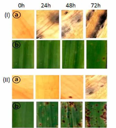
Histochemical detection of H2O2 in barley leaf tissues was carried out by following the method of Orczyk et al. (2010). Leaf samples were stained with DAB (3, 30-diaminoben- zidinetetrahydrochloride) and with Calcofluor White. The stained samples were exam- ined under a fluorescence microscope (Olympus-ix21 station, X400, Japan). H2O2 was localized due to dark blue coloration in the periplasmic space of the cells (Fig. 1). Obser- vations were made for at least 20 infection sites per leaf sample collected from two to five inoculated plants. The number of cells with H2O2 in 25 random microscopic fields was counted and percentage calculated (Bestwick et al. 1997). Five fields at high magnifica- tion (×200) were randomly chosen.
Figure 1. Localization of H2O2 (a) and SB symptoms (b) in tissues of barley leaves; resistant (I) and susceptible (II) at 0, 24, 48 and 72 hours post inoculation with C. sativus of three replicates SE
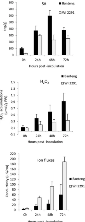
Figure 2. Changes in SA, H2O2 and ion fluxes in barley resistant ‘Banteng’ and susceptible ‘WI2291’ seedlings inoculated C. sativus. Data represents the mean of three replicates and SE
Quantification of endogenous salicylic acid
The leaf tissue (1.0 g) was collected at different time points; 0, 24, 48 and 72 hours post inoculation (hpi) and ground in 2.5 mL of 90% methanol using a pre-chilled pestle and mortar. SA was measured using the method described by Trapp et al. (2014) with minor modifications. Extraction was achieved by adding 1.0 mL of ethylacetate, dichlorometh- ane, isopropanol and MeOH:H2O (8:2) into each. The extract was centrifuged at 4°C at 16,000 g for 5 min. The supernatant was transferred into a new 1.5 mL micro-centrifuge tube and dried in speed vac. After drying, 100 μl of MeOH:H2O was added to each sam- ple, homogenized under vortex and centrifuged at 16,000 g and 4°C for 10 min.
SA was quantified by a high performance liquid chromatography (HPLC-MS/MS) sys- tem (Agilent Technologies, Germany). The ability of quantification by this method was tested using the procedure described by Green (1996). Since no additional MS/MS spec- trum peaks except for the band corresponding to the standards for the analyte were ob- tained, the method was considered selective.
Electrolyte leakage from leaf segments
Segments of barley leaves (ca. 1 cm) of each genotype put into 20 ml of deionised water in a test tube and washed slowly using a rotary shaker (100 rpm) at room temperature to remove solutes from both leaf surfaces and damaged cells due to cutting. Electrical leak- age was measured at 0, 24, 48 and 72 hpi using a LF 92 conductimeter (WTW GmbH, Weilheim, Germany) by as described by Kwon et al. (1996).
Data analysis
The data generated for H2O2, SA and electrolyte leakage were the average of the three replicates each. Comparison of means between genotypes was performed using analysis of variance (ANOVA) at the 5% level in the software package Statistica 6.1. When the main effect was significant, differences between means were evaluated for significance by using the Scheffe F-test. The standard deviation was calculated.
Results
SB severity symptoms (chlorosis and necrosis) was always more obvious in the compat- ible barley ‘WI2291’ compared with the incompatible one ‘Banteng’ (Figure 1). To better understand these interactions, changes in H2O2, SA and electrolyte leakage during infec- tion at 0, 24, 48 and 72hpi were investigated. Seedlings inoculated with distilled water were also analyzed at 0h. H2O2 and SA level were observed post inoculation of seedlings in both compatible and incompatible interactions.
However, significant variations and disparity (P=0.001) in the values of these param- eters were found among different time points (Figure 2). There was also a marked in- crease in electrolyte leakage following the interaction between barley leaves and the fun- gal pathogen, C. sativus. However, this leakage was higher in the susceptible barley com- pared with the resistant one.
Discussion
Inoculation with C. sativus virulent pathotype (Pt4) induced a marked increase in the amount of H2O2, SA and ion fluxes when compared to seedlings before inoculation, in both susceptible and resistant barley cultivars (Fig. 2). The earliest detection of H2O2 might be in relation to the earliest signs of HR after inoculation (Bindschedler et al.
2006). Similar results were found in rice (Xiong and Yang 2003) and wheat (Hulbert et al.
2007).
C. sativus is a hemibiotrophic with an initial biotrophic stage during disease develop- ment. Data showed that SA contents of compatible and incompatible barley leaves in- creased 24hpi in comparison with non-inoculated plants at the start of the experiment.
These results indicate that SA is be expected to play a role in signaling events during fungal infection by C. sativus, which might be attributed to the involvement of SA in plant defense responses which binds and inhibits catalase activity and consequently ele- vates cellular levels of H2O2 (Shim et al. 2003). However, oxidative burst largely pre- cedes the local and systemic accumulation of SA in pathogen-inoculated plants undergo- ing HR (Apel and Hirt 2004).
It is suggested from this study that host-generated H2O2 during the necrotrophic phase contributes to resistance since, when the total number of cells showing H2O2 localization in test seedling was considered, it was observed that the number of cells localized in the incompatible host ‘Banteng’ were greater than the ones in the compatible host ‘WI2291’.
Bradley et al. (1992) reported that the increase of H2O2 levels can affect plant defense in several ways, presumably by stimulating cross-linking of proline-rich proteins of the cell wall, and by inducing several plant genes involved in cellular protection and defense (Forman et al. 2003).
Over time, also ion fluxes increased but they were higher in the susceptible genotype WI2291 as compared with the resistant one, which might suggest somewhat enhanced cell death in the compatible reaction, even though the conductivity values were much lower than reported for typical HR (Keisa et al. 2011). Demidchik et al. (2014) reported that electrolyte leakage is mainly related to K+ efflux from plant cells, which is mediated by plasma membrane cation conductances.
In conclusion, this study illustrated that significant increases in SA and H2O2 were found upon barley challenged with C. sativus, with values being consistently higher in the incompatible host. The patterns of H2O2 accumulation clearly differentiated during the time course of inoculation and corresponded to SA and inversely to ion fluxes re- sponse profiles in compatible and non-compatible host tissue, which could indicate that these changes might have roles in barley compatible and incompatible interactions. This study serves to broaden our understanding of the molecular cellular basis of barley- C. sativus interactions but more studies, including also more susceptible and resistant barley genotypes, are needed to better understand the mechanisms for susceptibility and resistance.
Acknowledgements
The authors thank the Director General of AECS and the Head of Molecular biology and Biotechnology Department for their much appreciated help throughout the period of this research.
References
Al-Daoude, A., Jawhar, M., Arabi, M.I.E. 2013. Hydrogen peroxide induction in barley-Cochliobolus sativus interaction. J. Plant Pathol. 95:197–199.
Apel, K., Hirt, H. 2004. Reactive oxygen species: metabolism, oxidative stress, and signal transduction. Ann.
Rev. Plant Biol. 55:373–399.
Apoga, D., Akesson, H., Jansson, H.B., Odham, G. 2002. Relationship between production of the phytotoxin prehelminthosporol and virulence in isolates of the plant pathogenic fungus Bipolaris sorokiniana. Eur. J.
Plant Pathol. 108:519–526.
Arabi, M.I.E., Jawhar, M. 2003. Pathotypes of Cochliobolus sativus (spot blotch) on barley in Syria. J. Plant Pathol. 85:193–196.
Arabi, M.I.E., Jawhar, M. 2004. Identification of Cochliobolus sativus (spot blotch) isolates expressing differ- ential virulence on barley genotypes in Syria. J. Phytopathol. 152:461–464.
Belt, K., Huang, S., Thatcher, L.F., Casarotto, H., Singh, K.B., Aken, O.V., Millar, A.H. 2017. Salicylic acid- dependent plant stress signaling via mitochondrial succinate dehydrogenase. Plant Physiol. 173:2029–2040.
Bestwick, C.S., Brown, I.R., Bennett, M.H.R., Mansfield, J.W. 1997. Localization of hydrogen peroxide accu- mulation during the hypersensitive reaction of lettuce cells to Pseudomonas syringae pv phaseolicola. Plant Cell. 9:209–221.
Bestwick, C.S., Brown, I.R., Mansfield, J.W. 1998. Localized changes in peroxidase activity accompany hydro- gen peroxide generation during the development of a nonhost hypersensitive reaction in lettuce. Plant Physiol. 118:1067–1078.
Bindschedler, L.V., Dewdney, J., Blee, K.A., Stone, J.M., Asai, T., Plotnikov, J., Denoux, C., Hayes, T., Gerrish, C., Davies, D.R., Ausubel, F.M., Bolwell, G.P. 2006. Peroxidase-dependent apoplastic oxidative burst in Arabidopsis required for pathogen resistance. Plant J. 47:851–863.
Bradley, D.J., Kjellbom, P., Lamb, C.J. 1992. Elicitor- and wound induced oxidative cross-linking of a proline- rich plant cell wall protein: a novel, rapid defense response. Cell 70:21–30.
Demidchik, V., Straltsova, D., Medvedev, S.S., Pozhvanov, G.A., Sokolik, A., Yurin, V. 2014. Stress-induced electrolyte leakage: the role of K+-permeable channels and involvement in programmed cell death and metabolic adjustment. J. Exp. Bot. 65:1259–1270.
Forman, J., Demidchik, V., Bothwell, J.H.F., Mylona, P., Miedema, H., Torres M.A., Linstead, P., Costa, S., Brownlee, C., Jones, J.D.G., Davis, J.M., Dolan, L. 2003. Reactive oxygen species produced by NADPH oxidase regulate plant cell growth. Nature 422:442–446.
Green, J.M. 1996. Peer reviewed: a practical guide to analytical method validation. Anal. Chem. 68:305–309.
Häffner, E., Karlovsky, P., Splivallo, R., Traczewska, A., Diederichsen, E. 2014. ERECTA, salicylic acid, abscisic acid, and jasmonic acid modulate quantitative disease resistance of Arabidopsis thaliana to Verticillium longisporum. BMC PlantBiol. 14:71–85.
Hulbert, S.H., Bai, J., Fellers, J.P, Pacheco, M.G., Bowden, R.L. 2007. Gene expression patterns in near iso- genic lines for wheat rust resistance gene Lr34/Yr18. Phytopathology 97:1083–1093.
Jawhar, M., Shoiab, A., Arabi, M.I.E., Al-Daoude, A. 2017. Changes in transcript and protein expression levels in the barley – Cochliobolus sativus interaction. Cereal Res. Comm. 45:104–113.
Keisa, A., Kanberga-Silina, K., Nakurte, I., Laura Kunga, L., Rostoks, N. 2011. Differential disease resistance response in the barley necrotic mutant nec1. BMC Plant Biol. 11:66.
Kumar, J., Schafer, P., Huckelhoven, R., Langen, G., Baltruschat, H., Stein, E., Nagarajan, S., Kogel, H.K.
2002. Bipolaris sorokiniana, a cereal pathogen of global concern: cytological and molecular approaches towards better control. Mol. Plant Pathol. 3:185–195.
Kwon, C.Y., Rasmussen, J.B., Franc, L.J., Meinhardt, S.W. 1996. A quantitative bioassay for necrosis toxin from Pyrenophora triticirepentis based on electrolyte leakage. Phytopathology 86:1360–1363.
Leng, Y., Zhong, S. 2015. The role of mitogen-activated protein (MAP) kinase signaling components in the fungal development, stress response and virulence of the fungal cereal pathogen Bipolaris sorokiniana.
PLoS ONE10(5): e0128291. https://doi.org/10.1371/journal.pone.0128291.
Mathre, D.E. 1997. Compendium of barley diseases. American Phytopathological Society Press, St. Paul, MN, USA.
Nanda, A.K., Andrio, E., Marina, D., Pauly, N., Dunand, C. 2010. Reactive oxygen species during plant–
microbe early interactions. J. Integ. Plant Biol. 52:195–204.
Orczyk, W., Dmochowska-Boguta, M., Czembor, H.J., Nadolska-Orczyk, A. 2010. Spatiotemporal patterns of oxidative burst and micronecrosis in resistance of wheat to brown rust infection. Plant Pathol. 59:567–575.
Rodríguez-Decuadro, S., Silva, P., Bentancur, O., Gamba, F., Pritsch, C. 2014. Histochemical characterization of early response to Cochliobolus sativus infection in selected barley genotypes. Phytopathology 104:715–
Shim, I., Momose, Y., Yamamoto, A., Kim., D.W., Usui, K. 2003. Inhibition of catalase activity by oxidative 723.
stress and its relationship to salicylic acid accumulation in plants. Plant Growth Reg. 39:285–292.
Trapp, M.A., De Souza, G.D., Filho, E.R., Boland, W., Mithöfer, A. 2014. Validated method for phytohormone quantification in plants. Front Plant Sci. 5:417.
Vásquez, A.H., Salinas, P., Holuigue, L. 2015. Salicylic acid and reactive oxygen species interplay in the tran- scriptional control of defense genes expression. Front Plant Sci. 6:171.
Xiong, L., Yang, Y. 2003. Disease resistance and abiotic stress tolerance in rice are inversely modulated by an abscisic acid-inducible mitogen-activated proteinkinase. Plant Cell 15:745–759.