224
D-3-Phosphoglycerate, D-2-Phosphoglycerate, Phosphoenolpyruvate
Rudolf Czok and Lieselotte Eckert
D-3-Phosphoglycerate (3-PG) can be detected by paper chromatography
1
). A polarimetric
2
) and two enzymatic, spectrophotometric m e t h o d s
3
,
4
) are available for the quantitative determination of 3-PG. The last two are described here.
The principle of the spectrophotometric methods consists of the enzymatic conversion of 3-PG either to 1. D-Glyceraldehyde-3-phosphate ( G A P )
or to
2. L-Lactate
and the quantitative determination of the reduced diphosphopyridine nucleotide ( D P N H ) consumed in the process. Which of the two assay methods to choose depends on the type of sample and on the concentration of the substrates in the sample, which are to be determined together with 3-PG. With method 2. (p. 229) pyruvate, phosphoenolpyruvate (PEP), 2-phosphoglycerate (2-PG) and 3-PG can be measured in the same assay, while with method 1. (below) only 2-PG and 3-PG can be mea
sured. Phosphate inhibits the conversion of 3-PG to 2-PG and therefore the determination by method 2. takes longer.
D-2-Phosphoglycerate can also be detected by paper chromatography
1
). The assay for 2-PG descri
bed here depends o n its enzymatic conversion to phosphoenolpyruvate (PEP) by enolase. This method was first described by Rodwell et a/.
3
).
The chemical methods for the determination of phosphoenolpyruvate are based o n the lability of the phosphate b o n d
5
,
6
) . The phosphate is split off in alkaline iodine solution by mercury salts or by heating at J00°C in 1 N HC1, and determined as inorganic phosphate. PEP absorbs in ultraviolet light
7
) and has a molar extinction coefficient at 240 mu, and p H 7 of 1.44X 10
6
(cm.
2
/mole). Analysis by spectrophotometric measurements at 240 mu, is only suitable for pure solutions, because the extinction coefficient varies with the p H and magnesium ion c o n c e n t r a t i o n
8
,
9
) . The method described here for the determination of PEP depends on its enzymatic conversion to lactate with pyruvic kinase and lactic dehydrogenase.
D-3-Phosphoglycerate and D-2-Phosphoglycerate
Determination with phosphoglycerate kinase, glyceraldehyde-3-phosphate dehydrogenase and phosphoglycerate mutase.
Principle
Phosphoglycerate kinase ( P G K ) and glyceraldehyde-3-phosphate dehydrogenase ( G A P D H ) catalyse the following reactions:
D E. Gerlach, A. Fleckenstein and K. J. Freundt, Pfliigers Arch. ges. Physiol. Menschen Tiere 263, 682 [1956/57]; E. Gerlach, A. Fleckenstein and E. Gross, ibid. 266, 528 [1957/58].
2) O. Meyerhof, Biochem. Z. 297, 60 [1938].
3
) V. W. Rodwell, J. C. Towne and S. Grisolia, J. biol. Chemistry 228, 876 [1957].
4
) H. J. Hohorst, F. Kreutz and Th. Biicher, Biochem. Z. 332, 18 [1959].
5
> K. Lohmann and O. Meyerhof, Biochem. Z. 273, 60 [1934].
6
) G. Schmidt in S. P. Colowick and N. O. Kaplan: Methods in Enzymology. Academic Press, N e w York 1957, Vol. Ill, p. 223.
7) O. Warburg and W. Christian, Biochem. Z. 310, 384 [1941].
8) F. Wold and C. E. Ballou, J. biol. Chemistry 227, 301 [1957].
9
) /. F. Bealing, R. Czok, L. Eckert and /. Jager, unpublished.
PGK
(1) D-3-Phosphoglycerate + A T P ; ^ 1,3-diphosphoglycerate + A D P Mg2+
G A P D H (2) 1,3-Diphosphoglycerate + D P N H + H+ ^— —^
D-glyceraldehyde-3-phosphate + phosphate + D P N + A stoichiometric conversion is obtained by trapping the glyceraldehyde phosphate with hydrazine
4
).
By combining reactions (1) and (2) with the reaction catalysed by phosphoglycerate mutase ( P G M ) : PGM
(3) D-2-Phosphoglycerate D-3-phosphoglycerate D-2-phosphoglycerate can be determined in the same assay mixture.
Reagents
1. Triethanolamine hydrochloride, A. R.
2. Magnesium sulphate, MgSC>4-7H20, A. R.
3. Glutathione, GSH 4. Hydrazine sulphate, A. R.
5. Adenosine triphosphate, ATP
sodium salt, ATP-Na2H2-3HiO. Commercial preparation, see p. 1006.
6. Reduced diphosphopyridine nucleotide, DPNH
sodium salt, DPNH2-Na2- Commercial preparation, see p. 1011.
7. D-Glyceraldehyde-3-phosphate dehydrogenase, GAPDH
crystalline, from rabbit skeletal m u s c l e
1 0
) ; suspension in 2.5 M ammonium sulphate solution.
Commercial preparation, see p. 979.
8. Phosphoglycerate kinase, PGK
crystalline, from rabbit skeletal m u s c l e
1 1
) or y e a s t
1 2
) ; suspension in 2.4 M ammonium sulphate solution containing 0.04 M N a 4 P 2 ( > 7 . Commercial preparation, see p. 994.
9. Perchloric acid, A. R., sp. gr. 1.67; ca. 70% ( w / w ) 10. Potassium hydroxide, A. R.
11. Ethylene-diamine-tetra-acetic acid, EDTA
disodium salt, E D T A - N a
2
H2
- 2 H2
0 (Titriplex III, Trilon B, Versene).12. 2,3-Diphosphoglycerate, 2,3-di-PG
(brucine)5-salt prepared from pig blood according to
1 3
>, from D-3-phosphoglycerate and A T P with an extract of acetone-dried chicken breast muscle according t o
1 4
) or as the Bas-salt accord
ing t o
1 5
) .
13. Phosphoglycerate mutase, PGM
crystalline, from rabbit m u s c l e
1 6
.
1 7
) ; suspension in 2.4 M ammonium sulphate solution. Com
mercial preparation, see p. 995.
14. Hydrochloric acid, A. R., I N 15. Sodium hydroxide, A. R., I N 16. Diethyl ether, A. R.
io) G. Beisenherz, H. J. Boltze, Th. Biicher, R. Czok, K. H. Garhade, E. Meyer-Arendt and G. Pflei
derer, Z. Naturforsch. 8b, 555 [1953].
u
) F. W. Bube, R. Czok and /. Jager, unpublished.
12
) Th. Biicher, Biochim. biophysica Acta 1, 292 [1947].
13
> /. Greenwald, J. biol. Chemistry 63, 339 [1925].
14
) S. Grisolia and B. K. Joyce, J. biol. Chemistry 233, 18 [1958].
15
) E. Baer, J. biol. Chemistry 185, 763 [1950].
16
) R. Czok, L. Eckert and /. Jager, unpublished.
17
) R. Czok and Th. Biiche,, Advances Protein Chem. 7 5 , 3 1 5 [I960].
226 Section B : Estimation of Substrates Purity of the e n z y m e preparations
G A P D H : The preparation should have a specific activity of at least 1400 units*Vmg. protein.
P G K : The preparation should have a specific activity of at least 1.7X 10
4
units *)/mg. protein.
P G K commercial preparations may be contaminated with sufficient phosphoglycerate mutase to cause interference. The P G M can be removed from the yeast enzyme as follows: centrifuge 5 ml. of a crystalline suspension containing 3.2 X 10
5
units *) P G K / m l . and 875 units *) P G M / m l . , take up the sediment in 10 ml. of a mixture
1 2
> of 6 ml. saturated a m m o n i u m sulphate solution (20° C), 2 ml. 0.2 M Na-pyrophosphate solution, 0.17 ml. 2 N N H
4
O H and distilled water to 10 ml. With thorough mixing, slowly add 2 ml. distilled water until only a very slight turbidity remains. Centrifuge this off and discard. 3 0 % P G K and 4 5 % P G M is lost. Slowly add solid ammonium sulphate (recrystallized according t o1 0
) ) to the clear supernatant until the concen
tration is 3.0 M and then allow to stand overnight. Centrifuge, the clear supernatant still contains 3 0 % o f the P G K , but no P G M . The protein concentration is 3.2 mg./ml. A d d solid ammonium sulphate to the supernatant to bring the concentration up to 3.25 M (until turbid and crystalli
zation starts). The activity of the P G K obtained in this way is 1.7 X 10
4
units/mg. protein. The preparation is free from P G M .
P G M : The specific activity should be at least 30000 units*)/mg.
P G K and P G M should be free from enolase, pyruvic kinase and lactic dehydrogenase. G A P D H and P G K should be free from P G M .
Preparation of Solutions
I. Triethanolamine buffer (0.2 M; pH 7.6):
Dissolve 9.3 g. triethanolamine hydrochloride in ca. 200 ml. doubly distilled water, add 3.7 g. E D T A - N a 2 H 2 - 2 H 2 0 , adjust to pH 7.6 with ca. 9 ml. 2 N NaOH and dilute with doubly distilled water to 250 ml.
II. Magnesium sulphate (0.5 M):
Dissolve 12.3 g. MgS04«7H 2 0 in doubly distilled water and make up to 100 ml.
III. Glutathione (0.05 M):
Dissolve 15.4 mg. GSH in 1 ml. buffer (solution I).
IV. Hydrazine (0.2 M):
Dissolve 3.12 g. hydrazine sulphate in doubly distilled water and make up to 100 ml.;
just before use neutralize 1 ml. of this solution with 0.2 ml. 1 N NaOH.
V. Adenosine triphosphate (ca. 0.15 M ATP):
Dissolve 100 mg. ATP-Na 2 H 2 • 3 H 2 0 in l m l . doubly distilled water, neutralize to between pH 7.0 and 7.4 with about 0.03 ml. 10N KOH (indicator paper).
VI. Reduced diphosphopyridine nucleotide (ca. 0.01 M (3-DPNH):
Dissolve 7 mg. DPNH-Na 2 in 1 ml. doubly distilled water or buffer (solution I).
VII. 2,3-Diphosphoglycerate (ca. 0.01 M 2,3-di-PG):
Suspend 30 mg. (brucine)s-salt (molecular weight 2260) in 1.5 ml. doubly distilled water. Precipitate the brucine with 0.06 ml. 1 N NaOH (curd-like precipitate) and centrifuge for 5 min. (3000 g). Wash the precipitate with 0.5 ml. doubly distilled water, centrifuge, extract the combined supernatants twice with 10 ml. portions of diethyl ether (shake for 5 min.). Separate off the aqueous phase and free from residual ether by evacuating (water pump). Adjust to pH 6—7 with about 0.01 ml. 1 N HC1 (universal indicator paper, Merck).
*) Definition of the units, see p. 33.
VIII. Glyceraldehyde-3-phosphate dehydrogenase, GAPDH (ca. 10 mg. protein/ml):
Use as a suspension or centrifuge and dissolve the sediment in the original volume of doubly distilled water.
IX. Phosphoglycerate kinase, PGK (ca. 10 mg. protein/ml.):
Use as a suspension or centrifuge and dissolve the sediment in the original volume of doubly distilled water.
X. Phosphoglycerate mutase, PGM (ca. 10 mg. protein/ml.):
Use as a suspension or centrifuge and dissolve the sediment in the original volume of doubly distilled water.
XI. Perchloric acid (ca. 6 % w/v):
Dilute 52 ml. 70% (w/w) HC10 4 to 1000 ml. with doubly distilled water.
XII. Potassium hydroxide (ca. 10N):
Dissolve 56 g. KOH in doubly distilled water and make up to 100 ml.
Stability of the s o l u t i o n s
The A T P solution is stable for several weeks at p H 7. Prepare the G S H and hydrazine solutions freshly for each series o f measurements. The acid hydrazine solution is stable indefinitely. Prepare the D P N H solution freshly each week. All the other solutions are stable practically indefinitely at between 0 and 5°C.
Procedure
Experimental material a n d deproteinization
Deproteinize samples with perchloric acid solution (XI) and then adjust to pH 3—4 with KOH (solution XII) (cool in ice). For a complete description, see p. 254. For correction of the analytical results for the blood content of the tissue, see p. 549.
Spectrophotometric m e a s u r e m e n t s
Wavelength: 366 mu; light path: 1 cm.; final volume: 2 ml. Measure against air.
Pipette successively into the cuvette:
0.500 ml. buffer (solution I) 0.032 ml. M g S 0 4 solution (II) 0.100 ml. GSH solution (III)
0.030 ml. neutralized hydrazine solution (IV) 0.100 ml. ATP solution (V)
0.040 ml. DPNH solution (VI) up to 1.200 ml. deproteinized sample.
If 2-phosphoglycerate is to be determined in the same assay add 0.025 ml. 2,3-di-PG solution (VII)
and take correspondingly less sample.
Equilibrate the mixture at 25° C. Mix in
0.040 ml. GAPDH suspension or solution (VIII) (30 units)
and read the initial optical density Ei. There should be no significant change within 3 —5 min.
If a constant "drift" occurs, which is repeated after the complete reaction of the 3-PG and 2-PG, extrapolate to zero time (refer to p. 39). Start the reaction by mixing in
0.002 ml. PGK suspension or solution (IX) (50 units)
228 Section B: Estimation of Substrates
and after 4—5 min. read the optical density E 2 . To determine 2-phosphoglycerate mix in 0.002 ml. PGM suspension or solution (X) (ca. 100 units)
and after 5 — 10 min. read the optical density E3.
Ei — E 2 = AE 3 _ PG and E 2 — E3 = A E 2 . PG are used for the calculations.
The optimum concentration of both substrates is 0.06 to 0.4 pimoles/assay mixture. For measurements at 340 mu or 334 mu take only 0.020 ml. DPNH solution (optimum concen
tration range: 0.03 to 0.2 [xmoles 3-PG and 2-PG/assay mixture).
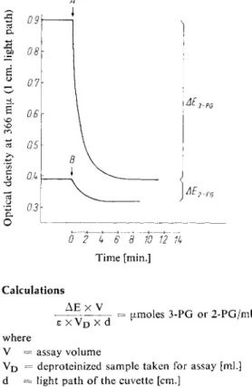
An illustration of the course of the reaction is given in Fig. 1.
A
O
0 2 4 6\ 8 10 12 1k Time [min.]
Fig. 1. Determination of 3-phosphoglycerate (3-PG) and 2-phosphoglycerate (2-PG) in a neutralized extract of rat liver; 1 g. liver (wet weight) in 5.2 ml. extract; 1.5 ml. extract/2 ml.
assay mixture.
A : Addition of phosphoglycerate kinase B: Addition of phosphoglycerate mutase
Calculations A E X V
77 r = umoles 3-PG or 2-PG/ml. deproteinized sample s x V
D
x dr
where
V = assay volume
Vpj = deproteinized sample taken for assay [ml.]
d = light path of the cuvette [cm.]
z — extinction coefficient of D P N H [cm.
2
/[xmole]
£ 3 6 6 = 3.30 18)
£ 3 4 0 = 6.22 19)
£334 = 6.0920)
Specificity
N o work has been done on the specificity of the determination.
18) H. J. Hohorst, Biochem. Z. 328, 509 [1957].
19) A. Kornberg and W. E. Pricer in E. E. Snell: Biochemical Preparations. Wiley, N e w York 1953, Vol. Ill, p. 20.
20) G. Beisenherz, Th. Biicher and K. H. Garbade in S. P. Colowick and N. O. Kaplan: Methods in Enzymology. Academic Press, N e w York 1955, Vol. 1, p. 391.
D-3-Phosphoglycerate, D-2-Phosphoglyeerate, Phosphoenolpyruvate
Determination with phosphoglycerate mutase, enolase, pyruvic kinase and lactic dehydrogenase
Principle
3-Phosphoglycerate (3-PG), 2-phosphoglycerate (2-PG) and phosphoenolpyruvate (PEP) can be determined by means of the following reactions:
PGM
(1) 3-PG ^ ^ 2-PG enolase
(2) 2-PG , - P E P
Mg2+
PK
(3) PEP + A D P ^ = = ^ pyruvate + A T P
Mg2+, K+
LDH
(4) Pyruvate + D P N H + H+ , lactate + D P N +
The measure of the over-all reaction is the decrease in optical density at 366 mpi due t o the oxidation of D P N H (last step in the series of reactions). Quantitative conversion is assured because of the positions of the equilibria of the reactions catalysed by pyruvic kinase (PK) and lactic dehydrogenase
(LDH) 4,2i).
Biological material m a y contain 10 times more 3-PG than 2-PG, P E P and pyruvate. In such cases, the accuracy o f the assay o f all four compounds is not very high. The accuracy can be increased by determining the 2-PG and PEP separately in larger samples or by carrying out the combined assay at 340 mu,, thus increasing the sensitivity o f the measurements of optical density.
3-PG can only be determined under the conditions described here if the assay mixture contains less than 10~
3
M inorganic phosphate
1 6
>. However, the inhibitory effect of phosphate can be considerably reduced if the same volume o f MnSC>4 solution (IV) is added to the assay mixture instead o f the MgSC>4 solution. The determination of 2-PG, PEP and pyruvate is not affected by phosphate.
Reagents
1. Triethanolamine hydrochloride, A. R.
2. Potassium chloride, A. R.
3. Magnesium sulphate, MgSC>4-7H20, A. R.
4. Manganous sulphate, MnSC>4-4H20, A. R.
5. Adenosine diphosphate, ADP
sodium salt, ADP-Na3; commercial preparation, see p. 1004.
6. 2,3-Diphosphoglycerate, 2,3-di-PG
(brucine)5-salt prepared from pig blood according t o
1 3
) , from D-3-phosphoglycerate and A T P with an extract of acetone-dried chicken breast muscle according t o
1 4
) or as the Bas-salt accord
ing to
1 5
>.
7. Reduced diphosphopyridine nucleotide, DPNH
sodium salt, DPNH-Na2- Commercial preparation, see p. 1011.
8. Ethylene-diamine-tetra-acetic acid, EDTA
disodium salt, E D T A - N a
2
H2
. 2 H2
0 (Titriplex III, Trilon B, Versene).21) / . T. McQuate and M. F. Utter, J. biol. Chemistry 234, 2151 [1959].
230 Section B : Estimation o f Substrates
9. Lactic dehydrogenase, LDH
crystalline, from rabbit skeletal muscle
1 0
\ suspension in 2.2 M a m m o n i u m sulphate solution.
Commercial preparation, see p. 986.
10. Pyruvic kinase, PK
crystalline, from rabbit skeletal m u s c l e
1 0
* , suspension in 2.1 M a m m o n i u m sulphate solution.
Commercial preparation, see p. 997.
11. Enolase
crystalline, from rabbit skeletal m u s c l e
9
.
1 7 )
, suspension in 2.6 M ammonium sulphate solution.
Commercial preparation, see p. 973.
12. Phosphoglycerate mutase, PGM
crystalline, from rabbit skeletal m u s c l e
1 6
.
1 7 )
, suspension in 2.4 M ammonium sulphate solution.
Commercial preparation, see p. 995.
13. Perchloric acid, A. R., sp. gr. 1.67; ca. 70% (w/w) 14. Potassium hydroxide, A. R.
15. Hydrochloric acid, A. R., I N 16. Sodium hydroxide, A. R., I N 17. Diethyl ether, A. R.
Purity of the e n z y m e preparations
The preparations should have the specific activities stated in the following Table and the amounts of the contaminants should not be greater than those laid down in columns 2 to 5.
Contaminants
Enzyme Specific activity Units as % of the total activity
P K Enolase P G M L D H
L D H 17000 units *)/mg. protein 0 0 0 -
PK 6500 units/mg. protein — 0.001 0 0.04
Enolase 2 500 units/mg. protein 0 0 0.04
P G M 30000 units/mg. protein 0.2 0 0.6
Preparation of Solutions
I. Triethanolamine buffer (0.2 M; pH 7.6):
Dissolve 9.3 g. triethanolamine hydrochloride in ca. 200 ml. doubly distilled water, add 3.7 g. EDTA-Na 2 H 2 , adjust to pH 7.6 with ca. 9 ml. 2 N NaOH and dilute to 250 ml. with doubly distilled water.
II. Potassium chloride (2 M):
Dissolve 14.9 g. KC1 in doubly distilled water and make up to 100 ml.
III. Magnesium sulphate (0.5 M):
Dissolve 12.3 g. M g S ( V 7 H 2 0 in doubly distilled water and make up to 100 ml.
IV. Manganous sulphate (0.005 M):
Dissolve 1.11 g. MnS04-4HiO in doubly distilled water and make up to 1000 ml.
V. Adenosine diphosphate (ca. 0.01 M ADP):
Dissolve 51.1 mg. ADP-Na3 in doubly distilled water and make up to 10 ml.
*) Definition o f the units according t o
1 0
) , see p. 33.
VI. 2,3-Diphosphoglycerate (ca. 0.01 M 2,3-di-PG):
Suspend 30 mg. (brucine)5-salt (molecular weight 2260) in 1.5 ml. doubly distilled water. Precipitate the brucine with 0.06 ml. 1 N NaOH (curd-like precipitate).
Centrifuge for 5 min. (3000 g). Wash the precipitate with 0.5 ml. doubly distilled water, centrifuge and extract the combined supernatants twice with 10 ml. portions of diethyl ether (shake for 5 min.). Separate off the aqueous phase and free from residual ether by evacuating (water pump). Adjust to pH 6—7 with about 0.01 ml.
1 N HC1 (universal indicator paper, Merck).
VII. Reduced diphosphopyridine nucleotide (ca. 0.01 M (3-DPNH):
Dissolve 7 mg. DPNH-Na2 in 1 ml. doubly distilled water or buffer (solution I).
VIII. Lactic dehydrogenase, LDH (ca. 15 mg. protein/ml.):
Use as a suspension or centrifuge and dissolve the sediment in the original volume of doubly distilled water.
IX. Pyruvic kinase, PK (ca. 10 mg. protein/ml.):
Use as a suspension or centrifuge and dissolve the sediment in the original volume of doubly distilled water.
X. Enolase (ca. 5 mg. protein/ml.):
Use as a suspension or centrifuge and dissolve the sediment in the original volume of doubly distilled water.
XL Phosphoglycerate mutase, PGM (ca. 10 mg. protein/ml.):
Use as a suspension or centrifuge and dissolve the sediment in the original volume of doubly distilled water.
XII. Perchloric acid (ca. 6% w/v):
Dilute 52 ml. 70% (w/w) HCIO4 to 1000 ml. with doubly distilled water.
XIII. Potassium hydroxide {ca. 10N):
Dissolve 40 g. KOH in doubly distilled water and make up to 100 ml.
Stability of the s o l u t i o n s
Prepare the D P N H solution freshly each week. Prepare the MnSC>4 solution freshly for each series of determinations. All the other solutions are stable practically indefinitely between 0 and 5°C.
Procedure
Experimental material and deproteinization
Deproteinize samples with perchloric acid (solution XII) and then adjust to pH 3 —4 with KOH (solution XIII) (cool in ice). For a complete description, see p. 254. For correction of the analytical results for the blood content of the tissue, see p. 549.
Spectrophotometric m e a s u r e m e n t s
Wavelength: 366 mu.; light path: 1cm.; final volume: 2.25 ml.*). Measure against air.
Pipette successively into the cuvette:
0.500 ml. buffer (solution I) 0.075 ml. KC1 solution (II) 0.032 ml. M g S 0 4 solution (III)
(or M n S 0 4 solution (IV))
*) The addition of the enzyme solutions or suspensions increases the assay volume by less than 1 %.
232 Section B : Estimation of Substrates
0.050 ml. ADP solution (V) 0.025 ml. 2,3-di-PG solution (VI) 0.040 ml. DPNH solution (VII) up to 1.500 ml. deproteinized sample.
Equilibrate the assay mixture at ca. 25°C (5 to 10 min. in a constant temperature cuvette holder). Mix in
0.001 ml. LDH suspension or solution (VIII) (130 units),
wait for the end of the reaction and then read the optical density E i . Mix in 0.002 ml. PK suspension or solution (IX) (about 60 units).
On completion of the reaction (5 — 10 min.) read the optical density E2. Mix in 0.02 ml. enolase suspension or solution (X) (40 units).
On completion of the reaction (5 to 10 min.) read the optical density E3. It should not change significantly within 3—5 min. If a small constant "drift" occurs, which continues after the complete conversion of the 3-PG, extrapolate to zero time (refer to p. 39). Mix in
0.002 ml. PGM suspension or solution (XI) (ca. 100 units) and after 10 — 15 min. read the optical density E4.
A
Fig. 2. Determination of 3-phosphoglycerate (3-PG), 2-phosphoglycerate (2-PG), phos
phoenolpyruvate (PEP) and pyruvate (Pyr.) in a neutralized extract of rat liver (1 g.
liver; wet weight, in 5.2 ml. extract; 1.5 ml.
extract/2 ml. assay mixture).
A : Addition of lactic dehydrogenase B: Addition of pyruvic kinase C: Addition o f enolase
D : Addition of phosphoglycerate mutase
0 2k 6 8 10 12 1k Time [min.]
The following are used for the calculations:
Ei — E
2
= A EP E P ; E2
— E3
= A E2
- P G ; E3
— E4
= A E3
. P G .The optimum concentration of all three metabolites is 0.06 to 0.4 [Jimoles/assay mixture.
The sensitivity of the analysis is doubled if the optical density is read at 334 or 340 ma (use only 0.020 ml. DPNH solution).
An illustration of the course of the reaction is given in Fig. 2.
Calculations
A E x V
u.moles 3-PG, 2-PG or PEP/ml. deproteinized sample e X VD X
d
where
V = volume of the assay mixture
V
D
= volume o f the deproteinized sample taken for assay [ml.]d = light path o f the cuvette [cm.]
e = extinction coefficient o f D P N H [cm.
2
/[xmole]
£ 3 6 6 = 3.30 18)
£
3
4 0 = 6.22 19)£334 = 6.09
2
0 )
Specificity
The reactions catalysed by enolase and pyruvic kinase guarantee the high specificity o f the determi
nation described here 22). L-2-PG and the homologues of D-2-PG do not react with enolase. Apart from P E P , no substrate of pyruvic kinase is k n o w n which can give a reaction product capable of reacting with lactic dehydrogenase.
22) F. Wold and C. E. Ballou, J. biol. Chemistry 227, 313 [1957].