Rickettsiae and Rickettsioses
ALOYSIUS KRIEG
Biologische Bundesanstalt für Land- und Forstwirtschaft, Institut für biologische Schädli?igsbekämpfung, Darmstadt, Germany
I. Introduction 577
II. Diagnosis and Identification 578
A. Morphological Examination 579
Β. Immunofluorescence Technique 580
C. Serological Examination 581
D. Bioassay 582
III. Cytology and Chemical Composition 583
IV. Metabolism 587
V. Resistance of Rickettsiae 588
A. Climatic Conditions (Temperature, Humidity) 588 B. Range of pH, Isotonic Media, and Nutritional Stress 589 C. Disinfectants, Chemotherapeutics 589
VI. Preparation and Cultivation 590
A. Isolation 590
B. Culturing 591
VII. Pathology 592
A. Developmental Cycle 592
B. Special Pathology 595
VIII. Epizootiology 608
A. Rickettsiae without Host Alteration 608 B. Rickettsiae with Host Alteration 609
References 611
I . INTRODUCTION
Rickettsiae are usually considered as intermediates between viruses and bacteria. There exist two theories as to their nature. T h e virus theory is expressed in the taxonomy of Zhdanov (1957) and also in that of Philip (1956). T h e taxonomy of Pinkerton (1936, 1942),
577
however, considered the rickettsiae as bacterialike organisms. Some authors concluded from the heterologous serological Weil-Felix reaction that many rickettsiae are closely related to some serotypes of Entero- bacteriaceae. Today, other arguments are quoted in support of the bacteria theory: the rickettsiae contain both nucleic acids and they also possess enzymes for metabolism which can be inhibited by chemo- therapeutics and antibiotics. In addition, they have a bacteriumlike structure and multiply by binary fission as demonstrated by electron micrographs. In contrast to viruses, some of the rickettsiae do not grow exclusively in living cells, but also extracellularly or better epicellularly.
There is no evidence that true rickettsiae, dealt with here, are a missing link between viruses and bacteria.
Many properties of rickettsiae resemble extensively those of Bru- cellaceae. Therefore, extracellularly growing rickettsiae may be placed between arthropod (and vertebrate) -adapted types of the genus Pas
teurella and intracellularly growing rickettsiae. Another possibility is the evolution of rickettsiae from obligate symbiotic (i.e., mutualistic) bacteria such as are found in insects. Since rickettsiae are obligate parasites or commensals of arthropods, there might possibly have been a coevolution of arthropod hosts and their rickettsiae with correspond
ing genetical adaptation. T h i s view is substantiated particularly by the existence of a balanced relation between pathogen and host, as reported for Dermacentroxenus spp. living in ticks and for Rickettsoides spp.
living in insects. For most of those rickettsiae which are closely related to their hosts, also vertical transmission is known.
Arthropod-borne rickettsiae have been found in Arachnoidea (e.g., Acarina), Insecta (e.g., Aphaniptera, Anoplura, Mallophaga, Diptera, Coleoptera), and also in various vertebrates (several birds, and mam
mals, including m a n ) .
According to their bacterialike nature Krieg (1961), in contradiction to Zhdanov and Philip, proposed Rickettsoideae as a new taxonomic class within the phylum Protophyta containing the order Rickettsiales.
T h e Rickettsiales comprise the two families Chlamydozoaceae and Rickettsiaceae. Only the latter family is treated here. In accordance with Philip (1956), the Rickettsiaceae may be divided according to their tropisms into three tribes: Rickettsieae, Ehrlichieae, and Wol- bachieae.
I I . DIAGNOSIS AND IDENTIFICATION
For the identification of rickettsiae, four types of laboratory tech
niques are available: (1) the morphological examination; (2) the immunofluorescence technique; (3) the serological test; and (4) the bioassay. T h e bioassay includes the use of arthropods and also of
small laboratory animals (guinea pigs, mice, etc.) in the case of rickettsiae pathogenic for mammals.
A. Morphological Examination
Morphological examination offers valuable evidence, but rickettsiae cannot be determined by simple morphological properties. They are minute microorganisms, 0.2 to 0.3 by 0.3 to 3.0 μ in size, and without any active movement. Except for the smallest forms (e.g., Coxiella spp., Rickettsiella spp., Enterella spp.), they are not filterable. For micro
scopic examination of fresh smears, dark-field illumination or phase contrast is used. T h e Brownian movement of the minute, highly refringent rickettsiae is very characteristic. "Early" stages of rickettsiae, however, often arrange themselves in chainlike structures and are larger than the older ones. Since the early stages are less refringent, it is often difficult to detect them in fresh smears. Otherwise, these forms are readily stained with aniline dyes, and are mostly Macchiavello-negative.
By using acridine orange as fluorochrome, younger forms give a strong green fluorescence in ultraviolet light whereas the older ones do not (Krieg, 1955a). Krieg proved the usefulness of fluorochrome auramine for staining "mature" rickettsiae. Staining of the latter with the usual basophilic fluorochromic or diachromic dyes, however, will give no satisfactory results. For distinguishing mature rickettsiae in smears, therefore, two special staining methods are recommended: the Macchia- vello technique and the Giemsa method. T h e first one is based upon the weak acid-resistance of rickettsiae, which resembles that of acid-fast mycobacteria. Therefore, basic fuchsin (as in the classical Ziehl-Neelsen method) is a useful dye for demonstrating rickettsiae and was accepted by Macchiavello for his technique.
A modified Macchiavello procedure for air-dried smears fixed for 5 to 10 minutes in methanol, is the following: Staining in 1 percent solution of basic fuchsin for 60 minutes; then 5 seconds' differentiation in a 0.5 percent solution of citric acid. Another modification: Staining in 1 percent solution of basic fuchsin under heating to 95 °C for 45 seconds; then 90 seconds' differentiation in a 0.5 percent solution of citric acid. In connection with Macchiavello's technique, counterstaining in a 1 percent solution of methylene blue (for 20 seconds) is used.
T h e Results are: rickettsiae, crystals, and globules of sphaeroidocytes are stained red, nuclei, albuminoid spheres, and also bacteria are counterstained blue.
In tissue sections the polychromatic Giemsa solution (azur-eosin) gives better results than basic fuchsin. Fixation in Susa's, Zenker's, or Helly's fluid is suggested. T h e sections are placed for 12 to 24 hours in
diluted (2:100 or 4:100) buffered dye solution (pH 7.4 to 7 . 6 ) . After dehydration in acetone-xylene they should be embedded over xylene in a permanent medium, e.g., Canada balsam. T h e results are: rickettsiae violet, nuclei violet, albuminoid spheres blue, crystals and globules of sphaeroidocytes red.
Using the Feulgen technique (or Giemsa staining after HCl hydrol
ysis) the rickettsiae react strongly positive (in contrast to intact granules or capsules of Bergoldiavirus spp.). T h i s technique is, therefore, proposed as a significant diagnostic tool. (It is important to know for differential identification that capsules of Bergoldiavirus also do not react positively to Macchiavello or Giemsa staining techniques.) With Gram's stain rickettsiae, and also capsules, give negative results.
B. Immunofluorescence Technique
This method is a specific one to detect antigens in tissues and cells.
It represents a combination of antigen-antibody reaction and histologic staining technique and was initiated by Coons et al. (1950). Air-dried smears or frozen sections, unfixed or fixed in ethanol at —70°C, are incubated at 37°C with antibody γ-globulin labeled with fluorescein isothiocyanate or other fluorochrome radicals.1 T h e n the slides are rinsed in buffered NaCl solution and examined microscopically with fluorescence equipment.
Coons et al. (1950) localized and identified by this technique Rickettsia prowazekii da Rocha-Lima and Dermacentroxenus rickettsii Wolbach in smears of exudates and in frozen sections of vertebrate tissues by means of labeled homologous antibody γ-globulin. Rickettsia prowazekii was also identified in smears from infected Pediculus humanus capitis deGeer. Further, it was possible to differentiate in this way Rickettsia prowazekii from other rickettsiae which accompany them in the gut of lice [e.g., Rickettsoides pediculi (Münk et da Rocha- Lima) Krieg and Rochalimaea quin tana (Schmincke) Macchiavello].
Burgdorf er and Lackman (1960) used the same method as a diagnostic tool for detection of Dermacentroxenus rickettsii in smears prepared from gut of infected Dermacentor andersoni Stiles. In smears of nymphal ticks fed on infected guinea pigs, the rickettsiae were seen in a bright fluorescence. Smears prepared from infected ticks molting into adults, however, emit a reduced fluorescent light. Using this technique, it is not difficult to differentiate Dermacentroxenus rickettsii from the harmless
ι For preparing antibody γ-globulin, hyperimmunized diagnostic sera from rabbits are used. Conjugation with the fluorochromic component is accomplished by a direct method according to Coons et al., or by an indirect one using fluorescein- conjugated anti-rabbit globulin from sheep.
Wolbachia dermacentrophila (Steinhaus) Philip which occurs through
out the gut-epithelium cells of the healthy ticks. Roberts and Downs (1959) used the immunofluorescence method for detection of Coxiella burnetii (Derrick) Philip in infected cells.
In connection with Rickettsiella infections, several authors have de
scribed associated crystals. Up until the present day, their nature and origin has been disputed (see Section V I I , B ) . It was stated recently in the immunofluorescence studies of Krieg (unpublished data) that the coat of crystals associated with Rickettsiella melolonthae is antigenically related to the surface antigen of typical rickettsiae. This result confirms the observations of Huger (1962) that the crystals originate within pleomorphical rickettsiae.
C. Serological Examination
Besides the immunofluorescence technique, the following classical serological tests are also employed for identification: Weil-Felix test, neutralization test, complement-fixation test and agglutination test.
Except for the Weil-Felix test (see below), they all are homologous reactions.
For classical serological examinations and for the immunofluores
cence technique, diagnostic antisera may be prepared by using rabbits (or guinea pigs). For this purpose, native or heat-inactivated rickettsiae isolated from arthropods or from yolk-sac cultures (see Section V I , A) are suspended in isotonic salt solution and injected twice into rabbits intravenously. I f the antigen is suspended in Freund's "Bacto-adjuvant incomplete"2 intramuscular administration into rabbits or intraperito
neal injection into guinea pigs is used. T h e injections are given 1 week apart. A further booster injection may increase the titer. Animals are bled by heart puncture 2 weeks later.
For detection of Rickettsia infections in guinea pigs, the comple
ment-fixation test is very useful as a diagnostic tool 21 to 30 days after infection. T h e neutralization test in guinea pigs is also available for identification of rickettsiae pathogenic for mammals, such as Derma- centroxenus rickettsii Wolbach and Zinssera tsutsugamushi (Hayashi) Macchiavello. Krieg used the homologous agglutination test especially for the identification of Rickettsiella melolonthae (Krieg) Philip and related species infecting insects (Krieg, 1955b, 1958b).
T h e discovery by Weil and Felix (1916) that the serum of patients infected with Rickettsia prowazekii, or other rickettsiae, may contain
2 This adjuvant contains mannide monooleate in paraffin oil (DIFCO Laboratories, Detroit, Michigan).
heterologous agglutinins which react specifically with Proteus vulgaris serotype O X , is important for practical diagnostic use.
It is interesting to note that the W F antibody induced by rickettsiae gives good agglutination with the Ο antigen of cells of distinct Proteus strains, but no comparable agglutination with the rickettsiae themselves.
T h e investigation of Westphal et al (1947) on this heterological sero- reaction suggests that Rickettsia prowazekii and Proteus OX-19 contain nearly the same polysaccharide antigen. It is localized on the surface of the cell wall of Proteus in contrast to rickettsiae in which this antigen is probably bound to an inner layer of the cell. Since parts of rickettsiae will distintegrate during typhus fever, the masked W F antigen becomes free and is now able to induce formation of W F antibody in patients.
Therefore, a serological reaction between the W F antibody and the intact rickettsiae is impossible.
T h e serological patterns of Rickettsieae according to the W F reaction have been described by Plötz et al (1948) : Rickettsiae which induce W F antibodies of the Proteus type OX-19 are grouped in the genera Rickettsia and Dermacentroxenus. Members of the latter genus also react as heterophiles with the Proteus type OX-2. Other rickettsiae induce W F antibodies which do not react with the Proteus type OX-19 or OX-2 but with the Proteus type OX-K. They belong to the genus Zinssera. T h e rickettsiae of the genera Rochalimaea and Coxiella do not induce Proteus O X agglutinins for W F reaction. After inoculation of Rickettsiella melolonthae, which is known to be less pathogenic for vertebrates (Krieg, 1955b), into mammals, no W F antibodies of the Proteus types OX-19, OX-2, or O X - K could be detected (Krieg, 1962).
D. Bioassay
According to Weigl (1924) and others, lice were used for bioassay (xenodiagnosis). T h i s technique is especially indispensable for the identification of Rochalimaea quintana, which cannot be tested in mammals (Weyer, 1948b). For xenodiagnosis, 30 to 80 lice are fed on patients. Another arthropod test is the rectal inoculation of infected material into lice. By feeding, only 30 percent may be successfully infected; by artificial application, however, 100 percent. After 4 to 6 days, smears from lice are examined microscopically.
According to Spencer and Parker (1930), Weyer (1948a), and Kordovä and Rehäcek (1959), ticks are also very useful for the xenodiagnosis of rickettsiae. Infectious material is introduced into the hemocoel of the ticks. After the latter are fed with blood of healthy animals, 10 to 30 days after inoculation, smears of hemolymph and preparations from different organs of the dissected arthropods are
examined. (For the inoculation technique see Section VI, B , and for pathological details see Section V I I , B.)
In addition, for aiding in the identification of vertebrate-related rick
ettsiae, an animal test with rodents is suggested: Species of the tribe Rickettsieae (with exception of Rochalimaea quintana), induce fever and scrotal reaction (the latter revealed as a periorchitis) in 60 to 70 percent of the injected guinea pigs or mice, after the first passage.
For testing Cowdria spp., however, ferrets are more suitable.
Intraperitoneal, subcutaneous, intracerebral, intravenous, intrapul- monary (intranasal route), and intratesticular application of rickettsiae can also be used. Blood, brain, liver, spleen, lungs, and testes (scrotal reaction) are examined. In the blood of most treated guinea pigs and other mammals antibodies for serological studies are present. When extracts of infected arthropods are injected into chick embryos, morpho
logical examination often may not be successful until after 3 to 6 successive passages. Quantitative tests (titrations) are carried out by determining the minimal infecting dose (ID) after inoculation of dilutions into suitable hosts.
I I I . CYTOLOGY AND CHEMICAL COMPOSITION
Light-microscope observations demonstrate rickettsiae to be very small rod-shaped microorganisms, growing singly or in chains. Sikora
(1942) stated that most of the rickettsiae show coccoid forms under favorable conditions and that they become bacilliform during multi
plication. Schaechter et al. (1957) studied the growth of Dermacen- troxenus rickettsii in cultures of rat fibroblasts by phase contrast microscopy. Transverse binary fission of D. rickettsii could be observed and recorded in series of microphotographs of single rickettsiae. Atypical (partly extreme pleomorphical) forms of rickettsiae were described by Sikora (1942), Gönnert (1948), and Huger (1962).
As in bacteria also in rickettsiae, nuclear equivalent structures can be demonstrated with basic dyes (Giemsa solution or methyl green) after removal of the cytoplasmic R N A with RNase or Ν HCl. Ris and Fox (1949) found this in Rickettsia prowazekii, and Krieg (1955a) in Rickettsiella melolonthae. Feulgen's technique and ultraviolet (UV) microscopy (at λ = 253.7 ιημ) too, are very useful for distinguishing nuclear equivalents. Giemsa stain after HCl hydrolysis reveals this as well. NaCl solution has no influence on staining of these structures.
Always one central nuclear equivalent is present in resting cells, two in cells which are in binary fission. Ris and Fox (1949) detected large amounts of R N A in the cytoplasm of Rickettsia prowazekii by means of ribonuclease (RNase). After fixation in formalin, rickettsiae were
stained uniformly red by pyronine. Preceding treatment of rickett
siae with RNase or physiological NaCl solution did not produce any reaction with pyronine. Polar granules in rickettsiae are often made visible by basic dyes. They are Feulgen negative, do not react with Neisser's stain, and must be clearly distinguished from Feulgen-positive nuclear equivalents in rickettsiae undergoing cell division. It is difficult to demonstrate all these details easily by means of the light microscope.
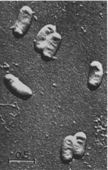
Only electron micrographs (Fig. 1) allow exact measurements of rickettsiae. Rickettsia cells range in size from about 200 to 300 πιμ in diameter and from 250 to 900 πιμ (maximum about 3000 πιμ) in length.
They are mostly opaque to the electron beam. Untreated or Os04-fixed air-dried rickettsiae, shadowed with metal or SiO, reveal a cell wall surrounding an often shrunken protoplast (Weyer and Peters, 1952;
Wissig et al, 1956).
FIG. 1. Electron micrograph: Entire cells of Rickettsiella melolonthae, shadowed with SiO ( χ 25,000).
T h e cell wall can be isolated by cautious treatment of rickettsiae with a 1 percent solution of sodium deoxycholate. Chemical analysis by acid hydrolysis of purified cell walls of Rickettsia typhi indicate that their constituents are amino acids (12 types), sugars (glucose, hexos- amine, glucuronic acid, galactose), and small amounts of ribonucleic
acid (RNA) (Schaechter et al., 1957). Diaminopimelic acid, a compo
nent of cell walls in many bacteria, has not been observed. On the other hand, glucuronic acid, found in rickettsiae, has so far not been reported as an element of bacterial cell walls. By using lysozyme, Allison and Perkins (1960) could break down the cell walls of Coxiella burnetii and Rickettsia typhi. One of the degradation products is muramic acid, which is known as a typical element of bacterial cell walls.
In ultrathin sections the cell walls of rickettsiae have been found to be approximately 25 ηΐμ thick [18 to 30 ιτιμ: Schaechter et al. (1957) in Zinssera tsutsugamushi; 5 to 10 ιημ: Stoker et al. (1956) in Coxiella burnetii; 15 to 30 ιημ: Krieg (1960) in Rickettsiella melolonthae].
Schaechter et al. and Krieg found the cell wall to consist of two membranes (thickness 4.5 to 7.0 ιημ each) separated by a less dense layer
(thickness 8 to 12 ιημ). According to electron micrographs of ultrathin sections of Rickettsia typhi (Wissig et al., 1956), Coxiella burnetii
(Stoker et al., 1956), and Rickettsiella melolonthae (Krieg, 1960), the protoplast of these rickettsiae appears inside its membrane in a very dense central mass surrounded by a zone of moderately dense material.
T h e central mass, the shape of which varies greatly (rod-shaped, comma- shaped, or sigmoid) often lies in an elongated vacuole, which may be an artifact. T h e dense central mass resists acid hydrolysis (as used in the Feulgen technique) and RNase. Therefore, it is assumed to be the nuclear equivalent of the rickettsial cell. It is possible that the DNA- containing structures within the rickettsiae are organized similarly as the chromosomelike nuclear equivalents of bacteria.
T h e structure of small rickettsiae (Coxiella spp. and Rickettsiella spp.) resembles somewhat that of virus capsules of Bergoldiavirus spp.:
a rod-shaped dense center surrounded by a capsulelike structure. In contrast to capsule protein of Bergoldiavirus, however, the protoplast of rickettsiae is not dissolved quickly by weak alkali (Krieg, 1957, 1960). For further differences see Section I I , A. Rickettsiae usually show denser areas near the poles of their cell, which look like globules in electron micrographs of shadowed preparations. These subpolar areas, also observed by light microscopists (see above), are dissolved by acid hydrolyses, but do not evaporate during intensive electronic bombardment. They are thought to be traces of R N A proteins, not metaphosphates (Krieg, 1955a).
Tovarnickij et al. (1946) and Cohen (1946) analyzed rickettsiae preserved in NaCl solutions or treated with phenol. They reported the presence of DNA in typhus rickettsiae, but not RNA, and the authors concluded that rickettsiae might be similar to viruses in containing only one type of nucleic acid. On the other hand, Smith and Stoker (1951),
Wyatt and Cohen (1952), Cohn et al. (1958), and Krieg (1958a) dem
onstrated R N A in Coxiella burnetii, Rickettsia prowazekii, Rickettsia typhi, and Rickettsiella melolonthae, respectively. Smith and Stoker
(1951) found a ratio of RNA:DNA in Coxiella burnetii near 1:3. Krieg (1958a), however, stated a ratio of 2.2:1 for Rickettsiella melolonthae, and Cohn et al. (1958) estimated it as 3.5:1 for Rickettsia typhi. T h e re
sults of Krieg and of Cohn et al. agree with statements of Leslie (1955) on classical bacteria. T h e low value observed by Smith and Stoker is due to unstable soluble RNA, according to Cohn et al. (1958). Smith and Stoker (1951) working with Coxiella burnetii and Wyatt and Cohen (1952) working with Rickettsia prowazekii found the DNA to contain the bases adenine, thymine, guanine, and cytosine; methylcytosine was never present. Smith and Stoker noted the DNA of Coxiella burnetii grown in chick embryo to be closely related in its composition to the DNA of the embryo; therefore, the authors suggested that Coxiella had incorporated the DNA of its host. Wyatt and Cohen (1952), how
ever, cultivated Coxiella burnetii and Rickettsia prowazekii in the same host (chick embryo) and found the DNA specific for each of the two rickettsiae. Both nucleic acids could be characterized by their different ratios (adenine -f- thymine : guanine - { - cytosine). This result contrasts with the hypothesis of Smith and Stoker, who supposed that equal ratios should be found with both rickettsiae. In addition, the results of Wyatt and Cohen demonstrate that in rickettsiae the molar ratios (adenine:thymine) and (guanine:cytosine) are nearly constant, whereas the ratio adenine -f- thymine : guanine -|- cytosine has a char
acteristic value, depending on the type of rickettsia DNA.
T A B L E I
MOLAR PROPORTIONS OF PURINE AND PYRIMIDINE BASES IN D N A OF RICKETTSIAE^
Host Rickettsia A : T G:C A + T : G + C
Chick embryo C. burnetii 1.13 1.02 1.25
Chick embryo R. prowazekii 1.12 1.11 2.08
a After Wyatt and Cohen (1952).
Morphology, chemical composition, and also biological activities of rickettsiae are sensitive to so-called physiological salt solutions. For Rickettsia prowazekii, Rickettsia typhi, and Rochalimaea quintana especially, Weyer and Peters (1952) reported that the protoplasm of these rickettsiae is turned into globular elements after staying in 0.14 m NaCl at 0°C for two hours. These authors, and also Ris and Fox
(1949), referred to the solubility of nucleic acids in NaCl solutions.
In agreement with this, Cohn et al. (1958) observed an emission of
unstable nucleic acids (RNA) from starving rickettsiae in isotonic media and, simultaneously, the biological inactivation of the rickettsiae.
T h e fact that some authors could not find R N A in their purified rickett
siae suspensions and that others observed only a very low RNA:DNA ratio may be explained by the solubility of rickettsia RNA.
There are only earlier analyses of the different constituents of purified rickettsiae. Tovarnickij et al. (1946) reported a partial quan
titative analysis of total rickettsial cells; their results are the following:
lipoids 46.6 percent, phospholipids 15.8 percent, neutral fat 29.7 percent, protein 34.7 percent, nucleic acids 12.0 percent, carbohydrates 4.1 per
cent, ash 3.0 percent. T h e percentage of nucleic acids may be greater since the authors did not find R N A (it may have been lost by purification or preservation in NaCl solutions).
IV. METABOLISM
In contrast to viruses and in analogy to bacteria, the rickettsiae show metabolic activity. However, it is so extremely heterotrophic that cultivation on simple artificial media has not been successful so far
(see Section V I , B ) .
Purified suspension of Rickettsia typhi oxidize L-glutamic acid on reactions of the tricarboxylic acid cycle (Bovarnick and Snyder, 1949).
This reaction is HCN susceptible. T h e rickettsiae so far examined are also able to consume some intermediate products of the tricarboxylic acid cycle, such as pyruvate, but no glucose. Furthermore, rickettsiae synthesize citrate from acetate or acetyl phosphate (Paretsky et al., 1958). Studies by Hayes et al., (1957) indicate that respiratory metab
olism of Rickettsia typhi to be similar to that of bacteria. T h e electron transport may be accomplished by a disphosphopyridine nucleotide
(DPN) -specific flavinenzyme-cytochrome system. T w o important enzymes of the energy and phosphate metabolism, adenosine tri- and diphos- phatase (ATPase and ADPase), were found by Paretsky et al. (1958) in cell suspensions of Coxiella burnetii. According to the same authors, metabolism of rickettsiae depends essentially on factors such as DPN, A T P , and CoA. This indicates the high degree of parasitism of rickettsiae and the linkage of their own metabolism with that of living host cells.
Bovarnick et al. (1959) and Bovarnick and Schneider (1960) worked with isotope-labeled amino acids such as methionine-S35 and glycine-l-C14 and registered the uptake of these amino acids by Rickettsia prowazekii and their incorporation in the rickettsia's own proteins (on the other hand, acetate-1-C14 was found again exclusively in the lipid fraction).
T h e incorporation of amino acids in the proteins of rickettsiae was
inhibited by chloramphenicol in a way similar to that in the case of protein metabolism of classic bacteria. According to the amino-acid me
tabolism of rickettsiae it is of interest that enzyme preparations (ultra
sonic extracts) of Coxiella burnetii are capable of synthesizing serine from formaldehyde and glycine-2-C14 in the presence of tetrahydrofolic acid (Myers and Paretsky, 1961).
V. RESISTANCE OF RICKETTSIAE
T h e longevity of rickettsiae is influenced by their environment.
Important factors of this are climatic conditions, and the reaction and contents of the medium.3
A. Climatic Conditions (Temperature, Humidity)
T h e differences between the temperature limits of rickettsiae are probably related to the temperature range of their hosts. So Rickettsiella species pathogenic for Coleoptera larvae have a temperature minimum for growth from 14 to 20 °C, an optimum from 25 to 28 °C, and a maximum at 32 °C. Rickettsiae pathogenic for man (Dermacentroxenus rickettsii, Rickettsia prowazekii, and Rickettsia typhi) also have their minimum from 16 to 20°C; their optimum, however, is from 32 to 35°C, and their maximum is at 37 °C. T h e thermal death points for rickettsiae suspended in water are: > 60 minutes at 60°C for Coxiella burnetii, which is surely killed only by boiling; 30 minutes at 60°C for Rocha
limaea quintana; 10 minutes at 60°C for Rickettsiella popilliae; 15 to 30 minutes at 50°C for Rickettsia prowazekii) 10 minutes at 50°C for Dermacentroxenus rickettsii and Zinssera tsutsugamushi.
T h e longevity of different species of rickettsiae differes widely in the temperature range from 4°C to 20°C in aqueous solutions. In some cases (Coxiella burnetii, Rickettsiella melolonthae), suspensions may be infective for a month. Rickettsia typhi survives at 4 ° C a week, Coxiella burnetii some months, and Rickettsiella melolonthae more than one year. Deep-frozen rickettsiae may retain their activation for some months: for example, Rickettsia prowazekii (at — 2 0 ° C ) , eight months; and Rickettsiella popilliae (at —80°C) for more than three years. Dried Coxiella burnetii, Rochalimaea quintana, and Rickettsia prowazekii survive more than one year, and Rickettsiella melolonthae several months, at room temperature. Also, lyophilization for preserva
tion is practical, but the ampules have to be kept at low temperatures (—10 to — 2 0 ° C ) . Addition of preservative colloids such as skimmed
3 T h e literature of Sections V, A and C is too extensive to be cited completely here. Special information has been assembled by Philip (1957) in Bergey's Manual and by Zdrodovskii and Golinevich (1960).
milk (pH 7 . 6 ) , albumin, or peptone protects purified rickettsiae from inactivation during freezing.
B. Range of pH, Isotonic Media, and Nutritional Stress
It has been shown that the hydrogen-ion concentration of the medium is a very important factor for preservation. T h e pH optimum lies near the neutral point (pH 6.4 to 7 . 2 ) . Most rickettsiae, e.g., Rickettsia prowazekii, are resistant to weak alkali (pH 8.0) for an hour, but they are inactivated by weak acid (pH 4.0) within the same time.
Alkali and acids in higher concentration kill the rickettsiae, including the very resistant Coxiella species, in a relatively short time: For example, Coxiella burnetii is inactivated within 80 minutes by 0.5 to 1.0 percent HCl or 1.0 to 2.0 percent NaOH (Zdrodovskii and Golinevich, 1960).
Longevity in aqueous suspensions is dependent on the composition of the medium. At room temperature, most rickettsiae are inactivated within 24 to 48 hours in blood or hemolymph. Physiological saline and distilled water are relatively poor preservatives for most rickettsiae:
in these solutions inactivation takes place at room temperature after 2 to 6 hours. Better results are obtained with Tyrode solution, Snyder solution, or skimmed milk. Bovarnick and Allen (1957) observed the activity of purified suspensions of Rickettsia typhi to decrease rapidly by "starvation" in isotonic media held at 0°C. This result was ascribed to emission of DPN into the medium because reactivation was possible by incubation of inactivated rickettsiae in DPN. Reactivation was effec
tive only when the rickettsiae were kept at 0°C or 4°C. However, Cohn et al. (1958) found that after inactivation by "starvation" at 36°C (3 hours in isotonic media) the rickettsiae could not be restored by incuba
tion in DPN. While glutamic acid, pyruvate, and A T P may prevent in
activation at 0°C (Bovarnick and Allen), but not at 36°C (Cohn et al.), the energy metabolism only interferes with the cold inactivation. Accord
ing to Cohn et al., the decrease of activity at 36°C is very complex and may be connected with an enzymatic decomposition of the nucleic acids of the rickettsiae.
C. Disinfectants, Chemotherapeutics
T h e activity of most rickettsiae is quickly destroyed (within 30 minutes) by 0.5 percent phenol, 0.5 percent formaldehyde, 0.5 percent chloramine, and 0.5 percent lysol. Coxiella burnetii, however, is very resistant to these disinfectants. Other effective germicides are high- molecular amino acids such as DL-(octylaminoethyl)glycine in a 1 percent solution. It is reported that the resistance of rickettsiae to lipid solvents, such as diethyl ether, chloroform, ethylene chloride,
carbon disulfide, is low; but they are resistant to carbon tetrachloride and benzene.
Some aromatic organic acids are able to stop metabolism and growth of rickettsiae. Inhibition by p-aminobenzoic acid is especially strong with most strains of Rickettsia prowazekii, lower with Zinssera tsutsugamushi, and not present in the case of Coxiella burnetii.
This inhibition is reversed by p-oxybenzoic acid. Some mutants of Rick
ettsia prowazekii are resistant to p-aminobenzoic acid, but then these are susceptible to acetylsalicylic acid. Inhibition by acetylsalicylic acid is reversed by ^-aminobenzoic acid. Furthermore, most rickettsiae patho
genic for man are susceptible to chloramphenicol and antibiotics of the tetracycline group (Aureomycin, Terramycin) and the erythromycin group (Erythromycin, Carbomycin, Oleandomycin); they are, however, resistant to penicillin and sulfonamides. On the other hand, Cowdria ruminantium (Cowdry) Moshkovskiy and Rickettsiella popilliae, are susceptible to sulfonamides. T h e latter is also sensitive to streptomycin, but resistant to penicillin and Aureomycin. These inhibitors and anti
biotics may be used as therapeutic agents.
Weiss and Dressier (1962) labeled strains of Rickettsia prowazekii genetically by serial passages in chicken embryos in the presence of in
creasing concentrations of chemotherapeutics. When resistance is re
tained after many drugless passages, it may be suitable as a genetic marker because it is a clearly demonstrable difference between two sub
strains. T h e authors isolated substrains of the Madrid-Ε strain resistant to Aureomycin or Erythromycin or ^-aminobenzoic acid. T h e resistance to these chemotherapeutics was specific; cross resistance was only shown to related drugs (e.g., between Aureomycin and Thiocymetin), but not to unrelated ones.
VI. PREPARATION AND CULTIVATION
A. Isolation
For isolation of rickettsiae from infected arthropods the surfaces of the latter are first treated with ethanol or ethyl ether, decontaminated in an aqueous solution of 0.01 percent merthiolate, and then rinsed in sterile water. T h e infected organ (for instance, the intestine of Pediculus spp. or Stethorus spp., and the fat body of Melolontha spp. or Tipula spp.) or the whole arthropod (especially ticks and mites) is homogenized in sterile water. For isolation and purification of rickettsiae grown in the yolk sac of chick embryo the extraction of the lipids with ethyl ether is necessary. From a 10 percent tissue homogenate, larger particles (e.g., cells, nuclei, mitochandria, albuminoid spheres, crystals, detritus,
etc.) are removed by low-speed centrifugation (1000 g for several times).
T h e turbid supernatant contains the rickettsiae. They are sedimented by high-speed centrifugation (7500 g for 1 hour). Differential centrifuga
tion is recommended to isolate the rickettsiae from the impurities. Small types of rickettsiae (Coxiella spp., Rickettsiella spp., Enterella spp.) may be purified by filtration through filter membranes of cellulose nitrate (mean porosity: 0.6 μ). Hara (1958) reported a method for partial purification of rickettsiae with a cation exchange resin (Amber- lite XE-64) and Hoyer et al. (1958) suggested using cellulose anion exchange (Ecteola-SF) . Dutky (1959) used Celite for the adsorption of rickettsiae, and water for their elution.
Most rickettsiae-infected arthropods can be preserved in a refrigera
tor at 4°C for several days. It should be repeated that purified rickettsiae are sensitive to "starvation" and the preservation in so-called physio
logical salt solutions (see Section V, B ) . B . Culturing
Rickettsiae do not grow in artificial media. Earlier reports of cultivation of Rickettsoides melophagi (Nöller) Krieg and other species in vitro on simple artificial media (e.g., glucose blood-nutrient agar or peptone-gelatin-blood medium) at 26°C to 28°C, as reported by Her tig and Wolbach (1924) and Kligler and Aschner (1931), could not be confirmed (Gubler, 1947; Krieg, unpublished). Therefore, propaga
tion of rickettsiae even today can be accomplished only in the manner of an obligate parasite, like that of viruses.
Cultivation in vivo is possible in arthropods such as lice and ticks and, in some cases, in coleopterous larvae. Infection of lice and ticks may be obtained by feeding them infected blood. For artificial applica
tion into the alimentary tract the paranal route, using glass capillaries, is preferred. About 0.001 ml is administered to one louse. Peroral administration of a rickettsial suspension to Coleoptera larvae is preferable after starvation of the host or by force-feeding with a micro- syringe. Intrahemocoelic applications to coleopterous larvae are carried out by injection through the intrasegmental cuticle of the middle segments (0.03 to 0.05 ml per larva). Introduction into the hemocoel of lice and ticks is possible, through their genital openings, with glass capillaries (0.001 ml per louse; 0.02 to 0.03 ml per tick) . For intra
hemocoelic inoculations 500 units of penicillin per milliliter of infective material might be added. Whereas it is possible to cultivate species of the tribes Rickettsieae and Ehrlichieae in living mammals, or in tissue cultures of mammal cells, this method is not successful in most cases
with species of the tribe Wolbachieae.
For the multiplication of Rickettsieae, also small laboratory animals (guinea pigs, mice, rats, and rabbits) are suitable. Stressors, such as X rays, benzol poisoning, and application of cortisone, enhances their susceptibility. For administering rickettsiae to animals parenterally, suspensions in isotonic salt solutions are necessary; 3 to 5 ml per animal is inoculated intraperitoneally or intratesticularly. T h e rickettsiae grow better at 35 °C than at 37 °C. Therefore, rickettsiae multiply better in the scrotum of mammals than on the peritoneum. After intranasal application rickettsiae thrive very well in lungs of mice and rats.
T h e yolk sac (in some instances even the chorioallantoic membrane) of 5- to 7-day-old chick embryos and other plasma-tissue cultures are very convenient for the cultivation of vertebrate-pathogenic rickettsiae
(Cox, 1938). Here, they grow at 35°C in chick embryos and at 32 °C in tissue cultures. For adaptation, 3 to 6 passages are usually required.
Entodermal cells of chick embryo (the same cells that support the growth of rickettsiae in the yolk sac of the intact embryo) were used for culturing Coxiella burnetii (Weiss and Pietryk, 1956) and Rick
ettsia prowazekii (Weiss and Dressier, 1958). Weiss and Dressier (1960) proved centrifugation to be profitable for application of rickettsiae to entodermal tissue cultures. Embryonated eggs have also been success
fully used (Steinhaus, 1942) to culture the nonpathogenic rickettsia Wolbachia dermacentrophila, in which case the organisms appeared to grow chiefly in the egg fluids.
V I I . PATHOLOGY
In this section, only the insect pathology of rickettsiae is treated.
For information concerning vertebrate pathology it is suggested that the reader consult treatises such as that by Zdrodovskii and Golinevich
(1960).
A. Developmental Cycle
Dutky (1959) observed a dosage-dependent incubation period in infections of Rickettsiella popilliae with the host Popillia japonica.
From this he concluded an exponential multiplication rate of the pathogen. T h e time of duplication was estimated to be from 6 to 11 hours. Hopps et al. (1959) determined the duplication rate of Zinssera tsutsugamushi to be about 8 hours in tissue culture.
An eclipse phase was never found among infections with Rickett
siella melolonthae (Krieg, unpublished). Other workers have referred to the fact that ticks perorally infected with Dermacentroxenus rickettsii are not able to transmit the infection prior to an incubation period
of 9 days. On the other hand, the permanent presence of intact virulent rickettsiae in the inoculated ticks can be demonstrated at any time by injection of tick extracts into guinea pigs.
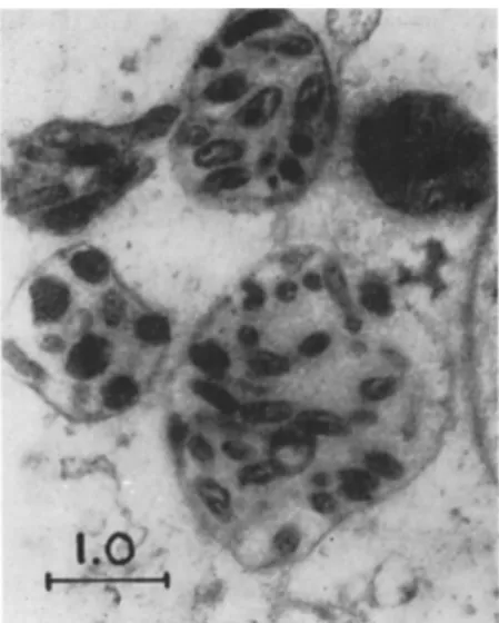
According to Krieg (1960) several individuals of Rickettsiella melolonthae lie in special vacuoles after penetration of the cellular membrane of the host cells. These vacuoles or "initial bodies" (see Fig. 2) swell and the rickettsiae inside them multiply rapidly. T h e number of initial bodies increases and, in the end phase of the cellular infection, the cells contain numerous large vacuoles (also called Herde, Bläschen, Infektionsnester, globular centers, colonies). They are filled with large amounts of rickettsiae (see Figs. 3 and 4) which are suspended in a fluid of low viscosity. Sometimes the vacuoles are reported to be stainable intra vitam with neutral red and are therefore called N R bodies. T h e morulalike cells burst and release the vacuoles or these flock together before the cell ruptures. Such cells, which afterward ap
pear homogeneous, are called Mooser cells. According to results of Wissig et al. (1956), however, a second pathway of rickettsia development in host cells may be suggested. These authors found, in ultrathin sections of chick embryo epithelial cells infected by Rickettsia typhi, that the rickettsiae are scattered throughout the entire cytoplasm between the nucleus and the cell membrane. Thus, the Mooser-cell state may be reached also in a direct manner.
During their passage through ticks, lice, fleas, or larvae of Coleoptera, rickettsiae may undergo morphological change. After invasion of the host cell and in the stage of rapid multiplication they are enlarged ("youth forms" according to Krieg, 1955a). In this early stage of infection chains of rickettsiae were also observed, for example, in the case of Coxiella burnetii (Herzberg et al., 1950), Rickettsia prowazekii (Weyer, 1954), Rickettsiella melolonthae (Wille and Martignoni, 1952;
Krieg, 1955a; Vago, 1959), and Rickettsiella tipulae Müller-Kögler (Müller-Kögler, 1958). It is supposed that these chains are formed dur
ing a phase of intensive reproduction and that they break down into typical ("mature") rickettsiae later on. Further development into atyp
ical or pleomorphical cells (spherical, club-shaped, or with protrusions) and giant forms is described for Rickettsia prowazekii by Sikora (1942) and Gönnert (1948) and for Rickettsiella melolonthae and Rickettsiella tipulae by Huger (1962).
A process of multiplication, such as that which takes place in vacuoles of the cytoplasm, may also occur on the surface of cells. Such epicellular growth is noted in the case of Rochalimaea quintana and Rickettsoides pediculi, both nonpathogenic for their arthropod hosts.
During their epicellular development rickettsiae like Wolbachia melo-
Melolontha melolontha infected with Rickettsiella melolonthae (early stage of the disease), ultrathin section, Os04-fixation, contrasted with U 02 ( χ 12,000). Note the vacuoles of different size (so-called initial bodies) in which the rickettsiae are enclosed.
FIG. 3. Electron micrograph: A portion of cytoplasm in a fat-body cell of Melolontha melolontha infected with Rickettsiella melolonthae (progressed stage of the disease); ultrathin section, O s 04 fixation, contrasted with U OQ ( χ 18,000). Note that the vacuoles are enlarged (one is greater than the sector shown in the figure) and that they contain many rickettsiae partly in binary fission.
594
phagi form chains which build up palisadelike patterns on the terminal cell membranes of the gut epithelium (Anigstein, 1927). In contrast
to the other rickettsiae, species of the genus Dermacentroxenus not only multiply in the cytoplasm of the infected cells of their tick or vertebrate host, but also enter the nuclei.
Fir,. 4. Light micrograph: Smear preparation of fat-body cells of Melolontha melolontha infected with Rickettsiella melolonthae (progressed stage of the disease);
phase contrast ( χ 800). Note that the cytoplasm of the cells contains many large vacuoles (so-called N R bodies) which are filled with rickettsiae (single rickettsiae are not visible) and associated crystals.
B . Special Pathology
Rickettsiae and rickettsialike organisms, as far as is known, are obligate commensals or parasites of arthropods, and frequently of vertebrates. Therefore, their taxonomy is closely related to their patho
genic properties: Rickettsialike organisms which have a distinct affinity only for arthropods belong to the tribe Wolbachieae; such rickettsiae harmful to vertebrates, too, are grouped in the tribe Ehrlichieae if they are pathogenic for monkeys, Carnivora, and Ruminantia, but not for men and Rodentia; and in the tribe Rickettsieae, if they were harmful to men, apes, and many Rodentia (guinea pigs, mice, rats).
T A B L E I I
ARTHROPODS AS VECTORS OF THE T R I B E R I C K E T T S I A E
Rickettsia Arthropod Host
Rickettsia prowazekii da Rocha-Lima
Rickettsia typhi (Wolbach and Todd) Philip
Rochalimaea quintana (Schmincke) Macchiavello
Dermacentroxenus akari (Huebner) Philip and Hughes
Dermacentroxenus australis Philip
Dermacentroxenus conorii (Brumpt) Steinhaus
Dermacentroxenus rickettsii Wolbach
Dermacentroxenus Sibiriens Zdrodovskii
Coxiella burnetii (Derrick) Philip
Pediculus humanus DeGeer
Bdellonyssus bacoti Hirst Nosopsyllus fasciatus (Bosc) Polyplax spinulosa (Burmeister) Xenopsylla astia Rothschild Xenopsylla cheopis (Rothschild) Pediculus humanus DeGeer
Alloder many ssus sanguineus Hirst Bdellonyssus bacoti Hirst
Ixodes holocyclus Neumann
Amblyomma hebraeum Koch Boophilus decoloratus (Koch) Haemaphysalis leachi (Audouin) Hyalomma aegypticum (Linnaeus) Rhipicephalus appendiculatus Neumann Rhipicephalus sanguineus (Latreille) Rhipicephalus simus Koch
Amblyomma americanum (Linnaeus) Amblyomma cajennense (Fabricius) Amblyomma hebraeum Koch Amblyomma maculatum (Linnaeus) Dermacentor andersoni Stiles Dermacentor occidentalis Neumann Dermacentor variabilis (Say)
Haemaphysalis leporispalustris (Packard) Rhipicephalus sanguineus (Latreille)
Dermacentor marginatus Sulzer Dermacentor nutalli Olenev Dermacentor pictus (J. F . Hermann) Dermacentor silvarum Olenev Haemaphysalis concinna Koch Haemaphysalis punctata Canestrini
Amblyomma americanum (Linnaeus) Amblyomma variegaturn (Fabricius) Dermacentor andersoni Stiles Dermacentor occidentalis Neumann
T A B L E II (Continued)
Rickettsia Arthropod Host
Coxiella burnetii (Derrick) Philip (Continued)
Zinssera tsutsugamushi (Hayashi) Macchiavello
Haemaphysalis humerosa Warburton and Nuttal
Haemaphysalis leachi (Audouin)
Haemaphysalis leporispalustris (Packard) Hyalomma detriticum Schulze
Hyalomma dromedarii Koch Hyalomma excavatum Koch Hyalomma lusitanicum Koch Hyalomma savignyi Gervais Ixodes ricinus (Linnaeus)
Ornithodorus hermsi Wheeler et al.
Ornithodorus moubata (Murray)
Rhipicephalus bursa Canestrini and Fanzago Rhipicephalus dentatus Marx
Rhipicephalus sanguineus (Latreille) Trombicula akamushi (Brumpt)
1. The Tribe Rickettsieae
T h e taxonomy of the tribe is influenced by the types of Proteus-OX agglutinins induced by rickettsiae in mammals, especially in human hosts (see Section I I , C ) . On the other hand, Weyer (1954) recom
mended the pathogenic behavior of the tribe Rickettsieae against lice as a taxonomic base. T h e hemolymph of lice is a most favorable medium for all species of Rickettsia, and most of them grow in the intestine of the lice, as well. In their behavior against this host, Rickettsia prowazekii and Rickettsia typhi are closely related and next to them Dermacentro- xenus spp.; however, Rochalimaea quintana, Coxiella burnetii, and Zinssera tsutsugamushi take exceptional positions. T h e classification is furthermore in agreement with the vector spectrum of the rickettsiae.
Species of the genera Rickettsia and Rochalimaea are transmitted by insects (lice or fleas) ; Coxiella spp., Dermacentroxenus spp. and Zinssera spp. are transmitted by arachnids (ticks or mites) (see T a b l e I I ) .
a. The genera Rickettsia and Rochalimaea (insect-borne group).
Rickettsia prowazekii and Rickettsia typhi are pathogenic for lice. They grow within the cytoplasm of the gut epithelium cells when applied perorally. T h e infected cells hypertrophy, become circular and are dis
charged from the gut-epithelium layer. T h e multiplication of rickettsiae within the cells is so intensive that these will burst eventually and liberate the rickettsiae into the lumen of the gut. T h e gut contents and feces show masses of infective fragments of gut cells plus rickettsiae.
Since th e destroye d cell s o f th e epitheliu m ar e no t regenerated , th e gu t wall i s irreparabl y damaged . I n thi s stage— a fe w hour s befor e death — the lic e becom e reddis h a s a resul t o f th e permeatio n o f sucke d huma n blood fro m th e gu t int o th e hemocoel . I n suc h "re d lice " rickettsia e are foun d i n th e hemolymph , too , wher e the y ma y multipl y extracel - lularly. Rickettsia prowazekii an d Rickettsia typhi, injecte d int o th e hemocoel o f a louse , gro w firs t extracellularl y i n th e hemolymph . Rick- ettsia prowazekii, i n addition , late r invade s th e gu t epitheliu m cells .
After intrahemocoeli c applicatio n o f Rickettsia prowazekii t o Pedic- ulus humanus, usin g th e genita l route , ofte n ovarie s an d egg s ar e in - fected. T h e n hatchin g larva e ar e heavil y contaminated ; the y di e no t later tha n a t th e fift h day , befor e th e firs t molt . I n thes e youn g larva e the rickettsia e gro w no t onl y i n th e stomach , bu t als o i n fat-bod y cell s and i n undifferentiate d cells . Al l rickettsia e studie d i n cell s o f lic e multiply onl y i n th e cytoplas m o f th e infecte d cells . N o invasio n o f nuclei ha s bee n observed , no t eve n wit h suc h rickettsia e a s gro w withi n the cel l nucle i o f tick s o r vertebrat e tissues . Althoug h Rickettsia spp . show rapi d multiplicatio n i n th e gu t o f lic e an d fleas , the y hav e no t been detecte d i n th e salivar y gland s o f thes e hosts , excep t tha t Sikor a (1920) claime d t o hav e observe d rickettsia e als o i n th e salivar y gland s of lice . So , al l transmissio n experiment s o n vertebrates , throug h th e bites o f infecte d lic e an d fleas , hav e bee n unsuccessful .
According t o Weye r (1954) , resistanc e o f lic e agains t infection s wa s observed: infectio n wit h virulen t Rickettsia prowazekii o r Rickettsia typhi i s no t alway s letha l t o th e insec t host .
Experiments b y Weye r (1950 ) t o infec t mosquitoe s [Culex molestus Forskäl, Aedes aegypti (Linnaeus) , Anopheles atropalpus (Coquillett) ] with rickettsia e ha d n o positiv e results . Weye r (1947 , 1950) , i n addi -
tion, teste d th e behavio r o f rickettsia e i n larva e o f th e mealwor m (Te- nebrio molitor Linnaeus ) afte r intrahemocoeli c inoculation . I n thi s insect h e observe d a n intensiv e multiplicatio n o f Rickettsia prowazekii and Rickettsia typhi an d foun d the m i n nondifferentiate d cells , youn g fat-body cells , an d phagocyte s fro m 6 t o 2 6 day s afte r inoculation . Rick- ettsia prowazekii an d Rickettsia typhi ma y b e inoculate d successfull y into tick s [Argas reflexus (Fabricius) , Ornithodorus moubata (Murray) , and Rhipicephalus bursa Canestrin i an d Fanzago ] parenterall y (Weyer ,
1948b). I n thes e hosts , rickettsia e multipl y fro m 1 0 t o 3 0 day s afte r inoculation. Als o ovarie s an d egg s ar e infected , an d rickettsia e coul d b e detected i n tick s o f th e ¥ 1 generation , too .
Rochalimaea quintana i n contras t t o th e specie s o f th e genu s Rickett- sia multiplie s onl y o n th e cel l surfac e i n th e gut s o f lic e withou t path - ological effec t whe n administere d perorally . Injecte d int o th e hemocoel ,
Rochalimaea first multiplies in the hemolymph and later on also upon epithelial cells of the intestine. Furthermore, this pathogen is able to grow in human lice, but not in lice of other animals nor in fleas. (Mul
tiplication of R. quintana also takes place only in man, not in mice or other mammals.) No multiplication takes place with R. quintana inoc
ulated into larvae of Tenebrio molitor. Weyer (1948a), however, re
ported growth of R. quintana in the tick Ornithodorus moubata after intrahemoeoelic inoculation. Philip (1957, in Bergey's M a n u a l ) , sug
gested that Rochalimaea quintana may be identical with Rickettsoides pediculi.
For epizootic and epidemic questions variations and fluctuations in virulence are very important. From the Madrid strain of Rickettsia prowazekii, for example, in passages through chick embryos, a stable variant (Madrid-Ε) dissociated which shows only low virulence to labo
ratory animals. An interesting report on fluctuations of virulence and localization of rickettsiae in lice was given by Weyer in 1947. He found changes from intracellular and virulent rickettsiae populations (of types Rickettsia prowazekii and Rickettsia typhi) to extracellular populations avirulent for mice and lice (similar to the species Rochalimaea quintana or Rickettsoides pediculi, respectively). These variations occurred spon
taneously or after treatments such as intrahemoeoelic inoculation into lice, passages through fleas, ticks, and larvae of the mealworm. In many cases, readaptation to intracellular and virulent rickettsiae was possible by passages through the intestines of lice. Weyer assumed that the rise and fall of epidemics may be related to such variations. Furthermore, he suggested that all lice-related rickettsiae may be variations of a single Rickettsia species which fluctuates between high and low virulence, as well as intracellular and extracellular localization. Since these sugges
tions are not in agreement with other pathogenic and serological prop
erties of rickettsiae, they are highly hypothetical. In this connection it is remarkable, however, that Price et al. (1958) could demonstrate trans
formation of Rickettsia prowazekii to Rickettsia typhi by application of a cell extract of the latter species to Rickettsia prowazekii. T h e authors assumed that the transforming agent of the cell extract contained rick- ettsia DNA. Several attempts were made by Weiss and Dressier (1962) in chicken embryos or entodermal cultures to demonstrate genetic inter
actions among different strains of R. prowazekii (for genetic markers, see Section V, C). But they have not been successful in isolating any recombinants.
b. The genera Dermacentroxenus and Coxiella (tick-borne group).
Species of the genera Dermacentroxenus and Coxiella, which are nat
urally associated with ticks, invade not only the intestinal epithelium of
their hosts, but also many other tissues including the gonads and salivary glands (Pinkerton, 1942). Therefore, it is remarkable that ticks and mites (not lice and fleas) are able to transmit the rickettsiae via the egg to the next generation and also, by biting, to mammals. Davis
(1943) reported the transmission of Dermacentroxenus rickettsii via the eggs to the F4 generation in Ornithodorus parken Cooley. Germinative transmission takes place in about 30 percent of Dermacenior-infected females (Price, 1954). Species of the genus Dermacentroxenus enter the cytoplasm as well as the nuclei of their host cells (Pinkerton, 1942).
In spite of this, these rickettsiae do not produce a harmful disease in ticks.
In contrast to species of the genus Dermacentroxenus, Coxiella burnetii does not enter the nuclei of infected cells. Eight to 18 days after infection Coxiella is present in the gut cells of the tick and also in the hemocytes, Malpighian tubes, fat body, epidermis, and ovaries. In the cytoplasm, typical vacuolelike inclusions are formed which are filled with rickettsiae (Kordovä and Rehäcek, 1959). Such vacuolelike inclusions have also been reported from diseased cells of vertebrate tissues (Herzberg et al, 1950; Roberts and Downs, 1959). Trans-ovum transmission of Coxiella burnetii was reported by Davis (1943) to the F2 generation of Ornithodorus hermsi Wheeler et al., and by Brezina and Rehäcek (1961) to the Fx generation of Dermacentor marginatus Sulzer.
Species of the tick-borne genera Dermacentroxenus and Coxiella are able to infect lice experimentally. According to Weyer (1952), Derma
centroxenus rickettsii, D. conorii, D. akari, D. Sibiriens, D. australis, and Coxiella burnetii grow in the cytoplasm of the gut cells when admin
istered by the peroral or the rectal route. Here, the damage of the cells is caused mostly by toxic reactions in contrast to damage by Rickettsia prowazekii. Swelling and decay of the epithelial cells induced by mas
sive invasion and reproduction of rickettsiae could not be observed.
After successful infection, most of the lice turn reddish and die within 6 to 7 days. T h e i r feces are infective. I f injected into the body cavity, the pathogens multiply in the hemolymph where some species show a high reproductive rate as, for example, does D. rickettsii. Others, like D. conorii, secondarily invade the gut epithelium, too. C. burnetii eventually is able to infect epidermal and fat-body cells. Larvae of
Tenebrio molitor are infected successfully by C. burnetii, and die within 11 days. Also, D. conorii is able to grow in the fat-body cells of mealworm larvae (Weyer, 1952).
Pinkerton (1942) and Price (1954) noticed fluctuations in the virulence of Dermacentroxenus rickettsii. Price isolated four representa-
tive strains of this species in North America which showed differences in scrotal reaction, severity of fever, fatality, and persistence in guinea pigs. One strain was avirulent for guinea pigs, the other three possessed virulent and avirulent phases. In naturally infected arthropod vectors [Dermacentor andersoni Stiles, Dermacentor variabilis (Say) and Haemaphysalis leporispalustris (Packard)] first only the avirulent phase was found. This agrees with observations of Pinkerton (1942), who detected reversible inactivation of rickettsiae in hibernating ticks.
Here, a blood meal and incubation temperatures near 37°C are necessary for restitution of the virulence of rickettsiae. Price (1954) suggested that molt hormones may play a role in controlling virulence. Also, additions of DPN and CoA are able to enhance the virulence in this case. T h e reversible decrease of activity may be related to starvation.
Existence of serological variations of Coxiella burnetii was first reported by Stoker and Fiset (1956). T h e factors which induce the phase phenomenon are unknown. Brezina and Rehäcek (1961) isolated C.
burnetii phase I from all instars of Dermacentor marginatus which were first infected parenterally by phase I I . Coxiella burnetii reisolated as phase I from the tick differed from phase I I not only in its serological properties, but also in reduced virulence for the guinea pig and for the yolk sac of the chick embryo.
In contrast to all other members of the Dermacentroxenus, the rickettsia Dermacentroxenus akari has mites (Aliodermanyssus san
guineus and Bdellonyssus bacoti) as vectors and is not transmitted by ticks.
c. The genus Zinssera (mite-borne group). Zinssera tsutsugamushi induces in its natural host, the mite Trombicula akamushi (Brumpt), a general infection. In this way also the ovaries are infected and, there
fore, transmission via the egg is possible. This rickettsia cannot grow in the intestine of the louse. After parenteral application, however, it may grow extracellularly in the hemocoel (Weyer, 1954). Ζ. tsutsuga
mushi persisted in the hemolymph of larvae of Tenebrio molitor up to 25 days. Whether or not multiplication occurs could not be established
(Johnson and Traub, 1952; Weyer, 1958).
2. The Tribe Ehrlichieae
A further group of rickettsia-like organisms connected with arthro
pods, mostly ticks, are species of the tribe Ehrlichieae. They are patho
genic for certain vertebrate hosts but (in contrast to the tribe Rickett
sieae) not for man. Philip (1956) distinguished two genera, Ehrlichia and Cowdria.
a. The genus Ehrlichia (tick-borne group). Ehrlichia spp. are para-
sites of the blood monocytes in vertebrates. T h e type species is Ehrlichia canis (Donatien and Lestoquard) Moshkovskiy, which induces a fatal infection in dogs (Donatien and Lestoquard, 1935). This microorgan
ism is also pathogenic for monkeys (e.g., Simia inuus Linnaeus, fam.
Cercopithecidae), but not for rodents. T h e arthropod vector is Rhipi
cephalus sanguineus (Latreille), all stages of which are able to transmit the pathogen. In this case the rickettsiae form typical vacuoles in the infected cells (often described as colonies) which measure 2 to 10 μ.
Trans-ovum transmission from one generation of the host to the next seems to be a special property of this genus in contrast to the genus Cowdria.
b. The genus Cowdria (tick-borne group). T h e type species of the genus Cowdria is Cowdria ruminantium (Cowdry) Moshkovskiy. It para
sitizes the endothelial cells and causes the fatal so-called heartwater disease in Ruminantia (sheep, goats, bovines) (Cowdry, 1925a, b ) . In most rodents the disease could not be induced. Ferrets (Putorius furo Linnaeus, fam. Mustelidae) are susceptible to experimental infection.
Cowdria ruminantium is transmitted, for example, by Amblyomma he
braeum Koch and Amblyomma variegatum (Fabricius). In these ticks rickettsiae are observed in the cytoplasm of the epithelial cells of the gut, but not in their nuclei. In the cytoplasm vacuoles are found filled with rickettsiae. T h e microorganisms do not pass by way of the egg from one generation to the next.
3. The Tribe Wolbachieae
These pathogens have only arthropods as natural hosts. In a few cases, however, vertebrates may be susceptible to experimental infection.
With regard to their different tropisms to organs or tissues Krieg (1961) distinguished four genera: Rickettsoides, Enter ella, Rickettsiella, and Wolbachia.
a. The genus Rickettsoides. Rickettsoides comprises such organisms as grow epicellularly on the gut epithelium of arthropods and may be harmless for their hosts (Krieg, 1961). T h e type species is Rickettsoides melophagi which is regularly found in Melophagus ovinus (Linnaeus).
This microorganism does not grow in the intestine or in the hemocoel of lice (Sikora, 1920). Most of the authors (e.g., Hertig and Wolbach, 1924) described only epicellular growth of R. melophagi, but Sikora
(1918) and others have suggested that intracellular forms also occur.
Similar types are observed in Linognathus stenopsis (Burmeister) and Pediculus humanus. When ingested, Rickettsoides pediculi multiplies without pathogenic effect on the gut wall of the louse. After injection into the hemocoel, however, it induces a fatal general infection (Sikora,
1920). Therefore, Philip (1957) in Bergey's Manual suggested that Rickettsoides pediculi may be identical with Rochalimaea quintana.
Other types grouped in the genus Rickettsoides are epicellular forms found by Kligler and Aschner (1931) in species of Hippoboscidae (e.g., Lipoptena caprina Austen, Hippobosca capensis Olfers, and Hippobosca equina Linnaeus). Intracellular forms are reported by the same authors in the gut cells of other pupipara. These rickettsiae found in some Nycteribiidae (Nycteribia kollari Frauenfeld, Nycteribia biarticulata Herman, Nycteribia blasii Kollar) may therefore be placed into the genus Enterella. Kligler and Aschner detected a rickettsia-like organism in Lynchia maura Bigot which attacks not only the cells of the gut, but also those of the fat body and the oocytes. In this case, transmission takes place via the egg. This microorganism is placed tentatively in the genus Wolbachia. Further species of the genus Rickettsoides are re
ported from Bovicola equi (Linnaeus) ( = Trichodectes pilosus Giebel) and Bovicola caprae (Gurlt) .
b. The genus Enterella. T h e genus Enterella comprises such rickett
siae as grow intracellularly in the gut epithelium of arthropods, causing more or less pathogenic effects (Krieg, 1961). T h e type species is En
ter ella culicis (Brumpt) Krieg, first found by Brumpt (1938) in Culex quin que fasciatus Say. It multiplies in the cytoplasm of the intestinal epithelium of the mosquitoes, and destroys it.
Micks et al. (1961) reported briefly some observations on a rickettsia
like organism in the midgut cells of newly emerged adults of Culex fatigans Wiedemann, Culex molestus Forskäl, Anopheles quadrimacu- latus Say, and Aedes aegypti (Linnaeus). These microorganisms may be closely related or identical with that type first reported by Brumpt (1938). T h e "mycetome-like bodies" in which the small organisms are arranged are simply intracellular vacuoles, filled with rickettsiae (as described in Section V I I , A ) . T h e microorganisms are, however, not limited to such vacuoles, but may occupy the whole cytoplasm. Nuclei of the midgut cells are not affected. In contrast to the authors' sugges
tion, the symbiotic effect attributed to this microorganism in the sense of mutualism seems to be improbable.
Enterella stethorae (Hall and Badgley) Krieg, first described by Hall and Badgley (1957) in larvae of Stethorus spp. beetles, induces high mortality. It grows exclusively in the cytoplasm of the gut epithelium and destroys it. Cells of the Malpighian tubes are not attacked. Peroral transmission is reported.
c. The genus Rickettsiella. T h e genus Rickettsiella contains rick
ettsia-like organisms which grow primarily in the cells of the fat body of arthropods. Formation of associated crystals are typical in connection