STRUCTURAL ELUCIDATION OF SODIUM HYALURONATE GEL FOR INTRA-
ARTICULAR APPLICATION
PhD thesis
Andrea Krüger-Szabó
Doctoral School of Pharmaceutical Sciences Semmelweis University
Supervisor: Dr. István Antal, Ph.D.
Dr. Romána Zelkó, D.Sc.
Official reviewers
Head of the Final Examination Committee:
Dr. Zsuzsanna Fürst, D.Sc.
Members of the Final Examination Committee:
Dr. Miklós Vecsernyés, Ph.D.
Dr. László Tóthfalusi, Ph.D.
Budapest 2015
1
Table of Content
Abbreviations ... 3
1. Introduction ... 5
1.1.Intra-articular delivery systems ... 5
1.1.1.Anatomy of articular joint ... 5
1.1.2. Rheumatic diseases ... 8
1.1.3. Drug therapies of rheumatic diseases ... 11
1.1.4. Intra-articular injections ... 12
1.1.4.1. General about intra-articular injections ... 12
1.1.4.2. Modified release intra-articular drug delivery systems ... 12
1.1.5. Requirements of intra-articular delivery systems... 13
1.2. Hyaluronic acid ... 18
1.2.1. History ... 18
1.2.2. Structure ... 19
1.2.3. Biological attributes ... 21
1.2.4. Physical and chemical properties ... 21
1.2.5. Stability ... 24
1.2.6. Hyaluronic acid-based hydrogel... 25
1.2.7. Studies on the characteristics ... 26
1.2.7.1. Viscometry ... 26
1.2.7.2. Thermal analysis ... 27
1.2.7.3. X-ray diffraction ... 28
1.2.7.4. Spectroscopy ... 28
1.2.7.5. Positron annihilation lifetime spectroscopy ... 30
1.2.8. Formulation aspects of hyaluronic acid ... 33
1.2.8.1. Sterilization ... 33
1.2.8.2. Freeze-drying ... 34
2. Aims ... 36
3. Materials and methods ... 37
3.1. Materials ... 37
3.1.1. Active ingredient ... 37
3.1.2. Excipients ... 37
3.2. Methods ... 37
3.2.1. Preparation of hydrogels ... 37
2
3.2.2. Heat sterilization ... 38
3.2.3. Freeze-drying ... 38
3.2.4. Thermal analysis ... 39
3.2.5. Reconstitution... 39
3.2.6. Rheological measurements ... 39
3.2.7. Surface morphology ... 40
3.2.8. Positron annihilation lifetime spectroscopy ... 40
3.2.9. Karl Fischer water content determination ... 41
3.2.10. X-ray powder diffraction ... 42
4. Results ... 43
4.1. Heat sterilization ... 44
4.1.1. Formulation and characterization of sodium hyaluronate gels ... 44
4.1.2. Structural elucidation of sodium hyaluronate gels ... 44
4.1.2.1. Positron annihilation lifetime spectroscopy ... 44
4.1.2.2. Viscoelasticity ... 48
4.2. Freeze-drying ... 50
4.2.1. Formulation and characterization of sodium hyaluronate gels ... 50
4.2.2. Structural elucidation of sodium hyaluronate gels ... 52
4.2.2.1. Reconstitution ... 53
4.2.2.2. Viscoelasticity ... 54
4.2.2.3. Thermal analysis ... 55
4.2.2.4. XRPD ... 57
4.2.2.5. SEM ... 59
4.2.2.6. o-Ps lifetime values ... 60
5. Discussion ... 61
5.1. Effect of the heat sterilization ... 61
5.2. Effect of the freeze-drying ... 62
6. Conclusions ... 66
7. Summary ... 67
8. Összefoglalás ... 68
9. References ... 69
10. List of own publications ... 79
11. Acknowledgements ... 80
3
Abbreviations
22NaCl sodium-22 radionuclide NaCl ATR attenuated total reflectance
AUC area under the curve
Bq Becquerel
Da dalton
DMARD disease-modifying antirheumatic drugs DSC differential scanning calorimetry EDTA ethylenediaminetetraacetate FTIR Fourier Transform Infrared
FWHM full width at half maximum
G´ storage modulus of shear
G´´ loss modulus of shear
H2O water
HA hyaluronic acid
HPC hydroxypropyl cellulose
i.a. intra-articular
IgG immunoglobulin G
IL interleukin
IR infrared
MALS multi-angle light scattering
Mw molecular weight
NaHA sodium hyaluronate
NMR nuclear magnetic resonance
NSAID non-steroidal antiinflammatory drugs NSARD non-steroidal antirheumatic drugs
o-Ps orto-positronium
PALS positron annihilation lifetime spectroscopy
PMMA polymethyl methacrylate
polyHEMA poly-(2- hydroxyethyl)-methacrylate
p-Ps para-positronium
4
PVA polyvinyl alcohol
QbD Quality by Design
rpm revolutions per minute
SEC size-exclusion chromatography
SEC-MALS size-exclusion chromatography multi-angle light scattering SEC-MALS-RI size-exclusion chromatography multi-angle light scattering and
refractive index
SEM scanning electron microscopy TBF-α tumor necrosis factor alpha
TG thermogravimetric
UV ultraviolet
XRPD X-ray powder diffraction
5
1. Introduction
One third of Europeans suffers at least once in their life from rheumatic diseases and furthermore all fifth people stands longer time under such treatment (Eular, 2011). By contrast, a disproportionately small number of products are marketed for intra-articular treatments. Several forms of rheumatic diseases are also known but only in a few cases there is the possibility of complete recovery or to prevent the disease. Besides surgical treatments nowadays a number of drugs are available for the systemic drug therapy.
However, it is not significant in the joint injectable (intra-articular) forms of the locally applied active ingredients. The compositions of synovial fluid space are very similar to those of the serum and they are in constant contact with the bloodstream, but it is difficult to have a systemic drug therapy without unnecessarily high load of the organization (Gerwin et al., 2006). By using intra-articular applications drugs can be directly injected into the joint, which can ensure a high local drug concentration and low systemic effect. The non-invasive therapies are more comfortable drug delivery for the patients but in the long run the intra-articular therapy causes less damage to health such as digestive tract and renal vascular side effects (Edwards et al., 2007).
1.1. Intra-articular delivery systems
1.1.1. Anatomy of articular joint
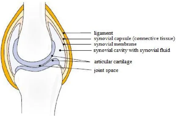
Human joint is one of the best units of the nature. Its role is to maintain stability and movement of the skeleton, accompanied by precise function through the life, even under extreme load capacity. However, the secret to this ongoing development of renewed bone is the cartilage and synovial fluid. For the joints function without friction an extremely smooth surface is required. This ensures the cartilage which is stably and elastically covers the bone. The synovial fluid as a lubricant protects the cartilage by supplying with nutriment and oxygen. The synovial fluid (synovia) is produced by the synovial membrane which covers the connective tissue in the joint capsule. The capsule
6
prevents the exit of the synovial fluid from the articular cavity. The joint is held together by the ligament (Figure 1) (Röder, 2010).
Sufficient oxygen and nutrients to absorb into the cartilage from the joint fluid it is essential to move. Thereby resulting pressure effect that from the cartilage depleted synovial fluid is pressed out and is replaced with fresh fluid. So the cast synovial fluid enters the bloodstream through the synovial membrane and fresh nutrients and oxygen from the blood comes into the synovial.
Figure 1 Schematic illustration of healthy joint (Röder, 2010)
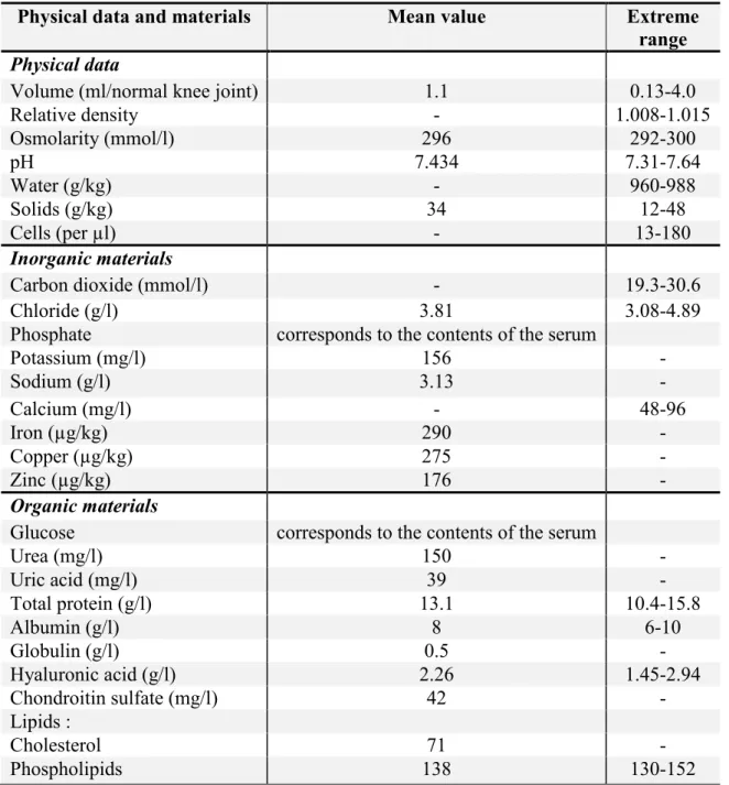
Intra-articular (i.a.) preparations go directly into the synovial fluid. Therefore it is important to know the exact composition of the synovial fluid (Table 1). The synovia is none other than the blood dialysate (Zeidler, 1986). The composition and content of electrolytes and low-molecular materials is identical to those of serum. In the healthy joint the synovial fluid amounts approximately 0.5-2 ml (Dewire et al., 2001; Mason et al., 1999). The synovial fluid is changed continuously and rapidly (approx. 2 hours) and one of the key components of the synovia is only renewed every 38 hours (Mason et al., 1999).
7
Table 1 Composition of healthy synovial fluid (Zeidler, 1986)
Physical data and materials Mean value Extreme
range Physical data
Volume (ml/normal knee joint) 1.1 0.13-4.0
Relative density - 1.008-1.015
Osmolarity (mmol/l) 296 292-300
pH 7.434 7.31-7.64
Water (g/kg) - 960-988
Solids (g/kg) 34 12-48
Cells (per µl) - 13-180
Inorganic materials
Carbon dioxide (mmol/l) - 19.3-30.6
Chloride (g/l) 3.81 3.08-4.89
Phosphate corresponds to the contents of the serum
Potassium (mg/l) 156 -
Sodium (g/l) 3.13 -
Calcium (mg/l) - 48-96
Iron (µg/kg) 290 -
Copper (µg/kg) 275 -
Zinc (µg/kg) 176 -
Organic materials
Glucose corresponds to the contents of the serum
Urea (mg/l) 150 -
Uric acid (mg/l) 39 -
Total protein (g/l) 13.1 10.4-15.8
Albumin (g/l) 8 6-10
Globulin (g/l) 0.5 -
Hyaluronic acid (g/l) 2.26 1.45-2.94
Chondroitin sulfate (mg/l) 42 -
Lipids :
Cholesterol 71 -
Phospholipids 138 130-152
8
Basically, the hyaluronic acid (HA) is responsible for the viscoelasticity of the synovia (Balazs et al., 1970; Balazs, 2009). The result of the viscoelasticity is that in case of low-frequency movement of the joint the viscous liquid works as a lubricant and in case of major mechanical stress the viscous liquid can store mechanical energy elastically. In the inflamed joints larger quantities (up to 100 ml) of synovial fluid can be produced wherein the hyaluronic acid lose its viscosity by reduction of the concentration and the molecular weight (Marshall et al., 1997). The viscosity of the synovial fluid of a healthy joint exceeds 300 mPa × s. However, after the outbreak of rheumatic symptoms the viscosity drops below 300 mPa × s (Dewier et al., 2001, Simkin, 1985).
1.1.2. Rheumatic diseases
Rheumatic diseases can be arthrosis (wear of joint) or arthritis (inflammation of joint).
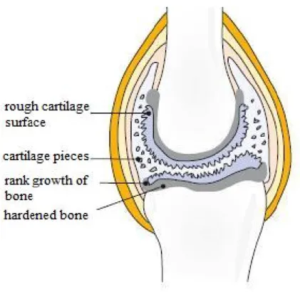
Arthrosis, osteoarthrosis and degenerated articular rheumatism class in articular wear diseases (Röder, 2010). Presumably, arthritis is caused by the imbalance between the demand and supply of nutrients. Other possible reasons are that the synovial fluid gets only a few nutrients or because of the lack of movement an inadequate nutrient circulation arises. If the cartilage does not get the proper quantity of nutrients from the joint fluid, cartilage cells die. The results are that the cartilage is torn into fibers and loses from the smooth surface. With any further movement a piece of cartilage drops out and the bone protected membrane becomes thinner (Figure 2) (Röder, 2010). The bone forms bulges below the cartilage which cause the breaking off further pieces of cartilage. The immune system treats these pieces in the synovial fluid as a foreign body which so the inflammation caused triggers a protective reaction. As a result, materials having a disastrous effect on cartilage are sent out which leads to rapid death of the cartilage cells. Because of the pain from inflammation patients move even less which leads to even less nutrient emissions. The resulting circulus vitiosus is difficult to curb.
Arthrosis affects most often the load bearing joints such as the hip (coxarthrosis) or knee (gonarthrosis) joint.
9
Figure 2 Schematic illustration of rheumatic joint caused by wear (Röder, 2010)
The inflamed articular disease is the arthritis. The most prevalent form is the chronic polyarthritis that calls most often rheumatoid arthritis. The inflammation becomes recognizable early because of the redness, warmth and swelling of the joint. At the beginning of the disease a high fever can occur in several times. Autoimmune reactions trigger the inflammation which results that the immune system replaces cartilage with foreign body material or it also attacks. The causes of the autoimmune reaction are not yet known to science but presumed causes could be bacteria, viruses or mechanical cell damage. During activation of the immune system B lymphocytes produce antibodies (lgG) which induce new auto antibodies (rheumatoid factor). The lgG and the rheumatoid factor form an immune complex which is phagocyted by the granulocytes and cells of the synovial membrane. As a result cytokines released to ensure the maintenance of the inflammatory reaction. Cytokines in the rheumatic joint can be divided into three groups (Mutschler, 2001):
- proinflammatory cytokines such as interleukin (IL)-1, -2, -6, -8, -15 and -18 or tumor necrosis factor alpha (TNF-α) regulate the inflowing and activation of the inflammatory effector cells (for exp. T-cells) and the proliferation of the chondrocytes and fibroblasts;
10
- anti-inflammatory cytokines such as IL-4, -10 and -13 inhibit purposefully the proinflammatory cytokines;
- anti-cytokine protein such as the soluble TNF-α-receptor or the IL-1-receptor regulate the inflammation by catching the proinflammatory cytokine.
In the joint of the patient with rheumatoid arthritis all of the three types of cytokine can be found, but during the diseases it shifts the balance to the cytokines.
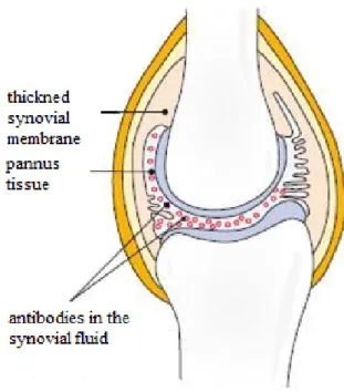
Then the occurring inflammation starts from the synovial membrane (synovitis) which at first of all becomes thick, forming bumps and fibrous, stringy tissue projects forward (Figure 3) (Röder, 2010). The resulting tissue called pannus. In the pannus tissue a large number of antibodies can be found to lead to the destruction of cartilage. The rheumatoid arthritis can occur in all joints. Firstly, in the most cases it is formed in joints of hand and finger, but later it can spread in the knee and hip joints. The symptoms can be very strong especially at night and in the morning. Another (rarely occurring) two forms of inflammatory rheumatic diseases are known: psoriasis arthritis and Bechterew's disease. In both cases, the diseases are triggered by autoimmune reactions.
Figure 3 Schematic illustration of rheumatic joint caused by inflammation (Röder, 2010)
11 1.1.3. Drug therapies of rheumatic diseases
In recent decades therapies and strategies have been pretty changed. During the drug therapies of rheumatic diseases the patient can have a shorter or longer period asymptomatically. The attenuation of pain is the most important aspect. Drug therapy especially considering the groups of active ingredients the following options are available:
non-steroidal anti-inflammatory drugs (NSAID);
non-steroidal antirheumatic drugs (NSARD);
glucocorticoids;
synthetic disease-modifying antirheumatic drugs (synthetic DMARD);
biological disease-modifying antirheumatic drugs (biological DMARD);
cartilage protector.
Cartilage protectors are the following substances: chondroitin sulfate, glucosamine, carrageenan and hyaluronic acid. They possess cartilage building characteristics. One of the most important is the HA. It is the lubricant for the cartilage and responsible for the viscosity of the synovial fluid. Due to the viscoelastic properties of HA, by the different moving of the joint (exp. sport), HA can have very different viscosity values. Thanks to this property, HA is a very versatile molecule.
There are a growing number of such active ingredients marketed as parenteral, solid or semisolid forms for rheumatic diseases. At this point it should be mentioned that parenteral biologicals make a greater number. According to an outlook to 2020 worldwide the Rx drug sales add up to $1 trillion and the biological make up 52 % (Evaluate, 2014).
12 1.1.4. Intra-articular injections
1.1.4.1. General about intra-articular injections
Oral compositions (e.g. glucocorticoids, NSARD) have a great disadvantage for the treatment of rheumatic diseases: unpleasant systemic side effects may occur when taking. Undoubtedly, because of the poor bioavailability other active ingredients (e.g.
HA) are used only for parenteral form. These problems can be avoided by using of i.a.
injections Thus, due to the natural endowments of the joints the i.a. form is a very optimal input gate. In most of the cases the hand, knee and foot joints are affected and their good accessibility favors the intra-articular injections. From the synovial fluid only a little active ingredient enters the bloodstream and therefore systemic side effects are negligible. A very important drawback is the extremely high risk of infection during the use of i.a. preparations, especially for several consecutive injections (Albert et al., 2006). However, the risk of infection is reducible by compliance with the standards, using sterile components and thorough disinfection of the injection site (Schumacher, 2003).
1.1.4.2. Modified release intra-articular drug delivery systems
During the injections, there is the risk for infection therefore the number of injections in a year should be reduced. Also a long-term drug exposure should be achieved with an injection, and if it is possible, a combination of active ingredients should be used. Many different active ingredients could have a long-term drug exposure with carrier systems like micro- and nanoparticles (Szabó A. et al., 2012), liposomes (Foong et al., 1988; Lopez-Garcia et al., 1993; Elron-Gross et al., 2009), hydrogels (Réeff et al., 2012; Shah, 2001) and physically activated delivery systems like thermoresponsive (Betre et al., 2006; Shamji et al., 2007; Kusanagi et al., 2007; Aly, 2008) or magnetically modulated carriers (Butoescu et al., 2009). Almost every third knee articular injection is inaccurate (Brandt et al., 2003), which unnecessarily exposes patient to invasive therapy without improving the condition.
13
Parenteral compositions also i.a. belong to the pharmaceutical formulations with the most stringent requirements. The following requirements shall be taken into account:
sterility;
pyrogen-free;
isotonic;
physiological pH;
viscosity;
compatibility of active ingredients and excipients;
contamination free;
compatibility with the packaging material;
stability during storage.
The injectable suspension is one of the most complex pharmaceutical formulation considering their stability, production and application. By parenteral suspensions several factors should be taken into consideration (Liebermann et al., 1996):
dispersibility /sedimentation rate;
stability;
particle size;
syringeability.
For the intra-articular compositions the sterility is essential. The sterility of solutions can be achieved by wet and dry heat if during the process all of the active ingredients and excipients remain stable. The sterility of the other intra-articular dosage forms can be achieved with adequate preparation of the materials and by keeping the fully aseptic conditions (Gerwin et al., 2006). Sterile filtration is not feasible by formulations containing particles or by solid, semisolid forms.
Ensuring isotonicity of the preparations is just as important as the sterility. Hypotonic or hypertonic parenteral preparations can cause pain or tissue damage (Bauer et al., 2002).
14
Therefore, it is very important to achieve an appropriate isotonic value which must be between 225 and 430 mOsm/kg.
The pH value should be set to the physiological pH (7.4). Greater deviations from the physiological value could cause pain. The minor differences can be compensated by the synovial fluid.
For the applicability of the product the viscosity plays a very important role. The higher viscosity can cause strong pain to the patients or the optimal syringeability could be difficult or even totally blocking. Syringeability is described by the Hagen-Poiseuille equation (Fry, 2014):
ܨ =
ଵଶ଼ொµగర (Eq. 1)
where:
F = syringe stopper (plunger) force;
Q = volumetric flow rate;
µ = dynamic viscosity;
L = needle length;
D= needle bore diameter;
A = syringe plunger area.
The syringe stopper or plunger force is dependent on numerous factors, including the volumetric flow rate, the dynamic viscosity, the needle length and bore diameter and the syringe plunger area, the relevant tissue.
The parenteral manufacturing industry pays great attention to particulate contamination. Particulate contaminations in parenterals are risk factors and can lead to damage to patients. Contaminations can be caused by interactions between the formulation, product packaging, packing materials, formulation ingredients, manufacturing process and environment (Langille, 2013). Quality by Design (QbD) is an important approach by parenteral manufactures. Quality is ensured by planning or design. The crises, risks and problems to get particulate contaminations can be avoided by well management of the different processes. The collaboration with logistic partners,
15
testing methods (final visual inspections, measurement of changes in conformation that could lead to aggregation, using instruments for sub visible particles etc.) to avoid contaminations, is indispensable (Challener, 2014). QbD pays also attention to speed up the processing time and to reduce the costs.
The formulations could lose their stability during storage. Therefore some compositions are marketed as a dry powder which must be dispersed in a solvent before use (Gerwin et al., 2006). On the pharmaceutical market some products can be found in ready-to-use pre-filled syringe. The application of pre-filled injections avoids the critical point when the content of the vial is pulled up into the syringe which has new infection risks (Ayral, 2001). On the other hand the pre-filled syringes are also contributor to the increased incidence of problems with visible particulate contaminations (Challener, 2014). The inadequate time and temperature storage can also cause particulate contaminations in the parenteral preparations.
In the case of intra-articular suspensions the good and rapid and the slow sedimentation have an important role in the correct dosage of drugs. The remaining active ingredient(s) in the syringe can have serious consequences for the therapy.
The stability of the suspension is good if the particles are dispersible and do not agglomerate.
Several scientific articles were published about the optimum size of particles of intra-articular suspensions. Results of phagocytose of suspended parts are very different. So it should be noted, most of the literature suggests the avoidance of phagocytosis. Thus, the particles must be at least between 5 - 15 µm that exceeded the size of the macrophages and thereby prevent the annexation (Howie et al., 1993).
In parenteral considering i.a. injections a high number of excipients can be used safely. Water works as a solvent or dispersant. In most cases for isotonic sodium chloride, mannitol or sorbitol can be used. Sodium hydroxide, sodium hydrogen phosphate, hydrochloric acid, phosphoric acid or citric acid adjust the optimum pH value. In some of the products other excipients can be found: polysorbate 80 such a surfactant, sodium EDTA such a stabilizer, propylene glycol or polyethylene glycol such cosolvents and preservatives (benzyl alcohol, methyl- or propyl-4-hydroxy
16
benzoate and cetylpyridinium chloride) (Gerwin et al., 2006). For the stabilization of suspensions croscarmellose sodium, hypromellose or gelatine can be used.
There are a small number of intra-articular injections on the pharmaceutical market (Table 2). Mainly, glucocorticoids are available for the i.a. administration.
However, an increasing number of publications have been published in the topic of i.a.
delivery systems with NSAI and NSAR active ingredients.
Table 2 Some commercially available i.a. products (without HA products)
Commercial name Company Formulation Drug Concentration (mg/ml) CELESTAN ® Depot MSD SHARP &
DOHME GMBH
suspension betamethasone 2.7
CELESTAN ® solubile MSD SHARP &
DOHME GMBH
solution betamethasone 4
Delphicort ® RIEMSER Pharma GmbH
suspension triamcinolone 25; 40
dexa-clinit ® Hormosan solution dexamethasone 4
Dexamethason-mp ® medphano Arzneimittel GmbH
solution dexamethasone 4
Diprosone ® Depot MSD SHARP &
DOHME GMBH
suspension betamethasone 2
Fortecortin ® Injekt Merck Serono solution dexamethasone 4; 8
Lederlon ® RIEMSER Pharma GmbH
suspension triamcinolone 5; 20
Lipotalon ® Recordati Pharma GmbH
liposome dexamethasone 2.5
Prednigalen ® GALENpharma GmbH
suspension prednizolone 10; 25; 50
Prednisolut ® mibe GmbH Arzneimittel
solution prednizolone 5; 12.5; 25; 50
In the last years the number of HA/sodium hyaluronate (NaHA) i.a. formulations increased (Table 3). Most of the preparation is in the form of a viscous solution, also known as hydrogel. HA is an innovative active ingredient with a lot of possible applications.
17
Commercial name Company Conc. Excipients
(mg/ml)
ARTROject ® ORMED GmbH 5 NaCl, Na2HPO4,
NaH2PO4, H2O
Curavisc ® curasan AG 10 NaCl, Na2HPO4,
NaH2PO4, H2O Fermathron ® Biomet Deutschland GmbH 10 NaCl, Na2HPO4,
NaH2PO4, H2O GO-ON ® Rottapharm|Madaus GmbH 10 NaCl, Na2HPO4,
NaH2PO4, H2O GO-ON ® matrix Rottapharm|Madaus GmbH 20 sorbitol
Hya-ject ® / -mini ORMED GmbH 10 NaCl, Na2HPO4, NaH2PO4, H2O
Hya-ject ® plus ORMED GmbH 20 mannitol, NaCl,
Na2HPO4, NaH2PO4, H2O
Hyalart ® /D MEDA Pharma GmbH & Co.
KG
10 NaCl, Na2HPO4, NaH2PO4, H2O Hyalubrix ® MEDA Pharma GmbH & Co.
KG
15 NaCl, Na2HPO4, NaH2PO4, H2O
Monovisc ® Plasmaconcept AG 22 NaCl, H2O
Orthovisc ® / -mini Plasmaconcept AG 15 NaCl, H2O Ostenil ® / -mini TRB Chemedica AG 10 NaCl, Na2HPO4,
NaH2PO4, H2O Ostenil ® plus TRB Chemedica AG 20 mannitol, NaCl,
Na2HPO4, NaH2PO4, H2O
Recosyn ® Recordati Pharma GmbH 10 NaCl, Na2HPO4, H2O Recosyn ® -m.d. Recordati Pharma GmbH 10 NaCl, Na2HPO4, H2O,
citric acid Recosyn ® -forte Recordati Pharma GmbH 20 NaCl, Na2HPO4,
NaH2PO4, H2O, Na2CO3
Sinovial ® / -Mini / - HighVisc
IBSA Farmaceutici Italia Srl 8; 16 NaCl, H20, sodium phosphate (unknown) Synvisc ® / -One Sanofi-Aventis DL GmbH 8 NaCl, Na2HPO4,
NaH2PO4, H2O Viscoseal ® TRB Chemedica AG 5 NaCl, Na2HPO4,
NaH2PO4, H2O
Nowadays, it is very difficult to classify which formulations or applications are medical devices or drugs. This special emphasis is made on the regulatory issues of HA, too. The different countries have different opinion about the regulatory. There is no specification about the concentration, Mw, type of the application, viscosity, condition etc. of HA to classify to the medical devices or to the drugs.
18
1.2. Hyaluronic acid
1.2.1. History
NaHA is the sodium salt of HA. There is a wide variety of names for HA which refers to the versatility of the material (Dicker et al., 2014). In 1934 Meyer et al. isolated it and called it HA because of his supposed mild acid properties. In the physiological medium, HA is present as a polyelectrolyte salt, calls “hyaluronate” with a cation, mostly with sodium, like “sodium hyaluronate”. Balazs et al. (1986) used the name
“hyaluronan”, which form can be always used.
They are of importance in epidermal pathology, tissue engineering, ophthalmic surgery, drug delivery systems, pulmonary and joint pathologies and aesthetic surgery (Price et al., 2007). HA is the most important substance in the synovial fluid of articular joints to be found in the extracellular matrix of the synovium. It is the lubricant for the cartilage and responsible for the viscosity of the synovial fluid. Healthy joints contain approximately 2.26 g/l HA (Zeidler, 1986). It is a normal process that for people over the age of 30 years starts to lose HA production in body. Therefore, characteristics and formulations of HA are increasingly important in pharmaceutical technology.
The history of HA goes back to 1934 when Karl Meyer purified HA from the vitreous of bovine eyes (Meyer et al., 1934). In 1958, Meyer et al. identified the structure of HA. At the beginning of the researches the biggest challenge was the biological purity of HA. It was very difficult to remove all of the impurities from the molecule which can cause inflammation, irritation or allergy. In the late 1960s the use of the “non-inflammatory fraction of sodium hyaluronate” was started for therapeutic proposes. In the mid-1970s 1 % HA solution (2 – 3 million Da) was used in veterinary medicine. In 1992 the first HA with 6 million Da was usable for therapeutic applications (Balazs, 2009).
The first NaHA product for i.a. pathway with the name Hyalart®/Hyalgan® was marketed by Fidia in Italy in year 1986 (Fidia Pharma, 2015). It was used for the osteoarthritis of the knee.
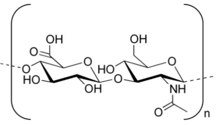
19 1.2.2. Structure
HA macromolecular chains are built from D-glucuronic acid and N-acetyl-D- glucosamine disaccharides (Figure 4) (Kogan et al., 2007). A molecule of 10 million Da contains 25,000 disaccharide units in the chains, which are held together by hydrophobic bonds (Romagnoli et al., 2008). The polysaccharide chains are linear and unbranched and roll up into a coil conformation. The intramolecular hydrogen bonds have a big role in the conformation of HA. In solution the surrounding water molecules of HA causes another formation of hydrogen bonds. According to the concept of Sheehan et al. (Sheehan et al., 2001) the hydrogen bonds are not responsible for the stiffening of the molecule. There is a little difference in hydrogen bond energy whether the molecule is hydrogen bonded to itself or to water molecules. The different conformations are deal subsequently.
Figure 4 Structural formula of HA
The length of the polysaccharide chains and the Mw of HA are very different in the various tissues. In normal tissues, a molecule of HA (10 million Da) has a thickness of 1 nm and a length of 25 µm (Romagnoli et al., 2008). In the biomatrix, HA has an Mw
in the range of 6 to 12 million Da (Balazs, 2009). The molecular weight of HA is approximately 7 million Da in healthy joints and 4.8 million Da in unhealthy joints (Wohlrab et al., 2004). The viscoelastic properties of HA under 1 million Da are
20
negligible because of the Mw in flamed joints (4.8 million Da). For that purpose, cross- linked HA is in demand (such as Hylan G-F 20, with an Mw of 6-7 million Da) for i.a.
injections (Migliore et al., 2010).
HA can have very different molecular weights, from hundreds up to millions of Daltons, it gets interesting to determine the molecular weight of Ha.
Size-exclusion chromatography (SEC) is used for polymer molecular weight characterization, but it not suitable for the determination of the molecular weight of HA.
It requires specific calibration standards. Multi-angle light scattering (MALS) detector can directly determine the molecular weight of a polymer without calibration standards.
If SEC is combined with a refractometer as a concentration detector and a viscometer for measuring intrinsic viscosity (SEC-Triple), it becomes a powerful technique to characterize HA and other polymers (Sabagh et al., 2015).
Šoltés et al. (2002) successfully used SEC-MALS coupled with capillary viscometer for the molecular characteristics of some commercial high-molecular-weight HA. Caspersen et al. (2014) studied the kinetics and mechanism of depolymerisation of solid sodium hyaluronate at elevated temperatures and various pH. To develop an improved correlation between Mw and intrinsic viscosity for sixty HA batches with Mw
ranging from 0.4 to 2.3 MDa, SEC with dual detection of multi-angle light scattering and refractive index (SEC-MALS-RI) was used.
21 1.2.3. Biological attributes
Naturally, the HA molecules is synthesized inside the cells, for example, by endothelial cells, adventitious cells or oocytes (Romagnoli et al., 2008). In the synovium it is produced by synoviocytes, fibroblasts and chondrocytes. In the nature, the most of the HA amount can be found in rooster comb (7500 μg/ml), in human umbilical cord (4100 μg/ml), in human joint fluid (1400 – 3600 μg/ml), in bovine nasal cartilage (1200 μg/ml) and human vitreous body (140 – 340 μg/ml). The whole list of the tissues and body fluids with different HA content was published by Shiedlin et al.
(2004). Approximately 11-17 g HA can be found in a human being (Laurent et al., 1991).
HA can be animal or bacterial origin. The most animal origin HA is produced from rooster comb and the bacterial origin HA can be prepared from the capsules of streptococcus bacterium. Therefore, each product has slightly different rheological properties (Manna et al., 1999). The drawback of the animal HA is the impurities. The bacterial HA has no problem with biological purity and that is why it has a better and repeatable quality. In the European Pharmacopoeia there is a request concerning the amount of the bacterial endotoxin or pyrogens. These substances have to be limited in parenteral products at most 0.5 and by intra-articular or intra-ocular parenteral preparations 0.05 I.E. pro mg NaHA (Ph. Eur. 8., 2014).
1.2.4. Physical and chemical properties
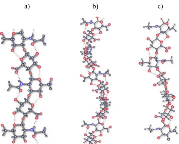
In the physiological medium, HA is present as a hyaluronate salt, negatively charged and highly hydrophilic (Vasi et al., 2014). In general, it has the cation sodium [(C14H20NNaO11)n], but other cations are possible (like zinc, potassium, magnesium, calcium) too. In solution HA has a much stiffened random coil structure. HA can have different conformations, which depend on the pH and the compounds of the solution. In 1983 Sheehan et al. (1983) reported about a left-handed four-fold helical structure by sodium hyaluronate and a three-fold helix by calcium hyaluronate. In 2001, Sheehan et
22
al. published results with potassium hyaluronate and their different helical structure (studied by X-ray fiber diffraction) (Figure 5). Mainly the electrolytes and the solvents have an effect on the chain flexibility of HA (Gribbon et al., 2000). Calcium and manganese has a greater impact on the flexibility of HA chains, while the chains are less flexible with sodium and potassium counter ions (Sheehan et al., 1983; Gribbon et al., 2000).
Figure 5 X-ray fiber diffraction of potassium hyaluronate a) 2-fold helix conformation with an axial rise per disaccharide of 0.98 nm; b) left-handed 4-fold helix conformation with an axial rise of 0.84 nm per disaccharide and c) left-handed 4-fold helix of axial rise of 0.95 nm per disaccharide (Sheehan et al., 2001)
a) b) c)
23
The pH of the i.a. formulations has to have about 7.4 (as the physiological pH value of the synovial fluid) or slightly lower, but not below pH ~ 5.5 (Gerwin et al., 2006). HA solution has pH between 6 and 7.2, therefore it is ideal for i.a. treatments. By decreasing pH (< 3) the carboxyl groups are protonated and HA form a water-insoluble gel (Wohlrab et al., 2004).
Under physiological condition HA with the carboxyl anion is able to bind a large amount of water. That is why the HA of 2 % in solution can bind 98 % water by forming a gel (Wohlrab et al., 2004). This gel - under physiological condition - shows viscoelastic and pseudoplastic characteristics and it is a non – Newtonian liquid.
In inflamed joints (e.g. in osteoarthritis, rheumatoid arthritis), the low viscosity is caused by increase of the volume of the synovial fluid and the reduction of the HA concentration and Mw (Marshall et al., 1997). Mw of HA has a significant effect on its half-life time. HA is metabolized very fast in the most tissues. The half-life time of HA with 6 million Da is about 13.2 h (Wohlrab et al., 2004). In different tissues HA has another length of the polysaccharide-chains, another Mw and that is why another half- life time. HA has a longer half-life time in vitreous of the eyes then in articular joints.
But this quantity depends on the replacing of the vitreous humor and the synovial fluids, too. Larsen et al. (Larsen et al., 2008) summarized the synovial disappearance half-lives and molecular weights of various solutes (lidocaine, diclofenac, hydrocortisone, hyaluronan, albumin). Hyaluronan has the longest half-life time in the synovial fluid, it is about 21.8 – 26.3 hours with 3 × 10⁶ g/mol molecular weights.
Various HA derivatives can be achieved by chemical modification. Carboxyl groups of HA is especially suitable for this preparation. HA receptors and hyaluronidase recognize the carboxyl groups of HA (Banerji et al., 2007). Therefore, these derivatives can modify the enzymatic degradation of the molecule and accordingly the effect of HA. Mainly carboxyl and hydroxyl groups of HA are used for chemical modification, for example, with carbodiimide, divinyl sulfone, glycidyl ether or dialdehyde (Oh et al., 2010). In order to produce hydrogels, HA derivatives are in strong demand, too. There are three types of HA whereby hydrogels are got, direct cross-linking of HA, cross- linking of HA derivatives and cross-linking between two different kinds of HA derivatives (Oh et al., 2010).
24
Maroda et al. (2011) prepared and characterized HA nanoparticles formed by amidation with an amine as a cross-linking agent in aqueous media. Cross-linked nanoparticles have a size less than 20 nm but during the purification it can be lost.
HA has an also great mucoadhesive property (Liao et al., 2005). HA creates bonds with the mucin in the mucous membrane and it causes longer residence time.
Because of these mucoadhesive properties, HA is able to increase the absorption capacity of different drugs (Lim et al., 2000).
1.2.5. Stability
HA is a very unstable and heat sensitive molecule, the molecular mass and the viscosity may be decreased due to the damage of bonds in the polymeric chains (Bailey et al., 1968). If HA in solution is warmed up to 100 ºC, the bonds between the chains are get damaged, and the Mw and viscosity decrease. That is the reason why sterilizer with high temperature cannot be used for HA.
Thermal effect or treatments, pressure, ultrasonic effects, filtration, mixing or using syringe can destroy the chains. These chain scissions cause the reduction of the Mw and accordingly the reduction of the viscosity (Bailey et al., 1968).
It is well known that hyaluronates are susceptible to degradation under a variety of conditions such as acid hydrolysis (Cleland, 1977; Longas et al., 1981), oxidative depolymerisation reactions (Matsumura et al., 1966; Kvam et al., 1993), and sonication (Kubo et al., 1993).
But there is also another natural destroyer for HA, it is the degradation enzyme, hyaluronidase. Hyaluronidase breaks down HA very fast in the articular joints. The speed of the degradation depends on the Mw and the structure of the HA. Improved stability of HA can be achieved with cross-linking. Cross-linked HA has a higher half- life time and can stay longer in the articular joints.
25 1.2.6. Hyaluronic acid-based hydrogel
Gels are one-phase systems, the most common group, in which the gel medium is the water, are the hydrogel. The gelling materials can be different polymers, like polymethyl methacrylate (PMMA), poly-(2-hydroxyethyl)-methacrylate (polyHEMA) or polyvinyl alcohol (PVA) (Li et al., 2009). The polymers of the hydrogels are held together by physical, ionic or covalent bonds and can absorb water (Davidovich-Pinhas et al., 2010; Sriamornsak et al., 2008). The gel carriers for i.a. uses play a dual role in articular therapy: on the one hand gel carriers ensure longer residence time for active ingredients in the joints compared to the drug solution forms, and on the other hand through their viscosity they maintain the motility of joints such an artificial lubricant.
Despite the many known artificial polymer systems in the development of pharmaceutical formulations, the endogenous HA and its various derivatives are the most preferred gelling material. Thus, the HA hydrogels used to help restore the physiological HA concentration, which increase the reduced viscosity. At present, in the therapeutic practice, only hyaluronic acid-based gels are applied (Orthovisc®, Monovisc®, Synvisc®, Biovisc®, Hyalgan®, Synocrom®, Synocrom Forte®) (Szabó A. et al., 2011). In in vitro studies, Réeff at al. (2012) with a HA gelling agent a release of several days (active ingredient clonidine) was reached. They used glyceryl monooleate as excipient for slowly soluble viscous phase in the synovial fluid (Shah et al., 2001).
26 1.2.7. Studies on the characteristics
1.2.7.1. Viscometry
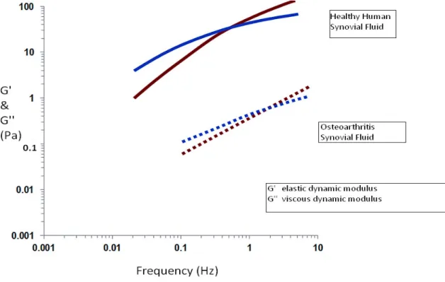
Due to the chemical structure of the polymer, HA shows interesting rheological properties. The coils of HA can straighten, and this behavior is the mechanism of action behind viscosupplementation (Balazs et al., 1970). The aqueous solutions of high- molecular-weight HA exhibit shear-dependent viscosity and frequency-dependent elasticity (Šoltés et al., 2002).
The viscoelastic behavior of HA in solution changes with exposure to different shear rates. The shear rate is dependent on the dynamic elastic and viscous moduli. The crossover point of these two moduli can be used for the rheological characterization of HA.
Figure 6 The viscoelastic behavior of HA and background of the viscosupplementation (brown: G' elastic dynamic modulus, blau: G'' viscous dynamic modulus; based on Balazs, 2009)
27
Above this frequency, the solution has elastic properties, and below this frequency it shows viscous behavior (Figure 6) (Balazs, 2009). This network, when sheared by flow or oscillation movements, can dissipate the energy in viscous flow or store it as elastic deformation (Gibbs et al., 1968). These rheological properties of HA provide viscous protection and elastic shock absorption in the joints (Phillips et al., 2013). The more suitable viscosupplementation product is the one with an elastic component similar to or greater than that of healthy young synovial fluid (Phillips et al., 2013).
In 1990 Hassan et al. have also used a simple viscometric method to quantify mucin-polymer bioadhesive bond strength. The mucoadhesive capacity was measured by a rheometer and this rheological behavior between the polymer and the mucin can be described by the following equation:
η
b= η
t– η
m– η
p (Eq. 2)where:
ηb = bioadhesive viscosity component;
ηt = viscosity of the mucin-polymer containing samples;
ηm = individual viscosity coefficients of the mucin;
ηp = individual viscosity coefficients of the bioadhesive polymer.
1.2.7.2. Thermal analysis
The thermal degradation of NaHA was evaluated by thermogravimetric analysis (Villetti et al., 2002) which indicated that low thermal stability can be observed for the polyanionic NaHA.
28 1.2.7.3. X-ray diffraction
Guss et al. (1975) examined the molecular conformations and interactions in two sodium salts of HA. The results of X-ray diffraction showed that the counterions form strong intermolecular interactions with neighbouring HA molecules. Water molecules are attracted by the charged sites to form hydration shells. Guss et al. with X-ray experiments convinced that increasing the amount of water present in the hydration shells causes the reduction of the mechanical stiffness of the HA molecule.
Sheehan et al. (1983) used X-ray fiber diffraction to study of conformational changes in hyaluronate induced in the presence of sodium, potassium and calcium cations. The effects of pH, humidity, temperature, ionic strength and hydrogen ionic activity were studied in physiological situations. X-ray fiber diffraction method is a monitoring technique to illustrate the conformational changes. The results showed that the conformations of the chains are held together by stronger or weaker intra-chain hydrogen bonds. HA has in the presence of counterion sodium a left-handed four-fold helix form and in the presence of calcium a three-fold helical structure. These results were confirmed by the results of NMR studies, too (Almond et al., 2006).
1.2.7.4. Spectroscopy
Lee et al. (1993) reported about the tightly bound water (primary water) of hydration in Li- and NaHA. Brillouin spectroscopy was used to examine dynamic coupling with the water of hydration and phase transitions in HA. Their results were that the relaxation time of the water relaxation mode is approximately the same in Li- and NaHA and the coupling to the biopolymer depends on the biopolymer itself.
Ultraviolet spectroscopic studies of Lee et al. (1995) examined the physical properties of hyaluronate films. Taken together, two results of electronic states were reported; the elastic moduli of Li- and NaHA decrease over the entire range of hydration (this indicates that the long-range Coulombic interactions are very sensitive to the water content) and the changes in the UV spectrum of a NaHA film (before and after being
29
cycled through the phase transition between 84 and 92 % relative humidity) were found to be irreversible.
Infrared (IR) spectroscopy is used for the identification and classification of compounds and because of this unique characterization, it is often called “fingerprint”
of the molecules. This experimental spectroscopy is very often used for polymers. In 1954 Orr et al. used conventional IR spectroscopy for partial assignments of bands of HA and other hyaluronates.
In 1976 Cael et al. recorded spectra with Fourier Transform Infrared Spectroscopy (FTIR) for two different crystalline forms of NaHA. It is an improved technique for the analysis of solid, liquid and gas. A review of research papers about the variety of molecules (mono- and oligosaccharides, polysaccharides) examining by FTIR have been presented (Kačuráková et al., 2001). HA-phosphate fiber mats were examined by Attenuated Total Reflectance Fourier Transform Infrared (ATR-FTIR) spectroscopy (Brenner et al., 2013). HA fiber mats were produced with glycerol phosphate, sodium phosphate and tripolyphosphate salts. All display the characteristic carbohydrate peaks for HA.
Gilli et al. (1994) studied NaHA and its oligomers by FTIR spectroscopy in the amorphous solid phase and in aqueous solution. Dřímalová et al. (2005) depolymerised HA by ultrasonic, microwave irradiation and conventional heating, tested the effect of pH and oxidants on HA. This degradation and the molecular weight changes of HA were studied by FTIR spectroscopy. The results are in agreement with the previous IR study on HA from Gilli et al. (1994), the overall spectral pattern has not changed by decreasing the molecular mass of HA. Dřímalová et al. also reported about NMR studies with the HA samples. The NMR and also FT-IR spectral analyses indicated that HA in the studied whole molecular weight range retained almost the backbone of the parent polysaccharide independently on the degradation method used.
30
The first time Lahajnar et al. (1986) reported on the proton nuclear magnetic resonance (NMR) spectroscopic study of water absorbed in solid, oriented NaHA. The results indicate a hydration similar to that of many other fibrous biopolymers at comparable relative humidity levels. Investigations using NMR in order to characterize the accurate solution conformation were mainly unsuccessful because of the overlap presents of the NMR spectra (Holmbeck et al., 1994; Donati et al., 2001).
Even so, Almond et al. (2006) successfully presented results about the solution conformation of HA determined by computer modeling (molecular dynamics simulations) and high-field nuclear magnetic resonance (NMR). The resulting data are used to test molecular dynamics simulations containing explicit water and ions at the atomic level. The results of this new description of the solution conformation of HA - a left-handed four-fold helix structure - are similar to that observed for potassium, sodium and calcium HA fibers by X-ray diffraction (Sheehan et al., 1983). The experiments of Almond et al. also pointed out that the intramolecular hydrogen bonds in HA exist in aqueous solution but they are constantly in motion and in rapid interchange with water- mediated interactions. This simulation can be used as a basis for constructing molecular models to understand the interaction between HA and proteins, while also explaining the viscoelastic properties of HA.
1.2.7.5. Positron annihilation lifetime spectroscopy
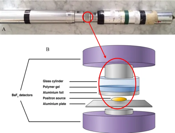
Variation of the free volume with temperature in the sol-gel transition of 2 % (w/w) κ-carrageenan showed the sol-gel transition to be an onset of the decreasing percentage of the free volume (Wakabayashi et al., 1996). Positron annihilation lifetime spectroscopy (PALS) (Figure 7) (Szabó B. et al., 2011; Szabó B. et al. 2012) is able to give important information about the free volume properties of polymers related to their molecular dynamics, volume relaxation and physical ageing.
31
Figure 7 Method of PALS (A - real construction of PALS; B - schematic construction of PALS (Szabó B. et al., 2012))
Sebe et al. (Sebe et al., 2012) have summarized the principles and the functions of PALS: the method is based on the annihilation of the electrons and positrons with high-energy γ-radiation. The electrons and positrons work as particle and antiparticle pairs and they destroyed mutually and finally they disappear or annihilate. During this process, the total mass of particle and antiparticle mutate to energy (photons without resting mass) according to Einstein equation E = mc2 (m is the mass of the particle, c is the speed of light and E is the released energy). This process produces energy without any side products, with 100 % efficiency.
A
B
32
During the annihilation of positron and electron resulting generally two photons leave each other at an angle of 180°. This phenomenon can be used for research purposes. Before the annihilation of positron and electron there is another intermediate form, the positronium atom. There are two forms of positronium atom: if the positron and electron spin are the same (+1/2 or -1/2) (the spin of positronium atom is ± 1), then called orto-positronium (o-Ps) or if the spins are contrary (the spin of positronium atom is 0), then called para-positronium (p-Ps). The life of p-Ps is too short, it annihilates in vacuum in 125 ps during resulting two photons, but the o-Ps survives (because the contrary spins inhibit the annihilation). But, after a time the inhibition is not strong enough and the o-Ps annihilate during resulting three photons. Its lifetime in vacuum is 142 ns. In polymers this lifetime can reduced to a few ns, because of the pick-off annihilation with an electron during resulting two photons as by p-Ps. This pick-off annihilation can be used to define the cavity size of polymers. The smaller the cavity of the polymer is, where the o-Ps passes, the faster it meets electron with contrary spin (Sato et al., 2008).
According Doppler-effect the velocity of the emitting source (in this case the electron during the annihilation) and the produced radiation (in this case the photons) may be measured. If in the polymer electron has energy (because of its velocity), it is added to the energy of produced photos. If the energy of annihilated photons can be measured, the momentum of electrons in the polymer cavity can be described.
Generally, 22NaCl is used as positron source and two scintillation detector with BaF2 are needed. The detectors must be placed opposite each other as close as that goes (between the detectors is the polymer (Figure 7). The time between the two detectors sign is the lifetime of the positron. The resulting curve can be evaluated with different computer programs.
Only one publication in the past affects the topic hyaluronic acid examined with PALS. Süvegh et al. (2000) have tested the positron lifetime technique as a tool of the structure study of sodium and zinc hyaluronates. By zinc hyaluronate the positron lifetime significantly increased comparing sodium hyaluronate. And normally it would mean that the free volume between the chains of zinc hyaluronate has also increased.
But this was not the case because in prior rheological studies (Burger et al., 2001) it was demonstrated that the free volume values in zinc hyaluronate decreased comparing
33
reported about the different bonds: sodium salt has intermolecular hydrogen-bridge- bonds and the exchange of cations (sodium to zinc) results intramolecular bonds. In the latter the chains curl up and that is why the free volume values decrease. This indicates that the intramolecular hydrogen-bridge-bonds are tauter and the o-Ps cannot access to the electrons so easy.
1.2.8. Formulation aspects of hyaluronic acid
1.2.8.1. Sterilization
Sterility for the injection dosage forms of the HA derivatives can be achieved only by sterile filtration of the solution or with the application of sterile solid HA treated by gamma irradiation (5 – 10 kGy) (Wohlrab et al., 2004).
Experiments with “ready-to-use pre-filled” HA syringe showed that by autoclaving for 20 min, the Mw decreased from 1.4 million Da to 0.8 million Da. By this gamma ray intensity the number of the chain scissions is acceptable and the results are repeatable.
Barbucci et al. (2002) investigated the sterilization of lyophilised cross-linked HA (synthesized from 50 % with COOH groups cross-linked HA). It was sterilized with steam, γ-rays and ethylene oxide. The swelling ability and morphology of HA were not modified by sterilization with ethylene oxide and γ-rays. The steam sterilization caused modifications of this type of HA.
However, data from simulated sterilization conditions suggest that higher concentrations of HA may have a protective effect on the stability of the long-chain molecule, such that heat treatment would not denature the hydrogel (Ali et al., 2009).
34
Furthermore, Novozymes Biopharma was dedicated to providing a Bacillus- derived high-quality HA. No animal-derived raw HA provides superior heat stability for more efficient sterilization process (Novozymes, 2012). The investigations of Szabó A.
et al. (2013) showed that the heat sterilization modified both the micro- and macrostructures of the NaHA (animal derived) gels depending on the concentration and therefore it is not a suitable method to achieve the sterility.
1.2.8.2. Freeze-drying
Freeze-drying process (lyophilisation) is a treatment to get chemically stable and sensitive substances more sustainable and it should be taken into consideration that it is also able to reduce the number of the bacteria in the formulation because of the very low temperature during the treatment. However, the freeze-drying and subsequent rehydration of thermosensitive polymer gels, like hydroxypropyl cellulose (HPC), could alter the microstructural properties of the gels in a way that leads to rapid shrinking rates (Kato et al., 2004).
Earlier data indicated that lyophilisation of hyaluronates as the free acid, in contrast to the sodium salt form, can have a detrimental effect on their physical characteristics (Doherty et al., 1994). The data of Doherty et al. (1994) indicate that the free acid form of medium molecular mass hyaluronate exhibited marked changes in molecular mass during lyophilisation whereas the NaHA appeared unchanged.
Measurements showed that the evolution of carbohydrate radicals on freeze- drying of HA is more than three times that of NaHA, which explained that HA is structurally less stable than NaHA (Tokita et al., 1997). Further, the calculations of Tokita et al. suggest that HA is much more labile against hydrogen abstraction as compared to NaHA.
35
against a distortion induced by freeze-drying is lower than that of NaHA. Concerning the stability of hyaluronate during freeze-drying, significant concentration effects would occur with respect to the hyaluronate, and it is possible that a combination of concentration effects and shifts in the effective pH of the amorphous reaction matrix could contribute to the apparent changes in molecular mass. Furthermore, this hypothesis concerning and pH effects is consistent with the observed stability of NaHA to lyophilisation as the pH of those solutions was near neutral (Doherty et al., 1994).
The changes of the NaHA microstructure were studied by positron annihilation lifetime spectroscopy (PALS) in the course of the lyophilisation that gives information about the free volume of the polymer systems. This measurement can be used for the analysis of amorphous materials like NaHA (Zelkó et al., 2006).
36
2. Aims
Many people have joint problems and therefore, the intra-articular delivery system provides a very promising solution for the therapies. An increasing number of HA injections appears on the international market, and this point shows the interest in this product. This molecule is also very manifold but the handling and the working with them are not so easy for the pharmaceutical industries. Because of the sensitivity of the molecule many processing and manufacturing processes are not cleared up.
The aim of this research work was to study the formulation of sodium hyaluronate gels of different concentrations for intra-articular application. Another purpose was to clarify the microstructural changes due to the manufacturing process, namely the effect of heat sterilization and freeze-drying.
The objectives of the research were as follows:
(i) to formulate NaHA hydrogels intended for parenteral (intra-articular) administration,
(ii) studying the effect of heat sterilization of NaHA hydrogels considering the microstructural changes (changes in the polymeric free volume and free volume distribution) and viscoelasticity,
(iii) evaluation of the effect of freeze-drying on the microstructure and rheological behaviour of NaHA hydrogels
37
3. Materials and methods
3.1. Materials
3.1.1. Active ingredient
NaHA of low molecular weight (Mw = 1,500 kDa) has an animal origin, and is produced from rooster comb. The pharmaceutical grade of NaHA was obtained from Gedeon Richter Ltd., Budapest, Hungary. The water content of the raw NaHA was 9,5 ± 1,37 %.
3.1.2. Excipients
Na2HPO4 and NaH2PO4 were used as isotonic sodium phosphate puffer (19.543 g Na2HPO4 x 2 H2O and 2.425 g NaH2PO4 x 0 H2O ad 1000 ml water). The puffer had pH 7.554 and an osmolarity of 286 mOsm/kg. The pharmaceutical grade was obtained from VWR International Ltd., Debrecen, Hungary.
3.2. Methods
3.2.1. Preparation of hydrogels
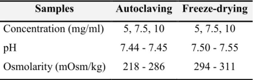
NaHA hydrogels of 5, 7.5 and 10 mg/ml concentration were prepared. At first the polymer was swelled in sodium phosphate buffer during 24 hours. Table 4 shows the measured parameters (concentration, pH, osmolarity) during the production.
38
Table 4 Product parameters of NaHA hydrogels Samples Autoclaving Freeze-drying Concentration (mg/ml) 5, 7.5, 10 5, 7.5, 10
pH 7.44 - 7.45 7.50 - 7.55
Osmolarity (mOsm/kg) 218 - 286 294 - 311
In the second part of the thesis 10 mg/ml NaHA hydrogels were prepared with water (without sodium phosphate puffer). The polymer was swelled in water during 24 hours. The pH and the osmolarity were 6.416 and 0.006 Osm/kg.
In both of the case, the hydrogels mixed with a glass rod at room temperature and stored foil sealed in refrigerator for 24 hours. After 24 hours the evaporated water was completed and again homogenized by a glass rod.
3.2.2. Heat sterilization
The samples were studied without any further treatment at two hours and at one week after autoclaving (Boxer autoclave 30075LR, Boxer Laboratory Equipment Ltd, UK) at 121 °C for 20 minutes, 103.4 kPa. The untreated and heat sterilized gels were stored at a temperature of 22 ±0.5 °C and a relative humidity of 55 ± 2 %.
3.2.3. Freeze-drying
The freeze-drying was performed in Scanvac CoolSafe 100-9 Pro type equipment (LaboGene ApS, Lynge, Denmark) equipped with a 3 shelf sample holder unit, recessed into the drying chamber. The process was controlled by a computer program (Scanlaf CTS16a02), the temperature and pressure values were recorded continuously.
The temperature of the drying chamber was between (-96) °C – (-92). Freezing of the samples was performed in the drying chamber.
39
Thermogravimetric (TG) analysis of NaHA was registered out on a METTLER Toledo TGA/DSC 1 (Mettler-Toledo AG, Greifensee, Switzerland) system, in nitrogen atmosphere with a flow rate of 50 ml/min, at a heating rate of 10 °C/min in the temperature range of 25 - 300 °C with sample weight of 8 - 15 mg in capsules of aluminium as sample containers.
The differential scanning calorimetry (DSC) measurements were carried out on a METTLER Toledo 821e DSC (Mettler-Toledo AG, Greifensee, Switzerland) system.
Under the Ar atmosphere with a flow rate of 100 ml/min samples with weight of 3-5 mg were measured at a heating rate of 10 °C/min in a temperature range of 25 - 300 °C and also in capsules of aluminium as sample containers. The measured data were registered with STARe software.
3.2.5. Reconstitution
For the reconstitution 5 g raw NaHA and 5-5 g the freeze-dried NaHA samples were dissolved in 5 ml water. The water was very slowly added by injection. It was measured how much time the dissolution and the hydration needed to get a hydrogel.
The timescale was noticed during 2 min.
3.2.6. Rheological measurements
Rheological measurements were carried out with a Kinexus Pro rheometer (Malvern Instruments Ltd, UK). The measured data were registered with rSpace for Kinexus Pro 1.3 software. A cone and plate geometry was used for the measurements.
The gap between the cone and plate of sample placement was 0.03 mm. The diameter of the cone was 50 mm. The quantity of the samples was 0.57 ml. The temperature of the samples was controlled within 37 ± 0.1 °C by a Peltier system. For the analysis of
40
viscoelasticity, the storage (G’) and loss modulus (G’’) of shear were plotted against the frequency, and their points of intersection were analyzed.
3.2.7. Surface morphology
The scanning electron microscopy (SEM) of the raw and freeze-dried NaHA was investigated by scanning electron microscopy (Hitachi S4700, Hitachi Scientific Ltd., Japan). A sputter-coating apparatus (Bio-Rad SC 502, VG Microtech, Uckfield, UK) was applied to induce electric conductivity on the surface of the samples. The air pressure was 1.3-13.0 mPa. Briefly, the samples were sputter-coated with gold- palladium under an argon atmosphere, using a gold sputter module in a high vacuum evaporator (0.1 Pa), and the samples were examined with the SEM instrument set at 10 kV (current 10 µA).
3.2.8. Positron annihilation lifetime spectroscopy
The source was covered by very thin foils. Due to the exponential penetration of the radiation, a larger intensity and smaller variation of the o-Ps lifetime could be obtained than with regular foil of 20-25 µm. Approximately 150 µl (2-3 drops) from each homogeneous polymeric gel was poured onto the foil. The solvent evaporation was prevented by a cylindrical metal cover.
For positron lifetime measurements, a positron source made of carrier-free
22NaCl was used. Its activity was around 106 Bq and the active material was sealed between two very thin foils. Lifetime spectra were measured with a fast-fast coincidence system based on BaF2/XP2020Q detectors and Ortec® electronics. Every spectrum was recorded in different channels of an analyzer card for different time and each contained about differentcoincidence events (Table 5). The number of coincidence counts was inevitably slightly lower than normal because of the increased distance between the two detectors. The detectors were arranged vertically in this experimental setup. An aluminium plate was placed on the lower detector. Although a great number