RESEARCH
Advantages of prophylactic
versus conventionally scheduled heart failure therapy in an experimental model of doxorubicin-induced cardiomyopathy
Mária Lódi1, Dániel Priksz2, Gábor Áron Fülöp1, Beáta Bódi1, Alexandra Gyöngyösi3, Lilla Nagy8, Árpád Kovács1, Attila Béla Kertész6, Judit Kocsis4,5, István Édes6, Zoltán Csanádi6, István Czuriga6^, Zoltán Kisvárday7,
Béla Juhász2, István Lekli3, Péter Bai8, Attila Tóth1, Zoltán Papp1 and Dániel Czuriga6*
Abstract
Background: Chemotherapy-induced left ventricular dysfunction represents a major clinical problem, which is often only recognised at an advanced stage, when supportive therapy is ineffective. Although an early heart failure treat- ment could positively influence the health status and clinical outcome, there is still no evidence of routine prophylac- tic cardioprotection for the majority of patients without previous cardiovascular history awaiting potentially cardio- toxic chemotherapy. In this study, we set out to investigate whether a prophylactic cardioprotective therapy relative to a conventionally scheduled heart failure treatment is more effective in preventing cardiotoxicity in a rodent model of doxorubicin (DOX)-induced cardiomyopathy.
Methods: Male Wistar rats (n = 7–11 per group) were divided into 4 subgroups, namely negative controls receiving intravenous saline (CON), positive controls receiving intravenous DOX (6 cycles; D-CON), and DOX-treated animals receiving either prophylactic (PRE, started 1 week before DOX) or conventionally applied (POST, started 1 month after DOX) combined heart failure therapy of oral bisoprolol, perindopril and eplerenone. Blood pressure, heart rate, body weight and echocardiographic parameters were monitored in vivo, whereas myocardial fibrosis, capillarisation, ultras- tructure, myofilament function, apoptosis, oxidative stress and mitochondrial biogenesis were studied in vitro.
Results: The survival rate in the PRE group was significantly improved compared to D-CON (p = 0.0207). DOX increased the heart rate of the animals (p = 0.0193), while the blood pressure (p ≤ 0.0105) and heart rate (p = 0.0029) were significantly reduced in the PRE group compared to D-CON and POST. The ejection fraction remained pre- served in the PRE group compared to D-CON or POST (p ≤ 0.0237), while none of the treatments could prevent the DOX-induced increase in the isovolumetric relaxation time. DOX decreased the rate of the actin-myosin cross-bridge cycle, irrespective of any treatment applied (p ≤ 0.0433). The myocardium of the D-CON and POST animals displayed pronounced ultrastructural damage, which was not apparent in the PRE group (p ≤ 0.033). While the DOX-induced apoptotic activity could be reduced in both the PRE and POST groups (p ≤ 0.0433), no treatment was able to prevent fibrotic remodelling or the disturbed mitochondrial biogenesis.
Conclusion: For attenuating DOX-induced adverse myocardial effects, prophylactic cardioprotection has many advantages compared to a late-applied treatment.
© The Author(s) 2019. This article is distributed under the terms of the Creative Commons Attribution 4.0 International License (http://creat iveco mmons .org/licen ses/by/4.0/), which permits unrestricted use, distribution, and reproduction in any medium, provided you give appropriate credit to the original author(s) and the source, provide a link to the Creative Commons license, and indicate if changes were made. The Creative Commons Public Domain Dedication waiver (http://creat iveco mmons .org/
publi cdoma in/zero/1.0/) applies to the data made available in this article, unless otherwise stated.
Open Access
*Correspondence: dczuriga@med.unideb.hu
^ Deceased.
6 Division of Cardiology, Department of Cardiology, Faculty of Medicine, University of Debrecen, Móricz Zs. krt. 22, H-4032 Debrecen, Hungary Full list of author information is available at the end of the article
Background
Worldwide, cancer is the second leading cause of death after cardiovascular diseases, but in 12 European Union countries it has recently taken the lead [1]. Although the survival rate of patients suffering from oncological diseases has significantly risen due to developments in modern oncotherapy, the cardiovascular side effects of chemotherapy and radiotherapy have placed limits on the success of oncotherapeutic strategies. Despite inten- sive ongoing research efforts, chemotherapy-induced cardiotoxicity remains an unresolved clinical problem, which may lead to cardiomyopathy and consequently, heart failure, with an incidence varying over a wide range (0.2–48%) [2]. In recent years, the use of classic cytotoxic agents such as anthracyclines has been re-evaluated due to the evolving role of targeted and immuno-oncology therapies, however, they still have basic and important role in the treatment of many malignancies. Unsurpris- ingly, most cardio-oncology related basic research has focused on doxorubicin (DOX), which belongs to the group of anthracyclines and is one of the most commonly applied chemotherapeutic agents with well-known car- diotoxic side effects that calls for efficient therapeutic approaches to circumvent cardiotoxicity.
In tumour cells, the primary cytotoxic effect of DOX is executed through the inhibition of topoisomerase II and DNA intercalation leading to reactive oxygen spe- cies production, DNA cross-linking and apoptosis [3].
The basic mechanism underlying DOX cardiotoxicity has not yet been completely elucidated, but one of the best accepted theories is that DOX interacts with iron metab- olism and this leads to the formation of an anthracycline- iron complex, which then induces lipid peroxidation, SH oxidation and DNA damage by reactive oxygen species production that in turn leads to contractile impairment, irreversible myocardial damage and fibrosis [4]. At the same time, other domains of DOX cardiotoxicity have also been proposed such as apoptosis, necrosis, inflam- mation, mitochondrial damage, myofilament protein dys- function, extracellular matrix remodelling, intracellular Ca2+ dysregulation, etc. [5, 6].
Although many previous studies tested the hypotheti- cal cardioprotective effects of various drug agents used in heart failure [7–16], as well as non-heart failure related substances in rodent models of DOX-induced cardio- myopathy [17–32], no systematic investigation has been conducted to evaluate the effects of a clinically relevant combination therapy and no optimum timing of pre- ventive measures has been established. Also, numerous
previous studies disregarded many clinical aspects of human cancer management, hence they did not closely mimic the human pathology (e.g. a single high dose instead of consecutive cycles of DOX, intraperitoneal instead of intravenous DOX administration, cardiopro- tective drug in the drinking water supply instead of oral gavage, etc.). Thus, a recent editorial stressed the need for horizontally integrating translational research for anthracycline cardiotoxicity and highlighted key criteria of experimental design, such as repetitive DOX cycles, intravenous administration, prophylactic cardioprotect- ant regimen, etc. [33].
As for clinical prevention, the prophylactic applica- tion of pharmacological agents with a mortality benefit in heart failure may in theory prevent or attenuate the degree of myocardial injury induced by DOX chemother- apy. Secondary prevention of cardiotoxicity has already entered clinical practice when symptoms, increase in car- diac biomarkers or overt heart failure develop, however, primary prevention is still in the research domain. One difficulty is the fact that a significant number of patients receiving chemotherapy are diagnosed with symptomatic heart failure already at an advanced stage, where the sec- ondary preventive cardioprotective approach is incapable of reverting the adverse myocardial changes. Apparently, the prophylactic use of some studied substances (angio- tensin converting enzyme inhibitors, angiotensin recep- tor blockers, β-blockers) have been suggested in recent position statements, but exclusively for high risk patients with multiple risk factors for cardiotoxicity, who repre- sent only a small fraction of the oncological population [2, 34, 35]. As a routine primary prophylaxis is not yet generally recommended in the larger population mainly due to the lack of sufficient randomised data from tri- als with an appropriate sample size, further studies are essential to elucidate the cardioprotective effects of these drugs, their effective primary preventive dosage, the opti- mum timing of therapy and possible unwelcome side effects as well [36]. Although smaller human trials on preventing cardiotoxicity mostly with single heart failure agents in patients undergoing chemotherapy are already available [37–46], the results are controversial and rou- tine prophylactic cardioprotection has not yet entered clinical practice in case of the vast majority of onco- logical patients without previous cardiovascular history.
At present, only patients with symptoms or increased cardiac biomarker levels, or those at higher risk would qualify for treatment with heart failure medication, as recommended by current consensus guidelines in effect Keywords: Animal model, Apoptosis, Cardio-oncology, Cardioprotection, Cardiotoxicity, Doxorubicin
[2, 34, 35]. With the current study, we challenge this practice and suggest that a prophylactic cardioprotective approach preceding the commencement of a potentially cardiotoxic chemotherapy may be beneficial in the unse- lected larger population as well.
To mimic the human pathology and clinical manage- ment we developed a rat model of DOX-induced car- diomyopathy by applying a consecutive intravenous dosing protocol directly extrapolated from human chem- otherapy. A set of in vivo and in vitro methodologies was employed to characterise myocardial changes of the ani- mals, as well as to test the cardioprotective effects of a prophylactic heart failure treatment relative to a conven- tionally scheduled one commenced only at a later stage.
Our primary hypothesis was that the preventive applica- tion of a combined supportive heart failure therapy pre- ceding the cardiotoxic exposure may be more protective than that scheduled at a time when overt cardiac disease has already developed.
Methods
Animal experiments and study design
In our study, efforts were made to mimic the human pathology as close as possible. Our translational con- cept and in vivo study protocol are presented on Fig. 1.
Timing and dosing of intravenous DOX cycles were cal- culated from existing human chemotherapy protocols and they were corrected to the lifespan, metabolism and body surface of rats [47]. Twelve-week-old Wistar rats (n = 7–11 per group) were used and divided into 4 sub- groups. To avoid previously described effects of hormo- nal differences in DOX cardiotoxicity [48], male rats were used exclusively in our study. The blood pressure (BP) and heart rate (HR) were monitored during the study by the tail-cuff method (CODA non-invasive blood pres- sure monitoring system, Kent Scientific Corporation, Torrington, CT, USA). Following baseline BP, HR meas- urements and echocardiography, animals in the prophy- lactic group received a daily combination of gradually uptitrated oral bisoprolol (2.5 mg/kg), perindopril (2 mg/
kg) and eplerenone (6.25 mg/kg) started a week before DOX (PRE), while those in the post-exposure group had the same therapy started 1 month after the intravenous DOX treatment (POST). According to our concept, the PRE treatment represents a prophylactic cardioprotec- tive approach for healthy subjects, which measure is currently missing from human practice, while the POST group represents subjects diagnosed with heart failure already at an advanced stage, where supportive therapy is often ineffective. The doses of the drugs applied were cal- culated from existing recommendations for human heart failure and they were corrected to the metabolism and body surface of rats [49]. To ensure effective serum levels
of the cardioprotective medications, drugs were applied in a mucous vehicle by oral gavage every day. Negative controls in the CON group and positive controls in the D-CON group received a drug-free vehicle (“placebo”;
mucilago hydroxyethylcellulosi) orally throughout the study, while animals in the POST group had it until day 51, then the drug-free vehicle was switched to an active heart failure therapy. The intravenous DOX exposure was carried out under light sedation (50 mg/kg keta- mine, 5 mg/kg xylazine) by administering 1.5 mg/kg intravenous DOX into the tail veins of the animals in the D-CON, PRE and POST groups on 6 occasions (on the 8th, 11th, 14th, 17th, 20th and 23rd days of the experi- ment). Animals in the CON group received intravenous saline on the same days. Follow-up echocardiography was carried out under deep sedation (100 mg/kg ketamine, 10 mg/kg xylazine) on days 51 and 80, while follow-up BP and HR measurements were performed on the 7th and 39th days of the experiment. Following echocardiogra- phy on day 80, animals were anaesthetised by intraperi- toneal thiopental (100 mg/kg), their hearts were excised and frozen in liquid nitrogen and stored at − 70 °C. In the D-CON group, 4 out of 11 surviving animals were pre- terminated on days 65–68 for clinical and ethical reasons to avoid imminent death and consequent loss of tissue material for the in vitro measurements. These animals were not included in the survival plot.
Echocardiography
Echocardiography measurements were performed using a General Electric Vivid E9 ultrasound system equipped with a linear 14.1 MHz i13L probe (General Electric, Fairfield, CT, USA). For M mode based systolic param- eters the parasternal long axis view was investigated.
For diastolic and Doppler based systolic parameters, the 4-chamber view was examined. To evaluate strain param- eters, a short cine loop was recorded from the 4-chamber view. Due to technical reasons and strict criteria of image quality, we used only two segments of the septum (basal and mid) to assess the strain parameters. All echocardi- ography images were obtained along with continuous electrocardiogram recording (limb leads).
Histology
A small piece of the left ventricular (LV) free wall was embedded into Shandon™ Cryomatrix (Thermo Fischer Scientific, Waltham, MA, USA). Fifteen µm thick sec- tions were cut using Cryotome™ Cryostat (Thermo Fis- cher Scientific, Waltham, MA, USA). After drying the sections, nuclei were stained with Mayer’s hemalum (VWR International, Radnor, PA, USA) for 10 min. Fol- lowing 10 min of bluing, picrosirius red staining was per- formed using a 0.1% solution. After rinsing in isopropyl
alcohol, sections were dehydrated and mounted using DPX (Sigma Aldrich, St. Louis, MO, USA). Slices were then investigated under an Olympus BX-50 microscope.
Signs of fibrosis and capillary density were analysed from representative images using the ImageJ program (National Institutes of Health, Bethesda, Maryland,
USA). Fibrosis was analysed on images at a magnifica- tion of 40×. The fibrotic area was expressed relative to the overall myocardial area using the colour threshold function of the ImageJ program. Capillary density was analysed in a blinded fashion on images obtained with a 40× objective, which met the following criteria: they Fig. 1 Translational concept and in vivo study protocol. On the upper timeline, cardiac deterioration, symptom development and clinical
management of a hypothetical oncological patient suffering from DOX-induced cardiotoxicity can be seen, who was diagnosed with heart failure at an advanced stage resulting in a poor prognosis. On the lower timeline, the in vivo study protocol of the animals can be seen based on a translational concept. Following baseline blood pressure, heart rate measurements and echocardiography, animals in the PRE group received a daily combination of gradually uptitrated oral bisoprolol, perindopril and eplerenone started a week before DOX, while those in the POST group had the same therapy started 1 month after the intravenous DOX treatment. According to our concept, the PRE treatment represents a prophylactic cardioprotective approach for healthy subjects, which measure is currently missing from human practice, while the POST group represents subjects diagnosed with heart failure already at an advanced stage, where supportive therapy is ineffective. Animals in the CON and D-CON groups received a drug-free vehicle (“placebo”) orally throughout the study, while those in the POST group had it until day 51, then the drug-free vehicle was switched to an active heart failure therapy. The intravenous DOX exposure was carried out on days 8, 11, 14, 17, 20 and 23 in the D-CON, PRE and POST groups, while animals in the CON group received intravenous saline on the same days. Follow-up blood pressure and heart rate measurements were performed on days 7 and 39, while follow-up echocardiography was carried out on days 51 and 80. AA aldosterone antagonist (eplerenone), ACEI angiotensin converting enzyme inhibitor (perindopril), BB β-blocker (bisoprolol), DOX doxorubicin, Echo echocardiography, HF heart failure, LV left ventricular
represented cross-sectional cuts of myocardial sections, free of tissue wrinkles or staining artefacts. The relative number of capillaries and the relative capillary area were expressed by outlining all capillaries on the surface of the sections and also the overall myocardial area by freehand selections.
TUNEL assay
To detect apoptosis, we used the terminal deoxynucleoti- dyl transferase (Tdt) nick end labelling test of the In Situ Cell Death Detection Kit, TMR (fluorescein-labelled cell markers) red (Roche, Mannheim, Germany). Apoptosis (DNA fragmentation) was detected by labelling the free 3′-OH termini with modified nucleotides in an enzy- matic reaction. The enzyme Tdt catalyses the template- independent polymerisation of deoxyribonucleotides to the 3′-end of single- and double-stranded DNA. The steps of the sample preparation and imaging for TUNEL are described in Additional file 1. Apoptosis was quanti- fied by the ratio of Tdt-positive nuclei/total nuclei in each section.
Electron microscopy
Tissue processing for electron microscopy was per- formed using a modified version of Somogyi’s technique [50], which is described in Additional file 1. Representa- tive pictures were taken using an Olympus Transmission Electron Microscope JEOL-1010 and iTEM software. The cardiomyocyte width was measured across the nucleus.
To characterise DOX-induced ultrastructural changes, densitometry was employed on images displaying lon- gitudinally sectioned myocardium acquired at a low magnification (3000×; 10–13 images/group) using the Image J program (National Institutes of Health, Bethesda, Maryland, USA) in a blinded fashion. Following a deter- mination of the background density of an image (for white balance), cardiomyocytes were outlined by free- hand selections, excluding all nuclei and any artefact. The area and mean density of each selection were then saved.
The outlined DOX-induced ultrastructural myocardial changes (including myofibrillolysis, mitochondrial disin- tegration and vacuolisation) all resulted in a lower overall density of the cardiomyocytes compared to an undam- aged, healthy myocardium segment. The averaged mean densities of apparently intact sarcomeres served for exposition control. The final, background and exposition corrected mean densities of myocardium selections were weight-averaged for their respective area on the image.
Force measurements in isolated cardiomyocytes
The technique for force measurements in single, per- meabilised cardiomyocyte preparations was described earlier [51–53]. Repeated activation–relaxation cycles
were performed in single cardiomyocytes at 15 °C (to maintain the stability of the preparations), at a sar- comere length of 2.2 μm. Isometric force values were normalised for the maximal Ca2+-activated active force, and Ca2+–force relations were fitted to a modified Hill equation to determine the Ca2+-sensitivity of isometric- force production, i.e. pCa50. The active isometric force (Fmax), Ca2+-independent passive force (Fpassive) and the rate constant of force redevelopment (ktr,max) were then assessed. Fmax and Fpassive were normalised for the car- diomyocyte cross-sectional area, which was measured by using optically directed light.
Oxidative status of contractile proteins
Protein carbonyl group investigations were adapted from Balogh et al. [53] using an Oxyblot Protein Oxydation Detection Kit (Merck Millipore, Burlington, MA, USA).
The detailed steps of sample processing and signal detec- tion are presented in Additional file 1.
Phosphorylation status of contractile proteins
To investigate protein phosphorylation, ProQ™ Diamond protein gel staining (Thermo Fisher Scientific, Waltham, MA, USA) was employed. The steps of tissue preparation and signal detection are described in Additional file 1.
Western immunoblot for mitochondrial proteins
Samples from the LV free wall were processed and labelled with the following antibodies being produced in a rabbit: anti-acetyl coenzyme A carboxylase (ACC), anti-phospho-ACC and anti-peroxisome proliferator- activated receptor-gamma coactivator 1 alpha (PGC1α) (Cell Signaling Technology, Boston, MA, USA). The detailed steps of tissue processing and imaging are given in Additional file 1.
Western immunoblot for caspase‑3
Approximately 100 mg of heart tissue was used and labelled with an antibody against caspase-3. The steps of tissue preparation and visualisation are given in Addi- tional file 1.
Data analysis and statistics
During the mechanical measurements, Ca2+-induced contractions of the preparations were recorded with a custom-built LabVIEW Data Acquisition platform.
The contractile parameters of the cardiomyocytes were analysed with the LabVIEW analysing software pack- age (Myo; National Instruments, Austin, TX, USA) and Origin 6.0 (Originlab Corporation, Northampton, MA, USA). The signal intensities of protein bands were quan- tified using the ImageJ (National Institutes of Health, Bethesda, Maryland, USA) and MagicPlot (MagicPlot
Systems, Saint Petersburg, Russia) software packages.
Laboratory variables were measured multiple times, averaged within each animal and used in an analysis as a single value characteristic of that animal (“mean of the mean”; except for body weight, body mass index, strain imaging and biochemical measurements on Fig. 8). The sample size of the experimental groups is indicated on the figure panels, while the number of measurements is stated in the figure and table legends. Between-groups comparisons for the survival outcome were based on an overall log-rank test and all pairwise variants thereof. For all other outcomes, analysis of variance or the Kruskal–
Wallis test was applied for overall, and Student’s two- sample t test or Wilcoxon’s rank-sum test for pairwise comparisons, as appropriate for distribution shapes.
Values are given as mean ± standard error of the mean (SEM). The criterion for statistical significance was p ≤ 0.05. In the experiment, the statistical package uti- lised was Stata (StataCorp LLC. 2017, Stata Statistical Software: Release 15. College Station, TX, USA).
Results
The clinical parameters of the animals are presented in detail in Table 1. The survival rate in the D-CON group was significantly worse compared to that in CON (p = 0.0247), but it remained preserved in the PRE group (p = 0.0207 vs. D-CON). Animals in the POST group were able to maintain their health status once combined heart failure therapy was commenced from the begin- ning of day 52 (Fig. 2a). The thriving of animals (i.e. put- ting on weight) exposed to DOX was significantly worse compared to that in CON, irrespective of any treatment applied (p ≤ 0.0018) (Fig. 2b). The heart rate and blood pressure were monitored until day 39. Afterwards the tail of the DOX-treated animals became stiff and sclerotic due to the direct effect of intravenous DOX, hindering
reliable tail-cuff measurements. This is why the D-CON and POST animals were pooled during these examina- tions, as up until then the treatment of the animals was identical (the POST treatment began only from day 52).
DOX significantly increased the heart rate of the animals compared to that in CON [464 ± 19 vs. 406 ± 11 beats per minute (BPM), p = 0.0193], while the prophylac- tic treatment of the PRE group significantly lowered the heart rate, the systolic and diastolic blood pressure com- pared to CON and/or D-CON + POST (347 ± 7 BPM, p = 0.0074 vs. CON, p = 0.0029 vs. D-CON + POST;
115 ± 5/85 ± 6 vs. 146 ± 6/110 ± 4 mmHg in PRE and D-CON + POST, respectively, p ≤ 0.0105) (Fig. 2c–e).
Echocardiographic data of the animals are presented in Table 2. At follow-up on day 80, a significantly reduced ejection fraction was seen in the D-CON group compared to that in CON (66.4 ± 3.6 vs. 84 ± 1.3%, p = 0.0043). This decrease was prevented by the prophylactic (81.3 ± 2%, p = 0.0046 vs. D-CON), but not by the conventionally scheduled treatment (72 ± 3.4%, p = 0.3886 vs. D-CON, p = 0.0237 vs. PRE) (Fig. 3a). A slight increase in the isovolumetric relaxation time (IVRT) was observed in the DOX-treated animals, which was statistically sig- nificant in the POST animals compared to that in CON (34.1 ± 1.3 vs. 27.8 ± 1.4 ms, p = 0.006) (Fig. 3b). A slight decrease in the systolic longitudinal strain rate was observed in the DOX-treated groups, which was more apparent in the D-CON and POST groups than that in PRE [− 3.5 ± 0.4, − 4.1 ± 0.3, − 3.3 ± 0.8 vs. − 5 ± 0.9 1/s in D-CON, PRE, POST and CON, respectively, p = 0.0321 (PRE vs. D-CON)] (Fig. 3c).
Picrosirius red staining revealed an increased fibrotic area (> 10%) in all DOX-exposed groups, irrespective of any treatment applied (p ≤ 0.0433) (Fig. 4a, b). A mild, statistically non-significant capillary rarefaction could be detected in the D-CON and POST groups compared to Table 1 Clinical parameters of the animals
BW body weight, DBP diastolic blood pressure, HR heart rate, SBP systolic blood pressure, tBMI body mass indexed for tibia length, n number of animals per group (5 measurements per animal, except for single body weight and tBMI measurements)
* p ≤ 0.05 vs. CON; + p ≤ 0.05 vs. D-CON/D-CON + POST; § p ≤ 0.05 vs. PRE; ^ p ≤ 0.05 vs. POST; # p ≤ 0.05 vs. day 0
CON (n = 7) D‑CON (n = 7) PRE (n = 8) POST (n = 7)
Day 0 Day 74 Day 0 Day 74 Day 0 Day 74 Day 0 Day 74
BW (g) 309.29 ± 5.25^ 481.71 ± 14.37+§^# 333.43 ± 20.42 328.29 ± 10.91* 331.38 ± 10.86 342.25 ± 49.7* 339.71 ± 10.04* 311.71 ± 22.85*
tBMI (kg/m2) 157 ± 3.57+§^ 248.87 ± 8.74+§^# 194.78 ± 18.392* 189.9 ± 9.71* 187.91 ± 10.62* 189.72 ± 15.98* 206.45 ± 11.16* 190.52 ± 7.88*
CON (n = 7) D‑CON + POST (n = 16) PRE (n = 5–8)
Day 0 Day 7 Day 39 Day 0 Day 7 Day 39 Day 0 Day 7 Day 39
SBP (mmHg) 123.31 ± 3.05§ 134.51 ± 7.85§ 132.77 ± 8.11 132.61 ± 3.27§ 136.11 ± 2.47§ 145.91 ± 5.54§# 149.73 ± 4.98*+ 112.63 ± 4.3*+# 115.44 ± 5.19+# DBP (mmHg) 92.23 ± 2.49§ 104.91 ± 7.35§# 103.34 ± 6.34 102.41 ± 3.23§ 103.64 ± 3.01§ 110.34 ± 4.39§ 116.75 ± 7.06*+ 83.3 ± 3.79*+# 85.24 ± 5.77+# HR (1/min) 418.31 ± 7.27§ 419.2 ± 13.05§ 405.51 ± 11.18+§430.86 ± 11.73 435.75 ± 12.98§ 464.06 ± 19.36*§ 451.25 ± 11.81* 380.33 ± 10.41*+# 347.24 ± 6.59*+#
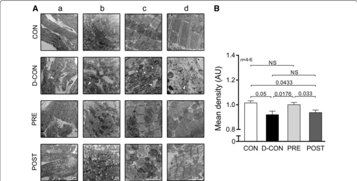
CON or PRE, which was evident from the slight decrease in the relative number and relative area of capillaries (Fig. 4c, d). On electron microscopical examination, pro- nounced ultrastructural changes were observed in the ultrathin myocardial sections obtained from the D-CON group, namely myofibrillolysis, vacuolisation, mitochon- drial damage, Z-disc degradation and chromatin disin- tegration in the nuclei. Samples from the POST animals displayed a visually similar ultrastructural pattern to those of the D-CON group, with less pronounced mito- chondrial damage, while in the PRE group most of the above ultrastructural changes were not apparent—except for a mild degree of myofibrillolysis (Fig. 5A). Quantifica- tion of the electron microscopic images by densitometry suggested a preserved myocardial ultrastructure in the PRE, but not in the POST group (p ≤ 0.0433) (Fig. 5B).
On further analysis, a mild cardiomyocyte hypertrophy could be seen in the D-CON group (17 ± 0.9 µm), which was significant compared to that in PRE (14 ± 0.6 µm, p = 0.0301) (data not shown).
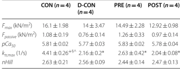
Mechanical measurements in single, permeabilised cardiomyocytes revealed no significant change in the Ca2+-sensitivity (pCa50), maximum Ca2+-activated
active or Ca2+-independent passive force parameters of cellular preparations isolated from the groups (Fig. 6a, c, e–f; Table 3). However, a significant decrease in the ktr,max was observed in all DOX-exposed groups com- pared to that in CON, which could not be prevented by any treatment applied (2.16 ± 0.20, 2.63 ± 0.42, 2.04 ± 0.08 vs. 4.41 ± 0.26 in D-CON, PRE, POST and CON, respectively, p ≤ 0.0433) (Fig. 6b, d).
The TUNEL assay of the myocardial samples con- firmed a significantly increased ratio of apoptotic nuclei in the D-CON group compared to that in CON (12.75 ± 2.35 vs. 4.85 ± 1.18%, p = 0.0209), which was not apparent in the PRE (1.79 ± 0.63%, p = 0.0209 vs.
D-CON) or POST groups (3.62 ± 1.77%, p = 0.0433 vs. D-CON) (Fig. 7a, b). A Western blot analysis revealed an increased level of caspase-3 in the myo- cardial samples of the D-CON animals relative to that in CON [0.85 ± 0.12 vs. 0.58 ± 0.06 arbitrary units (AU), p = 0.05], which was markedly reduced by the prophylactic treatment in the PRE group (0.59 ± 0.05 AU, p = 0.089 vs. D-CON), but not by the conven- tionally scheduled therapy applied in the POST group Fig. 2 Survival and clinical parameters of the animals. The survival rate in the D-CON group was significantly worse compared to that in CON, but it remained preserved in the PRE group (a). The body weight was significantly lower in groups exposed to DOX compared to that in CON, irrespective of any treatment applied (b). DOX significantly increased the heart rate of animals, while the prophylactic treatment in the PRE group significantly decreased the blood pressure and heart rate (c–e). DBP diastolic blood pressure, HR heart rate, SBP systolic blood pressure. Lines at top represent doxorubicin exposure (DOX), prophylactic (PRE) or conventionally scheduled heart failure treatment (POST), respectively. n number of animals per group (5 measurements per animal, except for single body weight measurements); Statistics: Wilcoxon’s rank-sum test except for survival (log-rank test); *p ≤ 0.05 vs. CON; +p ≤ 0.05 vs. D-CON/D-CON + POST; §p ≤ 0.05 vs. PRE; ^p ≤ 0.05 vs. POST; #p ≤ 0.05 vs. day 0
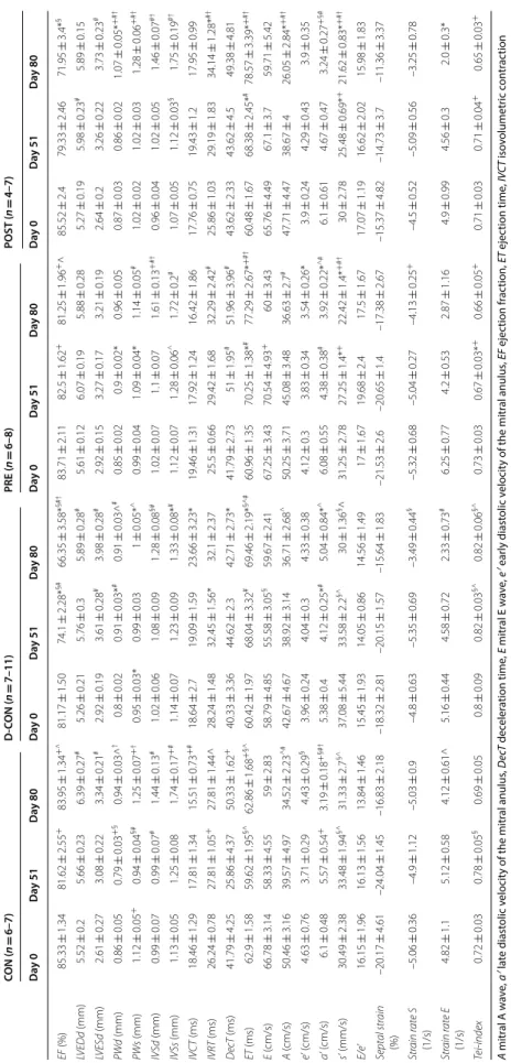
Table 2 Echocardiographic parameters of the animals A mitral A wave, a′ late diastolic velocity of the mitral anulus, DecT deceleration time, E mitral E wave, e′ early diastolic velocity of the mitral anulus, EF ejection fraction, ET ejection time, IVCT isovolumetric contraction time, IVRT isovolumetric relaxation time, IVSd diastolic thickness of the interventricular septum, IVSs systolic thickness of the interventricular septum, LVEDd end-diastolic diameter of the left ventricle, LVESd end-systolic diameter of the left ventricle, PWd diastolic thickness of the posterior wall, PWs systolic thickness of the posterior wall, s′ systolic velocity of the mitral anulus, Strain rate E strain rate measured at the time of the E wave (diastolic), Strain rate S strain rate measured at the time of the s′ wave (systolic), Tei-index myocardial performance index [(IVCT + IVRT)/ET], n number of animals per group (3 measurements per animal, except for single strain measurements) * p ≤ 0.05 vs. CON; + p ≤ 0.05 vs. D-CON; § p ≤ 0.05 vs. PRE; ^ p ≤ 0.05 vs. POST; # p ≤ 0.05 vs. day 0; † p ≤ 0.05 vs. day 51 CON (n = 6–7)D‑CON (n = 7–11)PRE (n = 6–8)POST (n = 4–7) Day 0Day 51Day 80Day 0Day 51Day 80Day 0Day 51Day 80Day 0Day 51Day 80 EF (%)85.33 ± 1.3481.62 ± 2.55+83.95 ± 1.34+^81.17 ± 1.5074.1 ± 2.28*§#66.35 ± 3.58*§#†83.71 ± 2.1182.5 ± 1.62+81.25 ± 1.96+^85.52 ± 2.479.33 ± 2.4671.95 ± 3.4*§ LVEDd (mm)5.52 ± 0.25.66 ± 0.236.39 ± 0.27#5.26 ± 0.215.76 ± 0.35.89 ± 0.28#5.61 ± 0.126.07 ± 0.195.88 ± 0.285.27 ± 0.195.98 ± 0.23#5.89 ± 0.15 LVESd (mm)2.61 ± 0.273.08 ± 0.223.34 ± 0.21#2.92 ± 0.193.61 ± 0.28#3.98 ± 0.28#2.92 ± 0.153.27 ± 0.173.21 ± 0.192.64 ± 0.23.26 ± 0.223.73 ± 0.23# PWd (mm)0.86 ± 0.050.79 ± 0.03+§0.94 ± 0.03^†0.8 ± 0.020.91 ± 0.03*#0.91 ± 0.03^#0.85 ± 0.020.9 ± 0.02*0.96 ± 0.050.87 ± 0.030.86 ± 0.021.07 ± 0.05*+#† PWs (mm)1.12 ± 0.05+0.94 ± 0.04§#1.25 ± 0.07+†0.95 ± 0.03*0.99 ± 0.031 ± 0.05*^0.99 ± 0.041.09 ± 0.04*1.14 ± 0.05#1.02 ± 0.021.02 ± 0.031.28 ± 0.06+#† IVSd (mm)0.99 ± 0.070.99 ± 0.07#1.44 ± 0.13#1.02 ± 0.061.08 ± 0.091.28 ± 0.08§#1.02 ± 0.071.1 ± 0.071.61 ± 0.13+#†0.96 ± 0.041.02 ± 0.051.46 ± 0.07#† IVSs (mm)1.13 ± 0.051.25 ± 0.081.74 ± 0.17+#1.14 ± 0.071.23 ± 0.091.33 ± 0.08*#1.12 ± 0.071.28 ± 0.06^1.72 ± 0.2#1.07 ± 0.051.12 ± 0.03§1.75 ± 0.19#† IVCT (ms)18.46 ± 1.2917.81 ± 1.3415.51 ± 0.73+#18.64 ± 2.719.09 ± 1.5923.66 ± 3.23*19.46 ± 1.3117.92 ± 1.2416.42 ± 1.8617.76 ± 0.7519.43 ± 1.217.95 ± 0.99 IVRT (ms)26.24 ± 0.7827.81 ± 1.05+27.81 ± 1.44^28.24 ± 1.4832.45 ± 1.56*32.1 ± 2.3725.5 ± 0.6629.42 ± 1.6832.29 ± 2.42#25.86 ± 1.0329.19 ± 1.8334.14 ± 1.28*#† DecT (ms)41.79 ± 4.2525.86 ± 4.3750.33 ± 1.62+40.33 ± 3.3644.62 ± 2.342.71 ± 2.73*41.79 ± 2.7351 ± 1.95#51.96 ± 3.96#43.62 ± 2.3343.62 ± 4.549.38 ± 4.81 ET (ms)62.9 ± 1.5859.62 ± 1.95§^62.86 ± 1.68+§^60.42 ± 1.9768.04 ± 3.32#69.46 ± 2.19*§^#60.96 ± 1.3570.25 ± 1.38*#77.29 ± 2.67*+#†60.48 ± 1.6768.38 ± 2.45*#78.57 ± 3.39*+#† E (cm/s)66.78 ± 3.1458.33 ± 4.5559 ± 2.8358.79 ± 4.8555.58 ± 3.05§59.67 ± 2.4167.25 ± 3.4370.54 ± 4.93+60 ± 3.4365.76 ± 4.4967.1 ± 3.759.71 ± 5.42 A (cm/s)50.46 ± 3.1639.57 ± 4.9734.52 ± 2.23^#42.67 ± 4.6738.92 ± 3.1436.71 ± 2.68^50.25 ± 3.7145.08 ± 3.4836.63 ± 2.7#47.71 ± 4.4738.67 ± 426.05 ± 2.84*+#† e’ (cm/s)4.63 ± 0.763.71 ± 0.294.43 ± 0.29§3.96 ± 0.244.04 ± 0.34.33 ± 0.384.12 ± 0.33.83 ± 0.343.54 ± 0.26*3.9 ± 0.244.29 ± 0.433.9 ± 0.35 a’ (cm/s)6.1 ± 0.485.57 ± 0.54+3.19 ± 0.18+§#†5.38 ± 0.44.12 ± 0.25*#5.04 ± 0.84*^6.08 ± 0.554.38 ± 0.38#3.92 ± 0.22*^#6.1 ± 0.614.67 ± 0.473.24 ± 0.27+§# s’ (mm/s)30.49 ± 2.3833.48 ± 1.94§^31.33 ± 2.7§^37.08 ± 5.4433.58 ± 2.2§^30 ± 1.36§^31.25 ± 2.7827.25 ± 1.4*+22.42 ± 1.4*+#†30 ± 2.7825.48 ± 0.69*+21.62 ± 0.83*+#† E/e’16.15 ± 1.9616.13 ± 1.5613.84 ± 1.4615.45 ± 1.9314.05 ± 0.8614.56 ± 1.4917 ± 1.6719.68 ± 2.417.5 ± 1.6717.07 ± 1.1916.62 ± 2.0215.98 ± 1.83 Septal strain (%)–20.17 ± 4.61–24.04 ± 1.45–16.83 ± 2.18–18.32 ± 2.81–20.15 ± 1.57–15.64 ± 1.83–21.53 ± 2.6–20.65 ± 1.4–17.38 ± 2.67–15.37 ± 4.82–14.73 ± 3.7–11.36 ± 3.37 Strain rate S (1/s)–5.06 ± 0.36–4.9 ± 1.12–5.03 ± 0.9–4.8 ± 0.63–5.35 ± 0.69–3.49 ± 0.44§–5.32 ± 0.68–5.04 ± 0.27–4.13 ± 0.25+–4.5 ± 0.52–5.09 ± 0.56–3.25 ± 0.78 Strain rate E (1/s)4.82 ± 1.15.12 ± 0.584.12 ± 0.61^5.16 ± 0.444.58 ± 0.722.33 ± 0.73#6.25 ± 0.774.2 ± 0.532.87 ± 1.164.9 ± 0.994.56 ± 0.32.0 ± 0.3* Tei-index0.72 ± 0.030.78 ± 0.05§0.69 ± 0.050.8 ± 0.090.82 ± 0.03§^0.82 ± 0.06§^0.73 ± 0.030.67 ± 0.03*+0.66 ± 0.05+0.71 ± 0.030.71 ± 0.04+0.65 ± 0.03+
(0.72 ± 0.01 AU, p > 0.9999 vs. D-CON, p = 0.0321 vs.
PRE) (Fig. 7c).
To explore underlying molecular events behind the changes in the mitochondrial system of cardiomyo- cytes—which seem to be in the centre of DOX-induced cardiotoxicity—we characterised members of the cellular energy sensor web, PGC1α and adenosine monophos- phate-activated protein kinase activity (by assessing the phosphorylation of ACC, its substrate). A Western blot analysis of PGC1α revealed significantly decreased levels in the DOX-treated animals, irrespective of any treatment applied (0.41 ± 0.1, 0.36 ± 0.11, 0.28 ± 0.07 vs. 0.96 ± 0.17 AU in D-CON, PRE, POST and CON, respectively, p ≤ 0.009) (Fig. 8a). Moreover, a non-significant increase of P-ACC/ACC ratios was observed in the D-CON group compared to that in CON (1.36 ± 0.18 vs. 0.89 ± 0.24 AU, p = 0.0881) (Fig. 8b). The ProQ™ Diamond gel staining revealed a non-significant increase in the phosphoryla- tion level of a ~ 31 kDa protein co-migrating with car- diac troponin I in the D-CON group compared to that in PRE (1.56 ± 0.3 vs. 0.76 ± 0.11, p = 0.0782) (Fig. 8c). No changes were observed in the total phosphorylation of
other contractile proteins. A gross analysis of the bands on the Oxyblot did not reveal any significant differences between the groups (data not shown).
Discussion
The cardiotoxicity of modern oncotherapy represents not only a great clinical challenge, but also a heavy bur- den on cancer patients, their treating physicians and on society as well. Myocardial dysfunction and heart fail- ure may be serious complications of chemotherapy with varying incidence [2]. Cardio-oncology is a relatively new and emerging discipline for the cardiovascular surveil- lance of oncological patients, in order to achieve success- ful oncotherapy with reduced myocardial harm, thereby improving the long-term survival rate and quality of life of patients. The present translational study sheds light on the importance of primarily applied prophylactic car- dioprotection, which resulted in (1) an improved survival rate, (2) preserved systolic LV function and (3) conserva- tion of the myocardial ultrastructure in our model. None of these beneficial effects were observed when the car- dioprotective therapy was applied at a later stage. In the Fig. 3 Echocardiographic parameters of the animals. At follow-up, a significantly reduced ejection fraction was seen in the D-CON and POST groups compared to CON, while the ejection fraction in the PRE group remained preserved (a). A slight increase in the IVRT was observed in the DOX-treated animals, which was statistically significant in the POST animals compared to that in CON (b). A slight decrease in the longitudinal strain rate was observed in the DOX-treated animals, which was more apparent in the D-CON and POST groups (c). Representative recordings of M-mode and Doppler mode echocardiography (d). Illustration of strain imaging in the rat heart (e). IVRT = isovolumetric relaxation time. n number of animals per group (3 measurements per animal, except for single strain measurements); Statistics: Wilcoxon’s rank-sum test; *p ≤ 0.05 vs. CON; +p ≤ 0.05 vs.
D-CON; §p ≤ 0.05 vs. PRE; ^p ≤ 0.05 vs. POST; #p ≤ 0.05 vs. day 0; †p ≤ 0.05 vs. day 51
current work, efforts were made to design a clinically rel- evant study concept by closely mimicking a current onco- therapeutic approach of patients. An array of in vivo and in vitro methods were employed, including the relatively novel strain imaging, which is rarely applied in small ani- mals mainly due to technical difficulties. We investigated several domains of DOX cardiotoxicity in vitro that may be interfered with the cardioprotective therapy applied (fibrosis, apoptosis, myofilament structure and function, oxidative stress, mitochondrial biogenesis).
Previously, Santos et al. investigated the structural and functional characteristics of mitochondria isolated from the hearts of DOX-receiving rats treated with carve- dilol. According to their results, carvedilol was capable of preserving mitochondrial respiratory parameters and prevented mitochondrial damage. They also showed via
electron microscopic imaging that the DOX-exposed myocardium displayed pronounced vacuolisation, which could not be seen in samples taken from the carvedilol group [54]. Similar beneficial effects on mitochondrial function and adenosine triphosphate production were shown by Hiona et al. when they tested the effects of enalapril in a rat model of DOX-induced cardiomyopa- thy [10]. In our model, we captured the above described electron microscopic pattern of vacuolisation in the D-CON group, but not in the PRE group. We also dem- onstrated a disturbed mitochondrial morphology in the D-CON group coinciding with reduced expression of a key nuclear orchestrator of mitochondrial biogenesis, PGC1α. These findings are consistent with previous data, where reduced expression of PGC1α accompa- nied cardiovascular lesions [55–57] and the induction of Fig. 4 Myocardial fibrosis and capillary density. a Representative images of myocardial sections stained with picrosirius red and Mayer’s hemalum in all groups. The red colour identifies fibrosis. b Increased fibrotic area was detected in all DOX-exposed groups, irrespective of any treatment applied.
c, d A mild, statistically non-significant capillary rarefaction could be detected in the D-CON and POST groups compared to CON or PRE, which was evident from the slight decrease in the relative number and relative area of capillaries. n number of animals per group (9 images per animal for fibrosis, 1–4 images per animal for capillary density); Statistics: Wilcoxon’s rank-sum test; numbers are p values; NS: non-significant
mitochondrial biogenesis was protective against DOX- cardiotoxicity [58]. Although the prophylactic treat- ment in our PRE group led to a preserved mitochondrial ultrastructure, it was not able to prevent the decrease in PGC1α.
Doxorubicin is thought to increase the production of reactive oxygen species [5]. These introduce oxi- dative stress and may damage various cellular struc- tures, including the mitochondria. As mitochondria are essential in cellular energetics, their dysfunction also leads to free radical production and this further increases the level of oxidative stress. In a prior study the free radical production of DOX-exposed cultured rat cardiomyocytes could be alleviated by carvedilol but not by atenolol [59]. Similar beneficial effects of enal- april were described in vivo in rats [10], while capto- pril and telmisartan were found to be equally effective in reducing the level of oxidative stress markers [16]. In our study, we observed a slight increase in ACC phos- phorylation in the D-CON group, which was normal- ised to some extent in the PRE and POST groups. This suggests that the pharmacological treatment applied may alleviate energetic stress presented to the car- diomyocytes. However, this effect was statistically not
significant and a gross analysis of oxidative stress by protein carbonylation did not reveal any differences between the groups. When comparing our results with previous literature findings, the divergence in certain parameters may be accounted for by the differences in the models and protocols used, and also by possible unique features of the previously studied drugs, which may not always be apparent with other agents used in our study.
Myofibrillar degeneration and myofilament protein dysfunction are both representants of DOX-induced car- diotoxicity. A number of studies demonstrated detailed histological and electron microscopic data of DOX- induced myocardial changes, such as myofibrillar disar- ray [60], myofibrillolysis, sarcomeric disintegration [9, 61], contraction band formation [10], and cardiomyocyte hypertrophy [61, 62]. Some of these abnormalities were successfully attenuated by inhibiting the renin–angio- tensin–aldosterone system [10, 15, 16]. In contrast with a conventionally scheduled therapy, we demonstrated a well-preserved ultrastructure of the myofilaments when applying a prophylactic treatment, which highlights the importance of the temporal aspects of cardioprotection in preserving myocardial ultrastructure.
Fig. 5 Electron microscopic imaging. A Pronounced ultrastructural changes were seen in the ultrathin myocardial sections obtained from the D-CON group: myofibrillolysis (d), vacuolisation (c, arrow), mitochondrial damage (c, arrowhead), Z-disc degradation (b, d, arrow) and chromatin disintegration in the nuclei (a, arrow). Samples from the POST animals displayed a visually similar ultrastructural pattern to those from the D-CON group, with less pronounced mitochondrial damage, while in the PRE group, most of the above ultrastructural changes were not apparent—except for a mild degree of myofibrillolysis. B Densitometry suggested a preserved myocardial ultrastructure in the PRE, but not in the POST group. AU optical density in arbitrary units. n number of animals per group (1-6 images per animal); Statistics: Wilcoxon’s rank-sum test; numbers are p values;
NS non-significant
In our study, we employed a unique mechanical meas- uring system to characterise contractile properties of isolated cardiomyocytes. Although we did not find any significant differences in most contractile parameters of the cardiomyocytes from the different groups, the sig- nificantly decreased ktr,max value measured in the DOX- exposed cardiomyocytes suggests that the actin-myosin cross-bridge cycle of these myofilaments might be dis- rupted by DOX. Although a slight increase in the phos- phorylation level of troponin I could be observed in the
D-CON group, it did not reach statistical significance and was not accompanied with any robust change in the Ca2+-sensitivity of the cardiomyocytes isolated from this group.
Fibrosis represents another domain of DOX cardiomy- opathy, which is also the subject of intensive preclinical research. Carvedilol [7], captopril [16], telmisartan [16]
and spironolactone [63] have all been found to decrease DOX-induced myocardial fibrosis in small animals.
Despite this, studies examining eplerenone gave either negative [12] or inconclusive results [13] when tested for the reversibility of DOX-induced interstitial fibrosis in mice. Interestingly, the combined application of our study drugs did not result in any significant improvement in the DOX-induced overall increase in fibrosis, regard- less of the time span of the treatment. However, based on the mild differences seen in capillary density among the groups, we assume that the increase in the amount of myocardial collagen in the D-CON and POST animals might be the result of a more pronounced replacement fibrosis in response to cardiomyocyte cell death. In con- trast, the relatively preserved capillarisation along with the increased collagen amount in the PRE group implies that the myocardial remodelling process in these animals could involve interstitial/perivascular fibrosis that might spare the cardiomyocytes. This hypothesis could also explain the preserved ejection fraction of these animals despite their increased fibrotic levels. However, the dif- ferences in capillary density among the groups were sta- tistically non-significant, hence further investigations are necessary to draw firm conclusions from it. Overall, fibrosis in the present study is likely to play only a mar- ginal role in the systolic LV dysfunction of the animals in the D-CON group, while other mechanisms, such as apoptosis could be more important here.
Apoptosis is often referenced as a main contribu- tor to DOX-induced myocardial damage. Apoptotic cardiomyocytes can be identified by various assays (e.g. TUNEL) detecting DNA damage and fragmenta- tion. Previously, many research groups examined the preventability of DNA damage in experimental DOX cardiomyopathy [7, 59, 63]. In accordance with their results, we successfully demonstrated that both the prophylactic and the conventionally scheduled therapy significantly reduce the degree of DNA fragmentation captured by TUNEL. At the same time, the caspase-3 activity, which is a biomarker of apoptosis, could only be substantially reduced by the prophylactic treat- ment, and not by the conventional therapy. Previously, Hiona et al. failed to demonstrate attenuating effects of enalapril on increased caspase-3 and caspase-9 activi- ties in a rat model of DOX cardiomyopathy [10], while Fig. 6 Force measurements in isolated cardiomyocytes. No
significant change in the Ca2+-sensitivity (pCa50, a, c), active (e) or passive force values (f) could be detected, but a significant decrease in the ktr,max was observed in all DOX-exposed groups, which could not be prevented by any treatment applied (b, d). Illustration of an isolated cardiomyocyte. pCa50 = Ca2+-sensitivity of isometric force production, ktr,max= rate constant of force redevelopment. n number of animals per group (2–4 cardiomyocytes per animal); Statistics:
Wilcoxon’s rank-sum test; numbers are p values; NS: non-significant