R E S E A R C H Open Access
A comprehensive survey of the mutagenic impact of common cancer cytotoxics
Bernadett Szikriszt1, Ádám Póti1, Orsolya Pipek2, Marcin Krzystanek3, Nnennaya Kanu4, János Molnár1, DezsőRibli2, Zoltán Szeltner1, Gábor E. Tusnády1, István Csabai2, Zoltan Szallasi3,5,6,8*, Charles Swanton4,7*and Dávid Szüts1*
Abstract
Background:Genomic mutations caused by cytotoxic agents used in cancer chemotherapy may cause secondary malignancies as well as contribute to the evolution of treatment-resistant tumour cells. The stable diploid genome of the chicken DT40 lymphoblast cell line, an established DNA repair model system, is well suited to accurately assay genomic mutations.
Results:We use whole genome sequencing of multiple DT40 clones to determine the mutagenic effect of eight common cytotoxics used for the treatment of millions of patients worldwide. We determine the spontaneous mutagenesis rate at 2.3 × 10⁻¹⁰per base per cell division and find that cisplatin, cyclophosphamide and etoposide induce extra base substitutions with distinct spectra. After four cycles of exposure, cisplatin induces 0.8 mutations per Mb, equivalent to the median mutational burden in common leukaemias. Cisplatin-induced mutations, including short insertions and deletions, are mainly located at sites of putative intrastrand crosslinks. We find two of the newly defined cisplatin-specific mutation types as causes of the reversion of BRCA2 mutations in emerging cisplatin-resistant tumours or cell clones. Gemcitabine, 5-fluorouracil, hydroxyurea, doxorubicin and paclitaxel have no measurable mutagenic effect. The cisplatin-induced mutation spectrum shows good correlation with cancer mutation signatures attributed to smoking and other sources of guanine-directed base damage.
Conclusion:This study provides support for the use of cell line mutagenesis assays to validate or predict the mutagenic effect of environmental and iatrogenic exposures. Our results suggest genetic reversion due to cisplatin-induced mutations as a distinct mechanism for developing resistance.
Keywords: Whole genome sequencing, Mutagenesis, Cisplatin, Cyclophosphamide, Etoposide, Cytotoxics, Cancer chemotherapy, Chemotherapy resistance, BRCA2, Spontaneous mutagenesis, DT40
Background
Cytotoxic drugs have been in use for cancer therapy since the 1950s, and remain the first line treatment for most cancers today. These drugs inhibit cell proliferation through a range of different mechanisms, including directly damaging DNA, interfering with DNA metabol- ism and interfering with the mitotic machinery. Success- ful treatments kill tumour cells, but also exert side effects attributable to a number of factors including the
inhibition of cell proliferation in healthy tissues. Treat- ments may also have long-term negative consequences through inducing genomic changes. In normal somatic cells, mutations induced by chemotherapy may acceler- ate tumorigenic processes. The development of second- ary malignancies is an especially significant issue following childhood cancers and epidemiological studies have associated treatment with alkylating agents and topoisomerase inhibitors with the later development of acute myoblastic leukaemia (AML) and other tumour types [1]. Moreover, treatment-induced mutations in surviving cancer cells increase the genetic heterogeneity of the tumour and may contribute to the development of resistance to further treatment.
Chemotherapeutics are tested for genotoxicity, the ability of the drug to cause DNA damage. The most
* Correspondence: zoltan.szallasi@childrens.harvard.edu;
charles.swanton@crick.ac.uk; szuts.david@ttk.mta.hu
3Center for Biological Sequence Analysis, Department of Systems Biology, Technical University of Denmark, 2800 Lyngby, Denmark
4CRUK Lung Cancer Centre of Excellence, UCL Cancer Institute, London, UK
1Institute of Enzymology, Research Centre for Natural Sciences, Hungarian Academy of Sciences, 1117 Budapest, Hungary
Full list of author information is available at the end of the article
© 2016 Szikriszt et al.Open AccessThis article is distributed under the terms of the Creative Commons Attribution 4.0 International License (http://creativecommons.org/licenses/by/4.0/), which permits unrestricted use, distribution, and reproduction in any medium, provided you give appropriate credit to the original author(s) and the source, provide a link to the Creative Commons license, and indicate if changes were made. The Creative Commons Public Domain Dedication waiver (http://creativecommons.org/publicdomain/zero/1.0/) applies to the data made available in this article, unless otherwise stated.
important currently approved tests are the comet assay for detecting DNA breaks, the chromosome aberration assay and the micronucleus formation test [2]. These as- says give indirect and imprecise predictions of carcino- genic potential [3], as a finding of genotoxicity only reveals that a compound has potential to cause genomic mutations, without measuring the outcome in a surviv- ing cell. Mutagenicity itself has primarily been assayed using reporter genes, including the Ames reverse muta- tion assay in bacteria [4] and HPRT mutagenesis in mammalian cell lines [5]. However, the comprehensive detection of all genomic changes of all types only became available with affordable whole genome sequencing.
Mutagenic effects have been attributed to a large pro- portion of cancer chemotherapeutic agents. Alkylating agents induce direct DNA adducts and nitrogen mus- tards such as cyclophosphamide have been shown to in- duce base substitution mutations in mutation reporters as well as chromosome rearrangements [6]. Platinum- containing crosslinking agents work by a similar mech- anism to alkylating agents. Cisplatin adducts have been shown to cause base substitutions in vitro and in re- porter genes [7], which were also detected in cisplatin- treated C. elegans worm genomes [8]. Topoisomerase II inhibitors such as etoposide and doxorubicin cause DNA breaks, which are the likely causes of chromo- somal translocations in secondary cancers induced by these drugs [9, 10]. Drugs of the diverse antimetabolite family interfere with DNA replication, leading to double strand breaks and chromosome aberrations [11–13]. The microtubule-targeted class of cancer chemotherapeutics
are not expected to have a direct impact on mutagenesis, though paclitaxel has been described to affect DNA repair through disrupting the trafficking of DNA re- pair proteins [14].
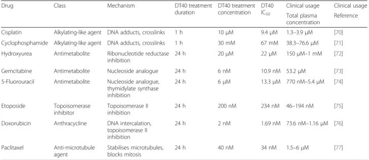
In summary, while genotoxic effects have been mea- sured indirectly for most cytotoxic drugs, sequence-based data for mutagenicity are only available for cisplatin, from an invertebrate model [8]. To acquire reliable data on gen- omic mutagenicity, we performed whole genome sequen- cing on cultured cells treated with representatives of each major category of cancer chemotherapeutics. Each of the chosen cytotoxic agents (Table 1) has been reported to give a positive result in the Ames test or the related bac- terial umu-test [15–19]. HPRT mutagenesis was reported for cisplatin, cyclophosphamide, doxorubicin and etopo- side [20–23], but absent for hydroxyurea [24]. We set out to determine how relevant these findings are to genomic mutagenesis in vertebrate cells. Such studies have not been performed previously, but a proof-of-concept is provided by a recent report on the genomic effect of three environmental mutagens in single sequenced mouse em- bryonic fibroblast clones [25] as well as earlier studies that used whole exome sequencing [26–28]. The main benefit of the obtained mutagenic spectrum data will be the abil- ity to use cancer genome sequences to determine whether the mutagenic drugs have contributed to the development of the tumour, and we provide an important example for this in the reversion of oncogenic gene mutations. The chicken DT40 lymphoblastoma cell line was chosen for treatments for the following reasons: (1) the genome size is about one-third compared to the human genome;
Table 1Cytotoxic drugs investigated in this study
Drug Class Mechanism DT40 treatment
duration
DT40 treatment concentration
DT40 IC50
Clinical usage Clinical usage Total plasma
concentration
Reference
Cisplatin Alkylating-like agent DNA adducts, crosslinks 1 h 10μM 9.4μM 1.3–3.9μM [70]
Cyclophosphamide Alkylating-like agent DNA adducts, crosslinks 1 h 30 mM 67 mM 38.3–76.6μM [71]
Hydroxyurea Antimetabolite Ribonucleotide reductase inhibition
24 h 20μM 22μM 150μM–1 mM [72]
Gemcitabine Antimetabolite Nucleoside analogue 24 h 6 nM 10.9 nM 53.2μM [73]
5-Fluorouracil Antimetabolite Nucleoside analogue, thymidylate synthase inhibition
24 h 6μM 13.3μM 770 nM–5.4μM [74]
Etoposide Topoisomerase
inhibitor
Topoisomerase II inhibition
24 h 200 nM 234 nM 46–194 nM [75]
Doxorubicin Anthracycline DNA intercalation, topoisomerase II inhibition
24 h 2 nM 1.69 nM 73.6 nM–1.16μM [76]
Paclitaxel Anti-microtubule agent
Stabilises microtubules, blocks mitosis
24 h 40 nM 34 nM 1.5–6μM [77]
The name, class and basic mechanism of each drug used in this study is shown, together with the duration and concentration of mutagenesis assay treatments, the estimated IC50concentrations under the same treatment conditions and data on the total plasma concentration range reported in clinical use, with the matching literature reference
(2) this cell line has been used very extensively for DNA repair studies and it models mammalian DNA repair well [29]; and (3) the availability of a wide range of isogenic DNA repair mutant cell lines will allow future comparisons on the influence of individ- ual repair factors on mutagenesis. This detailed gen- omic analysis of multiple post-treatment cell clones provides the most comprehensive survey of the muta- genic potential of commonly used cytotoxics in cancer medicine.
Results
In vitro use of eight chemotherapeutic agents
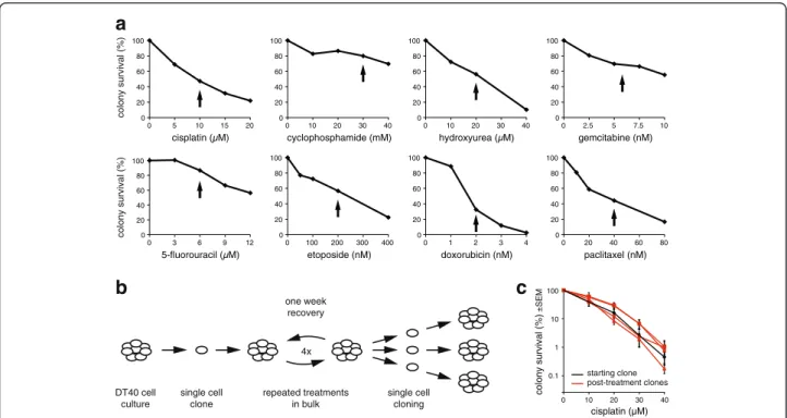
Isogenic wild-type DT40 cells derived from a single cell clone were treated with eight different commonly used cytotoxic agents representing each of the main classes of cancer chemotherapeutics. The agents are listed in Table 1. To select a treatment concentration, we mea- sured the sensitivity of DT40 cells to each drug using a clonogenic survival assay (Fig. 1a). We chose treatment conditions near the IC50concentration of each drug that induce only moderate cell death, with 30–85 % of the cells surviving, in order to avoid selecting for resistant clones that could behave differently during subsequent treatment rounds due to potential changes in, for ex- ample, drug transport or DNA repair. For the surviving
cells, treatments were repeated once a week through four cycles, mimicking cancer chemotherapy regimens and increasing the chance of inducing mutations (Fig. 1b). A comparison of the cisplatin sensitivity of sev- eral post-treatment clones to the starting clone shows that this moderate treatment regimen did not cause significant selection for resistance (Fig. 1c).
One of the tested drugs, cyclophosphamide, undergoes activation by hydroxylation by cytochrome P450 en- zymes [30]. While this is thought to mainly take place in the liver during therapeutic treatment, lymphocytes have also been shown to express the enzymes necessary for cyclophosphamide activation [31, 32]. Therefore, due to the instability and limited availability of the active me- tabolite 4-hydroxycyclophosphamide, cyclophosphamide was added to cells in its pro-drug form. Cisplatin and cyclophosphamide, the two drugs that are known to form DNA adducts, were added for 1 h with the reason- ing that their DNA damaging effect should be largely independent of cell cycle phase. The remaining drugs were used in 24-h treatments. This duration is twice the length of the DT40 cell cycle, ensuring that each cell would be affected by the treatment regardless of the cell cycle phase in which the drugs exert their main effect.
Single nucleotide variation (SNV) and short insertion/
deletion mutations were identified in three cell clones
0 20 40 60 80 100
0 5 10 15 20 cisplatin ( M)
0 20 40 60 80 100
0 10 20 30 40 cyclophosphamide (mM)
0 20 40 60 80 100
0 10 20 30 40 hydroxyurea ( M)
0 20 40 60 80 100
0 2.5 5 7.5 10 gemcitabine (nM)
0 20 40 60 80 100
0 3 6 9 12 5-fluorouracil ( M)
0 20 40 60 80 100
0 100 200 300 400 etoposide (nM)
0 20 40 60 80 100
0 1 2 3 4 doxorubicin (nM)
0 20 40 60 80 100
0 20 40 60 80 paclitaxel (nM)
colony survival (%)colony survival (%)
DT40 cell culture
single cell clone
single cell cloning repeated treatments
in bulk one week
recovery
4x
0 10 20 30 40 cisplatin (µM)
colony survival (%) ±SEM
0.1 1 10 100
starting clone post-treatment clones
a
b c
Fig. 1Cytotoxic treatments.aColony survival assay of DT40 cells treated with the indicated cytotoxic drugs for 1 h (cisplatin, cyclophosphamide) or 24 h. The concentration chosen for mutagenesis assays are indicated withblack arrows.bAschematic drawingof the mutagenesis assay.
Genomic DNA was sequenced from the pre-treatment starting cell clone and three post-treatment cell clones.cComparison of the cisplatin sensitivity of the starting clone (black) and clones isolated following four rounds of cisplatin treatment (red). Mean and SEM of three measurements is shown
derived from each treatment using the IsoMut method developed for this purpose [33, 34]. This approach pro- vides mutation information for the genomes of three in- dividual cells that went through the treatment regime (Fig. 1b). Briefly, we compared all the whole genome se- quences obtained in this study at each genomic position, and only accepted a mutation if it was present in exactly one sample, satisfying criteria on minimum mutated allele frequency, coverage of the mutated sample, and minimum reference allele frequency of each other sam- ple. Due to the lack of availability of validated SNP and short insertion or deletion mutations (indel) datasets, this mutation detection method performs much better on the chicken genome than other commonly used methods, identifying 90–95 % of all mutations with no more than 0–5 false-positive SNVs per genome [33].
Spontaneous mutations
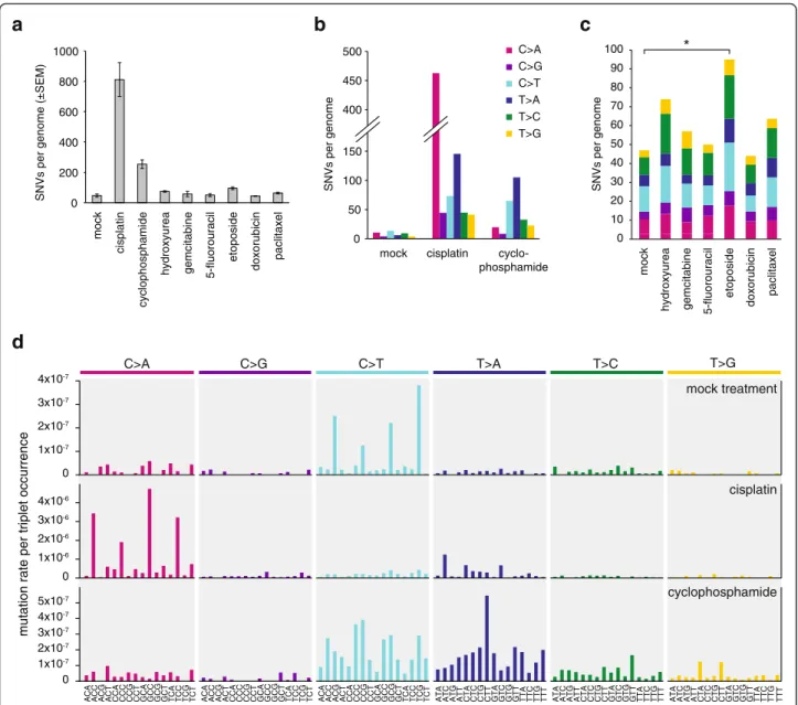
Following a mock treatment regimen spanning approxi- mately 100 cell generations, we detected 47 ± 20 (SD) novel SNVs in three post-treatment clones (Table 2, Additional file 1: Table S1). It is likely that almost all the identified mutations truly arose during the mock treat- ment, as these were identified as unique mutations among all the whole genome sequences obtained for this study, and the same mutation detection method found no unique SNVs – which would be false positives – in the pre-treatment starting clone (Table 2). Of the six possible base substitutions (C > A, C > G, C > T, T > A, T > C, T > G), C > T transitions and C > A transversions were the most common among the spontaneous muta- tions (Fig. 2c, d). The observed mutation number, pro- jected to the 2.06 × 10⁹ base pair diploid genome is equivalent to about 2.3 × 10⁻¹⁰ mutations per base per cell division. When mutations are viewed in the context of the neighbouring bases, and the spontaneous ‘triplet
mutation spectrum’ is normalised to the frequency of genomic occurrence of each triplet, it becomes apparent that NCG > NTG mutations are most common, presum- ably due to C > T base substitutions at methylated CpG sequences [35]. We calculated that NCG > NTG mu- tations were 15× more common than the mean muta- tion rate. Non-normalised triplet spectra are shown in Additional file 2: Figure S1.
Cisplatin induces base substitutions and short indels Cisplatin induced the greatest number of SNVs among the eight tested drugs (Fig. 2a). We performed a detailed analysis of cisplatin-induced mutations to better understand the mutagenic mechanisms. We detected 812 ± 193 SNVs per sequenced post-treatment clone.
C/G > A/T transversions were most common, ac- counting for 57 % of all SNVs, but all six classes of base substitutions increased at least fourfold (Fig. 2b).
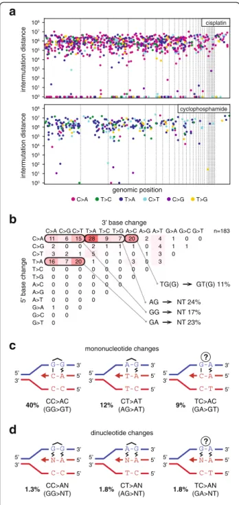
Looking at cisplatin-induced SNVs in the context of the neighbouring bases, it is apparent that NCC > NAC mutations are most common, accounting for 40 % of all SNV cases. Further common changes are NCT > NAT and NTC > NAC, arising in 12 % and 9 % of the SNV cases (Fig. 2d, Additional file 2: Figure S1 and Figure S3 and Additional file 1: Table S2). As the overwhelming majority of cisplatin-induced DNA lesions are intrastrand cross- links between neighbouring purines [36, 37], these three SNV types could represent mutations opposite the 3’base of crosslinked GG, AG and GA dinucleotides, respectively.
In case of GG and AG intrastrand crosslinks, these muta- tions arise through the incorrect incorporation of an ad- enosine opposite the 3’G of the lesion (Fig. 3c). However, GA crosslinks have not been observed in the above reports. Therefore, we catalogued the bases surround- ing the 211 observed TC > AC (GA > GT) mutations, and found that 159 incidences happened at TCC > ACC or TCT > ACT sequences, suggesting that the adjacent base pair 3’to a GG or AG intrastrand crosslink can also mutate. Of the remaining 52 mutations, ten happened at the 5’ base of potential AG crosslinks at CTC > CAC sequences, but in the remaining cases the only poten- tial site for a bipurine crosslink is at GA (Additional file 2: Figure S2). We conclude that cisplatin induces mutagenic lesions at GA dinucleotides, where the le- sions may be hitherto unobserved intrastrand GA crosslinks or monoadducts. To complete the analysis of cisplatin-induced single nucleotide mutations, we note an enrichment of CCA > CAA and CTN > CAN base changes, suggesting adenosine mis-incorporation opposite the 5’base of crosslinked GG or AG dinucleotides.
In agreement with finding mutations (pyrimidine to adenine) across both 3’and 5’ bases of putative cross- linked intrastrand cisplatin lesions, we also detected 61 ± 21 dinucleotide mutations per sample (Fig. 3a, Table 2Number of SNV and short insertion/deletion mutations
in the sequenced samples
Treatment n SNV Insertion Deletion
Mean ± SD Mean ± SD Mean ± SD
None (starting clone) 1 0 0 0
Mock 4 47 ± 20 4.5 ± 1.3 3.0 ± 0.8
Cisplatin 3 812 ± 193 49.0 ± 15.0 83.0 ± 21.2
Cyclophosphamide 3 254 ± 50 3.0 ± 1.7 5.0 ± 1.7
Hydroxyurea 3 74 ± 9 4.7 ± 1.5 3.7 ± 1.2
Gemcitabine 3 57 ± 31 2.3 ± 2.1 3.0 ± 1.7
5-fluorouracil 3 50 ± 16 3.0 ± 1.0 2.7 ± 1.5
Etoposide 3 95 ± 15 3.7 ± 1.2 6.7 ± 4.2
Doxorubicin 3 44 ± 15 3.3 ± 0.6 4.0 ± 4.6
Paclitaxel 3 64 ± 10 1.0 ± 1.0 3.0 ± 0.0
Additional file 1: Table S3). Seventy-five percent of these mutations were found at AG, GG or GA dinu- cleotides. Interestingly, these changed to a range of sequences, equivalent to the incorporation of dinucle- otides AA, AT, AC and AG opposite the putative intrastrand crosslink (Fig. 3d). A further common di- nucleotide mutation class was CA > AC and 18 of 20 cases were found at CCA sequences. On the opposite strand these TGG > GTG mutations could indicate base changes in the position 5’ to GG crosslinks. The
classification of different dinucleotide mutations is shown in Fig. 3b.
Taken together, we observed base substitution muta- tions at the 5’position and the 3’position, as well as the preceding and the following position of putative intras- trand crosslinks. Sequencing the replicated outcome of a GG crosslink in a shuttle plasmid only provided suffi- cient evidence of mutations at the 3’ position [38] and the number of mutations detected in cisplatin-treated C. elegans worms allowed the detection of the same
T>C T>A
C>T C>G
C>A T>G
0 200 400 600 800 1000
mock cisplatin cyclophosphamide hydroxyurea gemcitabine 5-fluorouracil etoposide doxorubicin paclitaxel mock hydroxyurea gemcitabine 5-fluorouracil etoposide doxorubicin paclitaxel
SNVs per genome (±SEM) SNVs per genome SNVs per genome
0 50 100 150 400 450 500
mock cisplatin cyclo- phosphamide
ACA ACC ACG ACT
CCA CCC CCG CCT GCA GCC GCG GCT TCA TCC TCG TCT ACA ACC ACG ACT
CCA CCC CCG CCT GCA GCC GCG GCT TCA TCC TCG TCT ACA ACC ACG ACT
CCA CCC CCG CCT GCA GCC GCG GCT TCA TCC TCG TCT ATA ATC ATG ATT
CTA CTC CTG CTT GTA GTC GTG GTT TTA TTC TTG TTT ATA ATC ATG ATT
CTA CTC CTG CTT GTA GTC GTG GTT TTA TTC TTG TTT ATA ATC ATG ATT
CTA CTC CTG CTT GTA GTC GTG GTT TTA TTC TTG TTT
C>G C>T T>A T>C C>A
T>G
a b c
d
mock treatment
cisplatin
cyclophosphamide 0
1x10⁻⁷ 2x10⁻⁷ 3x10⁻⁷ 4x10⁻⁷
0 1x10⁻⁶ 2x10⁻⁶ 3x10⁻⁶ 4x10⁻⁶
0 1x10⁻⁷ 2x10⁻⁷ 3x10⁻⁷ 4x10⁻⁷ 5x10⁻⁷
mutation rate per triplet occurrence
0 10 20 30 40 50 60 70 80 90
100 *
Fig. 2Number and spectrum of treatment-induced SNVs.aThe mean number of observed SNVs per genome following the described treatment regimen with the indicated drugs.Error barsindicate SEM.bBase substitution spectrum of mutations that arose from the mock treatment, as well as cisplatin and cyclophosphamide treatments.cThe mean number of mutations per sample and base substitution spectrum of the indicated treatments. Significant differences from the mock treatment (p<0.05, Student’s t-test) are indicated with anasterisk.dTriplet mutation spectra of the mock, cisplatin and cyclophosphamide treatments. The middle base of each triplet, listed at thebottom, mutated as indicated at thetop of the panel. The number of mutations of each type was normalised to the frequency of occurrence of that base triplet in the chicken genome, and the resulting mutation rates are shown
mutations, as well as dinucleotide mutations at prob- able AG crosslinks [8]. Our high resolution data indi- cate mutagenesis at each position of a 4-base pair stretch centred on crosslinked GG and AG lesions.
Cisplatin-induced interstrand crosslinks form at GC sequences [39], which can only be present in the triplet spectrum data as GCN. Assuming that by analogy with mutations seen at putative intrastrand crosslinks the most common cause of mutations at interstrand cross- link lesions would be adenosine misincorporation oppos- ite central crosslinked G, these mutations should present as GCN > GAN changes. Some of these triplet base changes were already counted above as potential intras- trand crosslink-induced mutations. Only the GCG > GAG combination could not happen at the site of an intrastrand crosslink, as GCG contains no neighbouring purines.
These mutations are very rare (0.2 % of all SNVs) after cisplatin treatment. In conclusion, our data do not show strong evidence of point mutations induced by cisplatin interstrand crosslink adducts.
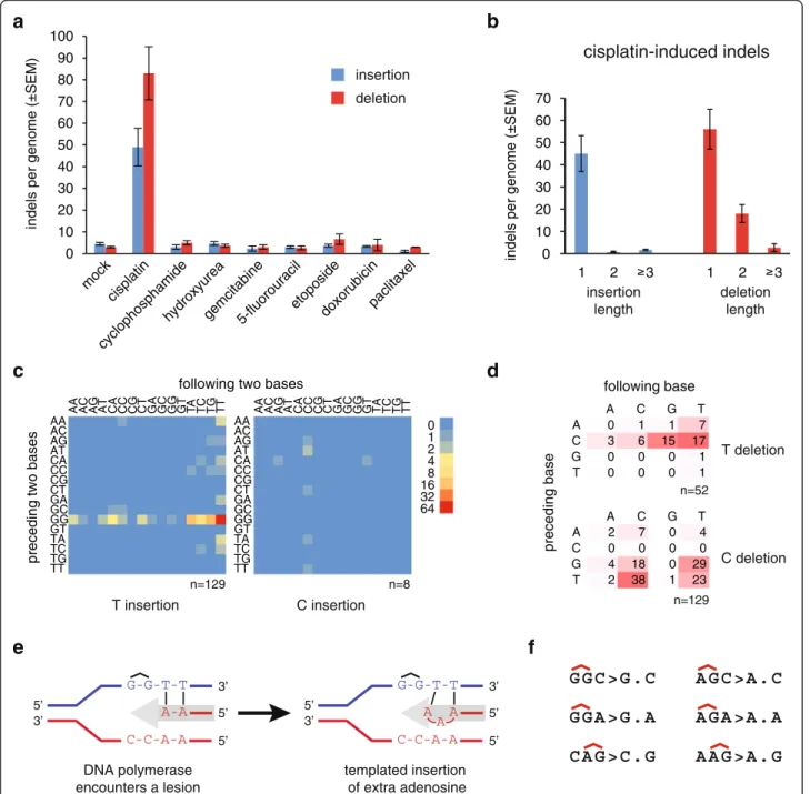
Cisplatin treatment also induced a remarkable number of short insertion and deletion mutations, totalling 132 ± 34 per sample (Fig. 4a, Table 2, Additional file 1:
Table S1). The insertions were almost exclusively one base long (95 % of all insertions, Fig. 4b). We classi- fied one-base insertions based on their sequence con- text (Fig. 4c, Additional file 1: Table S4). Ninety-four percent of one-base insertions were A/T base pairs.
On the strand with the thymidine insertion, the pre- ceding two bases were GG in 81 % of cases, presum- ably representing the site of an intrastrand crosslink Surprisingly, the bases following the insertion site also
G-G
C-C C-A
A-G
T-C T-A
G-A
C-T C-A
G-G
C-C N-A
A-G
T-C N-A
G-A
C-T N-A
Fig. 3SNV mutation spacing, dinucleotide mutations and proposed mechanisms of mono- and dinucleotide mutations.aThe distance of each SNV mutation from the previous SNV on the same chromosome is plotted against the genomic position of the mutation.
Thin dashed linesindicate chromosome boundaries. Chromosomes are shown in numerical order; chromosome Z is shown last on theright.
The colour of eachdotillustrates the type of mutation according to the key at the bottom of the panel. Mutations with an intermutation distance of one are part of dinucleotide mutations. One sequenced clone of each is shown.bSequence analysisof the 183 dinucleotide mutations detected following cisplatin treatment. The change in the 5’base is shown in the rows, while the 3’base in the columns. The equivalent mutations on the two strands are added together, e.g.
GG > TT is shown as CC > AA. The most common mutation types are grouped together below the table and their sequences are indicated using the purine-rich strand to aid interpretation.cSchematic models for the replicative process that may generate each of the most common classes of cisplatin-induced mononucleotide (c) and dinucleotide (d) mutations. Putative intrastrand crosslinks are marked, the uncertain lesion at mutated GA sequences is indicated with aquestion mark. Non-canonical base pairing is shown with azig-zag symbol. The contribution of each mutation class to the total number of observed SNVs is shown
showed strong sequence preference. The first base following a thymidine insertion was 84 % T, while the first two bases together were 51 % TT. If the muta- genic process is DNA synthesis using the damaged
strand as template, we can conclude that it preferen- tially inserts an extra adenosine when the bases 3’ to the template GG crosslink are thymines (see Fig. 4e for a model). Six of the eight observed C/G base pair
a b
0 10 20 30 40 50 60 70 80 90 100
mockcisplatin
cyclophosphamide
hydroxyureagemcitabine5-fluorouracil etoposide
doxorubicinpaclitaxel
indels per genome (±SEM) indels per genome (±SEM)
insertion deletion
0 10 20 30 40 50 60 70
1 2 3 1 2 3
insertion length
deletion length
cisplatin-induced indels
0 1 2 48 16 32 64 AA AC AG AT CA CC CG CT GA GC GG GT TA TC TG TT
AAAC AGAT CACC CGCT GAGC GGGT TATC TGTT
AA AC AG AT CA CC CG CT GA GC GG GT TA TC TG TT AAAC
AGAT CACC CGCT GAGC GGGT TATC TGTT
c
e
d
f
T insertion C insertion
preceding two bases
following two bases
A C G T
A 2 7 0 4
C 0 0 0 0
G 4 18 0 29
T 2 38 1 23
A C G T
A 0 1 1 7
C 3 6 15 17
G 0 0 0 1
T 0 0 0 1
following base
n=8 n=129
n=129 n=52
preceding base
C deletion T deletion
G G C > G . C G G A > G . A C A G > C . G
A G C > A . C A G A > A . A A A G > A . G
G-G-T-T
C-C-A-A A-A
C-C-A-A A A A G-G-T-T
DNA polymerase encounters a lesion
templated insertion of extra adenosine 5’
3’ 5’
5’
3’
5’
3’ 5’
5’
3’
Fig. 4Cisplatin-induced insertions and deletions.aThe mean number of observed insertions (blue) and deletions (red) per genome following the described treatment regimen with the indicated drugs.Error barsindicate SEM.bLength distribution of cisplatin-induced insertions and deletions.
cHeat mapof the frequency of one-base insertions, classified according to the preceding and the following two bases as indicated. The inserted base is shown below each panel. The equivalent mutations on the two strands are added and shown as T or C insertions.dTableandheat map of the frequency of one-base deletions, classified according to the preceding and the following base as indicated. The equivalent mutations on the two strands are added and shown as T or C deletions, shown to theright.eAschematic modelof the generation of the most common GGTT > GGTTT insertions during DNA replication. The incoming DNA polymerase (grey arrow) inserts adenosines opposite the thymine bases, then it inserts an extra adenosine upon encountering the cisplatin-induced GG intrastrand crosslink.fSequence context of the most common one-base deletions shown on the purine rich strand, with the position of putative intrastrand crosslinks indicated above the sequence
insertions occurred at CC/GG sites (Fig. 4c), also likely sites of intrastrand crosslinks.
Seventy-three percent of cisplatin-induced deletions were one base pair long. A classification of one base pair deletion based on the deleted base and the neighbouring two bases shows that the most common deletions af- fected GG or AG sequence motifs, which may be sites of intrastrand crosslinks (Fig. 4d, f, Additional file 1:
Table S4). In the case of AG, based on the AGC > AC and CAG > CG deletions, it is possible to conclude that ei- ther the 3’or the 5’base pair of a putative crosslinked AG dinucleotide may get deleted. In agreement with this, 40 of 59 (68 %) observed two-base deletions removed both base pairs of putative AG or GG intrastrand crosslinks (Additional file 2: Figure S4).
Cyclophosphamide primarily causes T > A and C > T mutations
Cyclophosphamide induced 254 ± 50 base substitution mutations, which is more than five times higher than the mock treatment (p= 0.025, Student’s t-test). The most common base changes were T > A and C > T, followed by a more modest increase in the number of T > C and T > G mutations (Fig. 2b). Cyclophosphamide has been shown to induce a range of adducts in the following proportions:
N7-guanine monoadducts (22 %), crosslinked adducts (6–12 %) and phosphotriester adducts (67 %) [6]. The N7-guanine monoadducts or the G-G interstrand cross- links may account for the C > T lesions. To look for evi- dence of crosslinked adducts, of which the most common have been observed as interstrand adducts between guanines at GNC sequences, we looked for sequence preferences two bases upstream from mutated cytosines (Additional file 2: Figure S3), but we could not find strong evidence for such changes. The prevalent T > A muta- tions, and also the rarer T > C and T > G mutations, pref- erentially occur at the centre of NTT triplets, with some further preference for CTT and TTT (Fig. 2d). These are unlikely to be caused by guanine adducts and may be due to phosphotriester adducts instead. Intermutation dis- tances indicated few dinucleotide mutations or other clus- tering of mutations (Fig. 3a). There was no clustering of mutations when comparing different treated clones in case of either cisplatin or cyclophosphamide, suggesting the lack of mutational hotspots and the largely random distri- bution of SNVs (Additional file 2: Figure S5).
In contrast to cisplatin, cyclophosphamide treatment did not cause a significant increase in the number of insertion or deletion mutations (Fig. 4a).
Etoposide treatment elevates the base substitution frequency
Six further drugs were investigated for their mutagenic potential: the antimetabolites hydroxyurea, gemcitabine
and 5-fluorouracil, plus the topoisomerase II inhibitor etoposide, the anthracycline doxorubicin and the anti- microtubule agent paclitaxel. A comparison of the total SNV numbers for the mock treatment plus these six treatments by ANOVA revealed a significant difference (p= 0.025). Indeed, etoposide induced more than twice the number of mutations as the mock treatment, which is a significant pairwise difference (p= 0.017, Student’s t-test). Each base substitution category increased, resulting in no major change in the overall mutation spectrum (Fig. 2c). In contrast to SNVs, the number of indels was not significantly elevated compared to the mock treatment (Fig. 4a).
No detectable mutagenic effect of hydroxyurea, gemcitabine, 5-fluorouracil, doxorubicin and paclitaxel Twenty-four-hour treatments with hydroxyurea, gemci- tabine, 5-fluorouracil, doxorubicin and paclitaxel did not induce a significant number of extra SNVs or indels in comparison to the mock treatment (Figs. 2a, 4a, ANOVA analysis). These treatments also did not change the spon- taneous SNV mutation spectrum (Fig. 2c). Also, none of the tested agents, including cisplatin, cyclophosphamide and etoposide, induced any larger indels (over 100 bp) or genome rearrangement events, except for a 3453-bp dele- tion in one etoposide-treated clone.
In conclusion, at concentrations that kill a moderate proportion of cultured cells, none of hydroxyurea, gem- citabine, 5-fluorouracil, doxorubicin or paclitaxel in- duced measurable genomic mutagenesis. In contrast, in the same assay, cisplatin and cyclophosphamide induced a large number of mutations with distinct mutation spectra, while etoposide treatment resulted in marginally elevated base substitution mutagenesis.
Lower mutagenesis rates in genes provides evidence of distinct repair rates
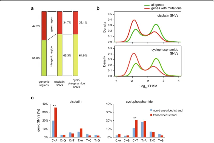
Chemotherapy-induced mutations have the potential of altering gene function and thereby contributing to the development of resistance or secondary tumours. To gauge the importance of this effect, we mapped cis- platin- and cyclophosphamide-induced SNVs with re- spect to gene sequences. A total of 44.2 % of the chicken genome is annotated to code for primary transcripts in genome version Galgal4.82. Interestingly, a smaller pro- portion of treatment-induced mutations appeared at genes (34.7 % and 35.1 %) than expected from a uniform distribution (Fig. 5a). Relative to a uniform genomic dis- tribution, mutations are 17 % more likely to occur at intergenic regions, while they are 22 % underrepresented at genic regions. The most likely explanation for the highly significant reduction of SNV numbers at genes versus intergenic regions (p<0.001 in case of both cis- platin and cyclophosphamide, χ2 test) is the activity of
transcription-coupled repair (TCR), which can remove single strand lesions in an error-free manner [40]. We made use of an RNA-seq dataset from the DT40 cell line to ask whether the gene expression level influences mu- tation density, as expected if it is influenced by TCR. In- deed, we found that the distribution of mutated genes is skewed towards low expression (Fig. 5b), suggesting that the error-free repair of lesions is more efficient in highly expressed genes. Moreover, highly significant (p <0.001, χ2 test) strand bias of cisplatin-induced C > A and cyclophosphamide-induced C > T mutations in genes specifically points to efficient repair of guanine adducts in the transcribed strand by TCR (Fig. 5c).
Correlation of the identified mutational patterns with mutational signatures in human cancer
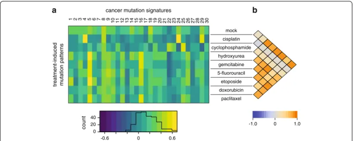
We compared the various treatment-induced mutational patterns to mutational signatures identified in human cancer (COSMIC signatures) [41–43]. The normalised
triplet spectrum of the mock treatment showed good visual similarity with the ageing-associated signature 1 (Fig. 1d) due to the presence of CG > TG mutations.
However, this was not borne out in Pearson correlation analysis (Fig. 6a) as in our mock treated samples there is a range of mutation types in addition to the 15-fold overrepresented CG > TG mutations, while signature 1 essentially contains no other mutation types. The broad- spectrum signature 5 is also associated with ageing [42]
and we observed positive correlation between this signa- ture and several treatments that did not change the spontaneous mutation profile (Fig. 6a). In agreement with the dominance of ageing-related mutational pro- cesses, the mutation profiles of all treatments except cis- platin and cyclophosphamide show good pairwise correlation (Fig. 6b). Cisplatin-induced mutations correl- ate well with the smoking-specific signature 4 and the aflatoxin-induced signature 24 (Fig. 6a), suggesting that these agents cause mutations by similar mechanisms.
0.0 0.1 0.2 0.3 0.4 0.5
Density
a b
55.8%
44.2%
intergenic region
65.3%
34.7%
64.9%
35.1%
-4 -2 0 2 4
Log10 FPKM 0.0
0.1 0.2 0.3 0.4 0.5
Density
genomic regions
cisplatin SNVs
cyclo- phosphamide
SNVs
all genes
genes with mutations
cyclophosphamide SNVs cisplatin SNVs
genic region
0%
10%
20%
30%
40%
C>A C>G C>T T>A T>C T>G 0%
10%
20%
30%
40%
C>A C>G C>T T>A T>C T>G
genic SNVs (%)
c
cisplatin
non-transcribed strand transcribed strand cyclophosphamide
**
**
Fig. 5Mutation density with respect to gene transcription.aThe proportion of genomic regions classified as intergenic (green) or genic (red) based on Ensembl genome annotation, shown on theleft, differs from the proportion of cisplatin or cyclophosphamide-induced SNVs found in the respective regions (middle and right columns).bThe distribution of the expression level of all genes, based on RNA-Seq coverage data (green) is shown against the distribution of the expression level of genes containing cisplatin or cyclophosphamide-induced SNVs (red). Expression levels are shown as the log10of FPKM values (fragments per kilobase of transcript per million of mapped reads).cStrand bias of genic mutations induced by cisplatin (left) or cyclophosphamide (right). Highly significant (p<0.001,χ2test) differences between the non-transcribed and the transcribed strands are indicated withdouble asterisks
Indeed, the three genotoxins all form bulky adducts at N7-guanines, which are generated by polycyclic aromatic hydrocarbons in the case of cigarette smoke [44]. Finally, cyclophosphamide-induced mutations show only weak correlation with the rare signature 25 of unknown aeti- ology (Fig. 6a). These results demonstrate that while mu- tagenesis analysis in cell lines can model the mutational processes observed in cancer, as also evidenced by ex- ome sequencing of mutagen-treated mouse and human cells [26–28] and whole genome sequencing of individ- ual mouse embryonic fibroblast clones [25], it is unlikely that mutations induced by cisplatin or cyclophospha- mide treatment significantly contributed to COSMIC signatures.
Mutagenic chemotherapy may induce resistance through genetic reversal of mutated genes
Mutagenic chemotherapy may have very significant con- sequences if the induced mutations contribute to the subclonal evolution of treatment resistance in surviving cells. We looked for evidence for such a process among documented mutations that restore functionality to mutated BRCA1 or BRCA2 genes [45]. Among seven frameshift mutations observed to restore BRCA2 gene function and cause resistance following cisplatin treat- ment of Capan-1 cells that carry a single base deletion inBRCA2[46], we found two instances of GGT > GGTT insertions, which is by far the most common sequence of cisplatin-induced insertions (Fig. 4c). These insertions, 18 bp downstream of the deleted base pair, restored the reading frame, the protein level and the function of
BRCA2 [46]. In a separate study, a nonsense mutation in BRCA2 became inactivated by a TAG > TAT SNV in a cisplatin-treated ovarian adenocarcinoma following a cisplatin-resistant relapse. In the PEO1 cell line estab- lished from the tumour before the emergence of cis- platin resistance, cisplatin selection led to TAG > TTG mutations of the stop codon in eight out of eight resist- ant clones that restored the BRCA2 protein [47]. Our re- sults show that mutation of either base to thymine at AG putative intrastrand crosslinks are a common conse- quence of cisplatin treatment, with CT > AT (AG > AT) and CT > CA (AG > TG) mutations making up 12 % and 7 % of all cisplatin-induced SNVs, respectively (Fig. 2d).
The finding of these newly identified cisplatin-induced insertions and SNVs in BRCA2 revertants emerging in vivo and in vitro very strongly suggests that therapy can directly induce resistance-causing mutations.
We attempted to calculate an estimate for the likeli- hood of drug-induced mutations generating the exact genetic changes required to revert frameshift or non- sense mutations. If a reverting frameshift needs to hap- pen within 20 base pairs of the original mutation and an SNV needs to change any one of the three bases of a stop codon, then 108randomly placed indels, or 7 × 10⁸ SNVs, would be required for a 50 % chance of the required mutation to occur in the human genome. Our experimental four-cycle cisplatin treatment regimen in- duced about 130 indels and 800 SNVs per Gb. If a clin- ical treatment regimen had the same mutagenic effect, it would take fewer than 1 million surviving treated tumour cells for a 50 % chance of the treatment-induced
mock cisplatin cyclophosphamide
hydroxyurea gemcitabine 5-fluorouracil etoposide doxorubicin
paclitaxel
1 2 3 4 5
40 20
-0.6 0
0
6 7 8 9 10 11 12 13 14 15 16 17 18 19 20 21 22 23 24 25 26 27 28 29 30
cancer mutation signatures
0.6
-1.0 0 1.0
treatment-induced mutation patterns count
a b
Fig. 6Correlation of drug-induced mutation patterns with mutation signatures identified in cancer.aHeat mapof the Pearson correlation coefficient between triplet base mutation patterns induced by the cytotoxic treatment adjusted to human triplet frequencies (rows) and the 30 confirmed mutational signatures identified in human cancer [43]. Theheat map keyis shown at thebottom, with an overlaid histogram indicating the number of cells in each value range.bHeat mapshowing Pearson correlation coefficients between each pair of treatment- induced mutational patterns
reversion of a frameshift or deletion of a nonsense muta- tion. Though partly based on speculative numbers, our approximations suggest that the effect of mutagenic treatments likely contributes to the evolution of drug re- sistance through the initiation of de novo mutations and indels in cancer subclones.
Discussion
We have determined in unbiased whole genome analyses the mutagenicity of eight different common chemothera- peutics. Cisplatin was found to induce many base substitu- tion mutations as well as very short insertion/deletion mutations, and the sequence context of these mutations suggests that they primarily arose at the site of intrastrand crosslinks. Cyclophosphamide also induces base substitu- tion mutations with a specific spectrum, while six further drugs have little mutagenic effect, with a slight elevation of base substitutions after etoposide treatment. Our find- ings may be relevant to assessing the long-term outcome of treatment with the investigated cytotoxic drugs.
Mutagenesis assays are essential to test the mutation- causing effect of chemical agents that humans are ex- posed to, be they medications or environmental agents.
In this study we used the genome of a vertebrate cell line for the purpose of a mutagenesis assay. Whole genome sequencing in the DT40 cell line far surpasses the cur- rently used mutagenesis tests in its relevance to human biology: the commonly used bacterial Ames test takes place in a different metabolic and DNA repair environ- ment, while reporter gene based tests in mammalian cells are affected by sequence bias due to the require- ment that detected mutations must affect protein-coding sequences. We believe that cell line whole genome se- quencing will become the new standard for mutagenesis testing as it is rapid, unbiased and very accurate. Human cell lines will be the most relevant for this purpose, but the chicken DT40 line is a good choice due to its stable diploid genome and its well-studied DNA re- pair properties [29, 48]. Indeed, the dependence of a particular mutagenic process on various DNA repair or replicative DNA damage bypass pathways is readily testable using the wide range of available DT40 mu- tant cell lines, which have been used for genotoxicity screening [49].
The first outcome of using the whole genome as a muta- genesis assay was the determination of the spontaneous mutation rate at 2.3 × 10⁻¹⁰ mutations per base per cell division, the first such measurement in a vertebrate cell line. Remarkably, this is the same order of magnitude as measurements obtained from budding yeast (3.6 × 10⁻¹⁰ [50]; 1.67 × 10⁻¹⁰[51]) orC. elegans(6.7 × 10⁻¹⁰[8]). The mutation rate in the human paternal germline is about two SNVs per year, while an average of 14.2 de novo mu- tations arise in the maternal germline in total [52]. Using
estimates that cell divisions take place every 15–16 days in the human paternal germline, and there are a total of 22–23 divisions in the maternal germline [53], the mean mutation rates can be estimated as 0.17 × 10⁻¹⁰ and 1.1 × 10⁻¹⁰ per base pair per cell division in the human paternal and maternal germlines, respectively. The lack of dependence on maternal age [52] suggests that spontaneous mutations mostly arise during cell prolifera- tion, and the similarity of these mutation rates throughout eukaryotes may be due to constraints of cellular metabol- ism and the mechanism of eukaryotic DNA replication.
The mutagenic effect of cisplatin has been extensively studied in prokaryotic and eukaryotic systems, as well as in vitro [7]. Studies ranging from the replication of a defined lesion in a shuttle vector in mammalian cells [38] to whole genome sequencing of cisplatin-treated C. elegans worms [8] identified CC > AC base substi- tutions as the most common cisplatin-induced muta- tion. In this study, we mapped a greater number of mutations than earlier investigations, presenting a fine resolution analysis of cisplatin-induced mutations. A detailed inspection of base substitutions and short indels revealed that the vast majority of such mutations are gen- erated at intrastrand crosslinks, the most common cis- platin DNA lesions. We showed that mutations can arise at either nucleotide of the intrastrand crosslinks as well as at the previous upstream and the next downstream pos- ition. Interstrand crosslinks, which may be more signifi- cant for the cytotoxic effect of cisplatin, had no detectable mutagenic effect.
Cyclophosphamide induced a markedly different SNV mutation spectrum than cisplatin, with the elevation pri- marily of T > A and C > T mutation numbers. It is chal- lenging to explain the mutation spectrum based on the available evidence of cyclophosphamide-induced lesions [6]. C > T mutations, which show strand bias in genes, may arise from N7-guanine adducts of the cyclophos- phamide metabolite phosphoramide mustard [54]. How- ever, while the N7-guanine adducts of cisplatin typically lead to C > A changes opposite the lesion, the C > T mu- tations caused by cyclophosphamide suggest a different mutagenic mechanism. As no adducts have been de- tected on adenine or thymine bases, the T > A muta- tions, which have also been observed in lacI reporter genes of cyclophosphamide-treated mice [55], may in- stead be caused by the common cyclophosphamide- induced phosphotriester adducts on the DNA backbone [56]. Indeed, phosphotriester adducts show some base preference for neighbouring thymines, and pyrimidine bases in general [57]. Phosphotriester adducts are very inefficiently repaired, which could explain the lack of strand bias of T > A mutations.
Single-strand adducts are repaired primarily by base excision repair and nucleotide excision repair. Both
mechanisms are expected to mostly produce an error- free outcome. Unless the lesions miscode directly, the main cause of mutagenesis is DNA replication that uses the damaged strand as template, termed translesion syn- thesis (TLS). This is typically performed by specialised translesion polymerases; indeed, while the replicative polymerasesδ orεcannot bypass a GG cisplatin adduct [58], polymeraseηandζtogether can bypass this lesion with a classical two-polymerase mechanism [59]. Our large dataset of cisplatin-induced mutations shows that mutations on the newly synthesised strand can appear in the position immediately upstream of the lesion as well as opposite the lesion, for example we observed NCC > ACC (GGN > GGT) mutations. Similarly, one-base insertions mostly appeared in the nascent strand upstream of the lesion, such as the common ACC > AACC (GGT > GGTT) insertions. The latter also suggests a mutagenic mechanism: the template base be- fore the crosslinked adduct may not fit perfectly into the active site of the replicative polymerase, which could lead to the base pairing of the incoming nucleotide with the previous template base, causing a templated insertion as seen here (Fig. 4e). Because all the cisplatin-induced indels are 1–2 base pairs only, and are generally located at puta- tive lesions, it is likely that they are mostly caused by translesion synthesis rather than the repair of DNA breaks or other mechanisms. TLS across cyclophosphamide lesions, which are mostly monoadducts, may be able to avoid similar template slippage, explaining the lack of indels induced by this treatment.
A significant finding of this study is that five of the investigated cytotoxic drugs were not mutagenic under the experimental conditions. Positive results were re- ported for these drugs in the Ames test or the HPRT assay [15, 17–19, 22, 24], which suggests that these as- says may overamplify the mutagenic signal. A limitation of interpreting the relevance of our finding for clinical use is that data are only available for plasma concentra- tions during clinical treatment, which are different from the concentration reaching the cancer cell. Still, cis- platin, 5-fluorouracil and etoposide were used at levels very near their measured plasma concentrations (Table 1). Cyclophosphamide was used at a much higher concentration, presumably due to the limited ability of cytochrome P450 enzymes in DT40 lympho- cytes to activate this prodrug. Hydroxyurea, gemcita- bine, doxorubicin and paclitaxel were used well below their measured clinical peak plasma concentrations.
However, at the treatment concentrations only 30–70 % of the cells survived, preventing us from using higher con- centrations. Overall, the near-lethal doses used in our ex- periments are probably a reasonable model for the conditions experienced by somatic and tumour cells dur- ing clinical treatment.
Do these results help estimate the oncogenic potential of the selected drugs? Cancers that arise as direct conse- quence of a known external mutagen, such as melanoma and different lung cancer types, typically contain the highest number of genomic mutations, about ten per megabase [41, 60]. If the induced SNVs are indeed the main contributors to carcinogenesis, we can conclude that a similar density of largely randomly spaced muta- tions is required for a tumour to develop somewhere in the body, in which case the mean density of the mutagen-induced somatic mutations in all affected cells is probably lower. Precise clinical data on the number of cytotoxic treatment-induced mutations are not available, while after a four-cycle treatment regimen, cisplatin in- duced an average of 0.8 mutations per megabase in DT40 cells. Thus the mutagenic consequences of such treatment are comparable to those of carcinogenic envir- onmental mutagens. The density of mutations induced by cisplatin treatment even surpassed the median muta- tion density of many common cancer types including breast, pancreatic and prostate cancer, AML and chronic lymphocytic leukaemia [60], suggesting that cisplatin treatment can make a major contribution to the devel- opment of secondary malignancies.
A recent report attributed CC > CA mutations to the mutagenic effect of cisplatin treatment in whole exome sequence data of cisplatin-resistant squamous cell car- cinoma of the head and neck [61]. This mutation pattern does not agree with the predominance of CC > AC cis- platin mutations demonstrated by our study and an earl- ier report [8]. For a treatment-derived mutation pattern to be observable, significant clonal expansion must hap- pen between the treatment and the sampling, which is less likely if the original tumours were resistant to the treatment. Consequently, we suspect that the observed CC > CA changes appeared due to a sample prepar- ation artefact, as has been reported previously [62].
Treatment-derived mutations will be easier to detect or validate when mutation spectra from controlled experiments are available. Following in the footsteps of initial whole genome mutagenesis studies usingE. coli, S. cerevisiaeandC. elegans[8, 63, 64], our study is the first to use whole genome sequencing of vertebrate cell clones to provide clinically relevant data on the mutagenicity of pharmaceutical agents.
Treatment-induced mutations will contribute to the further evolution of the tumour and could be relevant for the evolution of resistance. The finding of newly defined cisplatin-specific mutation types as causes of the reversion of BRCA2 mutations supports this notion, accompanied by our estimate that one genome among as few as a million cells surviving the treatment could contain any particular specific mutation. Treatments could not only cause the reactivation of mutated genes,
but any other genetic change advantageous for tumour growth or treatment resistance.
Conclusions
This study demonstrated the utility of whole genome se- quencing in cell lines as a mutagenesis assay. We deter- mined the spontaneous mutation rate in a cultured vertebrate cell line, and found it as low as the mutation rate of a range of organisms. We measured the muta- genic effect and defined the mutation spectrum caused by eight common cytotoxic agents. Our results suggest that cytotoxic treatment with the mutagenic cisplatin or cyclophosphamide can make a major contribution to the development of secondary malignancies and also directly contribute to the development of resistance. The defin- ition of the precise mutagenic signature of these drugs will help assaying their mutagenic effect in post-treatment tumour samples to provide further information. Based on the lack of a detectable increase in genomic mutations following treatment with hydroxyurea, gemcitabine, 5- fluorouracil, doxorubicin or paclitaxel, it is less likely that base substitution and small insertion/deletion mutations caused by these drugs make a significant contribution to tumorigenesis. Further confirmation of these results and the expansion of mutagenesis studies to other cancer ther- apeutics could influence the choice of curative cancer treatment regimens, particularly in childhood cancer.
Methods
Cell culture and drug treatments
The wild type DT40 cell line used in this study was obtained from the laboratory of Dr Julian E. Sale, MRC Laboratory of Molecular Biology, Cambridge, UK, and its complete genome sequence has been published [48].
Cells were grown at 37 °C under 5 % CO2in RPMI-1640 medium supplemented with 7 % fetal bovine serum, 3 % chicken serum, 50μM 2-mercaptoethanol and penicillin/
streptomycin. Drug sensitivities were measured using col- ony survival assays; treated cells were plated in medium containing 1 % methylcellulose using a tenfold dilution series and surviving colonies were counted 10 days later.
For the mutagenesis experiments, four rounds of drug treatments were performed in weekly intervals. One million cells were treated each time. The chemicals were obtained from Sigma. Etoposide, 5-fluorouracil and pac- litaxel were diluted from stock solutions in DMSO. The remaining drugs were dissolved in water. Cyclophospha- mide and 5-fluorouracil were dissolved freshly each time before treatments. Mock-treated cells were handled in parallel without the addition of any drug. Single-cell clones were isolated by limiting dilution and grown prior to sample preparation. Six clones were selected from 96- well plates at random. Genomic DNA was prepared using the Gentra Puregene Cell Kit (Qiagen) and three
of the preps were sequenced. Cisplatin sensitivity mea- surements were performed on four clones which in- cluded the three sequenced clones.
Whole genome sequencing and mutation detection Library preparation was done using either the TruSeq DNA Nano Library Preparation Kit (Illumina) or the NEBNext Ultra DNA Library Prep Kit for Illumina (New England Biolabs). Seven library pools (the starting clone and one each of mock, cisplatin, hydroxyurea, gemcita- bine, etoposide and paclitaxel treated clones) were loaded on Illumina HiSeq 2500 Rapid Run flow cells (v1) and sequenced in a 2 × 150-bp paired end (PE150) for- mat using Rapid SBS reagents. The remaining 22 sam- ples were loaded on Illumina HiSeq 2500 v4 High Output flow cells and sequenced in a 2 × 125-bp paired end format using HiSeq SBS v4 reagents. Library prepar- ation and DNA sequencing were done at the Research Technology Support Facility of Michigan State University, USA and at Novogene, Beijing, China.
The reads were aligned to the chicken (Gallus gallus) reference sequence Galgal4.73, using a method already described [48]. Duplicate reads were removed using the samblaster program [65]. Additionally, the aligned reads were realigned by the GATK IndelRealigner [66].
SNVs and indels were identified using the IsoMut method developed for multiple isogenic samples, using a downloadable tool [34]. Briefly, a pileup of all samples by genomic position was produced using the samtools mpi- leup command and a base quality filter of 30 was used to reduce sequencing noise. Data from 120 different se- quenced DT40 clones were used at this step, which in- cluded the 29 samples presented in this article. To identify SNVs and indels, the pileup data were filtered at each gen- omic position by minimum mutated allele frequency (0.33), minimum coverage of the mutated sample [10] and minimum reference allele frequency of each other sample (0.9). These parameters were determined in an optimisa- tion procedure using a test set [33]. The test set was ob- tained by comparing two sets of whole genome sequences from DT40 clones of two different genotypes, and the par- ameter optimisation resulted in the identification of 95 % of the test set (true-positive rate). A detailed description is given in Additional file 3. Structural variations were de- tected using the CREST algorithm [67].
Mutation analysis
Insertions and deletions in homopolymer or other repeat regions were aligned to the leftmost possible position.
During the analysis of indel sequence context, before adding events with complementary sequences, identified indels on the opposite strand were realigned to the right.
Cisplatin-specific and cyclophosphamide-specific muta- tions localising in genes and intergenic regions according