Accepted Manuscipt
Published: Struct. Chem. 2018, 29, 113–118., DOI: 10.1007/s11224-017-1008-x
Structural characterization of a sodium perchlorate−acridino-18- crown-6 ether complex
Tamás Németh1, József Levente Petró1, Ibolya Leveles2,3, Tünde Tóth1, György Tibor Balogh4, Beáta G. Vértessy2,3, Péter Huszthy1*
1Department of Organic Chemistry and Technology, Budapest University of Technology and Economics, H-1521 Budapest, PO Box 91, Hungary
2Institute of Enzymology, RCNS, Hungarian Academy of Sciences, Budapest, Hungary
3Department of Applied Biotechnology and Food Sciences, Budapest University of Technology and Economics, Budapest, Hungary
4Compound Profiling Laboratory, Gedeon Richter Plc., Budapest, Hungary
*Corresponding author: Tel.: +36-1-463-1071; fax: + 36-1-463-3297 E-mail:huszthy@mail.bme.hu
Abstract - This paper describes the X-ray crystal structure of a complex of acridino-18-crown-6 ether (S,S)-2 and sodium perchlorate. The structure shows a π–π bonded homodimer in the crystal. The average distance of the two tricyclic units (3.49±0.1 Å) indicates a strong π–π interaction. Fluorescence titration was performed in order to determine the stoichiometry and stability constant (Ks) of the sodium ion-(S,S)-2 complex. Based on the global fitting of the fluorescence spectra we suggest the formation of a complex with 1:1 ligand to metal ion ratio and the logK value determined by nonlinear regression analysis was 5.23.
Keywords: Crown ether, Acridine, Sodium complexation, X-ray analysis, π–π interaction, cation–π interaction, Fluorescence spectroscopy
2 Introduction
Sodium ion is essential to all living organisms. In humans, for example it regulates blood volume, blood pressure, and it helps the cells to transit nerve signals. The latter is accomplished by an active transporter molecule, Na+/K+-ATPase, which pumps ions against the ion gradient in the sodium/potassium channels [1]. Sensor molecules capable of selective discrimination of metal ions are of great significance due to their potential application in pharmaceutical or food industries, also in environmental chemistry [2]. During these selective interactions a generally occurring natural phenomenon called molecular recognition plays a vital role. The complexes, formed by the host and guest molecules, are held together by secondary interactions, such as hydrogen bonding, π–π [3-5] and cation–π [6-8] interactions. Receptors based on various macrocycles, such as monensin [9], bispyrrolidone [10] and crown ether derivatives [11-20], were capable of selective recognition of sodium ions.
We have studied the complexation ability of chiral dimethyl-substituted crown ether type sensor molecules containing an acridine fluorophore unit [(R,R)-1 and (S,S)-2, see Figure 1] toward the enantiomers of 1-phenylethylamine hydrogen perchlorate (PEA), 1-(1-naphthyl)ethylamine hydrogen perchlorate (1-NEA), phenylglycine methyl ester hydrogen perchlorate (PGME) and phenylalanine methyl ester hydrogen perchlorate (PAME) by UV/Vis and fluorescence spectroscopies [18]. Recently, we reported the preparation and X-ray analysis of the crystalline complexes of the dimethyl-substituted acridino-18-crown-6 ether (R,R)-1 and the enantiomers of 1-NEA [21]. We found that the heterochiral complex [(R,R)-1–(S)-1-NEA] is more stable than the homochiral one [(R,R)-1–(R)-1-NEA]. In the case of (S,S)-2, based on the fluorescence spectroscopic studies the macrocycle showed the highest enantiomeric discrimination toward the enantiomers of PGME [18]. In continuation of our research in this area we attempted to prepare the single crystals of (S,S)-2 and the enantiomers of PGME for X-ray analysis. To our surprise, in the cases of both enantiomers of PGME complexes of (S,S)-2 and sodium ions were formed.
UV/Vis spectroscopic study and fluorescence titration was performed in order to determine the stoichiometry and stability constant (Ks) of the sodium ion-(S,S)-2 complex. Also, to further study this complex, suitable single crystals for X-ray analysis were prepared from (S,S)-2 and sodium perchlorate.
3 Figure 1. Schematics of the acridino-18-crown-6 ether host molecules.
Experimental
Infrared spectrum was obtained on a Bruker Alpha-T FT-IR spectrometer. 1H (500 MHz) and 13C (125 MHz) NMR spectra were taken on a Bruker DRX-500 Avance spectrometer. Optical rotation was taken on a Perkin-Elmer 241 polarimeter that was calibrated by measuring the optical rotations of both enantiomers of menthol. Elemental analysis was performed on a Vario EL III instrument (Elementanalyze Corporation) in the Microanalytical Laboratory of the Department of Organic Chemistry, L. Eötvös University, Budapest, Hungary. Melting point was taken on a Boetius micro melting point apparatus and was uncorrected. Reagents were purchased from Sigma–Aldrich Corporation. All chemicals were of analytical grade, NaClO4.H2O was used in the complexation study. UV/Vis spectra were taken on a Multiskan Spectrum Microplate Spectrophotometer controlled by SkanIt Software for Multiscan version 2.1. Fluorescence spectra were recorded on a BMG Labtech CLARIOstar spectrophotometer. Spectrophotometric titrations were carried out according to the literature [22]. The stability constants of the complexes were determined by global nonlinear regression analysis using the ReactLabTM Equilibria spectral analyses suite (Jplus Consulting, www.jplusconsulting.com). The concentrations of the solutions of sensor (S,S)-2 were 100 µM for the UV/Vis measurements and 20 µM for the fluorescence titrations.
Synthesis of the crystalline (S,S)-2-Na+ complex
Crown ether (S,S)-2 [(8S,16S)-8,16-dimethyl-6,9,12,15,18-pentaoxa-25- azatetracyclo[21.3.1.05,26.019,24]heptacosa-1(26),2,4,19,21,23(27),24-heptaene] was prepared
4 according to the literature [18]. Crown ether (S,S)-2 (70 mg, 0.18 mmol) was added to the solution of NaClO4.H2O (22 mg, 0.18 mmol) in ethanol (8 mL). The mixture was refluxed for 10 mins and filtered hot. Suitable single crystals for X-ray crystallographic studies were obtained from the almost saturated solution which was allowed to stand at room temperature in a glass ampoule. This way 35 mg (37%) of bright red plates were obtained.
Mp 139–142°C; [α]D25 = +14 (c 0.14, EtOH); IR (KBr) νmax 3427, 3099, 3089, 3029, 2977, 2938, 2884, 1631, 1594, 1567, 1509, 1492, 1467, 1422, 1413, 1364, 1326, 1292, 1199, 1130, 1095, 1078, 956, 890, 790, 759, 721, 624 cm-1; 1H-NMR (CD3CN, 500 MHz) δ 1.37 (broad s, 6H), 3.55–3.91 (m, 8H, OCH2), 4.15–4.37 (m, 4H, OCH2), 4,43 (d, J = 8Hz, 2H, OCH2), 7.13–7.35 (m, 1H, Ar-H), 7.41–7.61 (m, 2H, Ar-H), 7.62–7.95 (m, 3H, Ar-H), 8.89 (s, 1H, Ar-H) 13C-NMR (CD3CN, 125 MHz) δ 15.80, 64.81, 68.90, 70.62, 70.66, 71.37, 73.08, 74.07, 74.41, 109.78, 121.80, 122.04, 127.46, 128.71, 129.57, 138.00, 140.92, 149.11, 150.01, 153.66; Anal Calcd for C23H27ClNNaO9: C 53.14; H 5.23; N 2.69. Found: C 52.85; H 5.51; N 2.62.
Crystal structure determination
A suitable crystal was selected and the X-ray dataset has been collected at 102 K on a single source micro-focus Cu X-ray sealed tube SuperNova diffractometer (Agilent Technologies) with monochromated Cu-Kα radiation (λ = 1.5418 Å) and Eos CCD detector. The data reduction was performed with program package CrysAlisPro SXRED [23]. The space group determination was performed via GRAL module by applying the Laue symmetry. The structures were solved by direct methods using Olex2 [24] and refined using fullmatrix least-squares. All calculations were performed using Olex2[24] and SHELXL97 [25] programs. The crystal data and refinement parameters are presented in Table 1. The data (CCDC 1548555) can be obtained at www.ccdc.cam.ac.uk/conts/retrieving.html.
Results and Discussion
The enantiomeric discrimination ability of the dimethyl-substituted acridino-18-crown-6 ether (S,S)-2 toward the enantiomers of the perchlorate salts of primary aralkylamines and α-amino acid esters was studied by Kertész et al. The fluorescence spectroscopic studies demonstrated that in the case of (S,S)-2, the highest enantiomeric discrimination was observed for the enantiomers of PGME [18]. In order to better understand the secondary interactions governing
5 the enantiomeric recognition of crown ether based sensor and selector molecules containing an acridine moiety our aim was to prepare suitable single crystals for X-ray analysis from (S,S)-2 and the enantiomers of PGME. However, instead of the diastereomeric complexes, the complexes of macrocycle (S,S)-2 and sodium ions were obtained in both cases. The presence of sodium ions can be explained by the fact that the last step of the preparation of (S,S)-2 [18] is a Bouveault–Blanc reduction [26], which employs sodium metal as a reducing agent, and during the reduction the oxidation of the metal takes place and sodium ions are formed. Presumably, crown ether (S,S)-2 formed strong secondary interactions with sodium ions, and this complex withstood the purification of the crude product. It can also be assumed that crown ether (S,S)-2 forms a complex with sodium ions with a higher stability constant (Ks) than with the enantiomers of PGME. In order to prove this assumption and to determine the stoichiometry and stability constant (Ks) of the sodium ion-(S,S)-2 complex, UV/Vis spectroscopic study and fluorescence titration were performed. Also, the single crystals of (S,S)-2 and sodium perchlorate were prepared and studied by X-ray analysis.
UV/Vis and fluorescence spectroscopic studies
The complexation ability of (S,S)-2 was first studied by UV/Vis spectroscopy in acetonitrile.
Figure 2. shows that no changes could be observed in the absorbance spectra even upon addition of a twenty-five-fold excess of sodium ions. However, the binding of sodium ions by (S,S)-2 was associated with significant fluorescence enhancement upon addition of these ions (Figure 3), thus fluorescence titration was performed. The fluorescence spectra were measured at 15 different cation to (S,S)-2 ratios (Figure 3). The latter fluorescence changes were used to determine the stability constant and stoichiometry of the complex. Upon being treated with sodium ions the fluorescence enhancement of the sensor molecule (S,S)-2 followed the Benesi-Hildebrand equation [22, 27], therefore we could assume the formation of a complex with 1:1 ligand to metal ion ratio. The changes in the spectra were further analyzed using nonlinear regression analysis and global fitting of the fluorescence spectra, which also suggested the 1:1 stoichiometry. The logK value determined by ReactLabTM Equilibria program suite was 5.23. Kertész et al.
determined the logK values for the complexation of (S,S)-2 with the enantiomers of PGME, in the same solvent (MeCN) [18]. Macrocycle (S,S)-2 formed a more stable complex with (S)-
6 PGME (logK 4.87) than with (R)-PGME (logK 4.61). It means that ligand (S,S)-2 is 2.3-fold more selective toward sodium ions than toward the (S) enantiomer of PGME.
Figure 2. UV/Vis spectra of (S,S)-2 (100 µM) in the presence of 0, 1, 5, 10, and 25 equiv. of sodium ions.
Figure 3. Fluorescence emission series of spectra upon titration of (S,S)-2 (20 µM) with sodium ions (0–20 equiv.) in MeCN, λex = 380 nm.
7 Crystal structure of the sodium-crown ether complex
In the crystal, the complex is found as a homodimer (Figure 4) consisting of two acridino-crown ethers and two sodium ions. The complex crystallized in the orthorhombic crystal system (Table 1). The distances of the two acridine rings of the monomers (3.41–3.59 Å) indicates a strong π–π interaction. The X-ray studies also suggest the existence of cation–π interaction, at a distance of 3.96–4.85 Å, between the sodium ions and the electron rich acridine moieties (Table 2).
The complexation environment of both sodium ions is well defined: among the six coordination partners, one nitrogen and five oxygen atoms are provided from the acridine unit and the macroring of the appropriate crown ethers, additionally two perchlorate ions are also present (Figures 4 and 5, Table 3). The strength of the π–π and cation–π interactions overcompensated the electrostatic repulsion between the sodium ions, thus the ions are drawn together (Figure 4).
In accordance with the XRD measurements the fluorescence titration suggested the formation of a complex with 1:1 ligand to metal ion ratio, which was also confirmed by elemental analysis (see Experimental Section). Due to the coordination the more flexible parts of the macroring (O3, O7, O11 or O17, O29, O34 of the appropriate monomer) are drawn toward the complexed sodium ions (Figure 5, Table 3).
NMR spectra
The 1H-NMR and 13C-NMR spectra were recorded in CD3CN due to the poor solubility of the complex in CD3OD. The signals doubled in the 13C spectrum of the complex comparing to the spectrum of free ligand (S,S)-2 [18], which also suggests the complexation of sodium ions.
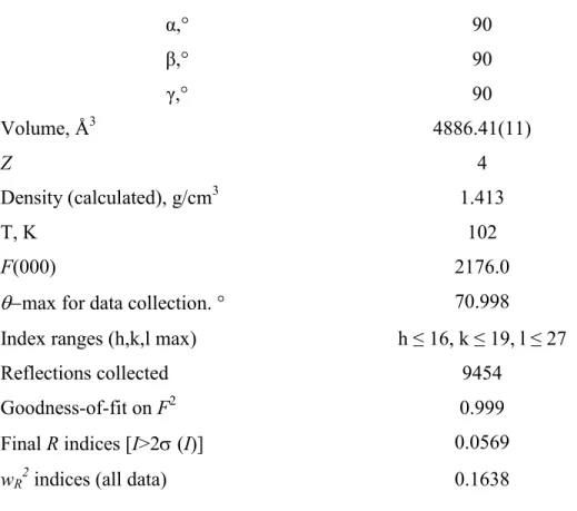
Table 1.: Crystallographic data for (S,S)-2-sodium complex
Compound (S,S)-2-sodium complex
Empirical formula C46H54Cl2N2Na2O18
Formula weight 1039.79
Crystal system orthorhombic
Space group P 212121
Unit cell dimensions a, Å 13.77(19)
b, Å 15.90(2)
c, Å 22.33(3)
8
α,° 90
β,° 90
γ,° 90
Volume, Å3 4886.41(11)
Z 4
Density (calculated), g/cm3 1.413
T, K 102
F(000) 2176.0
max for data collection. ° 70.998
Index ranges (h,k,l max) h ≤ 16, k ≤ 19, l ≤ 27
Reflections collected 9454
Goodness-of-fit on F2 0.999
Final R indices [I>2 (I)] 0.0569
wR2 indices (all data) 0.1638
Table 2.: Distances of the appropriate atoms that may indicate the presence of π–π and cation–π interactions.
π–π interaction cation–π interaction C· · ·C C· · ·C Na· · ·C/N Na· · ·C/N
(Å) (Å)
C33 · · ·C38 3.49 Na8 · · ·N18 4.05
C23 · · ·C51 3.59 Na8 · · ·C15 4.76
C14 · · ·C19 3.48 Na8 · · ·C19 4.85
C15 · · ·C22 3.41 Na8 · · ·C26 4.76
C21 · · ·C43 3.43 Na8 · · ·C27 4.54
C19 · · ·C28 3.54 Na3 · · ·C2 4.37
Na3 · · ·N12 3.96
Na3 · · ·C6 4.43
Na3 · · ·C25 4.65
Na3 · · ·C5 4.70
9 Figure 4. Structure of the dimer containing two monomers. Other species (two perchlorate ions) are also shown. Atomic coloring is as follows: C: grey, O: red, N: blue, Cl: green, Na: lilac, H:
white.
Figure 5. Separate representation of the two monomers. Note the differences in the lower parts of the macrorings of the appropriate crown ethers. Atomic coloring is as follows: C: grey, O: red, N: blue, Na: lilac, H: white.
10 Table 3.: Geometry of the complexed sodium ions.
Monomer O/N· · ·Na O/N· · ·Na O/N· · ·Na3· · ·O34
(Å) (°)
Monomer 1 O3 · · ·Na8 3.0
O11 · · ·Na8 3.0 O10 · · ·Na8 3.0 O1 · · ·Na8 3.0 O7 · · ·Na8 2.9 N12 · · ·Na8 2.9
Monomer 2 O4 · · ·Na3 2.6 101.2
O17 · · ·Na3 2.5 66.8
O34 · · ·Na3 2.5 -
O29 · · ·Na3 2.7 66.1
O9 · · ·Na3 2.5 107.3
N18 · · ·Na3 2.5 107.3
Monomer 1 and 2 Na8 · · ·Na3 2.4
Conclusions and further aims
We succeeded in preparing suitable crystals for X-ray analysis from the sodium complex of acridino-18-crown-6 ligand (S,S)-2. Macrocycle (S,S)-2 forms a π–π bonded dimer in the crystal.
Fluorescence titration was also performed in order to determine the stoichiometry and stability constant (Ks) of the sodium ion-(S,S)-2 complex: global fitting of the fluorescence spectra and elemental analysis indicated 1:1 stoichiometry.
Acknowledgements
Financial supports of the National Research, Development and Innovation Office (former OTKA, grant numbers: K112289, K109486 and NK84008), the CRP/HUN14-01 ICGEB Research Grant and the New Széchenyi Development Plan (TÁMOP-4.2.1/B-09/1/KMR-2010- 0002) are gratefully acknowledged. The research has also been supported by the ÚNKP-16-3-III.
New National Excellence Program of the Ministry of Human Capacities .
11 References
1. Reece JB, Urry LA, Cain ML, Wasserman SA, Minorsky PV, Jackson RB (Eds) (2014) Campbell Biology, 10th Edition, Pearson, Boston, USA.
2. Desvergne JP, Czarnik AW (Eds) (1997) Chemosensors of ion and molecule recognition NATO ASI Series C; Kluwer: Dordrecht, The Netherlands, Vol. 492.
3. Hunter CA, Sanders JKM (1990) The nature of π–π interactions. J Am Chem Soc 112:5525–
5534.
4. Anslyn EV, Dougherty DA (2005) In: Murdzek J (Ed) Modern physical organic chemistry, University Science Books, Sausalito, California, USA.
5. Pecsi I, Leveles I, Harmat V, Vertessy BG, Toth J (2010) Aromatic stacking between nucleobase and enzyme promotes phosphate ester hydrolysis in dUTPase. Nucleic Acids Res 38:7179–7186.
6. Mahadevi AS, Sastry GN (2013) Cation−π interaction: its role and relevance in chemistry, biology, and material science. Chem Rev 113:2100–2138.
7. Nagy GN, Marton L, Contet A, Ozohanics O, Ardelean LM, Révész A, Vékey K, Irimie FD, Vial H, Cerdan R, Vértessy BG (2014) Composite aromatic boxes for enzymatic transformations of quaternary ammonium substrates. Angew Chem Int Ed Engl 53:13471–13476.
8. Priyakumar UD, Sastry GN (2003) Cation-π interactions of curved polycyclic systems: M+ (M=Li and Na) ion complexation with buckybowls. Tetrahedron Lett 44 (32):6043–6046.
9. Pinkerton M, Steinrauf LK (1970) Molecular structure of monovalent metal cation complexes of monensin. J Mol Biol 49 (3):533–546.
10. Perrin A, Myers D, Fucke K, Musa OM, Steed JW (2014) N-Alkyl pyrrolidone ether podands as versatile alkali metal ion chelants. Dalton Trans 43 (8):3153–3161.
11. Izatt RM, Pawlak K, Bradshaw JS, Bruening RL (1991) Thermodynamic and kinetic data for macrocycle interaction with cations and anions. Chem Rev 91:1721–2085.
12. Izatt RM, Pawlak K, Bradshaw JS, Bruening RL (1995) Thermodynamic and kinetic data for macrocycle interaction with cations, anions and natural molecules. Chem Rev 95:2529–2586.
13. Gokel GW, Leevy WM, Weber ME (2004) Crown ethers: sensors for ions and molecular scaffolds for materials and biological models. Chem Rev 104:2723–2750.
12 14. Tong A-J, Song Y-S, Li L-D, Hayashita T, Teramae N, Park C, Bartsch RA (2000) Selective extraction of alkali metal cations with proton-ionizable dibenzo-16-crown-5 fluoroionophores.
Anal Chim Acta 420:57–64.
15. Gunnlaugsson T, Nieuwenhuyzen M, Richard L, Thoss V (2002) Novel sodium-selective fluorescent PET and optically based chemosensors: towards Na+ determination in serum. J Chem Soc Perkin Trans 2:141–150.
16. Jóźwiak M (2002) Complex formation of crown ethers with cations in water–organic solvent mixtures. III. Thermodynamics of interaction between Na+ ion with 15-crown-5 ether in water–
acetonitrile mixtures at 25°C. J Solution Chem 31:589–599.
17. Móczár I, Peragovics Á, Baranyai P, Tóth K, Huszthy P (2010) Synthesis and fluorescence studies of novel bis(azacrown ether) type chemosensors containing an acridinone unit.
Tetrahedron 66:2953–2960.
18. Kertész J, Móczár I, Kormos A, Baranyai P, Kubinyi M, Tóth K, Huszthy P (2011) Synthesis and enantiomeric recognition studies of dialkyl-substituted 18-crown-6 ethers containing an acridine fluorophore unit. Tetrahedron Asymmetry 22:684–689.
19. Łowicki D, Huczyński A, Stefańska J, Brzezinski B (2011) Spectroscopic, semi-empirical and antimicrobial studies of a new amide of monensin A with 4-aminobenzo-15-crown-5 and its complexes with Na+ cation at 1:1 and 1:2 ratios. Tetrahedron 67:1468–1478.
20. Skelton AA, Agrawal N, Fried JR (2015) Quantum mechanical calculations of the interactions between diazacrowns and the sodium cation: an insight into Na+ complexation in diazacrown-based synthetic ion channels. RSC Adv 5:55033–55047.
21. Tóth T, Németh T, Leveles I, Vértessy BG, Huszthy P (2017) Structural characterization of the crystalline diastereomeric complexes of enantiopure dimethylacridino-18-crown-6 ether and the enantiomers of 1-(1-naphthyl)ethylamine hydrogen perchlorate. Struct Chem 28:289–296.
22. Kádár M, Biró A, Tóth K, Vermes B, Huszthy P (2005) Spectrophotometric determination of the dissociation constants of crown ethers with grafted acridone unit in methanol based on Benesi-Hildebrand evaluation Spectrochim Acta Part A 62:1032–1038.
23. CrysAlisPRO (2011), Oxford Diffraction /Agilent Technologies UK Ltd, Yarnton, England.
24. Dolomanov OV, Bourhis LJ, Gildea RJ, Howard JAK, Puschmann H (2009) OLEX2: a complete structure solution, refinement and analysis program. J Appl Cryst 42:339–341.
13 25. Sheldrick GM (1997) SHELX97, Programs for crystal structure analysis. University of Gottingen, Germany.
26. Bouveault L, Blanc G (1903) Préparation des alcohols primaires au moyen des acides correspondants [Preparation of primary alcohols by means of the corresponding acids]. Compt Rend 136:1676–1678.
27. Benesi HA, Hildebrand JH (1949) A spectrophotometric investigation of the interaction of iodine with aromatic hydrocarbons. J Am Chem Soc 71:2703-2707.