A C Y L A T I O N A N D D I A Z O N I U M C O U P L I N G IN PROTEIN CYTOCHEMISTRY
W I T H SPECIAL REFERENCE T O THE B E N Z O Y L A T I O N - T E T R A Z O N I U M METHOD
By E . A . BARNARD
King's College, London, England
I . I n t r o d u c t i o n . . . . . . . . . . 2 0 3
I I . T h e o r y o f t h e M e t h o d s 2 0 4
A . P r e d i c t e d R e a c t i v i t i e s . . . . . . . . 2 0 4
B . T h e C y t o c h e m i s t r y o f t h e B e n z o y l a t i o n - D i a z o n i u m M e t h o d . . 2 0 8 C. S p e c t r a l C h a r a c t e r i s t i c s o f t h e R e a c t i o n P r o d u c t . . . . 2 1 1 D . R e l a t i o n t o O t h e r C y t o c h e m i c a l M e t h o d s . . . . . 2 1 3 I I I . I n s t r u m e n t a t i o n a n d P r o b l e m s of M e a s u r e m e n t . . . . 2 2 2
A . M i c r o - s p e c t r o p h o t o m e t r y . . . . . . . . 2 2 2
B . S p e c i m e n R e q u i r e m e n t s . . . . 2 2 3 C . M o u n t i n g R e q u i r e m e n t s . . . . 2 2 4 I V . P r o c e d u r e s , a n d t h e E f f e c t s o f V a r i a b l e s T h e r e i n . . . . 2 2 6 A . S t a n d a r d P r o c e d u r e s . . . . 2 2 6 B . E f f e c t of V a r i a b l e s i n t h e C y t o c h e m i c a l P r o c e d u r e . . . 2 2 9 C . P r o c e d u r e s f o r t h e E s t a b l i s h m e n t o f t h e C h e m i c a l B a s i s o f t h e
C y t o c h e m i c a l R e a c t i o n . . . . 2 3 2
V . C r i t i q u e of t h e M e t h o d 2 3 6 A . N a t u r e o f t h e C y t o c h e m i c a l R e a c t i o n . . . . 2 3 6
B . T h e A n a l y t i c a l P r o c e d u r e s . . . . 2 3 8 C . C y t o c h e m i c a l M e a s u r e m e n t s . . . . 2 3 9
D . F i x a t i o n A s p e c t s . . . . . . . . . 2 3 9
V I . A s s e s s m e n t o f R e s u l t s t o D a t e . . . . . . . 2 4 0
A . T h e P a t t e r n of D i s t r i b u t i o n i n T i s s u e s . . . . . 2 4 0
B . Q u a n t i t a t i v e R e s u l t s . . . . . . . . 2 4 7
C. I n t e r p r e t a t i o n s . . . . 2 5 0
V I I . A p p e n d i c e s 2 5 2 A p p e n d i x 1 : R e a g e n t s . . . . 2 5 2
A p p e n d i x 2 : P r e p a r a t i o n a n d S t a n d a r d i z a t i o n o f S t a b i l i z e d T e t r a z o t a t e 2 5 2 A p p e n d i x 3 : A n A p p a r a t u s f o r t h e F r e e z e - d r y i n g o f T i s s u e S m e a r s . 2 5 4
R e f e r e n c e s . . . . . . . . . . . 2 5 6
I. INTRODUCTION
I t is useful in protein cytochemistry to distinguish between methods providing information (a) on different protein species defined by their
203
2 0 4 Ε. A. BARNARD
activity (at present, this must be enzymic or antigenic or, possibly, meta
bolic) and (b) on different protein side-chain groups. Of the methods available for this latter purpose, the most promising would be the use of reagents selective for particular chemical groups. Such reagents must form stable covalent bonds at specified sites, and meet various other, often rather stringent, requirements for adequacy (cf. Danielli, 1 9 5 3 ; Barnard, 1958).
These latter reagents may usefully be chromogenic (i.e. introduce a selective absorption of light, either visible or ultraviolet, at the acceptor site) or, for other techniques, fluorescent or isotopic or electron-scattering.
Only the chromogenic case has so far been developed to any significant extent. Due to the overlapping reactivities of different protein groups, sequences using non-chromogenic blocking agents may be valuable for narrowing the range of reaction of a chromogenic reagent, as proposed by Danielli ( 1 9 5 0 ) . In the present report, some investigations of acylation and diazonium coupling as such blocking and chromogenic methods, will be discussed.
In methods of this type, it is hazardous to rely entirely on predictions from simple organic chemical considerations as to which groups will react with particular reagents. Knowledge and interpretation of the reactiv
ities of groups in proteins in simple solution is itself, at present, at a rather unsophisticated level, while complexities are introduced by the molecular combinations and physico-chemical conditions present in tissue specimens. In addition to the reactivity factors, steric factors are often of great importance here, as in the difficult problem of the avail
ability of particular protein groups in various conditions.
In the face of these uncertainties, the best approach in interpreting any particular case would appear to be, after taking into account such relevant information on protein behaviour as is available, to determine by micro-analytical methods on reacted material which groups have actually reacted. This empirical approach, in which the stages of deduc
tion and cytochemical analysis are followed by a biochemical validation, is illustrated in the case of the method described here.
II. THEORY OF THE METHODS A . PREDICTED REACTIVITIES 1. Diazonium Coupling in Cytochemistry The familiar coupling reaction of diazonium compounds
A r . N J O H - + R . H - > A r . N = N . R + H20
is of potential value in cytochemistry since it occurs relatively rapidly in
A C Y L A T I O N A N D D I A Z O N I U M C O U P L I N G 205 aqueous solution at slightly alkaline p H and at 5°, to yield coloured azo compounds.
When a diazonium compound is applied thus to a tissue specimen, the following may react :
(i) Certain groups in protein side-chains (to be discussed in detail below).
(ii) Some aliphatic — N H2 and —NH— groups, to form triazenes ( A r . N = N . N H . R ) . These will in practice generally occur in proteins (see below).
(iii) Naturally occurring phenols and amines, e.g. adrenaline, 5- hydroxy-tryptamine, oestrogens, etc. These will normally be removed in the pre-treatment of the tissue specimen. In exceptional cases such a low molecular weight compound might become attached in some way to a macro-molecular structure. A reaction from this source may occur in the enterochromaffin cells of the alimentary tract.
(iv) Other potential couplers do not, so far as is known, occur natur
ally. An important group of possible exceptions includes the purine and pyrimidine bases of polynucleotides. Some free pyrimidines do in fact couple (Burian, 1907 ; Johnson and Clapp, 1908 ; Fischer, 1909) but in an abnormal variant of the reaction occurring only in strong NaOH solution.
Thymine reacts with diazobenzene sulphonic acid (Hunter, 1936), but only in the presence of hydroxylamine and strong NaOH. Even so, nucleotides do not appear to react at all in these cases (Burian, 1907;
Fischer, 1909). Nucleic acids in the isolated state do not appear to react with diazo compounds at p H 9 (Gomori, 1952 ; Stuart-Webb, unpublished observations in this laboratory). The slight possibility remains, of course, that some structural feature in the in situ state introduces the capacity for coupling at moderate pH.
( v) Diazonium coupling is frequently applied to form a colour at sites where a reactive acceptor group, e.g. a naphthol, has been artificially introduced by enzyme action or by a prior reaction method (e.g. at SH or aldehyde groups). The occurrence of case (i) must then be borne in mind in designing sequences and in spectrophotometry.
Case (i), which can normally be expected to predominate, must be considered in more detail. Unambiguous, detailed analyses of the diazo coupling behaviour of proteins themselves have not yet been reported, most studies being on model compounds.
Free histidine and tyrosine were shown to couple in the classical investigations of Pauly (1904,1915), and it has since been confirmed that imidazoles, including histidine derivatives, form stable, true C-azo dyes (Fargher and Pyman, 1919 ; Pyman and Timmis, 1922 ; Diemair and Fox, 1938). Tyrosine residues in synthetic polypeptides couple as expected in
206 Ε . A . B A R N A R D
the 3-position (Sela and Katchalski, 1955). The spectra of azo-proteins support mono-coupling at tyrosine and histidine residues (Gelewitz et al, 1954).
Triazenes may be formed at amino groups in proteins (Fraser and Higgins, 1953). Diazo compounds can react with free amino acids to deaminate them (Zahn et al., 1954; Howard and Wild, 1957) or to form a triazene which may unexpectedly be stable to dilute HC1 (Busch et al., 1934). e-Aminocaproic acid, a model for a lysine residue, forms a bis- triazene (Howard and Wild, 1957). Thus, the coupling reaction of a- and e-NH2 groups in a given protein is uncertain. If triazenes are formed, and if they should remain after dilute acid treatment, they will nevertheless introduce relatively little absorption of visible light. I t has also been claimed that protein arginine groups will form triazenes (Howard and Wild, 1957), but this is on the basis only of the reaction of methylguan- idine. There is no direct evidence yet that these highly basic groups are affected in proteins.
Simple thiols (Duffin and Kendall, 1954; Howard and Wild, 1957) and cysteine (Zahn et al., 1954) react with diazo compounds, but the pro
ducts vary greatly in stability.
Tryptophane coupling has previously been the subject of contradic
tory and inconclusive reports. I t has recently been found, however, that free tryptophane couples with diazobenzene sulphonic acid to a slight extent, but in N-acetyl tryptophane and gramicidin to a considerable extent (Barnard, 1959). These stable dyes are red in acid and pale yellow- orange in alkali.
In summary, a diazonium reagent might conceivably modify any one of the more reactive protein side-chain groups, and it would be unsafe to exclude this possibility in any given protein case without further evi
dence. However, reaction to form strongly-coloured products (observed at alkaline or neutral pH) can be expected only in the cases of tyrosine and histidine, and to a smaller extent tryptophane, residues.
2. Acylation
Acylating agents, e.g. benzoyl chloride [PhCOCl] and acetic anhydride [(CH3. CO)20], are capable of reacting at sites (e.g. —NH2, —OH, —SH and others) in a large number of cellular components, including proteins, lipids, polysaccharides and nucleic acids. The multiplicity of these reactions reduces the usefulness of these reagents for cytochemical localizations, but they are valuable as blocking reagents. Chemically, acylation is usually performed in aqueous alkali, but the alternative method with an anhydrous medium containing an organic base has been found to be milder and more efficient in cytochemical applications. In
A C Y L A T I O N A N D D I A Z O N I U M C O U P L I N G 207 particular, dry acetonitrile with one equivalent of pyridine has been found here to be a very satisfactory vehicle. When applied thus in protein cytochemistry, acylating reagents should react at — N H2 (lysine and N- terminal), phenolic (tyrosine), —OH (serine and threonine) and —SH (cysteine) groups. In addition, some arginine side-chains may react (see Section D, 3 below).
The stability of these products will vary. The N-acyl compounds will normally be of high stability, requiring heating in acid for their hydro
lysis. Protein S-acyl derivatives can in general be expected to be of lower stability, and should be split readily in aqueous alkali at room tempera
ture (cf. Neuberger, 1938; Fraenkel-Conrat, 1944). Their ease of hydro
lysis is probably markedly dependent on their precise molecular environment ; migration of the acyl radical to neighbouring amino groups in slightly alkaline solution may also occur in some cases (Wieland et al., 1953). The O-acetyl groups can be expected to undergo hydrolysis at significant rates at about p H 10 and above at room temperature (cf. Herriott, 1935; Olcott and Fraenkel-Conrat, 1947; Ram and Maurer, 1958), although it is possible that some might be split at a lower pH.
The important case of the reactivity of the imidazole ring of histidine is slightly more complicated. In aqueous alkali, reaction with benzoyl chloride gives a stable product in which the imidazole ring is opened, while in anhydrous medium normally an N-acyl imidazole is formed, which decomposes in water leaving the imidazole ring unchanged (for a fuller account and literature references, see Barnard and Stein, 1958).
However, in the present studies it has been found that by reacting with anhydrous benzoyl chloride in the acetonitrile-pyridine medium, protein histidine groups in a large number of sites can be fully and stably blocked.
I t is not yet clear whether this is so because a type of ring-opening reac
tion occurs in this medium, or (less probably) because a stable N-acyl imidazole derivative is formed in the proteins.
Those groups in proteins which can react with diazonium hydroxides lose this capacity when they are acylated (since their nucleophilic charac
ter is then in all cases lost). Hence, in principle, effective acylation should block the diazonium coupling of proteins.
I t is clear, however, that firm, general predictions cannot be made about the reaction with acylating agents of the protein groups concerned, nor about the stability of the products. In addition, various acylating agents, applied cytochemically, show differing reactivities (see Section D, 4 below). Hence, analysis is required of each particular case for accurate interpretation of the observed results of cytochemical acylations.
208 Ε. Α. BARNARD
Β . T H E CYTOCHEMISTRY OF THE BENZOYLATION-DIAZONITJM METHOD
Application of the diazonium coupling reaction to cytochemistry was made quite early (Clara and Canal, 1932; Lison, 1936). In these early methods the method was regarded as showing phenolic groups only, and the colours obtained were generally orange or yellow. A more satisfactory procedure is that used by Danielli (1947) in which tetrazotized benzidine is used. This forms an azo link at one end of the molecule with, say, a tyrosine group in the protein. After suitable washing, the second, free diazo group is coupled with a naphthol, giving a strong red-purple colour
due to the bis-azo dye : 7
I +ff-naphthol p r o t e i n . , ^ ^ _ O H ^
No dye is applied to the tissue ; excess reagent can be washed away at each stage and the naphthol component can be varied as a check on adsorption artefacts. This method, referred to as tetrazonium coupling, has been used in most of the work to be discussed here.
The blocking of diazonium coupling by benzoyl chloride was first applied cytochemically in the pioneering attempt of Mitchell (1942), who at that time proposed it as a method for nucleotides. He employed the normal chemical method using benzoyl chloride in 2 Ν NaOH solution
(6 to 12 hr), followed by coupling using diazotized sulphanilic acid. The alkali treatment is drastic and the results were patchy ; the reaction was generally negative in normal cells (though a red stain was noted in the cytoplasm of some irradiated cells). Mitchell correctly suggested, how
ever, that tyrosine and histidine groups give rise to an overall coupling reaction, and that this should be blocked by benzoyl chloride.
The use of berfzoyl chloride in pyridine (12 hr) was proposed by Danielli (1950). This is clearly a much less drastic method. He has shown that a decrease is thus produced in the coupling ability of the bands in DrosophUa salivary gland chromosomes.
In the investigations to be discussed here, 10% benzoyl chloride in acetonitrile containing one equivalent of pyridine, applied at room tem
perature for 3 hr, was normally used and found to be highly efficient.
1. Applications to Cytochemistry
A C Y L A T I O N A N D D I A Z O N I U M C O U P L I N G 209 Acetonitrile is readily dried (as is required, see below) and has solvent properties very similar to those of its analogue, ethanol, to which, in use, the tissue has already been exposed ; it seems less likely to damage the tissue on long exposures than some other possible solvents.
2. Total Coupling Reaction
A diazonium reagent applied to a tissue section produces a strong colour reaction throughout all the cells. This, of course, simply indicates the general distribution of proteins. I t might be possible to apply this reaction for the micro-spectrophotometric estimation of total protein, but only in an approximate manner, since the content of tyrosine and histidine groups varies between protein types, as does the availability of those groups when present. The method would, less imprecisely, demon
strate the sum of available tyrosine and histidine, but even for this purpose there would be complications due to the possible contribution at some sites of tryptophyl and other groups, and to variations in the ratio of tyrosine to histidine (since the extinction coefficients of their products differ). Spectral studies might lead to a modification whereby the contributions of different groups could be distinguished.
3. Effect of the Blocking Reaction
When the diazonium reaction is preceded by anhydrous benzoylation, the coupling colour is, in general, no longer developed in the cell cyto
plasm. In the nuclei of all cells (and in one or two specific cytoplasmic sites), however, a strong and characteristic coupling reaction persists.
This is not affected even by prolonged benzoylation, and is the character
istic BDC reaction. (To avoid cumbersome repetition, this abbreviation will be used here to denote the use of benzoylation and diazonium coupling in sequence. Included here is the use of a tetrazonium reagent as a special case of diazonium coupling at a tissue group).
The efficiency of the benzoylation method cannot be in doubt since renewed application of the reagent gives no change in the result, and the medium after use will at once react with water, aniline or phenol ; in any case, the benzoylation is quite effective on the cytoplasmic proteins.
Thus, cytoplasmic proteins behave as predicted, but some component in the cell nucleus shows an unexpected resistance to benzoyl chloride, but not to diazonium coupling. This component has been identified (Barnard and Danielli, 1956) by reaction of tissue in bulk under con
ditions similar to those used cytochemically, followed by fractionation and analysis (see Section IV, C, for the procedures used). The component concerned has been shown to consist of protein histidine residues in a nucleoprotein fraction of the nucleus.
210 Ε . Α . B A R N A R D
4. The Water Effect
During the examination of this reaction, a remarkable effect of water on the reactivity was discovered. The reaction is normally obtained in a frozen-dried tissue specimen, fixed in alcohol. Exposure of such a speci
men to water for only one minute, prior to benzoylation and coupling, abolishes the colour reaction. On the other hand, specimens can remain in water for long periods after benzoylation without apparent effect on the coupling. The abolition is independent of the p H of the aqueous medium, and is obtained even after 1 min in 70% alcohol. Short exposures to still higher alcohol concentrations weaken the reaction. Water abolishes the subsequent reaction in all types of cell investigated ; at the most, a trace of exceptionally faint, diffuse staining is obtained.
This effect is not due to the removal of the chromogenic component in the water, as shown by the results of the following sequences (all on material frozen-dried and fixed in alcohol) :
(1) A l c o h o l - > B e n z o y l a t i o n - > W a t e r - > T e t r a z o n i u m c o u p l i n g .
(2) W a t e r - ^ A l c o h o l - > B e n z o y l a t i o n - > W a t e r - > T e t r a z o n i u m c o u p l i n g . (3) A l c o h o l - > B e n z o y l a t i o n - > W a t e r - ^ A l c o h o l - ^ B e n z o y l a t i o n - > W a t e r - >
T e t r a z o n i u m c o u p l i n g .
(4) A l c o h o l - ^ B e n z o y l a t i o n - > - A l c o h o l - > B e n z o y l a t i o n - > W a t e r - > T e t r a z o n i u m c o u p l i n g .
The nuclei stain in (1) and (4), but,not in (2) and (3).
For any interpretation in terms of the removal of the chromogenic component in water, sequence (3) shows tfyis removal must be assumed to occur in water after benzoylation as well as before it, whereas a longer water exposure occurs after benzoylation routinely, (1), with positive results. A lengthy renewed dehydration in alcohol after the water exposure, and before benzoylation, does not restore the reactivity.
This effect of small amounts of water explains the irreproducibility found in the reaction before fully anhydrous conditions were applied, and perhaps why failure to repeat it has occasionally been reported by other authors (e.g. Gomori, 1952; Burstone, 1955). Moisture must be avoided at all stages until after benzoylation.
5. Interpretation of the Reaction
The reaction has been interpreted (Barnard and Danielli, 1956;
Barnard, 1960a) as showing protective bonds at histidine groups in the nucleoprotein. These bonds, having properties similar to fairly strong hydrogen bonds, protect the imidazole ring of these histidine residues from benzoylation while elsewhere histidine (and other) groups react.
In water, these labile bonds are split, and coupling can occur. Similarly,
ACYLATION AND DIAZONIUM COUPLING 211 after water, benzoylation can occur. The bonds apparently cannot re
form in alcohol after a water treatment. The relation to other similar components, and to the in vivo state, are discussed in Section VI.
CO
NH I /
C H - C H2- C
CO CH I ^
NH I
ι ι
N H - C - R
Ph I CO I
NH N H - C - R
1 7 II
C H - C H2- C β COOH CH
R = -N=N
S03H
F I G . 1. D y e s p r o d u c e d b y t e t r a z o n i u m c o u p l i n g (using d i a n i s i d i n e a n d Η acid) a t a h i s t i d i n e g r o u p in t h e p r o t e i n c h a i n (I), a n d a t N - b e n z o y l h i s t i d i n e (II). ( T h e a s s i g n m e n t of t h e c o u p l i n g t o t h e 2-position in t h e i m i d a z o l e r i n g , a s s h o w n , is n o t e s t a b l i s h e d . )
C. SPECTRAL CHARACTERISTICS OF THE REACTION PRODUCT
The two chromophoric azo groups and the conjugated aromatic system present in the molecule of the reaction product I (Fig. 1) will give rise to a high absorption peak towards the red end of the visible spectrum.
The colour actually obtained in the cytochemical reaction varies with the
T A B L E I
N A P H T H O I C U S E D A S T H E S E C O N D C O U P L I N G C O M P O N E N T I N T H E T E T R A Z O N I U M M E T H O D
F i n a l c o l o u r o f N a p h t h o l S y n o n y m r e a c t i o n p r o d u c t β - n a p h t h o l
2 - n a p h t h o l - 3 : 6 - d i s u l p h o n i c a c i d l - a m i n o - 8 - n a p h t h o l - 4 : 6 -
d i s u l p h o n i c a c i d l - a m i n o - 8 - n a p h t h o l - 3 : 6 -
d i s u l p h o n i c a c i d - 1 : 8 - d i h y d r o x y n a p h t h a l e n e -
3 : 6 - d i s u l p h o n i c a c i d 8 - h y d r o x y q u i n o l i n e
— D e e p r e d
R a c i d R e d - b r o w n Κ a c i d P u r p l e ( s l i g h t l y b r o w n ) Η a c i d D a r k p u r p l e - v i o l e t C h r o m o t r o p i c G o l d e n b r o w n
a c i d
O x i n e R e d
212 Ε . Α . B A R N A R B
naphthol component used (Table I). The absorption curve of the BDC reaction product in nuclei has not yet been determined. A model for this product is the compound II, obtained by coupling with a-N-benzoyl histidine. The latter is used in preference to free histidine, to mimic a peptide, and to avoid the presence of the free amino group which has been found here in practice to react immediately with tetrazotized dianisidine.
The preparation of compounds of type I I in a pure state, for use in spectroscopic calibrations of the methods, is not altogether simple. When N-benzoyl histidine is reacted in free solution with one equivalent of tetrazotized dianisidine, followed later by Η acid, several by-products are also formed. These must be separated chromatographically, to follow
400 500 600 700 W a v e l e n g t h (m//,)
F I G . 2 . S p e c t r a of bis-azo d y e s d e r i v e d from t e t r a z o t i z e d d i a n i s i d i n e . I I (see F i g . 1 ) is f o r m e d w i t h 1 m o l e c u l e of N - b e n z o y l h i s t i d i n e a n d 1 m o l e c u l e of Η acid. I l l is t h e s y m m e t r i c a l b i s - a z o - H a c i d d e r i v a t i v e . S o l v e n t s : c a r b o n a t e buffer, p H 1 0 ( I I I ) ; 5 0 % p y r i d i n e ( I I ) . Ε is a b s o r b a n c e a t a n a r b i t r a r y c o n c e n t r a t i o n : t h e t r u e m o l a r e x t i n c t i o n coefficient h a s n o t b e e n d e t e r m i n e d , b u t is of t h e o r d e r of 5 0 , 0 0 0 (for I I I ) a t t h e a b s o r p t i o n m a x i m u m .
the course of the reaction and to determine the true spectrum of I I (Fig.
2). No evidence for coupling occurring twice in the imidazole ring has so far been found in this particular system by paper chromatographic analysis, although such bis-coupling has been observed in similar histidine derivatives with some simple diazonium compounds.
The spectrum of the dye product I I is found to contain a high peak in the region of 600 ταμ in alkaline or pyridine solution. A quite similar spectrum is found with other related bis-azo dyes derived from dianisi-
ACYLATION AND DIAZONIUM COUPLING 2 1 3
dine. It would appear, therefore, that micro-spectrophotometric deter
minations of the absorption curves on cells after such tetrazonium reactions are unlikely to give direct information on the nature of the sites of attachment. Such discriminations might be possible in some cases if suitable diazonium compounds were employed, but the micro-spectro- photometry would be difficult to accomplish with the sensitivity then required.
Solutions of these bis-azo dyes obey Beer's Law fairly well over the concentration ranges that can conveniently be examined (Table II).
T A B L E I I
V A R I A T I O N O F A B S O R B A N C E O F S O L U T I O N S O F D Y E C O M P O U N D I I W I T H C O N C E N T R A T I O N A N D P A T H L E N G T H
R e l a t i v e P a t h l e n g t h A b s o r b a n c e % T h e o r e t i c a l c o n c e n t r a t i o n8 . ( c m ) ( 5 8 0 ταμ) a b s o r b a n c eb
1 4 0 0 - 2 2 4 1 0 0
2 4 - 0 0 - 4 4 0 1 0 0
4 2 0 0 - 4 2 6 9 8
16 0 - 5 0 - 4 0 3 9 1
4 0 0 - 5 0 - 9 8 6 9 0
2 0 0 0 1 0 - 9 1 7 8 4
4 0 0 0 - 1 l - 8 2c 8 3
•
a T a k i n g l o w e s t c o n c e n t r a t i o n e m p l o y e d a s 1. T h e h i g h e s t c o n c e n t r a t i o n is r o u g h l y 4 - 6 χ 1 0- 4 M ( m e a s u r e m e n t s i n H i l g e r U v i s p e k s p e c t r o p h o t o m e t e r ; p H 1 0 ) .
b R e l a t i v e t o a b s o r b a n c e o f m o s t d i l u t e s o l u t i o n . T h e o r e t i c a l v a l u e i s t h a t a s s u m i n g B e e r - L a m b e r t L a w h o l d s .
c M a y b e i n a c c u r a t e d u e t o h i g h a b s o r b a n c e .
D . RELATION TO OTHER CYTOCHEMICAL ME T H O D S
1. DNA
A relationship to the Feulgen reaction (for D N A ) has been recognized from the start of work on the B D C reaction. The intra-nuclear distribu
tion of the two stains is similar in all types of cell examined. For a detailed comparison, the large nuclei of active pancreatic acinar cells may be taken. I t has been found possible to develop the B D C stain and the Feulgen stain separately in sequence in the same nucleus. Thus, after B D C reaction and photography, reduction (by titanous chloride) splits off the azo compound leaving the nucleus colourless and unchanged ; the Feulgen sequence is then performed, followed by re-photography. The
214 Ε. Α. BARNARD
two stains in any given nucleus are found to be superimposable, as illustrated in Fig. 3.
To discover whether the BDC reaction can occur after the removal of DNA from the nucleus is rather difficult. Removal prior to benzoylation, e.g. by desoxribonuclease (DNase), would involve an aqueous medium and therefore automatic abolition of the reactivity. Removal by DNase after benzoylation might be affected by the benzoyl groups present, but in any case it would be difficult to interpret the subsequent reaction, since newly available groups in the protein may be revealed in structural alterations involved in the removal of DNA from the nucleoprotein complex.
Alternatively, the effect of removal of the purine and pyrimidine bases of the DNA can be studied. Again, the complication of changes in the availability of protein groups is present to some extent, and the results can provide only a very approximate comparison with the normal BDC reaction.
The Feulgen-type hydrolysis (N HC1 at 60°) removes initially mainly purines only, and later mainly pyrimidines and other parts of the DNA molecule (Ely and Ross, 1949; Woods, 1957; Walker and Richards, 1957), this leading to the well-known bell-shaped curve for the variation of Feulgen intensity with hydrolysis time. After benzoylation, followed by hydrolysis, the Feulgen staining reaction is apparently unaltered, but the rate of decrease in intensity after the optimal hydrolysis time appears (by eye) to be notîbeably slower, presumably due to introduced benzoyl groups. Hydrolysis after benzoylation (in rat pancreas and intestine and fowl erythrocytes) appears to leave the subsequent coupling reaction almost unimpaired up to a 20-30 min hydrolysis period. At this point the Feulgen reaction (after benzoylation) is very weak, and it can be pre
sumed that the majority of the purines and pyrimidines have been removed. Even after 60 min hydrolysis, when the Feulgen reaction is abolished, some coupling reaction can be obtained (Fig. 4). I t seems probable that the basic proteins, which become highly benzoylated, are insoluble in the N HC1. The total coupling reaction in nuclei in parallel unbenzoylated sections is considerably decreased after similar hydrolysis.
There is also, in benzoylated material, a slight cytoplasmic coupling after 60 min hydrolysis, probably indicating the removal of a small number of blocking benzoyl groups.
Due to the complications mentioned, only a broad qualitative assess
ment can usefully be made, in these over-hydrolysis studies, but they were of value in indicating that after benzoylation the DNA bases and the coupling component can be distinguished and separated. The ques
tion of some initial association between these components in the intact nucleus remains, of course, unaffected.
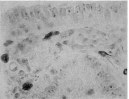
(Α) (Β) FIG. 3. Rat pancreas. Nucleus, with prominent nucleolus. A: BDC reaction. B: the same nucleus re-photographed, after reductive removal of tetrazo stain followed by the Feulgen reaction. Magnification : χ 3200.
ACYLATION AND DIAZONIUM COUPLING 215
216 Ε . A . B A R N A R D
FIG. 4. R a t i n t e s t i n e . B e n z o y l a t e d , followed b y h y d r o l y s i s in N - H C 1 a t 60°, 60 m i n . , t h e n t e t r a z o n i u m coupling. M a g n i f i c a t i o n : χ 480.
A C Y L A T I O N A N D D I A Z O N I U M C O U P L I N G 217 2. Carbohydrate Groups
The BDC reaction is found to be positive, and often intense, at certain non-nuclear sites which are known to be characterized by the presence of muco-proteins, e.g. intestinal goblet cells, cartilage, etc. (see Section VI, A). Some light is thrown on these exceptions by studies (Barnard, 1959) on the effect of the same benzoylation treatment on the periodic- acid-Schiff (PAS) reaction at these sites.
The PAS method (Hotchkiss, 1948) reveals the distribution of non- diffusible carbohydrate containing the 1:2 glycol group. Carbohydrate hydroxyls of this type normally react readily with benzoyl chloride, and the products no longer give an aldehyde with the periodate oxidation treatment (cf. Gersh, 1949); hence benzoylation should block the PAS reaction. In practice, when frozen-dried cells are subjected to anhydrous benzoylation followed by the PAS reaction, two types of behaviour are observed :
(i) A large number of characteristic PAS-positive sites lose the capacity for this reaction after benzoylation ; usually 30-60 min suffices.
(ii) In certain other sites, which include intra-cellular mucus in the duodenum, colon, stomach, and some sites in salivary glands and in cartilage, the PAS reaction cannot be blocked thus, even with very pro
longed benzoylation times. But a prior brief exposure to water (or aqueous alcohol) allows benzoyl chloride to block the PAS reaction there. This second group also, in all cases, gives the BDC reaction.
Further, both reactions show the same sensitivity to prior water treat
ment, with both disappearing at the same point. I t was shown (Barnard, 1959) by in fcerferometric measurements that this effect of water cannot be ascribed to the loss of material, to any significant extent, from the cells.
Hence it seems likely that at these latter sites in the frozen-dried cell, carbohydrate hydroxyls are bonded on to protein. I t is assumed that histidine groups are involved although no analysis has been made here ; it is not unlikely that other protein groups, not detectable by the coupling reaction, are also involved. The complex remains intact in anhydrous conditions, and these two components are thus mutually protected from benzoylation. But in a more polar medium the combination is irreversibly disrupted ; hence the histidine reacts with a diazo compound in aqueous buffer, and the glycol reacts with periodate in aqueous alcohol, or both components react with benzoyl chloride after water treatment.
What significance this carbohydrate-protein combination has in the intact tissue is unknown. For mucus, the newly synthesized or native form appears to be required. In cartilage, interesting discriminations between different stages of cellular development have been observed by this reaction.
218 Ε . Α . B A R N A R D
3. Arginine Groups
The anhydrous benzoylation treatment appears also to block cyto
plasmic arginine groups, while many nuclear arginine groups are not so blocked and can then be revealed and measured by the Sakaguchi reac
tion in alkaline medium (Barnard and Bell, 1960). These latter groups are believed to be those associated with DNA in the nucleoprotein.
These constitute, then, a further set of protein groups differentially protected from benzoyl chloride. The involvement of these arginine groups in bonding in the nucleoprotein clearly differs from that of the protected histidine groups : the arginine reaction is not affected by water (or alkali) pre-treatment, and is obtained equally after freeze-drying, freeze-substitution and several chemical fixatives. A relatively stable, salt-linked structure is probably concerned.
The versatility of the anhydrous benzoylation treatment is thus further demonstrated, in that the guanidino group, in spite of its high affinity for protons, is nevertheless acylated in this medium when not held in the charged form by neighbouring phosphate groups.
4. Reaction with other Blocking Agents a. Acetylation
Anhydrous acetylation, like benzoylation, should block tyrosine groups to form phenolic esters, normally stable at room temperature up to about p H 10 (see p. 207). With protein histidine residues, whether re
action will be observed is less certain. The product might be either the N-acetyl imidazole derivative, highly unstable to water (cf. Barnard and Stein, 1958) or a stable acetylated product in which the imidazole ring has been destroyed. The latter reaction is well known in the reactions of benzoyl chloride with certain histidine derivatives (see p. 207), but does not appear to have been established in the case of acetic anhydride.
However, it occurs similarly with iso-valeryl chloride in aqueous alkali (Windaus et al., 1921) and with the acetylating agent ketene on imidazole in dry, cold ether (Neuberger, 1938). The possibility that it may occur in the reaction of proteins with acetic anhydride cannot, therefore, be dismissed.
With benzoyl chloride, tetrazo (and diazo) coupling is generally blocked in the cytoplasm, and after water treatment in the nucleus ; it follows that the product of benzoylation at both tyrosine and histidine residues in cellular proteins must be stable under the conditions of the first coupling reaction (pH 9, 4°, about 20 min). I t is therefore of interest that acetic anhydride, applied in the same acetonitrile-pyridine medium, does not block coupling to the same extent. Coupling after acetylation gives a positive reaction in both cytoplasm and nucleus. The cytoplasmic
ACYLATION AND DIAZONIUM COUPLING 219 reaction is decreased, however. This may be only a kinetic difference, but if so it is considerable. Acetic anhydride (3 M, 6 hr or 1 M, 18 hr) leaves unblocked (at room temperature) many sites that are blocked by benzoyl chloride (0-7 M, 2 hr). Still longer exposures or elevated temperatures (cf. Pearse, 1953) might therefore be required, but would be undesirable.
Alternatively, it might be that some protein histidine groups are not effectively blocked by acetic anhydride, and are thus demonstrated.
Prior water treatment does not noticeably increase the blocking obtained, and the sequence
B e n z o y l a t i o n - W V a t e r - > A c e t y l a t i o n - V T e t r a z o c o u p l i n g
which presents an opportunity for the acetic anhydride to react at the shielded histidine groups in the nucleus, still produces a positive nuclear reaction (although it is not known whether this is quantitatively as great as the normal BDC stain). Recently, the rate and the extent of the total reaction of acetic anhydride at cellular sites have been measured abso
lutely in one cell type, using an isotopic cytochemical method (Barnard and Marbrook, 1961).
The acetylation treatment can be as effective as benzoylation in blocking the PAS reaction, although a true kinetic difference is then observed. At a number of sites, with 2 · 7 Μ acetic anhydride 1-2 hr is required to obtain the blocking produced by 0 · 7 M benzoyl chloride in i - l hr.
b. p-Nitrobenzoyl Chloride
This reagent has been employed in these studies (i) as a blocking agent, and (ii) as a chromogenic reagent. I n the latter case, the colour is developed by a procedure following the general method proposed by Danielli (1950, 1953) for the use of nitro compounds as chromogenic reagents.
^p-Nitrobenzoyl chloride (2 g) is applied in dry acetonitrile (50 ml) containing pyridine (3 · 2 ml), for 2 hr, followed by washes in acetonitrile, hydration and the colour development procedure (reduction with chromous chloride, 15 min, diazotization and coupling with H acid;
details are described by Maddy, 1961). Applied thus, the reagent is of value in indicating the sites and extent of reaction of benzoyl chloride.
I t produces an intense red-purple colour throughout the nucleus and cytoplasm of all cells, with the nuclei staining particularly intensely.
This reaction should give the sum of all groups available and reactive towards an acylating agent. The reaction is not confined to proteins.
Thus, it is noticeable that plant cell walls react strongly due t a reaction at carbohydrate hydroxyl groups ; similarly, extra-cellular mucus and basement membranes also react strongly.
220 Ε . A . B A R N A R D
However, it cannot be assumed that the cytochemical reaction of 39-nitrobenzoyl chloride is identical with that of benzoyl chloride and, indeed, this has now been shown not to be the case. It is of interest, as a case of differential reactivity, to examine the comparative cytochemical properties of these two reagents :
(i) ^9-Nitrobenzoyl chloride, like benzoyl chloride, blocks tetra- zonium coupling in the cytoplasm, but not in the nucleus. Similarly, brief prior exposure to water prevents this nuclear reaction.
(ii) When benzoylation is followed by ^p-nitrobenzoyl chloride used chromogenically, a colour reaction persists in the nuclei only, with a distribution very similar to that of the BDC reaction. Brief prior exposure to moisture abolishes this reaction too.
(iii) The same cytoplasmic sites (e.g. goblet cells, pancreatic acinar basal cytoplasm) that, exceptionally, give the BDC reaction, are positive also in reaction (ii) here, and a similar water effect is shown.
Reactions (ii) and (iii) show that there is some component, mainly nuclear, which can react with ^-nitrobenzoyl chloride (without water treatment) but which fails to react with benzoyl chloride. The water effect shows that this is a shielded component (in the frozen-dried tissue), and that when revealed, benzoyl chloride itself is capable of reaction with it. This is not necessarily surprising, since the more activated ^9-nitro- benzoyl reagent might compete more effectively with a bonded group at the acceptor site. Reaction (i) shows that the reactive component is not, however, the same as that responsible for the BDC reaction. This is confirmed by the result of a further experiment :
(iv) Benzoylation followed by coupling with diazotized sulph- anilic acid (cf. Section 5 below) and then by p-nitrobenzoyl chloride used chromogenically, again produces nuclear staining similar to the BDC reaction. Hence, the site of coupling after benzoylation is not the site of p-nitrobenzoyl reaction. I t should be noted here that the reduction stage in the colour development removes the azobenzene- sulphonate groups (as shown by controls) ; their colour does not inter
fere, therefore, but the sites to which they were attached remain vacant.
The identity of the component concerned here is still unknown. I t may be surmised that after benzoylation shielded groups exist in the nucleoprotein other than those within the limited range of the diazonium reagent, and that one or more of these groups is revealed by this more reactive acylating agent. Nucleoprotein arginine groups do not appear
ACYLATION AND DIAZONIUM COUPLING 221 to be involved, since they do not show the water effect with benzoyl chloride, and since it has been observed (v) that jp-nitrobenzoyl chloride, with or without prior water treatment, does not (at least qualitatively) block the nuclear (benzoyl chloride-resistant) Sâkaguchi reaction for arginine.
c. Other Reagents
Trials with a few other acylating (and related) agents on several tissues have been made, only to the extent of showing further variations in reagent reactivity, with semi-qualitative assessment by eye only.
^p-Iodo-benzoyl chloride (3-2%) and 3:4:5-triiodobenzoyl chloride (6%) (applied in benzene, for solubility reasons; these reagents were syn
thesized by Dr. M. M. Coombs for electron microscope staining studies) give little or no blocking (up to 20 hr) of the ^)-nitrobenzoyl chloride colour reaction or of the tetrazo coupling reaction, the triiodo compound being the least reactive of the two. I t seems likely that iodinated reagents of this particular type have severe limitations due to insolubility in suitable solvents, and to low reactivity. Phenyl isocyanate (10%, in pyridine, 2 hr) gives considerable blocking of both colour reactions but leaving a strong nuclear (and weak cytoplasmic) reaction. 3:4:5-triiodo- phenylisocyanate (6%, in benzene, up to 20 hr) gives no apparent block
ing. m-Nitrobenzene sulphonyl chloride (5%, in pyridine-acetonitrile, 3 hr) used chromogenically gives a rather weak reaction at all sites, but red cells react strongly; the reaction is completely blocked by benzoyl chloride. Similarly, ^-toluene sulphonyl chloride (7-5%, 3 hr) produces rather feeble blocking of the two colour reactions. Isocyanates may prove useful, but a relatively slow rate of reaction is apparently shown by the sulphonyl chlorides. Their stronger reaction in red cells, where histidine concentration (from haemoglobin) is high, may indicate preferential reaction at histidine groups.
5. Use of Alternative Diazonium Compounds
The use of tetrazotized dianisidine in the standard BDC method is dictated, as noted earlier, by the need for an intense colour in the reaction product. I t is of interest to enquire whether the characteristic behaviour observed with this method is dependent on the particular diazo reagent employed.
Reaction with each of the diazotized amines, sulphanilic acid,
^-nitroaniline and a-naphthylamine (0-02 M, p H 9, 5°, 20 min) has been found to produce a weak yellow colour, which appears to show the same distributions, with and without prior benzoylation, as the corresponding colour reactions obtained with dianisidine. Moreover, when each of the
222 Ε . Α . B A R N A R D
three mono-diazo reagents is applied, either with or without prior benzoylation, and is followed by the dianisidine coupling procedure, the intense red-purple colour reaction from the latter appears to be entirely blocked, only the initial pale yellow colour being observed. Hence, these three different diazonium compounds appear to reabt at the same sites, in the benzoylated and unbenzoylated cases respectively, as tetrazotized dianisidine (though measurements to test for an exact equivalence have not been made, and would be difficult to obtain with the required sensitivity).
III. INSTRUMENTATION AND PROBLEMS OF MEASUREMENT
A. M l C R O - S P E C T R O P H O T O M E T R Y
I t would be out of place here to discuss in detail the requirements and methods available for cytochemical micro-spectrophotometry with vis
ible light. Recent pertinent reviews include those of Swift and Rasch (1956), Leuchtenberger (1958) and Walker and Richards (1959).
The distribution within a cell nucleus of the stain produced by the BDC reaction is often not homogeneous, and in general resembles that of the Feulgen stain There will accordingly be the risk of distributional error (discussed, for example, in the reviews just cited).
One solution to this problem is that of Deeley (1955), involving the use of automatic scanning of the field by a small aperture. The Deeley apparatus, which employs mechanical scanning and electronic integra
tion of the signals, has been used in all the measurements discussed here of the BDC stain. At the same time, the crushing condenser of Davies et aL, (1954) has been employed in these measurements. With this latter device, the nucleus in question is flattened to any required degree by pressure applied through a sheet of cellophane. The combined effect of these measures is (a) to reduce local high absorbances to values which permit accurate measurement, (b) to minimize distributional error, and (c) to minimize out-of-focus errors.
Other solutions to the optical problems could no doubt be applied for measurement of this stain. In particular, the two-wavelength method (Ornstein, 1952 ; Patau, 1952) would be expected to be suitable. The use of a monochromator would in some respects confer an additional advan
tage since measurements could then be made at the absorption maxi
mum, and different coupling components (with different Am a x) could be compared.
With the Deeley apparatus, filters isolating a fairly narrow spectral band are used. Using tetrazotized dianisidine and H acid as the coupling components, filters 77A and 58 (Wratten) are suitable. Other require
ments (light source, etc.) are as for Feulgen micro-spectrophotometry.
ACYLATION AND DIAZONIUM COUPLING 223
B . SPECIMEN REQUIREMENTS
Methods used in Feulgen micro-spectrophotometry are not necessarily sufficient, without modification, for all cases in protein cytochemistry.
In the present state of techniques, micro-spectrophotometry is most usefully applied, in the case of a nuclear component, to the determination of the total amount of that component per nucleus, i.e. the entire nucleus is taken, as a readily identifiable and physiologically distinct entity.
Various methods of estimating this quantity have been employed.
Measurement of the stain contained in a plug within the nucleus has often been used, but this requires assumptions about the homogeneity of the nuclear stain and about the shape of the nuclei, both limiting the scope of application. These assumptions are not required in the measure
ment of entire nuclei by either the scanning technique or the two-wave
length technique. However, in all the methods two additional problems are met in the measurement of the total stain per nucleus :
1. Gut Nuclei in Tissue Sections
To minimize errors from this source, it is necessary to cut sections thick enough, and to rely on the recognition of any cut nuclei present.
This creates difficulties due to the overlap of nuclei (see below) in a number of tissues.
2. Stain in Overlying, Underlying or Adjacent Regions
This becomes a major problem in protein cytochemistry, in the cases where the stained component is present in the cytoplasm as well as the nucleus. Even for a stain present in the nucleus only, difficulty still arises from this source from near-by, out-of-focus nuclei in thick sections. In some cases, e.g. spleen or thymus, overlap of nuclei is always serious.
To measure the total nuclear stain by either the scanning or the two-wavelength methods, it is necessary to have an unstained region surrounding the measured nucleus, in order that the image of the nucleus may be totally enclosed within the diaphragm without contributions from extraneous absorption. Further, the background reading, giving the incident light I0 over the same field, should be made on a clear area as close as possible to the original area to avoid variations in I0 due to variation in the specimen, the mounting conditions, etc. These require
ments at present greatly restrict the application of these two accurate methods of measurement when the protein component is not confined to the nucleus. Failing an advance in photometric technique, these cases can be tackled by some method of isolation of the nuclei, providing significant losses are not thereby incurred.
224 Ε. Α. BARNARD
In the case of an exclusively nuclear component, as with the BDC stain in most tissues, the last-named requirement can often be satisfied in tissue sections, if overlapping and cut nuclei can be carefully excluded.
But the crushing condenser method, as used with the Deeley apparatus, cannot be employed with most sections of the required thickness, since overlap becomes prohibitive on crushing.
For the reasons mentioned above, tissue smears containing separated whole cells or disrupted cells have been employed here (see p. 226), thus ensuring that entire and spatially separated nuclei are measured. Smears have been found preferable in certain cases previously for similar reasons, in Feulgen or arginine micro-spectrophotometry (e.g. Ris and Mirsky,
1949; Swift, 1950; McLeish et al., 1957; Richards et al., 1956). An additional advantage of tissue smears is that the nuclei are normally rather flattened and the cytoplasm can be dispersed thinly, thus reducing scattering errors. Smears carry the disadvantage, of course, that the organization of the tissue is lost. The method is valuable at present for giving an adequate survey of classes of nuclei from a tissue, but in later, more sophisticated studies, some means of utilizing the information contained in the structure of the intact tissue will doubtless be required.
C. MOUNTING REQUIREMENTS
Three main factors have determined the choice of mounting medium : (i) the normal optical requirement for a mountant, that the refractive index is sufficiently close to that of the tissue to reduce light scatter to a negligible level ;
(ii) extraction or diffusion of the stain must not occur in this medium ;
(iii) for use in the crushing system, the medium must facilitate the flattening of the nuclei when they are sheared.
For measurements of the Feulgen stain with the Deeley apparatus, mounting in glycerol is normally employed. With the BDC stain, glycerol has been found excellent with respect to requirement (iii), adequate for (i), but gives appreciable, though slow, extraction losses.
Several other possible media have been tested. Immersion oil (Shil- laber's) is excellent for (i) and (ii), but flattening is inadequate in this medium, and similarly in liquid paraffin (see Fig. 5). I t is not entirely clear what properties the medium must possess for a crushed nucleus to show the full flattening effect. Normal lubricating properties alone are not sufficient as is shown by the results with the two last-named media and, further, with a fluorinated hydrocarbon lubricant (Perfluorolube
A C Y L A T I O N A N D D I A Z O N I U M C O U P L I N G 225 oil), which gives partial, but still inadequate, flattening. Light scatter is also higher with this medium, although if this were the sole defect it might be countered by an additive of suitable refractive index. No medium has yet been found (though the search has not been exhaustive) which gives the same desirable effect in the crushing method as glycerol, where nuclei can readily be flattened one by one to any required degree
G.I.
G.2.
(2n-4n) (4n-8n) PFL
^ 71 132 Stain (log scale)
F I G . 5. D i s t r i b u t i o n of B D C s t a i n i n n u c l e i of m o u s e liver cells, a s m e a s u r e d i n v a r i o u s m o u n t a n t s . T h e s a m e s p e c i m e n w a s m e a s u r e d i n t u r n i n paraffin ( P ) , glycerol {G.l) a n d a g a i n after s t a n d i n g 4 d a y s in glycerol (G.2). O n l y n u c l e i a p p a r e n t l y t e t r a p l o i d ( b y a p p r o x i m a t e r e l a t i v e size) w e r e m e a s u r e d . A n o t h e r s p e c i m e n from t h e s a m e a n i m a l w a s m e a s u r e d in p e r f l u o r o l u b e oil (PFL) ; a p p r o x i m a t e size r a n g e s a r e s h o w n . C r u s h i n g w a s o b s e r v e d t o b e fully efficient o n l y i n t h e glycerol case. (Cf. F i g . 1 4 B for full d i s t r i b u t i o n i n m o u s e liver.) F o r c o m p a r i s o n w i t h G.2, a s p e c i m e n of m o u s e k i d n e y , m e a n 59-9
± 1-9, s h o w e d a d e c r e a s e t o 44-3 ± 1-2, a f t e r 7 d a y s in glycerol a t 0°.
down to a thin sheet. Some molecular interaction between tissue com
ponents and glycerol is presumably involved, perhaps related to the swelling of proteins by glycerol (Caspersson, 1950). The elastic properties of the cell must be changed in such a manner as to allow plastic flow to occur when stressed. However, detectable losses in stain occur on standing in glycerol (Fig. 5), again possibly related to the protein swelling effect.
Fortunately, these losses are slow enough to allow measurement to be made in this medium.
The best procedure is to store the specimen in immersion oil from the time of staining until measurement, and then to wash and transfer to
8
226 Ε. A. BARNARD
glycerol. Readings are reproducible up to at least 10 hr in glycerol, but after about 2 days losses begin to occur slowly. A correction cannot be applied for these later losses, since they are detectable in some cells but not others in the same specimen, and they vary with the tissue and with the specimen treatment. I t has been confirmed on all tissues used that it is safe to measure in glycerol throughout the first day of mounting, since the same cells measured initially and finally give unchanged values.
Further, in cases where the nuclei do not contain regions of very high extinction and are reasonably flat initially, mounting in paraffin or immersion oil can be satisfactory since crushing is then not necessary ; in these cases, the same values are obtained as subsequently in glycerol, confirming that no initial extraction occurs in that medium. I t is not safe, however, after measuring a specimen in glycerol, to wash it (in water, alcohol and xylene) and return it to immersion oil for storage, in order to continue measurements later : it has been found that some losses are detectable then even after such storage a t 0°, presumably arising from bound glycerol.
IV. PROCEDURES, AND THE EFFECTS OF VARIABLES THEREIN A . STANDARD PROCEDURES
1. Freeze Drying
Freeze-drying is the most desirable method for the preparation of the specimens, both on the theoretical grounds mentioned later and on the empirical ground that a high and reproducible level of reaction is thus attained.
For tissue sections, standard methods of freeze-drying (cf., e.g. Bell, 1956) small pieces of tissue are satisfactory. The problem of freeze- drying thin smears will receive attention here, since (i) some specimens, e.g. ascites tumour, must be examined thus, and (ii) more importantly, normal tissues can often with advantage be examined in smear form for exact micro-spectrophotometry. A small fragment of the tissue (e.g. liver, kidney, etc.) is tapped with a perfectly flat-ended rod in a little isotonic saline for disruption, and smeared on a cover-slip, which is immediately quenched in a stirred bath of ^o-pentane : propane (1:2) at liquid nitro
gen temperature. The pressure exerted during the tapping and the smearing determines whether nuclei are liberated, and to what extent, from the separated cells. The saline can be replaced by other media, e.g.
Tyrode solution, sucrose solutions, etc., depending on the degree of retention required. No medium at all need be used, although free tissue fluids are always present ; a smearing-squashing technique is then still
A C Y L A T I O N A N D D I A Z O N I U M C O U P L I N G 227 satisfactory for most tissues, but some thick regions may be present and must be ignored. I n the BDC reaction, the intensity has been found in practice to be the same whether the nuclei are in intact cells, or liberated in saline or without external medium. A similar smearing technique has been used by Β. M. Richards in Feulgen measurements of the DNA content per nucleus (cf. Richards et al., 1956).
The quenched smears are washed by dipping in liquid nitrogen and are rapidly transferred to the drying chamber (see Appendix 3). Drying is carried out at —50°, although any temperature below about —40° is permissible, the rate of drying decreasing with decrease in temperature.
I t is essential to maintain the smear surface at a fairly uniform tempera
ture. Unevenly dried or poorly preserved specimens give a decreased or variable reaction. When dry, the smears are raised to room temperature in vacuo, and rapidly removed to a desiccator for storage.
The reaction cannot be obtained on smears or sections prepared by the method of freezing-substitution (Simpson, 1941) using absolute methanol or ethanol.
2. Specimen Pre-treatment
Smears for micro-spectrophotometry are simply fixed in absolute ethanol immediately prior to reaction. Frozen-dried tissue blocks are embedded in wax in vacuo and sectioned. Mounting on slides must be performed either by slight warming alone, or by flattening on acetonitrile or on 95% alcohol (e.g. at 47°, 30 sec). The water present in the latter medium does not appear to affect the wax-infiltrated tissue in these conditions. The wax is removed by xylene when required.
Fixation methods other than freeze-drying have not been studied in detail on account of their uncertain effects on the nucleoprotein. Tissue fixed in bulk in absolute alcohol, or in Lewitsky's fluid, gives the reaction, but to what quantitative extent is undetermined. After some fixatives, e.g. Carnoy's fluid, the reaction is weak or variable.
3. Reaction Method
(i) Fixation in absolute alcohol, two baths . . 20 min (ii) Wash in dry acetonitrile, two baths . . . 6 min (iii) Benzoylation: Dry acetonitrile (50 ml), benzoyl
chloride (4-2 ml) and dry pyridine (2-2 ml). Used at room
temperature, in a desiccator (CaCl2) . . . . 3 hr (iv) Alcohol wash, two baths . . . 6 min
(v) Take down to water through 90, 70, 50 and 25%
alcohols.
228 Ε. Α. BARNARD
(vi) Coupling (all operations in this stage are performed in an ice bath: specimens must be at 2°-4° throughout).
Wash in ice cold water . . . 4 min Tetrazotized dianisidine: a solution (0-04%) of the
stabilized salt (Appendix 2) in sodium veronal (2%) buffer, p H 9-0. This solution is made immediately before use and filtered quickly in the cold through a coarse paper. It is a clear yellow solution, which darkens on standing. Normal exposure is 18 min.
Washes: Veronal buffer (0-2%, p H 9) . . 1 min Water, two baths . . . . 1 min each
0-05 N HC1 l m i n Water, two baths \ min and 1 min
Second coupling: ΙΪ acid ( 0 - 1 % solution of the Na^salt)
in carbonate-bicarbonate buffer, p H 9 . . . . 1 0 min Or β-naphthol (0-1%) in N a2C 03 solution (0· 5%) . 10 min The specimens are then allowed to warm to room temperature, in the second coupling bath, over a period of 20 min.
(vii) Wash in N a2C 03 solution (0*5%), two baths . 6 min Wash in water, two baths . . . . 6 min (viii) Dehydrate through 25, 50, 70 and 90% alcohols.
Absolute alcohol, two baths . . . 6 min
Alcohol-xylene (1:1) l m i n
Xylene, two baths . . . 6 min
(ix) Mount in balsam, or for measurement mount and store in Shillaber's immersion oil under cellophane.
Some of these periods are flexible (see B, 3 below). Precautions must be taken to ensure that no moisture comes into contact with the specimen from the stage of freeze-drying to the end of benzoylation.
4. Micro-spectrophotometric Method
Measurements on this reaction have so far been carried out using the scanning, integrating micro-spectrophotometer of Deeley (1955).
The procedure used, described below, is similar to that of Deeley etal., (1954) and of Richards et al., (1956) for measurements of Feulgen stain.
Other micro-spectrophotometric methods, of proven reliability else
where, can no doubt be applied here.
The specimen (here, in all cases, a smear on a cover-slip) is taken from the temporary mounting in immersion oil, washed in xylene and alcohol and in 90, 70, 50 and 25% alcohols and water, and drained. I t is mounted
ACYLATION AND DIAZONIUM COUPLING 229 in glycerol under cellophane, and secured to a brass holder by wax.
Objective and condenser lenses are immersed in Shillaber's oil. Nuclei are crushed by the crushing condenser, then totally enclosed by the dia
phragm, and measured. The reading, at the same diaphragm setting, for an adjacent blank area is subtracted, to give the integrated total absorp
tion (in arbitrary units) for that nucleus. The mean of three readings is taken in every case.
Nuclei are measured in a number of different areas selected at random on the same specimen. Where significant variability among similar cells is found between different regions of one specimen, it is discarded : this can occur if drying was not satisfactory and is usually correlated with inferior cytological preservation.
B . E F F E C T OF VARIABLES IN THE CYTOCHEMICAL PROCEDURE
In establishing the conditions giving the most satisfactory and reproducible results with the maximum production of colour, frozen- dried rat liver and kidney, and chicken or frog blood smears have been used, in both qualitative and quantitative observations.
1. Coupling
Reaction using o-dianisidine gives, as expected, a rather stronger colour than using benzidine. p H around 9 seems optimum; adsorption of decomposition products becomes appreciable at higher pH, necessitat
ing further washing with the risk of some loss of the free diazo group.
70 60 50
Mean stai
n 40
30 20 10
0 10 20 30 40 50 60
Exposure to TDA (min.)
F I G . 6 . V a r i a t i o n of B D C s t a i n i n g w i t h l e n g t h of e x p o s u r e t o s t a n d a r d t e t r a z o s o l u t i o n ( c o n c e n t r a t i o n of free t e t r a z o n i u m d i c h l o r i d e , 0 - 0 1 7 % ) . F r o g e r y t h r o c y t e s ; e a c h p o i n t r e p r e s e n t s t h e m e a n s t a i n ( + S.E.), i n a r b i t r a r y u n i t s , for a b o u t 3 0 nuclei.
230 Ε . Α . B A R N A R D
Concentrations of (free) tetrazotized dianisidine much above 0-02% also incur this danger. The wash with dilute HC1 is intended to destroy the acid-labile triazenes (cf. Section I I , A). The concentration used, 0-05 N, is that which was shown, in work on tissue in bulk, to be the lowest that would give maximum splitting at those sites.
With other conditions standardized, the extent of reaction has been measured, in the nuclei of frog red cells, as a function of the exposure to the tetrazonium reagent (Fig. 6). The stain increases with length of reaction time to reach a plateau of maximum stain. At 60 min, however, there are signs that decomposition products are accumulating. Fourteen to 30 min is optimal.
Of the naphthols tried (Table I), H acid gives a reaction product with the most suitable light absorption properties for micro-spectrophoto
metry of the present type. In measurements oji frog red cells, it has been found that the intensity is unchanged for exposures to H acid solution, from 4 min to 12 min at 2°, followed by warming up to room temperature,
the total period in H acid being constant at 30 min. Coupling in H acid immediately at room temperature, however, gives a decrease (15%) in mean intensity.
2. Benzoylation
Qualitatively, the same pattern of dependence on length of benzoyl
ation has been observed on a number of different tissues, namely the cytoplasmic stain (apart from the special cases described in Section VI, B) decreases rapidly over the first 30 min and appears very slight or neglig
ible at 1^ hr and nil thereafter. The nuclear stain persists after at least 20 hr benzoylation. The concentration of benzoyl chloride can be reduced to as low as 1*5%: 18 hr treatment then gives the same result as 10%
benzoyl chloride for 3 hr.
Quantitatively, the conclusions with regard to time dependence have been confirmed in the case of frog red cells (Fig. 7). I t is seen that from 2 to 20 hr the nuclear stain remains constant. Cytoplasmic stain is zero at
2 hr. Hence the difference in reactivity involved is not merely kinetic, but is so great as qualitatively to distinguish this nuclear component.
Measurements after less than 2 hr benzoylation are complicated by the cytoplasmic stain remaining, i.e. the nuclear stain cannot be measured in whole cells by the present method without the inclusion of stain (where present) in some adjacent or overlying cytoplasm. I t might be of value to measure nuclei isolated by the non-aqueous method (Allfrey et al., 1952) to examine the initial total nuclear stain and its change with benzoyla
tion. Routinely, 3 hr benzoylation is satisfactory. Exposures as long as 20 hr give detectable swelling of some structures.