Clinical relevance of vascular distribution in the palate and vestibule – establishing the theoretical foundation of novel flap
designs for graft harvesting and reconstructive procedures Ph.D. Thesis
Arvin Shahbazi Irani
School of Clinical Medicine Semmelweis University
Supervisor:
Péter Windisch DMD, Ph.D.
Official reviewers:
Attila Szűcs DMD, Ph.D.
István Varga DMD, Ph.D.
Head of the Final Examination Committee:
Féhér Erzsébet MD, Ph.D., D.Sc.
Members of the Final Examination Committee:
Árpád Joób Fancsaly DMD, Ph.D.
Tamás Andrea MD, Ph.D.
Budapest
2019
TABLE OF CONTENTS
1. LIST OF ABBREVIATIONS………..
2. PREAMBLE ………..
3. INTRODUCTION ………..
4. OBJECTIVES ………
5. MATERIALS AND METHODS ………
5.1 Study I (clinical/case report study) ………
5.1.1 Treatment approach ……….
5.1.2 Case 1 ………..
5.1.3 Case 2 ………..
5.1.4 Case 3 ………..
5.1.5 Post-operative maintenance ………
5.1.6 Clinical evaluation ………..
5.1.7 Radiographic evaluation ……….
5.2 Study II (human cadaver study) ………
5.2.1 Latex milk injection ………
5.2.1.1 Thiel solution ……….
5.2.1.2 Process of latex milk injection ………..
5.2.2 Corrosion casting ……….
5.2.2.1. Process of corrosion casting ……….
5.2.2.1.1. Pre-casting phase ………..
5.2.2.1.2. Casting phase ………
5.2.2.1.3. Corrosion phase ………
6. RESULTS ………..
6.1. Study I ……….
6.1.1. Postoperative findings ………..
6.1.2. Intraoperative findings at membrane removal ………..
6.1.3. Intraoral radiographs out comes ………
6.1.4. Cone beam computed tomography out comes ………..
3 5 8 15 16 17 18 18 21 22 22 24 25 25 26 26 29 31 34 34 35 36 38 38 38 38 41 42
6.2. Study II ……….
6.2.1 Results of the vascular survey analysis in the vestibule ……….
6.2.2 Results of the vascular survey analysis in the palate and maxillary tuberosity ..
7. DISCUSSION ………..
8. CONCLUSIONS ………..………
9. SUMMARY ………..
10. ÖSSZEFOGLALÁS ………
11. BIBLIOGRAPHY ………
12. BIBLIOGRAPHY OF THE CANDIDATE’S PUBLICATIONS ………
13. ACKNOWLEDGEMENTS ……….
42 42 45 52 59 60 61 62 72 73
1. LIST OF ABBREVIATIONS
ASAA- Anterior superior alveolar artery BA - Buccal artery
BDX - Bovine derived xenograft BOP - Bleeding on probing CAL - Clinical attachment level
CBCT - Cone beam computed tomography CCA - Common carotid artery
CT - Computed tomography DPA - Descending palatine artery ECA - External carotid artery
ePTFE - expanded polytetrafluoroethylene FA - Facial artery
FGG - Free gingival graft
GBR - Guided bone regeneration GPA - Greater palatine artery GR - Gingival recession
HVC - Horizontal vertical and combination IAA - Inferior alveolar artery
ILA - Inferior labial artery IOA - Infraorbital artery LPA- Lesser palatine artery MA - Maxillary artery MEA - Mental artery MR - Mucosal recession NPA - Nasopalatine artery PBS - Phosphate buffered saline PD - Probing depth
PSAA - Posterior superior alveolar artery SA - Submental artery
SCTG - Subepithelial connective tissue graft SEM - Scanning electron microscopy
SLA - Superior labial artery SUA - Sublingual artery V - Volum
W - Weight
2. PREAMBLE
During my undergraduate academic years, I was impressed with the recent progression of various surgical techniques in dento-alveolar and periodontal surgeries. Between the third and fifth year of dental school, I was contributing as a teaching assistant in the Department of Anatomy, Histology and Embryology of Semmelweis University. My interest shifted towards the distribution of blood vessels and their possible influence on planning of incisions/flap designs in the oral cavity.
After my graduation, I was honored to receive an invitation to be a practice supervisor and lecturer from the English Course Director of Semmelweis University, Department of Anatomy, Histology and Embryology, Dr. Andrea Dorottya Székely, and the chair of the Department, Dr. Gábor Gerber. At the same time, I was accepted into the Dental Research Programme of the Semmelweis University School of Clinical Medicine, led by Professor Gábor Varga. The initial objective of my PhD research was to investigate the detailed analysis of blood vessels and conduct a survey in the oral vestibule and palate macroscopically, with the aim of introducing innovations related to the surgical flaps/incisions in those particular areas.
I undertook a literature search related to the morphology of blood vessel distribution in the oral cavity, collected data for a literature review and analyzed cadavers during my first year of studies under my PhD supervisor, Professor Péter Windisch, Chair of the Periodontology Department, Semmelweis University. The results of the clinical and cadaver analysis were compared to the collected data from the literature relating to the different flap/incision designs in periodontal, implant placement, bone augmentation, impacted wisdom/canine teeth, sinus floor elevation and sub-epithelial connective tissue graft surgeries. It became clear to me that the knowledge that could be gained from a pre-surgical arterial survey of anastomoses and critical points of blood distribution should be considered as one of the most important factors when designing a proper incision/flap procedure that would avoid damaging critical vessels. Such preplanned surgeries would be less invasive and attain appropriate levels of blood circulation and angiogenesis, promoting wound healing and reducing intra-operative bleeding and post-
operative complications. Therefore I started a challenging set of experiments, utilizing different visualization methods (Latex milk injection & Corrosion casting) on the vessels and dissecting them. By using these methods, I was able to convert the theoretical knowledge into practical findings and give a clear explanation about the morphological pattern of vascular distribution. During my research work, I was fortunate to be introduced to Dr. Georg Feigl, Acting Head of the Anatomy Department of the Medical University of Graz, Austria, and Dr. Bálint Molnár from the Department of Periodontology, Semmelweis University. Together with their anatomical and clinical knowledge, experience and support, we, as a team, achieved significant progress in mapping, analyzing and conducting our findings related to the course of the main arterial branches, their subdivisions and anastomoses relevant to the morphological aspects of the palate and vestibule, and their effect on clinical outcomes.
In the first phase of my PhD, I attended and assisted several surgeries utilizing different types of flap/incision design. This allowed me to clinically observe the path of blood vessels, and their possible influence on complications intraoperatively and postoperatively, under the supervision of Prof. Péter Windisch and Dr. Bálint Molnár.
I contributed as one of the co-authors to an innovative clinical research project led by Prof. Péter Windisch, which was published in a clinical case series article in the Quintessence International Journal. In this article, a novel split-thickness flap design without periosteal and vertical releasing incisions for horizonto-vertical ridge augmentation was introduced, with favorable wound healing due to undisturbed vascular supply.
After that, I directly investigated the theoretical findings related to the course, location and distribution of vessels on human cadavers. In the second phase of my PhD, I started to stain the blood vessels of the oral vestibule and the palate by corrosion casting and latex milk injection techniques and dissect them, in collaboration with Dr. Georg Feigl, Dr. Andrea Dorottya Székely and Dr. Gábor Gerber. We published the results of our team in the Journal Clinical Oral Investigations, describing the arterial supply of the palate, maxillary tuberosity and their clinical implications for flap design as well as soft tissue graft harvesting using this combination of different staining methods. We
performed the mapping of blood vessels on the hard palate and maxillary tuberosity, also discussing their influence on surgical procedures. For my publication, I was humbled to receive an award from Apáthy István foundation, Anatomy Department, Semmelweis University, as an appreciation of my research. After, I had an opportunity to collaborate with Prof. Péter Windisch and Dr. Bálint Molnár and contributed to the photographic illustrations of ‘Gingival Recession Management’ (chapter 8: Recession Coverage Using Autogenous Grafts, pages 100-101 - edited by Prof. Dr. Adrian Kasaj) with some of my palate arterial distribution works. During our still-ongoing investigations, we obtained valuable new information related to the blood supply of the oral vestibule, which may contribute to a better understanding of the clinical healing patterns of vestibuloplasty and ridge augmentation procedures.
It has always been a great veneration that I have had the chance to be a member of these clinical and anatomical studies. I was involved in providing new data either for mapping of blood vessels or for applied clinical studies, such as the novel split-thickness flap. In my thesis, I want to present an overview of the related literature on the arterial distribution of the vestibule and palate. This includes anastomoses, angiogenesis, wound healing, complications and novel approaches related to the flap designs for graft harvesting on the palate, and reconstructive procedures on the vestibule. I will then present and discuss the findings from our clinical and cadaver studies.
3. INTRODUCTION
In periodontal and implant dentistry, the oral vestibule and palate are considered as target areas for various types of surgical flaps. In order to achieve successful surgical results, the knowledge of blood vessel distribution, which will affect the angiogenesis, circulation and primary wound healing, is crucial (Arnold & West, 1991; Polimeni et al., 2006). During the design of different types of incisions, the mapping of blood vessels should be acknowledged by the surgeon to avoid any complications (Kleinheinz et al., 2005; Koymen et al., 2009; Shahbazi et al., 2018).
Morphologically the vestibule presents a slit-like space which is bordered laterally by the cheeks, anteriorly by the lips and internally by the alveolar arch, gingivae and teeth (Gray & Lewis, 1918; Berkovitz et al., 2009). The gingivae are formed by dense connective tissue, tightly attached to the periosteum of the bony alveolus, and cover the cervical areas of the teeth (Gray & Lewis, 1918). They contain a complex vascular distribution. The gingivae present interdental papillae which are located coronal to the gingival margin (Berkovitz et al., 2009; Lindhe et al., 2015). In the mucosa of the vestibule, the labial glands produce fluid that empties through small ducts. Similarly, the maxillary vestibule receives the salivary secretion from Stensen’s duct at the level of the upper second molar.
The oral vestibule is supplied mainly by sub-branches of the maxillary and facial arteries which are originating from the external carotid artery (ECA). The maxillary artery (MA) perfuses the upper and lower jaws including hard palate, maxillary tuberosity, maxillary sinus, vestibule, upper and lower teeth/gingivae with many branches (Rahpeyma & Khajehahmadi, 2017). These are the infraorbital artery (IOA), the greater palatine artery (GPA), the posterior superior alveolar artery (PSAA), the anterior superior alveolar artery (ASAA), the inferior alveolar artery (IAA), the buccal artery (BA) and the mental artery (MEA). The facial artery (FA) delivers different branches. The two branches of the FA which are named as, inferior labial artery (ILA) and submental artery (SA) participate in blood supply of the lower vestibule. The ILA originates at the level of the labial angle, together with the third branch, which is the
oris muscle. They move to the medial direction, give branches to the mucosa of the upper and lower vestibule, anastomose with the contralateral arteries and form the vascular network around the oral cavity (Pilsl et al., 2016). The SA passes beneath depressor labii inferioris and forms anastomoses with the ILA and mylohyoid branch of the IAA. This convoluted vascular circle, with an abundance of collateral sources of blood flow, is essential when different incisions/flaps are planned in the oral vestibule.
The upper gingiva is mainly supplied by the branches coming from the PSAA, IOA, ASAA, SLA and GPA. The branches of the BA, IAA, ILA, SA and the sublingual artery (SUA) mainly supply the lower gingiva. These arteries by giving supra-periosteal branches supply the gingivae (via their terminal branches) and form meshwork with blood vessels of the periosteum and periodontal ligaments (Lindhe et al., 2015). Below the epithelium of the gingivae they build a sub-epithelial plexus which creates thin capillary loops (diameter of approximately 7 µm) in each connective tissue papilla (Lindhe et al., 2015). Below the junctional epithelium, the dento-gingival plexus (mainly formed by venules) can be found that contains small vessels without capillary loops (in healthy gingiva) (Egelberg, 1966; Lindhe et al., 2015).
The most demanding interventions in the upper/lower jaw include implant placement (Buser et al., 2013) together with a horizontal reconstruction of lost hard tissue guided bone regeneration (GBR) techniques (Donos et al., 2008; Buser et al., 2009), ridge augmentation procedures (Tinti et al., 1996; Urban et al., 2015b), sinus floor elevation (Simion et al., 2004; Niu et al., 2018), as well as periodontal pocket surgeries (Cortellini
& Tonetti, 2015; Graziani et al., 2018), root coverage (Langer & Langer, 1985;
Zucchelli & De Sanctis, 2000; Zucchelli et al., 2006) and vestibuloplasty (Han et al., 1995; Urban et al., 2015a) procedures. The vast majority of these surgical indications are established by full thickness mucoperiosteal flaps (Simion et al., 1998), with uni- or bilateral full thickness horizontal and vertical periosteal releasing incisions, allowing for a tension-free flap design. However, despite predictable treatment success, there are some well-known complications related to extensive flap mobilization, e.g. partial disruption of the periosteal blood supply, intraoperative haemorrhage, bone loss, partial flap necrosis, vertical scars, and shrinkage and distortion of the vestibule (Fickl et al., 2011; Lim et al., 2018). These complications may impair the final functional and
esthetic outcome of surgical interventions in both of the jaws. On the other hand, such postoperative complications after periodontal plastic surgery interventions performed with a split thickness flap design (e.g. coronally advanced flaps for root coverage procedures and vestibuloplasty procedures) are not frequently reported, possibly due to the minimally-invasive approach. Consequently, there have been several authors suggesting alternative approaches, instead of the classical mucoperiosteal flap by Tinti et al. (1996) (with two full thickness vertical and horizontal periosteal releasing incisions) for ridge augmentation procedures. These include split thickness flap designs, characterized by the absence of periosteal incisions and, hypothetically less compromised postoperative flap circulation. Hur et al. (2010) and Ogata et al. (2013) introduced a split thickness flap for ridge augmentation with one split thickness vertical incision. Windisch and co-workers (2017) published a split thickness flap design without vertical releasing incisions and with a bilaminar two-layer flap closure, and reported a low number of postoperative complications and minimal vestibular distortion following the healing period. However, detailed anatomical data are still scarce in the literature, and this might limit clinicians desiring to overcome blood supply disturbances related to various mucosal dissection techniques. Therefore, establishing a solid anatomical basis for designing surgical interventions in the vestibule by detailed mapping of the arterial pathways is needed to allow for more advanced and sophisticated surgical interventions.
Another particular area of oral and periodontal surgeries is the palate, which forms the roof of the oral cavity. The palate is divided into two parts, the hard palate (anteriorly), the soft palate (posteriorly) (Gray & Lewis, 1918; Berkovitz et al., 2009). The hard palate is bordered by the alveolar/dental arch and gingiva. As Gray & Lewis (1918) explained, the covering of the hard palate is made, “by a dense structure, formed by the periosteum and mucous membrane of the mouth, which are intimately adherent. Along the middle line is a linear raphe, which ends anteriorly in a small papilla corresponding with the incisive canal” (p. 1112). The anterior aspect of the palatal mucosa on both sides of the raphe is rough, thick and pale, but the posterior aspect is smooth, thin, and deeper in color (Gray & Lewis, 1918). The hard palate is wrapped by stratified squamous epithelium; it contains many glands which are positioned between periosteum
and mucous membrane (Gray & Lewis, 1918). Approximately from the upper second premolar toward the incisors the palate contains numerous fatty tissue, but behind the second upper premolars the palate contains more glands in its connective tissue.
Generally, the suggested area for taking a subepithelial connective tissue graft (SCTG) is between the distal side of the canine to the mesial side of the first molar. The blood supply of the oral mucosa, and the palate in particular, shows a complex pattern, mainly supplied by branches of the maxillary, facial and ascending pharyngeal arteries, taking their origins from the ECA.
The third segment of the MA in the pterygopalatine fossa gives off a branch called the descending palatine artery (DPA) (Choi & Park, 2003), which descends and subdivides into the GPA and the lesser palatine artery (LPA). The GPA emerges from the greater palatine foramen, located on the hard palate between the second and third maxillary molars (Chrcanovic & Custódio, 2010; Kim et al., 2014). Further behind, on the horizontal plate of the palatine bone, the lesser palatine foramina can be found, where the branches of the LPA emerge. These arteries supply the majority of the hard palate, together with the soft palate. The branches of the GPA travel within the palatal bony groove, divided into medial and lateral palatine grooves by the palatine spine (Klosek
& Rungruang, 2009; Fu et al., 2011). The medial palatine groove contains the greater palatine nerve, whereas the GPA lies in the lateral groove to supply the mucosa, periosteum and palatal gingiva (Yu et al., 2014) before entering the incisive canal to form an anastomosis with the nasopalatine artery (NPA) (Shahbazi et al., 2018). The NPA enters the incisive canal to supply the anterior region of the hard palate (i.e.
intermaxillary segment). Here an anastomotic network is formed between the NPA and the GPA, supplying the majority of the palatal mucosa and periosteum.
Following oral surgical interventions, it is of high importance to be aware of the interrelation between these different anatomical entities, in order to aid the surgeon in flap design and to offer the most optimal circumstances for wound healing and revascularization. Information provided by anatomical atlases does not present clinically-relevant details of the palatal vascular network (i.e. ipsi- and contralateral anastomoses, individual changes of vascular pathways due to loss of dentition).
Providing a solid anatomical basis for local characteristics of the hard palate might
enable clinicians to avoid intra- and post-operative complications when planning oral surgical interventions by optimizing incision and flap designs. In order to elevate a flap, which allows surgical intervention to be followed by undisturbed wound healing, accurate information regarding the size and division of muscular, mucosal and periosteal vasculature is necessary. Oral cavity vestibule needs predictable surgical care due to the numerous vascular distribution in movable mucosa, keratinized gingiva and the periosteum.
The course of blood vessels in the palate and oral vestibule has a significant influence on the result of reconstructive surgical interventions by affecting intraoperative haemorrhage and postoperative healing. There are several surgery types of maxillofacial/oral surgical or periodontal treatment for which a well-established knowledge of secure incision lines and surgical approaches would be highly beneficial:
harvesting of SCTG (Langer & Calagna, 1980; Langer & Langer, 1993; Benninger et al., 2012), free gingival graft (FGG) (Sullivan & Atkins, 1969; Edel, 1974; Oh et al., 2017), removal of impacted canines (Abrams et al., 1988; Köşger et al., 2009), and flaps to allow implant placement (Kleinheinz et al., 2005; Koymen et al., 2009). Among all surgical interventions in the oral cavity, especially the front maxilla/mandible, requires predictable surgical care because of plausible esthetic commotion following surgeries disrupting the vestibular circulation. Although the morphological features of vestibular and palatal structures have previously been thoroughly investigated, clinicians still frequently face anatomical challenges during surgeries, and there is a growing need for a comprehensive macroscopical mapping of the palatal mucosal blood supply in order to avoid dangerous intraoperative and postoperative complications (Harris et al., 2005;
Griffin et al., 2006).
According to the literature, investigation of the blood flow in oral mucosa can be performed by in vivo angiography (Mörmann & Ciancio, 1977), laser Doppler or laser speckle analysis (Hoke et al., 1994; Molnár et al., 2017). These approaches provide valuable clinical data on functional changes in blood circulation following thermal, mechanical or chemical stimuli, and might be used for monitoring postoperative wound healing patterns following oral surgery or periodontal surgery interventions.
information on blood vessel function and structures. Thus more accurate ex vivo macro- and microscopical investigations are required to achieve a solid anatomical background for the physiological observations made by blood flow analysis methods.
Several staining methods exist that might be used to visualize the blood vessels of the palate for macroscopical analysis, such as latex milk injection (Alvernia et al., 2010) or corrosion casting (Rueda Esteban et al., 2017). These can all be used in human cadavers by injecting a substance through the ECA.
Latex milk is a flexible material, and it is composed of proteins, resins, tannins, oils, sugars, alkaloids and gums. (Haenssgen et al., 2014). It endures as an emulsion of polymer micro-particles which is mixable with water (Haenssgen et al., 2014). In general, the colored latex milk is injected into the vessels to analyze the path of the branches and sub-branches of the different vessels. This material is kept in an alkaline medium and will solidify when it is converted to an acid medium. The latex becomes solidified when it dries out. Also, by applying high pressure or keeping the latex under low temperature, it can become hardened as well. The latex hardens rapidly in the presence of formalin (Bergeron et al., 2006), this process is called emulsion polymerization (Haenssgen et al., 2014). After a proper embalming period of the cadaver by Thiel's solution and flushing of the vessels, they are injected with the latex milk. Thiel's solution has no detectable odor and results in life-like flexibility of body parts, excellent color preservation of muscle and vasculature, as well as superior antimicrobial preservation of cadavers (Thiel, 1992a; Thiel, 1992b; Thiel, 2002; Ottone et al., 2016). The addition of diluents can modify the viscosity and the setting time of latex (Alvernia et al., 2010; Haenssgen et al., 2014). Diluents for latex can be water (Alvernia et al., 2010; Haenssgen et al., 2014), ammonium hydroxide (NH4OH), or triethylamine ((C2H5)3N) (Thiel, 1992b; Haenssgen et al., 2014).
The corrosion casting method uses solidifying material such as methacrylates in order to discover the three dimensional structure of blood vessels within the tissue (Hossler &
Douglas, 2001; Haenssgen et al., 2014). In particular, the methacrylates are monomers in polymer plastics and can create the acrylate polymers, that are elastic, lucid and defiant to breakage (Haenssgen et al., 2014). In this method, the injection process is
suggested to be performed on fresh specimens, which are without any previous fixation or formalin (Rueda Esteban et al., 2017). Having a fresh corpse is essential before casting in order to prevent complications in the steps of the injection due to solidification and retraction, mainly in vascular tissues (Rueda Esteban et al., 2017).
After casting procedures, the corrosion, dissection, washing and drying will be the next steps before achieving the final outcome. The vessels can be identified during dissection. In addition, the latex milk and corrosion casting display excellent architecture when the specimen is to be X-rayed or studied under a microscope.
4. OBJECTIVES
The goal of my PhD dissertation is two-fold: firstly, I evaluate the existing evidence available in the literature related to the analysis of blood vessel distribution and their clinical relevance in the palate and oral vestibule. Secondly, I present novel information related to flap/incision designs in hard- and soft tissue reconstructive surgeries. The current available novel findings might affect the incision techniques in soft tissue graft harvesting in the future. Available data related to the pattern of mapping and subdivisions of the vessels have raised a number of fundamental questions regarding the possible future clinical impact of the presented novel flap/incision designs for ridge augmentation procedures. My research is focused on establishing the methodological basis to develop proper incision/flap designs, as well as angiogenesis, reduced complications with intra operative bleeding, wound healing and other post-operative problems. My PhD research in clinical and human cadaver studies was conducted to find answers to the main question: How can an incision/flap, designed according to accurate anatomical knowledge of the vascular distribution, contribute to acceptable angiogenesis, wound healing and less intraoperative bleeding beyond current therapeutic approaches?
The clinical and human cadaver studies were performed with the following aims:
• To introduce a novel surgical technique without damaging the collateral blood vessels, together with reconstruction of lost hard and soft tissues around dental implants, describing a partial thickness flap design with predictable two layer periosteal- mucosal wound closure.
• To establish a detailed macroscopic mapping of the anastomoses of the vestibular and palatal blood vessels by applying anatomical methods on cadavers, in order to bridge the gap between basic structural and empirical clinical knowledge.
• To provide clinicians with a good basis to understand the anatomical background of intra- and postoperative complications, as well as early wound healing events in SCTG and proper incision/flap design, depending on anatomical location.
5. MATERIALS AND METHODS
The present thesis reports on a clinical study (I) and a human cadaver research study (II), which are summarized. The clinical case report study was conducted at the Department of Periodontology, Semmelweis University, and involved patients undergoing treatment of alveolar hard and soft tissue defects. Cadaver research was carried out at the Department of Anatomy, Histology and Embryology, Semmelweis University, Budapest, Hungary and the Department of Macroscopical and Clinical Anatomy, Medical University of Graz, Austria.
Summary of study I (clinical/case report study)
Case report analyses of three patients (2 females, 1 male) which were 52-63 years of age with generalized chronic periodontitis presented posterior partial edentulism with class C alveolar defects according to the horizontal, vertical and combination (HVC) classification. A novel split thickness flap for guided bone regeneration was applied without vertical releasing incision to maintain the blood vessels. The flap was buccally and lingually mobilized in a full thickness manner. After removal of the granulation tissues simultaneously, a bone block was fixed supracrestally into prosthetically determined implant sites and implants were inserted. Harvested autogenous bone with a xenograft were mixed and grafted to the deficient areas. A nonresorbable titanium membrane was trimmed and adapted over the grafted area. The lingual flap and the buccal periosteal layer were sutured with horizontal mattress sutures. The buccal mucosal layer was sutured above the periosteal layer with horizontal mattress sutures.
Noninterrupted sutures were added on the flap margins to achieve complete primary closure. Soft tissue augmentation at membrane removal was applied in two of the patients. Following abutment connection, fixed implant-retained partial dentures were fabricated (Windisch et al., 2017).
Summary of study II (human cadaver study)
a. Ten head specimens from Austrian cadavers (six males, four females, two edentulous, eight dentate, 43-95 years of age) were prepared for analysis of vascular pathways of the oral vestibule (mucosa and periosteum) with their clinical impact on incision and flap design in oral surgery and implant dentistry. In this study most of the cadavers were injected with latex milk (manuscript under preparation).
b. Ten head specimens from six Hungarian cadavers (three males, three females; one dentate, five edentulous, 65-84 years of age) and four Austrian cadavers (two males, two females; two dentate, two edentulous, 59–90 years of age) were prepared for the macroscopic analysis of the blood vessels supplying the palatal mucosa. Their clinical implications for flap design and soft tissue graft harvesting were inspected.
Four cadavers were stained with the corrosion casting method and the other six cadavers with latex milk injection (Shahbazi et al., 2018).
The corpses were fixed with Thiel’s solution. The ECAs were dissected. In both techniques, before injection, the vessels were rinsed with phosphate buffered saline (PBS) and other solutions. Then careful injection was continued.
5.1 Study I (clinical/case report study)
Three nonsmoking patients with generalized chronic periodontitis were treated: a 63- year-old woman (Case 1), a 52-year-old man (Case 2), and a 56-year-old woman (Case 3). Patients presented posterior partial edentulism (Applegate-Kennedy Class II, mandible; Class I, mandible; Class II, mandible, respectively) with class C alveolar defects according to the horizontal, vertical and combination (HVC) classification (Wang & Al-Shammari, 2002). In each case, non contained periodontal defects were found at neighboring teeth (mandibular right first premolar, one-wall defect; mandibular right first premolar, one-wall defect; mandibular right canine, one-wall defect, respectively). Patients presented good general health, completed initial periodontal treatment, and maintained proper oral hygiene. Full mouth plaque and bleeding scores were less than 20% in all cases prior to surgeries.
The patients were treated in full accordance with ethical principles, including the World Medical Association Declaration of Helsinki (version 2008). Retrospective evaluation and publication of pre- and postoperative clinical and radiographic data was approved by the Semmelweis University Regional and Institutional Committee of Science and Research Ethics (Approval Number: 77/2011). Surgical interventions were undertaken with the understanding and written consent of each subject.
5.1.1 Treatment approach
In all three cases, horizonto-vertical ridge augmentation utilizing a novel split-thickness flap design was performed to ensure optimal three-dimensional implant positioning and long-term stability of peri-implant hard tissues. The same surgical technique was utilized in all cases: implant placement with a simultaneous ridge augmentation procedure. If optimal peri-implant soft tissue stability could not be ensured upon thinned alveolar mucosa, hard tissue reconstruction was followed by additional soft tissue grafting at membrane removal. Following abutment connection, fixed implant- retained partial dentures were fabricated (Windisch et al., 2017).
5.1.2 Case 1
• Local anesthesia (Ultracain DS Forte, Sanofi-Aventis) was given.
• A midcrestal incision on the edentulous ridge was extended by intrasulcular incisions at two neighboring teeth for additional mobilization to ensure tension-free wound closure.
• On the lingual side, a full-thickness flap was elevated down to the level of the mylohyoid line. Subsequently, inserting muscles and fibers were released from the inner aspect of the flap by blunt dissection, resulting in buccal displacement of the lingual flap.
• On the buccal side, the flap was elevated in full thickness from the midcrestal incision up to the mucogingival junction. Subsequently, flap elevation was continued in partial thickness by blunt dissection towards in the apical direction. Inserting muscles were released from the intact periosteum, resulting in a tension-free mobilization of the buccal partial-thickness flap.
• No vertical releasing incisions were made in order to avoid collateral periosteal blood supply disturbance.
• After granulation tissue removal, exposed root surfaces were scaled and planed by means of hand and ultrasonic instruments.
• The vertical and horizontal positioning of the fixtures was determined by means of a prefabricated prosthetic guide.
• After drilling with a proprietary predrill (Screw system, Hager & Meisinger), a bone block fixation screw was inserted 3 mm supracrestally into one of the prosthetically determined implant sites.
• Subsequently, a 3-mm-high bone cylinder was retrieved by a trephine bur (Hager &
Meisinger) from the same site. The previously inserted block fixation screw was included in the retrieved cylinder. The diameter of the trephine did not exceed the diameter of the last twist drill (inner diameter 2.5 mm/outer diameter 3.5 mm) used for subsequent implant osteotomy.
• After bone cylinder retrieval, at implant insertion, fixtures were left to protrude up to 3 to 5 mm from the crestal bone.
• Cover screws were placed.
• The previously harvested autogenous bone cylinder was fixed onto the alveolar ridge by screwing down the included bone block fixation screw 3 mm following pre-drilling to achieve further buccal and vertical tissue support in the inter-implant area. Thus, fixtures and the supracrestally fixed bone cylinder served as space maintainers outlining the three-dimensional extent of hard tissue reconstruction.
• Supracrestal implant surfaces were covered by locally retrieved autogenous bone particles using bone scrapers (Buser #1/2, Hu-Friedy). Since the amount of harvested autogenous bone was not sufficient to fill the created supracrestal defect completely, a bovine-derived xenograft (BDX; Bio-Oss, particle size 0.25 to 1.0mm, Geistlich) was used as additional grafting material.
• Autogenous bone and BDX were mixed in a 1:2 ratio. Subsequently, a nonresorbable titanium membrane (FRIOS Bone Shield, Dentsply Friadent) was trimmed and adapted over the grafted area, fixed by titanium pins (FRIOS Membrane Tacks, Dentsply Friadent).
• Following membrane fixation, as a result of the increased flap elasticity due to horizontal extension, the buccal periosteal layer and lingual flap could be coronally mobilized to achieve complete closure above the membrane.
• The lingual flap and the buccal periosteal layer were sutured with orally positioned horizontal mattress sutures (4/0 Supramid, Braun; “periosteal sutures”).
• The buccal mucosal layer was sutured to the oral flap above the periosteal layer with buccally positioned horizontal mattress sutures (5.0 Supramid, Braun; “mucosal sutures”).
• Finally, flap margins were adapted with noninterrupted sutures (6.0 Supramid, Braun;
“marginal sutures”) to achieve complete primary closure. If possible, all sutures were placed in keratinized mucosa. If the width of the keratinized mucosa did not allow, then the periosteal and mucosal sutures were placed approximately 2 mm from the incision line into nonkeratinized mucosa.
• Nine months postoperatively, soft tissue augmentation was carried out upon insufficient mucogingival conditions. Proper implant soft tissue coverage could not be ensured because of thinned alveolar mucosa following the augmentation procedure (underlying titanium membranes became transparently visible). The width and thickness of keratinized tissue was less than 2 mm over the grafted area, as confirmed by direct intraoperative measurements. Therefore, soft tissue augmentation was
performed during stage-two surgery 9 months after ridge augmentation and implant placement. The same flap design was applied as for the augmentation procedure. Flap elevation was, however, only minimally extended to allow for membrane and titanium pin removal.
• A free connective tissue graft was harvested from the palate using the single incision technique (Hürzeler & Weng, 1999) and sutured to the lingual full-thickness flap by horizontal mattress sutures (5/0 Supramid, Braun).
• Then, the vestibular mucosal layer was mobilized to achieve full coverage over the connective tissue graft. Oral and buccal flaps were adapted with mattress and non- interrupted sutures as described above.
• Three months after soft tissue augmentation, bone block fixation screws and cover screws were removed following elevation of a minimally invasive partial-thickness flap.
• Healing abutments were connected.
• The mucosal layer of the buccal partial-thickness flap was sutured to the oral flap by horizontal mattress sutures (5/0 Supramid, Braun) without primary wound closure in the inter-implant areas. Secondary epithelialization of these areas resulted in the formation of new keratinized tissue.
5.1.3 Case 2
• The surgical protocol followed the same technique as described in Case 1.
• Due to the favorable horizontal dimensions of the alveolar ridge, a larger diameter bone cylinder could be retrieved (diameter of trephine bur: inner diameter 3 mm, outer diameter 4 mm).
• A sufficient amount of autogenous bone particles could be locally harvested to be used as the sole filling material without the need for the application of any BDX.
• Connective tissue grafting was not indicated, since the width and thickness of keratinized tissue exceeded 2 mm over the grafted area.
• For the abutment connection, the same surgical approach was utilized as described in Case 1. Titanium membranes, titanium pins, bone block fixation screws and cover screws were removed at abutment connection, i.e. 9 months following ridge augmentation and simultaneous implant placement.
5.1.4 Case 3
• The surgical protocol followed the same technique as described in Case 1, except that two bone cylinders (inner/outer diameter: 3/4 mm and 2.5/3.5 mm) were retrieved.
• The expanded polytetrafluoroethylene (ePTFE) suturing material (CV5, Gore) was used as periosteal suture (Fig. 1, 2).
• The connective tissue grafting was indicated, since the width and thickness of keratinized tissue was less than 2 mm over the grafted area, as confirmed by direct intraoperative measurements. Soft tissue augmentation was conducted in a similar manner to Case 1 over the grafted area. For the abutment connection, the same surgical approach was utilized as described in Case 1.
• The titanium membrane, titanium pins and cover screw were removed at abutment connection, i.e. 9 months following ridge augmentation and simultaneous implant placement.
5.1.5 Post-operative maintenance
• Patients were not allowed to wear partial removable dentures on the operation site during the whole healing period of 9 months.
• Postoperative care consisted of 0.2% chlorhexidine mouth rinse (Corsodyl, GlaxoSmith- Kline) twice a day for 4 weeks.
• Mucosal and marginal sutures were removed 5 to 7 days after surgery, periosteal sutures 10 to 14 days after surgery.
• Patients were not allowed to brush or chew at operation sites for 14 days after surgery.
• Patients were recalled for professional tooth cleaning twice a week in the first 2 weeks postoperatively, and once a month until second surgery.
• Neither subgingival instrumentation nor periodontal probing was performed during this time interval.
• Screw-retained fixed partial dentures were placed 4 weeks after abutment connection.
• After prosthetic loading of implants, patients were recalled as part of supportive periodontal therapy every 6 months for professional oral hygiene.
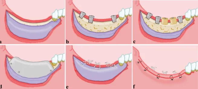
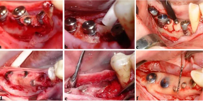
Fig. 1 Surgical treatment approach.
a) Split thickness flap preparation, b) Supracrestal implant positioning, c) Placement of bone cylinders, d) Fixation of titanium membrane, e) Periosteal suturing, f) Mucosal suturing.
a b c
d e f
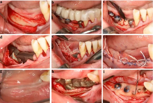
Fig. 2 Surgical treatment approach for horizontovertical ridge reconstruction (Case Three). a) Split thickness flap preparation, b) Supracrestal implant positioning, c) Placement of bone cylinders, d) Fixation of titanium membrane, e) Periosteal suturing, f) Mucosal suturing, g) Healing at 9 months, h) Re-entry and membrane removal at 9months, i) 9 months intraoperative view, complete defect reconstruction.
5.1.6 Clinical evaluation
At the time of the augmentation procedure and abutment connection, direct intraoperative measurements were registered to evaluate hard tissue formation around the implants. At each implant, the distance between the top of the implant shoulder and the alveolar crest was measured at four sites (mesial, distal, oral, vestibular aspect) at baseline (i.e. subsequent to implant placement) and at 9 months, i.e. immediately after membrane removal. All intraoperative measurements were recorded using a standard periodontal probe (UNC 15, Hu-Friedy). The thickness of keratinized mucosa over the grafted area was measured at baseline and 9 months following the augmentation procedure via sterile Kerr-files. All clinical measurements were rounded to the nearest 0.5 mm. Follow-up measurements were performed at 12, 24, 36, 48, and 60 months
f
g
b
d e
h i
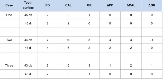
after prosthetic loading. Probing depth (PD), mucosal recession (MR), and bleeding on probing (BOP) were recorded around implants. PD, gingival recession (GR), and clinical attachment level (CAL) were recorded at neighboring natural teeth preoperatively and 9 months later to evaluate the outcome of the horizontally extended flap design involving teeth adjacent to the reconstructed ridge.
Periodontal measurements were also performed on a yearly basis during the follow-up period for 60 months; PD was less than 4 mm in each site (data not shown).
5.1.7 Radiographic evaluation
Panoramic radiographs were recorded at baseline. To assess crestal bone changes, intraoral radiographs were taken at baseline, immediately after surgeries, as well as at abutment connection and 12, 24, 36, 48, and 60 months after prosthetic loading. Images were digitized, and the distance between implant shoulder and the crestal bone was measured at the mesial and distal aspects of each fixture. Digital images were examined under an 8-fold magnification. Implant height was used for calibration. In Case 1, conventional computed tomography (CT) analysis was made prior to augmentation; in Cases 2 and 3, cone beam CT (CBCT) analysis was performed. In all three cases, additional CBCT analysis was performed 9 months postoperatively. Only qualitative radiographic evaluation was performed on CT scans.
5.2 Study II (human cadaver study)
The methods of latex milk injection (fourteen specimens) and corrosion casting (six specimens) were used bilaterally on human cadavers. These were donated to the Department of Anatomy, Semmelweis University, Budapest, Hungary, according to Hungarian approval rules of anatomical donation, and to the Department of Anatomy of the Medical University of Graz, Austria, complying with the Anatomical Donation Program of the Medical University of Graz and in accordance with the Austrian law.
The course and distribution of blood vessels in relation to soft and hard tissues, used in combination with a layer-by-layer dissection protocol, were studied. Digital
photographs were taken of each specimen from multiple directions with a magnification range of 1:1 to 1:3 using a macro lens mounted to a digital single lens reflex camera equipped with a ring light (Canon 600D, Canon 100 mm 2.8 macro lens, Canon MR-14 EX ring light). Photographs were analyzed by visual inspection on a calibrated monitor at 1:1 magnification.
5.2.1 Latex milk injection
Latex milk stains the blood vessels and makes them macroscopically remarkable. It facilitates the dissection of the vessels. By this method, the anatomical and physiological relation of blood vessels can be analyzed (Alvernia et al., 2010). In this method, the cadavers can be fixed in Thiel solution (Thiel, 1992a; Thiel, 1992b; Thiel, 2002) tanks for about a year. This solution permits prolonged preservation, keeps the natural color, texture, plasticity and flexibility of the tissues, same as a fresh specimen, and it allows easier injection of vessels till the thinnest branches (Thiel, 1992a; Thiel, 1992b; Thiel, 2002; Ottone et al., 2016).
5.2.1.1 Thiel solution
Since ancient times, the goal of preservation of the body has existed. This was mainly due to religious and scientific reasons. In 1992, Walter Thiel introduced a novel method which allowed the preservation of the body with natural colors.
The Thiel’s technique is composed of two steps:
1) Intravascular injection formula.
2) Fixation of the cadavers for a determined period in immersion solution inside a sealed container to disinfect, preserve tissue plasticity and avoid dehydration without the usage of preservation fluid (Thiel, 1992a; Thiel, 1992b; Thiel, 2002; Ottone et al.
2016). By this method, the cadaver handling is more effective, and it eludes the formation of vexatious gases because of low formaldehyde concentrations used in the solution (Thiel, 1992a; Thiel, 1992b; Thiel, 2002; Ottone et al. 2016) (Table 1).
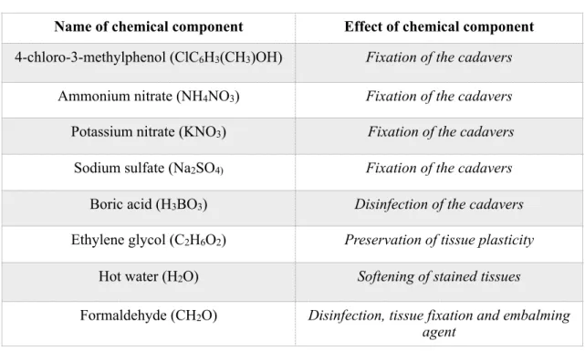
Table 1 Basic chemical components used in the Thiel embalming method.
The carotid arteries were washed with an intravascular Thiel solution, “containing 14300 mL of solution A, plus 500 mL of solution B and the addition of 700 g of sodium sulfite as well as 300 mL formalin” (Ottone et al., 2016, p. 1443) for an average body weight of 80 kg (Table 2). Then, the cadavers were injected with the latex milk, or they were fixed for around 6 months in the immersion solution (Table 2 and 3). The specimens were retained in zipper polyethylene bags and they were used for a long time (Thiel, 1992b; Thiel, 2002; Ottone et al., 2016). The outcomes of the utilization of the Thiel solution were non-irritating, almost odorless, maintaining the color and flexibility of the cadavers (Thiel, 1992a; Thiel, 2002; Ottone et al., 2016). The specimens were comparable to the living body, with adequate temporomandibular joint mobility and maintained tissue elasticity which were suitable for surgical interventions (Ottone et al., 2016). After embalming of the cadavers for about 6 months, those head specimens which were not injected by latex milk previously, were ready for the next step, which was latex milk injection.
Name of chemical component Effect of chemical component 4-chloro-3-methylphenol (ClC6H3(CH3)OH) Fixation of the cadavers
Ammonium nitrate (NH4NO3) Fixation of the cadavers Potassium nitrate (KNO3) Fixation of the cadavers Sodium sulfate (Na2SO4) Fixation of the cadavers
Boric acid (H3BO3) Disinfection of the cadavers Ethylene glycol (C2H6O2) Preservation of tissue plasticity
Hot water (H2O) Softening of stained tissues
Formaldehyde (CH2O) Disinfection, tissue fixation and embalming agent
Table 2 Basic composition of injection and immersion solutions described by W. Thiel in 1992. Note: Adapted from “Walter Thiel’s Embalming Method. Review of Solutions and Applications in Different Fields of Biomedical Research” by Ottone NE, Vargas CA, Fuentes R, Del Sol M, 2016, Int. J. Morphol, 34, p.1443. Copyright 2016 by Nicolás Ernesto Ottone; Claudia A. Vargas;
Ramón Fuentes; Mariano del Sol (INTERNATIONAL JOURNAL OF MORPHOLOGY), Attribution- NonCommercial, (https://creativecommons.org/licenses/by-nc/4.0/deed.en) CC BY-NC 4.0.
Solution A Injection solution Immersion solution Boric acid 3 g Solution A 14300 ml
Ethylene glycol 30 ml Solution B 500 ml Ammonium nitrate 20 g Formaldehyde 300 ml Potassium nitrate 5 g Sodium sulfate 700 g Hot water 100 ml Solution B
Ethylene glycol 10 ml 4-chloro-3-methylphenol 1 ml
The process of latex milk injection together with the dissection of the blood vessels in the oro-maxillofacial region was technique-sensitive. The equipment and chemicals to be used in latex milk injection were strictly checked and measured (Table 4). Any mistake or improper functioning of equipment would damage the entire process and the macroscopic analysis of the vessels.
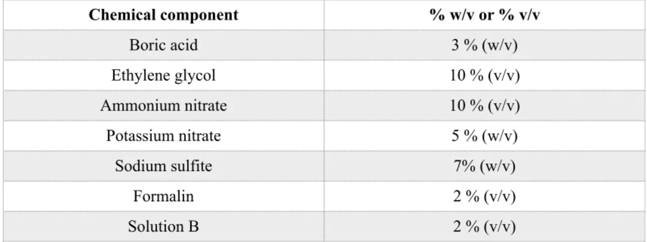
Table 3 Calculation of the percentage of weight/volume (w/v), and percentage of volume/volume (v/v) used in the immersion solution (according to Thiel, 1992a; Thiel, 1992b; Thiel, 2002; Ottone et al., 2016).
Chemical component % w/v or % v/v
Boric acid 3 % (w/v)
Ethylene glycol 10 % (v/v)
Ammonium nitrate 10 % (v/v)
Potassium nitrate 5 % (w/v)
Sodium sulfite 7% (w/v)
Formalin 2 % (v/v)
Solution B 2 % (v/v)
Ethylene glycol 10 ml Formaldehyde 2 ml Solution B 2 ml Boric acid 3 g
Ammonium nitrate 10 g Potassium nitrate 10 g Sodium sulfate 7 g Hot water 100 ml
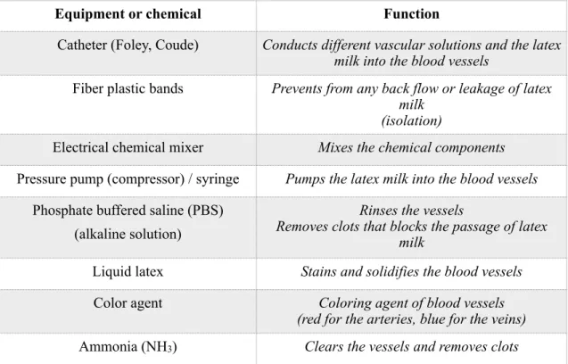
Table 4 Equipment and chemical components, with their functional properties, used for latex milk injection.
5.2.1.2 Process of latex milk injection
• The carotid trigones were carefully dissected. The common carotid arteries (CCAs), and subsequently the ECAs, were gently revealed.
• The CCA/ECA was rinsed with PBS under the pressure of minimum 27 kPa, and then perfusion of Thiel’s solution via the CCA/ECA was performed.
• Following perfusion with Thiel’s solution, the arteries were subsequently flushed again with PBS.
• Indirect anatomical study of the arterial orientation during irrigation with PBS through the CCA/ECA was made.
• After embalming, the CCA/ECA was cannulated carefully with either a Foley or a Coude catheter. A proper sized catheter was chosen.
Equipment or chemical Function
Catheter (Foley, Coude) Conducts different vascular solutions and the latex milk into the blood vessels
Fiber plastic bands Prevents from any back flow or leakage of latex milk
(isolation)
Electrical chemical mixer Mixes the chemical components Pressure pump (compressor) / syringe Pumps the latex milk into the blood vessels
Phosphate buffered saline (PBS) (alkaline solution)
Rinses the vessels
Removes clots that blocks the passage of latex milk
Liquid latex Stains and solidifies the blood vessels
Color agent Coloring agent of blood vessels
(red for the arteries, blue for the veins) Ammonia (NH3) Clears the vessels and removes clots
• Then the catheter was placed in the CCA/ECA. The fixation of the catheter was made precisely to prevent any leakage of the substance under pressure. The fixation (isolation) was performed by alternating knots (with fiber plastic bands) on different aspects of the vessel.
• Prior to injection of latex, 20-30 ml of diluted ammonia (NH3) was injected to clear the vessels.
Note: According to Echt (1998), ammonia can be used as a ‘preservative’ in the latex because it agitates the molecules of rubber and provides a two-phase product consisting of 30 - 40% solids; the product can be concentrated to 60% solids, producing ammoniated latex concentrate, that includes 1.6% ammonia by weight. By usage of low-ammonia latex concentrate (0.15 - 0.25% ammonia) and adding of secondary preservatives such as Sodium pentachlorophenate (C6Cl5ONa), Te t r a m e t h y l t h i u r a m d i s u l f i d e ( C H3)2N C S S2C S N ( C H3)2, S o d i u m d i m e t h y l d i t h i o c a r b a m a t e ( C3H6N N a S2) a n d Z i n c o x i d e ( Z n O ) the coagulation and contamination can be avoided (Echt, 1998).
• The latex milk (Creato Latexmilch, Zitzmann Zentrale, Baden, Germany) was colored red and it was injected by the pressure pump, syringe or pasteurized bottle with a delivery tube into the arteries. During the injection, the same and constant pressure is sufficient, but a higher pressure can be used to provide a complete distribution into the finer vessels.
• These arteries were “full form” and capable of resisting the rigidity of dissection thereafter. About 150 ml of latex was sufficient for an adult cadaver injection. After 20-30 min of injection, the latex started to solidify and imparted a red color to the arteries.
• Cadaver head specimens were ready for dissection about 4-6 weeks following injection, during which the latex would have time to set. Cadavers were sealed in plastic bags for a certain period with anti-fungal agents.
• After the embalming period, the specimens were dissected under 2.5x magnification using Nr. 15 and Nr. 15C surgical blades. The mucosa was elevated, the injected vessels were dissected in each layer, and the path of the arteries, along with their network, were macroscopically examined.
5.2.2 Corrosion casting
The method was mainly followed according to Lametschwandtner et al. (1990), Verli et al. (2007), and Rueda Esteban et al. (2017). This process, associated with or without scanning electron microscopy (SEM) is utilized to investigate the vascular orientation of organs and tissues (Lametschwandtner et al., 1990; Verli et al., 2007). The corrosion casting involves two main distinct procedures:
• Casting: Formation of the casts by the vessel lumens and solidification of vessels with a low-viscosity resin (Hodde et al., 1990; Lametschwandtner et al., 1990; Verli et al., 2007; Rueda Esteban et al., 2017). Casting reveals the complexity of blood vessels in three-dimensions in relation to bony anatomical landmarks as a result of complete maceration of soft tissues (Verli et al., 2007; Haenssgen et al., 2014, Shahbazi et al., 2018). The most crucial aspects of this method are the usage of head cadavers 1-2 days post mortem, isolation and selection of high quality vascular casting material (Table 5).
• Corrosion: Maceration of tissues with an alkaline solution surrounding the polymerized resin (Hodde et al., 1990; Lametschwandtner et al., 1990; Sims &
Albrecht, 1993; Verli et al., 2007). This process can be conducted by Sodium hydroxide (NaOH) and Potassium hydroxide (KOH) solutions, with/without detergents, at various concentrations (Hodde et al., 1990; Lametschwandtner et al., 1990; Sims & Albrecht, 1993; Verli et al., 2007; Rueda Esteban et al., 2017). The method results in the total dissolution of soft tissues located around the previously- casted vessels.
Table 5 Criteria for vascular casting materials (according to Verli et al., 2007).
The corrosion casting is more technique-sensitive than the latex milk injection, so the equipment, chemical components (Table 6) and freshness of the specimen should be strictly controlled. It is suggested that the cadavers without any previous fixation or formalin injection to be selected (Rueda Esteban et al., 2017). This method indicates a solidification in vascular tissues, so precise rinsing with water for several minutes is essential to eliminate clots and increase the visibility of the blood vessels (Rueda Esteban et al., 2017). After careful dissection of carotid trigones, the ECAs are rinsed with heparin or hydrogen peroxide (H2O2) and then by PBS which are described in pre- casting phase.
Low viscosity Polymerization time: 3-15 min
No shrinkage during polymerization Maintaining the structural configuration while drying
Small particles to fill the capillaries Permiting quantitative analysis
No morphological alterations in the tissues and vessels No penetration of tissues and their interstitial spaces
Resistant to the corrosion process Permitting microdissection
No damage to the surrounding tissues Atoxic
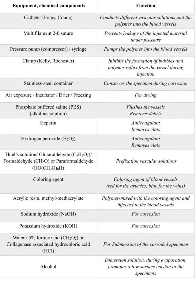
Table 6 Equipment and chemical components, with their functional properties, used in corrosion casting.
Equipment, chemical components Function
Catheter (Foley, Coude) Conducts different vascular solutions and the polymer into the blood vessels
Multifilament 2-0 suture Prevents leakage of the injected material under pressure
Pressure pump (compressor) / syringe Pumps the polymer into the blood vessels Clamp (Kelly, Rochester) Inhibits the formation of bubbles and
polymer reflux from the vessel during injection
Stainless-steel container Conserves the specimen during corrosion Air exposure / Incubator / Drier / Freezing For drying
Phosphate buffered saline (PBS) (alkaline solution)
Flushes the vessels Removes debris
Heparin Anticoagulant
Removes clots
Hydrogen peroxide (H2O2) Anticoagulant
Removes clots Thiel’s solution/ Glutaraldehyde (C5H8O2)/
Formaldehyde (CH2O) or Paraformaldehyde (HO(CH2O)nH)
Prefixation vascular solutions
Coloring agent Coloring agent of blood vessels
(red for the arteries, blue for the veins) Acrylic resin, methyl-methacrylate Polymer-mixed with the coloring agent and
injected to the blood vessels
Sodium hydroxide (NaOH) For corrosion
Potassium hydroxide (KOH) For corrosion
Water / 5% formic acid (CH2O2) or Collagenase associated hydrochloric acid
(HCl)
For Submersion of the corroded specimen
Alcohol
Immersion solution, during evaporation, promotes a low surface tension in the
specimens
5.2.2.1. Process of corrosion casting 5.2.2.1.1. Pre-casting phase
• Careful preparation of the specimen, dissection and identification of the CCA/ECA was performed and then isolation (Hill & McKinney, 1981) was made.
• After choosing the proper sized catheter/tube (Foley or Coude), which would allow the injection of the polymer, it was fixed very securely into the CCA/ECA. The size of the tube was depending on the vessel that was going to be injected. It was preferred to use the same or slightly smaller sized tubes for the injection.
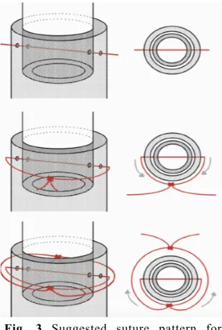
Once the tube was positioned in the artery, any leakage of the injected material under pressure was avoided by transfixation suture (Rueda Esteban et al., 2017), with alternating knots on each side of the artery (Fig. 3). Based on suggestion of Rueda Esteban et al.
(2017), multifilament 2-0 sutures were used. This suture prevented knot loosening and leakage of the polymer during the injection. Also, in some cases, for further isolation dental floss was used as well. Due to the high probability of damaging the arteries during injection, this process was carried carefully.
Fig. 3 Suggested suture pattern for cannulation. Note: Adapted from “Corrosion Casting, a Known Technique for the Study and Teaching of Vascular and Duct Structure in Anatomy” by Rueda Esteban RJ, López McCormick JS, Martínez Prieto DR, Hernández Restrepo JD, 2017, Int. J. Morphol, 35, p.1150.
Copyright 2017 by Roberto Javier Rueda Esteban;
Juan Sebastián López McCormick; Daniel Ricardo Martínez Prieto; Juan David Hernández Restrepo ( I N T E R N A T I O N A L J O U R N A L O F MORPHOLOGY), Attribution-NonCommercial (https://creativecommons.org/licenses/by-nc/4.0/
deed.en) CC BY-NC 4.0.
• Administration of anticoagulants; heparin or hydrogen peroxide (H2O2) (1-2%) were essential to remove clots that could otherwise meddle with the polymer’s entry, to expedite the injection (Lametschwandtner et al., 1990; Hodde et al., 1990; Verli et al., 2006; Rueda Esteban et al., 2017). After using H2O2, it was crucial to wash the vessels with the water, due to production of foam which would influence sufficient injection, the compound and could harm surrounding tissue (Rueda Esteban et al., 2017).
• ECAs were flushed by PBS under the pressure of minimum 27 kPa to clean away debris.
• Vascular prefixation with Thiel’s intravascular solution was performed after flushing (Shahbazi et al., 2018). Based on literature glutaraldehyde (C5H8O2), formaldehyde (CH2O) and paraformaldehyde (HO(CH2O)nH) at various concentrations (Hodde et al., 1990; Selliseth & Selvig, 1995; Ojima et al., 1997; Verli et al., 2007) could be used, to evade resin extravasating from the vessels into the tissues (Lametschwandtner et al., 1990), to promote withstanding of vascular walls (Selliseth & Selvig, 1995) and to reduce vascular expansion throughout injection of the resin (Kishi et al., 1990; Verli et al., 2007).
• Following prefixation, spontaneous washing with PBS solution was required, to withdraw the fixative and increase resin penetrability.
• To enhance the clarity of the injected polymer in the blood vessels, a red color code was given to differentiate the arteries.
5.2.2.1.2. Casting phase
• Acrylic resin (methyl-methacrylate) was selected as a polymer to be injected into the ECA. Before injection it was noted, as the resin viscosity decreases, the polymerization shrinkage increases, so if the resin would be diluted then the probability of shrinkage would be higher (Lametschwandtner et al., 1990).