DOI: 10.1556/066.2018.47.2.14
Preliminary communication
ASSESSMENT OF SUBACUTE GENOTOXIC AND HISTOPATHOLOGICAL EFFECTS OF A FOOD FLAVOUR INGREDIENT, 4-ETHYLBENZALDEHYDE (EBA) ON ZEBRAFISH
(DANIO RERIO) MODEL
D. BENCSIKa,b, GY. GAZSIb, B. URBÁNYIb, B. SZENDEc, G. RÁCZc, A. VÉHAa and ZS. CSENKIb*
aDepartment of Food Engineering, Faculty of Engineering, University of Szeged, H-6725, Szeged, Moszkvai krt.
5–7. Hungary
bDepartment of Aquaculture, Faculty of Agricultural and Environmental Sciences, Szent István University, H-2100, Gödöllő, Páter Károly u. 1. Hungary
c1st Department of Pathology and Experimental Cancer Research, Semmelweis University, H-1085, Budapest, Üllői út 26. Hungary
(Received: 28 July 2017; accepted: 20 October 2017)
Modern food industry widely uses a variety of fl avour and fragrance materials. One of the most used compound groups is the aldehydes. The benzaldehyde, also known as artifi cial almond oil, is one of the most commonly used fl avouring in food industry nowadays. The effects of this compound on different species are well known, a lot of toxicological information can be found in the literature. 4-ethylbenzaldehyde is also a member of aldehyde group, the physical properties are similar to benzaldehyde and also has almond scent. Unlike benzaldehyde, it has no chemical safety assessment according to its chemical safety sheet, and only one experiment can be found on its effects on vertebrates. This compound can also be found at the group of fl avours and fragrances. The aim of this study was to examine the subacute DNA and tissue damaging effects of EBA. The genotoxic effects of EBA in zebrafi sh were evaluated by using micronucleus assay. Signifi cant increase in the micronucleus frequency had been described for all tested concentrations. Alterations were found in the liver of the fi sh group treated with 11 mg l–1 EBA for 21 days.
Keywords: 4-ethylbenzaldehyde, fl avours, fragrances, genotoxicity, micronucleus assay, zebrafi sh
Modern food industry widely uses a variety of fl avours and fragrances. Among these numerous compounds are of natural origin, but some of them are synthetically produced.
Many of these substances have been proven to be toxic, although these compounds are still used in the industry or were used until they were banned. For example, various sweetening agents and artifi cial dyes (WEIHRAUCH & DIEHL, 2004; HUFF & LADOU, 2007; AMCHOVA et al., 2015) as additives play important roles in modern food processing, an average consumer take them in every day, it is a high-priority to know its risks for human health.
Food industry uses the group of aldehydes for a long time. The benzaldehyde, also known as artifi cial almond oil, is the second most commonly used artifi cial aroma after artifi cial vanilla (KRINGS & BERGER, 1998). Benzaldehyde has comprehensive toxicological literature (AMERICAN COLLEGE OF TOXICOLOGY, 2006), its chemical safety sheet contains a considerable amount of useful data (CAS 100-52-7). Acute results are very important, but
* To whom correspondence should be addressed.
Phone: +36 28 522 000/2310; fax: +36 28 522 927; e-mail: Csenki.Zsolt@mkk.szie.hu
246
people might get in contact with this material every day, like with EBA, so great attention should also be given to the chronic and subacute effects. It is less toxic to mammals (rabbit, rat, mouse, guinea pig), acute oral LD50 values are really high (higher than 1000 mg kg–1), except mice (oral LD50 28 mg kg–1). ABRAMOVICI and RACHMUTH-ROIZMAN (1983) had studied the effects of benzaldehyde on chicken embryos, they have found an increasing number of abnormal development of skeleton and limbs in a concentration dependent manner. According to the WHO (1996), the benzaldehyde causes delayed development, decreased fetal and neonatal weights in mice, rats, hamsters, and rabbits, but only in concentrations proved to be toxic to the parents. Acceptable daily intake of benzaldehyde is 5 mg kg–1 body weight and estimated daily intake of benzaldehyde is 9300 μg/capita/day in Europe and 36 000 μg/capita/day in the USA (WHO, 2002).
The compound (4-ethylbenzaldehyde) belongs to the group of aldehydes. Its physical properties are identical to benzaldehyde. It is a colourless, almond scented liquid. It can be found during analysis of fragrance components of different kind of foods, for example in lettuce and cabbage (LONCHAMP et al., 2009), in French beans (BARRA et al., 2007), and also in green tea (SHIMODA et al., 1995). It has also been described as a scent component of foodstuffs of animal origin, for instance in matured anchovy (TRIQUI & REINECCIUS, 1995), in mussels (LE GUEN et al., 2000), and in different kind of sea-fi sh (MORITA et al., 2003; SILVA et al., 2012). Less known is the fact that EBA is also known as a water disinfection by-product (RÁCZ et al., 2012). The study of this compound is very important for the preservation of human health, because we can get in contact with it in many areas of life.
Unlike benzaldehyde, according to the safety data sheet of EBA (CAS 4745-78-1), it has no chemical safety assessment or toxicity data. The compound is included in the scientifi c opinion on fl avouring group evaluation issued by the European Food Safety Authority (EFSA) (2012). In this EFSA publication, there is no security concern with EBA, it has been classifi ed as fl avouring agent for foodstuffs. There is only one publication mentioning this compound in scientifi c sense, an unpublished report from 1984, but the MSDI value of EBA had been determined (0.37 μg/capita/day) (EFSA, 2012). In contrast, according to the WHO (2002), threshold for human intake for the structural class of EBA is 1800 μg/day, and there is no safety concern at current level of intake when used as a fl avouring agent. In Regulation (EC) No 1272/2008 of the European Parliament and Council, it is not considered dangerous substance (EC, 2008). The currently available limited information on EBA is worrying. Up to now, it has only been published in one study on vertebrates. In that study (RÁCZ et al., 2012), the subacute effects of the compound have been studied on zebrafi sh model. The main goal of this paper is to increase knowledge on the effects of EBA on living organisms, thus contributing to consumer’s safety.
1. Materials and methods
1.1. Experimental substance
Stock solution of 4-ethylbenzaldehyde (EBA) (purchased from Sigma Aldrich Hungary, CAS 4745-78-1) has been prepared in distilled water, by using ultrasonication (amplitude: 20%, time: 4 min, Branson Digital Sonifi er 250, Branson Ultrasonics Corp., USA). Solutions for treatments have been prepared with the water of ZebTEC (Techniplast S.p.a.) fi sh maintenance system.
1.2. Treatment
This experiment has been made on laboratory cultured ‘AB’ zebrafi sh line, in compliance with the applicable animal welfare regulations. Before the experiment, fi sh had been kept in Techniplast ZebTEC laboratory recirculation system (at 27 °C water temperature, with 14 h light and 10 h dark periods, pH 7.0±0.2, conductivity: 525±50 μS). Fish were fed twice a day with complete SDS Small Gran dry fi sh food (Dietex International Limited Special Diets Services G.B.). This feeding method was used during the treatment. The treatment was done in semi-static system, test solutions were changed every second day. The experiment has lasted 21 days. Concentrations were defi ned according to the results of RÁCZ and co-workers (2012). The highest applied concentration was the LC10 value (calculated concentration of compound at which 10% of treated fi sh is expected to die) of that study, calculated by authors of the cited article as 11 mg l–1 to avoid dying of the test animals and to induce sublethal symptoms. Also, 5.5 and 2.75 mg l–1 concentrations were examined. In case of control groups only the water of fi sh keeping system has been used. Adult (approximately 6–8 months) male and female fi sh were also used during the experiment in 3 groups/dose level with 8 fi sh/
group.
1.3. Micronucleus assay
Induction of micronuclei (MNi) formation has been determined in erythrocytes isolated from fi sh exposed to 4-ethylbenzaldehyde for 1, 2, and 3 weeks (4 fi sh per concentration). After obtaining peripheral blood samples, they were immediately smeared on microscope slides (Menzel-Gläser). Slides were left to air-dry and then stained with Hoechst 33342 dye (5 μM).
The stained slides were examined under an epifl uorescence microscope (Olympus BX-51, Tokyo, Japan) at a magnifi cation of ×400, and evaluated for the presence of MNi exhibiting blue fl uorescence in the peripheral blood erythrocytes. A total of 2000 randomly selected cells with complete cytoplasm were examined from each slide. The criteria for the identifi cation of fi sh micronucleated erythrocytes were as follows: (a) MNi should be smaller than one-third of the main nuclei, (b) they should be on the same plane of focus, (c) MNi must be of the same colour and intensity as the main nuclei, (d) they should have oval or round shape, and (e) they should be clearly separated from the main nucleus.
1.4. Statistics
Data were analysed by using descriptive statistics and the results are presented as mean±SD.
Statistical evaluation of the micronucleus assay data was performed with STATISTICA 12 software package (StatSoft, Tulsa, OK, USA). For the data from the micronucleus assay, Poisson regression was used to compare with controls and varying concentrations of 4-ethylbenzaldehyde. P<0.05 was considered signifi cant.
1.5. Histopathology
Zebrafi sh were fi xed in 4% buffered formaldehyde for 24–48 h at 4 °C with opened abdominal cavity, washed with phosphate buffered saline (PBS), and tissues were dehydrated in a series of graded ethanol solutions and xylene before embedment in paraffi n. Fish were placed into the cassette for sectioning. Sections were 4–6 μm thick and were stained with hematoxylin and eosin (HE). Two males and two females had been used from both treatment concentrations.
248
2. Results and discussion
2.1. Micronucleus assay
The genotoxic effects of 4-ethylbenzaldehyde in zebrafi sh were evaluated by using micronucleus assay. The main goal of the 21-day experiment was to examine subacute chromosome damaging activity of the compound.
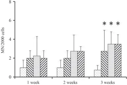
Poisson regression showed signifi cant increase in the micronucleus frequency for all concentrations tested, compared with control groups. Increasing number of MNi were found after the fi rst week, compared to the control group (1.00±0.82), even in the lowest applied concentration (2.00±0.82). Signifi cant increase was noticed only after 3 weeks exposure for all concentrations tested. In case of 2.75 mg l–1 the number of MNi formed was 2.75±2.22, for 5.5 mg l–1 and 11 mg l–1 treatments the numbers of formed MNi were 3.50±1.29 and 3.50±1.00, respectively, at the end of the experiment (Fig. 1, Table 1). No statistically signifi cant differences were observed among treated groups during the experiment in case of applied concentrations, so MNi formation was not concentration-dependent. Applied concentration range was probably not suffi ciently wide to detect concentration-dependent changes.
This method has been widely applied for a long time (ALMÁSSY et al., 1987; NERSESYAN
et al., 2014; SHIMADA et al., 2015). According to the results of this study, it is suitable to detect the genotoxic effects of EBA in low concentrations.
Similarly to acute studies, due to its structural and physico-chemical similarity, it is assumed that the chronic effects of EBA may be similar to benzaldehyde.
Fig. 1. 4-Ethylbenzaldehyde-induced micronuclei (MN) in zebrafi sh erythrocytes. Zebrafi sh were exposed to different concentrations of 4-ethylbenzaldehyde (0, 2.75, 5.5, and 11 mg l–1). The micronuclei frequency was determined in the F1 zebrafi sh treated with 4-ethylbenzaldehyde for 1, 2, and 3 weeks. Data are presented as mean±SD from four individuals. * Statistically signifi cant difference compared to corresponding control (P<0.05)
: 0 mg l–1; : 2.75 mg l–1; : 5.5 mg l–1; : 11 mg l–1
In case of chronic exposure, benzaldehyde has teratogenic (ABRAMOVICI & RACHMUTH- ROIZMAN, 1983), mutagenic (HAWORTH et al., 1983), and carcinogenic (NTP, 1990) effects. In spite of all these, US Food and Drug Administration (US FDA) has generally recognised it as safe (GRAS). EFSA also assessed the potential carcinogenicity of EBA and indicated that the compound is not carcinogenic and non-genotoxic (EFSA, 2012).
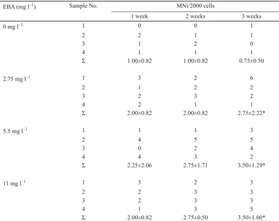
Table 1. 4-Ethylbenzaldehyde-induced genome damage. The frequency of 4-ethylbenzaldehyde-induced micronuclei (MNi) in zebrafi sh erythrocytes was assessed with the MN assay
EBA (mg l–1) Sample No. MNi/2000 cells
1 week 2 weeks 3 weeks
0 mg l–1 1 0 0 1
2 2 1 1
3 1 2 0
4 1 1 1
Σ 1.00±0.82 1.00±0.82 0.75±0.50
2.75 mg l–1 1 3 2 6
2 1 2 2
3 2 3 2
4 2 1 1
Σ 2.00±0.82 2.00±0.82 2.75±2.22*
5.5 mg l–1 1 1 1 3
2 4 5 5
3 0 2 4
4 4 3 2
Σ 2.25±2.06 2.75±1.71 3.50±1.29*
11 mg l–1 1 3 2 3
2 2 3 3
3 2 3 3
4 1 3 5
Σ 2.00±0.82 2.75±0.50 3.50±1.00*
Zebrafi sh were exposed to 4-ethylbenzaldehyde (0, 2.75, 5.5, and 11 mg l–1) for 1, 2, and 3 weeks as described in Materials and methods. Data are presented as mean±SD number of MNi/2000 from four individuals. Signifi cant difference between treated fi sh and the corresponding control is indicated by asterisk (*) (P<0.05).
2.2. Results of histopathology
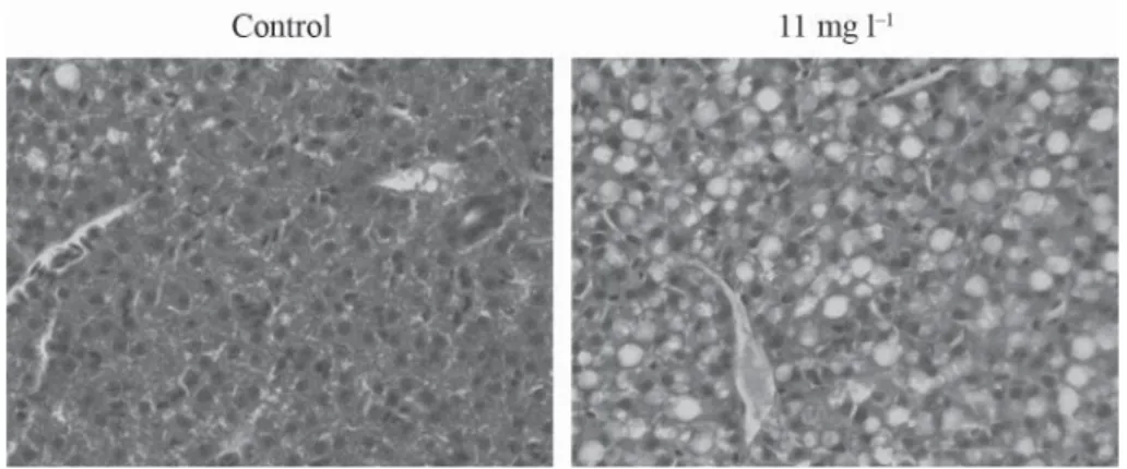
Alterations were found in the liver of the group of fi sh treated with 11 mg l–1 EBA for 21 days.
Within the liver parenchyma cells, changes were observed in the distribution and relative content of fat. Fat droplets nearly fi lled the whole cytoplasm and varied in size (Fig. 2). These slight lesions were observed in both sexes. No serious lesions, like adenofi brosis or hepatocyte megalocytosis were found. RÁCZ and co-workers (2012) applied three months treatment time and lower concentrations (2.5 mg l–1 and 5 mg l–1). Symptoms observed in the recent study are similar to their 5 mg l–1 results. According to these, the substance in a shorter treatment time
250
with higher treatment concentration also has signifi cant effect on the liver, the main detoxifying organ. In case of other organs (gill, kidney), considerable lesions were not experienced. Presumably to the development of these lesions takes more time, because RÁCZ
and co-workers (2012) described lesions in the mentioned organs.
Fig. 2. Histological sections of control and treated fi sh. Images showing liver of a control fi sh with moderate fatty change (left) and severe diffuse fatty change in the liver of a fi sh treated for 21 days with EBA (right). (H-E ×400)
3. Conclusions
Based on our results, 4-ethylbenzaldehyde has toxic effect at the tested concentrations. In case of micronucleus assay, EBA caused signifi cant increase in MNi formation in treated groups compared to control group, but it was not concentration-dependent. We can conclude that EBA has DNA damaging effect at lower concentrations and applied concentration range was not wide enough to detect concentration-dependent effects. At the highest applied concentration (11 mg l–1) it caused fat infi ltration in liver after 21 days of treatment. Applying longer treatment time may cause alterations in other organs.
In the future, it would be useful to examine a wider range of concentrations with this method and also to apply other genotoxicity tests, for example Comet Assay, on adult fi sh and embryos. It would be useful to examine the effects of this compound with molecular toxicological methods, for instance microarray assay.
Examined concentrations in this study are higher than EFSA limit, but there are diffi culties in determination of real human and environmental expositions. It may also occur in some foods naturally or as an additive in cosmetics and in drinking water. Our results have shown that it is important to examine compounds recognised as safe, because they may have hidden dangers.
*
The publication of this paper is supported by the EFOP–3.6.3–VEKOP–16–2017–00008 project co-fi nanced by the European Union and the European Social Fund.
References
ABRAMOVICI, A. & RACHMUTH-ROIZMAN, P. (1983): Molecular structure-teratogenicity relationships of some fragrance additives. Toxicology, 29, 143–156.
ALMÁSSY, ZS., KREPINSKY, A.B., BIANCO, A. & KÖTELES, G.J. (1987): The present state and perspectives of micronucleus assay in radiation protection. A review. Appl. Radiat. Isotopes, 38(4), 241–249.
AMCHOVA, P., KOTOLOVA, H. & RUDA-KUCEROVA, J. (2015): Health safety issues of synthetic food colorants. Regul.
Toxicol. Pharm., 73, 914–922.
AMERICAN COLLEGEOF TOXICOLOGY (2006): Final report on the safety assessment of benzaldehyde. Int. J. Toxicol., 25 (Suppl. 1), 11–27.
BARRA, A., BALDOVINI, N., LOISEUAU, A.-M., ALBINO, L., LESECQ, C. & LIZZANI CUVELIER, L. (2007): Chemical analysis of French beans (Phaseolus vulgaris L.) by headspace solid phase microextraction (HS-SPME) and simultaneous distillation/extraction (SDE). Food Chem., 101, 1279–1284.
EC (2008): Regulation No 1272/2008 of the European Parliament and of the Council on classifi cation, labelling and packaging of substances and mixtures, amending and repealing Directives 67/548/EEC and 1999/45/EC, and amending Regulation (EC) No 1907/2006. The European Council
EFSA (2012): Scientifi c opinion on fl avouring group Evaluation 20, Revision 4 (FGE.20REV4): Benzyl alcohols, benzaldehydes, a related acetal, benzoic acids, and relates esters from chemical groups 23 and 30. European Food Safety Authority, EFSA J., 10(12), 2994, 1–140.
HAWORTH, S., LAWLOR, T., MORTELMANS, K., SPECK, W. & ZEIGER, E. (1983): Salmonella mutagenicity test results for 250 chemicals. Environ. Mol. Mutagen, 5 (Suppl. 1), 3–142.
HUFF, J. & LADOU, J. (2007): Aspartame bioassay fi ndings portend human cancer hazards. Int. J. Occup. Env. Heal., 13, 446–448.
KRINGS, U. & BERGER, R.G. (1998): Biotechnological production of fl avours and fragrances. Appl. Microbiol. Biot., 49, 1–8.
LE GUEN, S., PROST, C. & DEMAIMAY, M. (2000): Characterization of odorant compounds of mussels (Mytilus edulis) according to their origin using gas chromatography-olfactometry and gas chromatography-mass spectrometry.
J. Chromatogr. A, 896, 361–371.
LONCHAMP, J., BARRY-RYAN, C. & DEVEREUX, M. (2009): Identifi cation of volatile quality of ready-to-use lettuce and cabbage. Food Res. Int., 42, 1077–1086.
MORITA, K., KUBOTA, K. & AISHIMA T. (2003): Comparison of aroma characteristics of 16 fi sh species by sensory evaluation and gas chromatographic analysis. J. Sci. Food Agr., 83, 289–297.
NERSESYAN, A., KUNDI, M., FENECH, M., BOLOGNESI, C., MISIK, M., WULTSCH, G., HARTMANN, M. & KNASMUELLER, S.
(2014): Micronucleus assay with urine derived cells (UDC): A review of its application in human studies investigating genotoxin exposure and bladder cancer risk. Mutation Research –Reviews, 762, 37–51.
NTP (1990): NTP TR 378 Technical Report on the toxicology and carcinogenesis studies of benzaldehyde (CAS No 100-52-7) in F344/N rats and B6C3Fi mice. National Toxicology Program, U.S. Department of Health and Human Services, 191 pages.
RÁCZ, G., CSENKI, ZS., KOVÁCS, R., HEGYI, Á., BASKA, F., SUJBERT, L., ZSÁKOVICS, I., KIS, R., GUSTAFSON, R., URBÁNYI, B. & SZENDE, B. (2012): Subacute toxicity assessment of water disinfection byproducts on zebrafi sh. Pathol.
Oncol. Res., 18, 579–584.
SHIMADA, K., YAMAMOTO, M., TAKASHIMA, M., WAKO, Y., KAWASAKO, K., AOKI, Y., SEKI, J., MIYAMAE, A. & WAKATA, A. (2015): Repeated-dose liver micronucleus assay of mitomycin C in young adult rats. Mutat. Res. Gen. Tox.
En., 780–781, 85–89.
SHIMODA, M., SHIGEMATSU, H., SHIRATSUCHI, H. & OSAJIMA, Y. (1995): Comparison of volatile compounds among different grades of green tea and there relations to odor attributes. J. Agr. Food Chem., 43, 1621–1625.
SILVA, M.C.E., SILVA, L.R., GUEDES-DE-PINHO, P. & COSTA, R. (2012): Volatile compounds in salted dried codfi shes from different species. Acta Alimentaria, 41, 375–388.
TRIQUI, R. & REINECCIUS, G.A. (1995): Changes in fl avor profi les with ripening anchovy (Engraulis encrasicholus).
J. Agr. Food Chem., 43, 1883–1889.
WEIHRAUCH, M.R. & DIEHL, V. (2004): Artifi cial sweeteners – do they bear a carcinogenic risk? Ann. Oncol., 15, 1460–1465.
WHO (1996): Benzyl acetate, benzyl alcohol, benzaldehyde, and benzoic acid and its salts. WHO Food Ad., 37, 31–79.
WHO (2002): Technical Report series 909, Evaluation of certain food additives and contaminants, pp. 73–95.