Imaging and aberrometric analyses of diseases affecting biomechanical properties of the cornea
Modern disgnostic methods of keratoconus
Ph.D. thesis
Dr. Miháltz Kata
Semmelweis University Clinical Medicine, Ph.D. School
Supervisor: Dr. Nagy Zoltán Zsolt DSC
Reviewers: Dr. Kerényi Ágnes Ph.D.
Dr. Farkas Ágnes Ph.D.
Chairman of comprehensive exam: Dr. Matolcsy András D.Sc.
Members of comprehensive exam: Dr. Füst Ágnes Ph.D.
Dr. Erdei Gábor Ph.D.
Budapest 2011
Contents
Table of contents………...2
Abbreviations………..………..4
1. Introduction………..……….6
1. 1. Epidemiology of keratoconus………..………..7
1. 2. Clinical Features………..………..7
1. 3. Histopathology………..………10
1. 4. Etiology and Pathogenesis………..………..11
1.4. 1. Associated disorders………..………..11
1.4. 2. Biochemical studies………..………...12
1.4. 3. Genetics………..……….13
1.5. Diagnosis of keratoconus………..………14
1.5.1. Photokeratoscopy and keratometry………..………14
1.5.2. Computer-assisted videokeratoscopy………..……….15
1.5.3. Elevation based topography………..………...19
1.5.3.1. Orbscan………..………..19
1.5.3.2. Scheimpflug camera………..………...20
1.5.4. Wavefront aberrometry………..……….21
1.6. Treatment of Keratoconus………..………23
1.6.1. Contact lenses………..……….23
1.6.2. Corneal crosslinking………..………...23
1.6.3.1. Surgical interventions: ICRS………..………..…….24
1.6.3.2. Surgical interventions: Keratoplasty………..………..….25
1.7. Other diseases affecting biomechanical properties of the cornea……….26
1.7.1. Pellucid marginal degeneration………..………..26
1.7.2. Keratoglobus………..………...28
1.7.3. Post-LASIK ectasias… ………..………..28
2. Purposes………..………..30
3. Methods………..………..32
3.1. Demographic data………...32
3.2. Patient inclusion criteria………..………33
3.3. Patient exclusion criteria……….33
3.4. Corneal topographic measurements………..………….……….34
3.5. Pentacam measurements………..………...34
3.6. Hartmann-Shack aberrometry measurements………..………..35
3.7. Statistical analyses………..………38
4. Results………..……….40
4.1. Evaluation of elevation parameters of keratoconus with Pentacam………...40
4.2. Anterior chamber characteristics of keratoconus ………..45
4.3. Shifting of the line of sight in keratoconus measured by a Hartmann-Shack sensor….50 5. Discussion………..………...56
5.1. Keratoconus detection with Scheimpflug imaging………....56
5.1.1. Characteristics of elevation based topography……….56
5.1.2. The role of refernce body selection in the diagnosis of keratoconus………...58
5.1.3. The use of elevation data in the diagnosis of keratoconus………...61
5.1.4. Comparative analysis of anterior chamber characteristics of keratoconus………..63
5.2. Keratoconus detection with aberrometry………...65
5.2.1. Optical and visual axes of the eye………66
5.2.2. Spherical aberration in keratoconus ……….68
5.2.3. The pupil apodizing and Stiles-Crawford effect……….………..70
6. Conclusion………72
7. Summary………...74
8. References………75
9. List of publications………....84
Acknowledgement………89
Abbreviations
ACD: anterior chamber depth AIC: Akaike information criterion ANOVA: analysis of variance
ASCRS: American Society of Cataract and Refractive Surgery AUROC: area under the receiver operating characteristic curve BCVA: best corrected visual acuity
BFS: best-fit sphere
BFTE: best fit toric ellipsoid reference body CCT: central corneal thickness
CFA: confirmatory factor analysis CL: contact lens
CRF: corneal resistance factor CH: corneal hysteresis
CXL: corneal crosslinking D: diopter
DLK: diffuse lamellar keratitis GEE: generalized estimating equation
HORMS: higher-order root mean square ICRS: intrastromal corneal ring segments
IOL: intraocular lens K: keratometry
KISA: central keratometry (K) inferior (I) minus superior (S) average (A) keratometry LASIK: laser-assisted in situ keratomileusis
LKP: lamellar keratoplasty LOS: line of sight
OPD: optical path difference ORA: ocular response analyzer OSA: optical society of America pIOL: phakic intraocular lens PKP: penetrating keratoplasty
PMD: pellucid marginal degeneration PRK: photorefractive keratectomy
QICC: quasi-likelihood under independence model criterion
RSB: residual stromal bed RMS: root mean square
ROC: receiver operating characteristic ROS: reactive oxygen species
SA: spherical aberration
SCE: Stiles-Crawford effect SD: standard deviation
TCT: thinnest corneal thickness TMS: topographic modeling system UCVA: uncorrected visual acuity US: ultrasound
1. Introduction
The cornea, a transparent tissue that covers the front of the eye, performs approximately 2/3 of the optical refraction and focuses light towards the lens and the retina.
Thus, even slight variations in the shape of the cornea can significantly diminish visual performance. It is reported though, that localized loss of corneal thickness, and likely also degradation of corneal mechanical properties, cause gradual tissue protrusion, which results in a more conical appearance of the cornea that imposes blurred vision. Surgical interventions and diseases of corneal tissue have been found to result in substantial changes in corneal tissue structure, which can then also alter the biomechanical properties of the cornea. The surgical procedures used to perform corneal refractive surgery result in changes in the corneal tissue structure, which affect the corneal thickness (CCT) and curvature of the cornea.
Corneal refractive surgery and corneal diseases thus can alter corneal biomechanics.
Keratoconus is the most frequently occurring disease of the cornea caused by a non- inflammatory deterioration of the corneal structure (1).
Keratoconus, which was first described in detail in 1854, derives from the Greek words Kerato (cornea) and Konos (cone) (2). Keratoconus is the most common primary ectasia. It is a bilateral and asymmetric corneal degeneration characterized by localized corneal thinning which leads to protrusion of the thinned cornea (3,4). Corneal thinning normally occurs in the inferior-temporal as well as the central cornea, although superior localizations have also been described. The disease induces myopia, irregular astigmatism and has well defined slit lamp findings (5,6). It has well-described clinical signs, but early forms of the disease may go undetected unless the anterior corneal topography is studied. The diagnosis of more advanced keratoconus is not complicated, because of the typical biomicroscopic and topographic findings, but the detection of subclinical or forme fruste cases may impose difficulty. Early disease is now best detected with videokeratoscopy or elevation based topography (7,8). It is particularly important to detect the disease among refractive surgery candidates, as keratorefractive procedures may worsen their condition (9).
Corneal protrusion causes high myopia and irregular astigmatism, affecting visual quality.
1. 1. Epidemiology
The incidence and prevalence of keratoconus in the general population has been estimated to be between 5 and 23, and 5.4 per 10,000, respectively. Differences on the rates reported are attributed to different definitions and diagnostic criteria employed between studies. However, it would not be surprising to expect an increase in the incidence and prevalence rates of this disease over the next few years with the current wide spread use of corneal topography leading to improved diagnosis(10). Keratoconus affects both genders, although it is unclear whether significant differences between males and females exist (11).
Keratoconus, classically, has its onset at puberty and is progressive until the third to fourth decade of life, when it usually arrests. It is most commonly an isolated condition, despite multiple singular reports of coexistence with other disorders. Commonly recognized associations include Down syndrome, Leber‘s congenital amaurosis, and connective tissue disorders. For example, patients with advanced keratoconus have been reported to have a high incidence of mitral valve prolapse. Atopy, eye rubbing, and hard contact lenses have also been reported to be highly associated with this disorder (3, 12).
1. 2. Clinical Features
The ocular symptoms and signs of keratoconus vary depending on disease severity. At incipient stages, also referred to as subclinical or frustre forms, keratoconus does not normally produce any symptoms and thus can go unnoticed by the patient and practitioner unless specific tests (i.e., corneal topography) are undertaken for diagnosis (2,3). Disease progression is manifested by a significant loss of visual acuity which cannot be compensated for with spectacles. Therefore, eye care practitioners should be suspicious about the presence of keratoconus when a visual acuity of 1,0 is difficult to achieve with increasing against-the- rule astigmatism. Near visual acuity is generally found to be better than expected from the refraction, distance visual acuity and age of the patient (13,14). The appearance of ―scissor‖
shadows while performing retinoscopy suggests the development of irregular astigmatism.
Through retinoscopy it is possible to estimate the location of the cone's apex and its diameter, and the adjustable spectacle corrected visual acuity achievable. The Charleux oil drop that is observed by backlighting the mydriatric pupil also poses a warning sign (2,3).
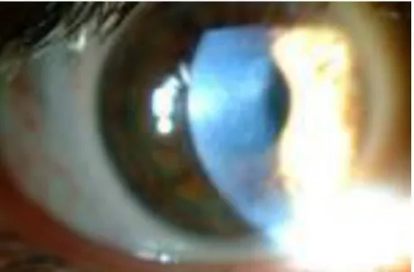
In moderate and advance cases of keratoconus, a hemosiderin arc or circle line, commonly known as Fleischer's ring (Figure 1), is frequently seen around the cone base. This line has been suggested to be an accumulation of iron deposits from the tear film onto the cornea as a result of severe corneal curvature changes induced by the disease and/or due to modification of the normal epithelial slide process (15,16).
Figure 1. Fleischer's ring, a hemosiderin arc around the cone base (from our own database ).
Another characteristic sign is the presence of Vogt's striae (Figure 2), which are fine vertical lines produced by compression of Descemet's membrane, which tend to disappear when physical pressure is exerted on the cornea.
Figure 2. Vogt's striae, fine vertical lines produced by compression of Descemet's membrane (from our own database).
The increased visibility of corneal nerves and observation of superficial and deep corneal opacities are also common signs, which can be present at different severity stages of the disease. The majority of contact lens patients eventually develop corneal scarring. Munson's sign (Figure 3), a V-shape deformation of the lower eyelid when the eye is in downward position, and Rizzuti's sign, a bright reflection of the nasal area of the limbus when light is directed to the temporal limbal area, are signs frequently observed in advanced stages (2,3).
Figure 3. Munson's sign, a V-shape deformation of the lower eyelid when the eye is in downward position (from our own database).
Acute hydrops (Figure 4) is a well-known complication, occurring in approximately 3% of patients with keratoconus. Hydrops occurs after rupture of the posterior cornea leads to an influx of aqueous humor into the cornea, resulting in edema. Corneal edema typically resolves in 6 to 10 weeks; therefore, hydrops is usually not an indication for emergency corneal transplantation (17).
Figure 4. Acute hydrops, rupture of the posterior cornea with a consequent influx of aqueous humor into the cornea (17).
1. 3. Histopathology
Thinning of the corneal stroma, breaks in Bowman‘s layer, and deposition of iron in the basal layers of the corneal epithelium comprise a triad of the classical histopathologic features found in keratoconus (Fig. 5). Depending on the stage of the disease, every layer and tissue of the cornea can, however, become involved in the pathological process. Fine details of these processes are most clearly appreciated by electron microscopy (2).
Figure 5. Breaks in Bowman’s layer (arrow) (3).
The epithelium may show degeneration of its basal cells, breaks accompanied by downgrowth of epithelium into Bowman‘s layer, particles within a thickened subepithelial basement membranelike layer and between basal epithelial cells, and accumulation of ferritin particles within and between epithelial cells most prominently in the basal layer of the epithelium.
Histopathologic features detected in Bowman‘s layer may include breaks filled by eruptions of underlying stromal collagen, periodic acid Schiff–positive nodules, and Z-shaped interruptions, possibly due to separation of collagen bundles and reticular scarring. Features noted in the stroma are compaction and loss of arrangement of fibrils in the anterior stroma, decrease in the number of collagen lamellae, normal and degenerating fibroblasts in addition to keratocytes, and fine granular and microfibrillar material associated with the keratocytes.
Descemet‘s membrane is rarely affected except for breaks seen in acute hydrops. The endothelium is usually normal. However, some abnormalities have been reported, including intracellular ―dark structures,‖ pleomorphism, and elongation of cells with their long axis toward the cone. Gross histopathologic analysis of corneal buttons undergoing penetrating keratoplasty for keratoconus has revealed the presence of two types of cone morphology:
―nipple‖-type cones, located centrally, and ―oval‖-(sagging) type cones, located inferiorly or inferotemporally. These types of cones often can be distinguished on slit-lamp examination or evaluation of the anterior corneal topography in keratoconus patients (2,3).
Histopathologic examination of corneal buttons in patients who have had acute hydrops reveals stromal edema (Figure 6). Descemet‘s membrane separates from the posterior surface and retracts into scrolls, ledges, or ridges. During the repair process, corneal endothelium extends over the anterior and posterior surfaces of the detached Descemet‘s membrane and denuded stroma: endothelial integrity is usually reestablished 3–4 months after the acute event (2-4).
Figure 6. Corneal hydrops and the break in Descemet's membrane with scrolled edges (4).
1. 4. Etiology and Pathogenesis
1.4. 1. Associated disorders
Keratoconus commonly develops as an isolated condition, although it has also been described in association with many syndromes and diseases (2,3,18). Studies have reported that 0.5–15% of subjects with Down's syndrome suffer from keratoconus, leading to an association 10–300 times higher than that of the normal population. This association has been suggested to occur as a result of eye rubbing owing to the increased rate of blepharitis seen in approximately 46% of Down's syndrome individuals (19). It has also been found that 30–41%
of subjects with Leber's congenital amaurosis, a rare genetic disorder, also suffer from keratoconus (20). Although keratoconus in Leber's congenital amaurosis has been documented as an oculo-digital sign (i.e., patients rub their eyes with the fingers in a strongly and compulsively manner), genetic rather than eye rubbing mechanisms for keratoconus have also been identified (2,3). Certain studies suggest that keratoconus patients do rub their eyes more often than normal (21). Contact lenses are also suggested as a source of mechanical trauma related to keratoconus (2,3). Because early in the disease process patients have mild myopic astigmatism with clinically normal-looking corneas and their vision is best corrected
with rigid contact lenses, it is extremely difficult to determine which came first, the keratoconus or contact lens wear.
Other associations between keratoconus and connective tissue disorders, such as Ehlers-Danlos syndrome subtype VI, Osteogenesis imperfecta and joint hypermobility have previously been reported (2). Additionally, some studies have found an association between advanced keratoconus and mitral valve prolapse (22).
Atopy is also reported to be highly associated with keratoconus (2,21).
1.4. 2. Biochemical studies
Despite the intensive research activity over the last few decades into the etiology and pathogenesis of keratoconus, the causes and possible mechanisms for its development remain poorly understood. Corneal thinning appears to result from loss of structural components in the cornea, but why this occurs is not clear. Theoretically, the cornea can thin because it has fewer collagen lamellae than normal, fewer collagen fibrils per lamella, closer packing of collagen fibrils, or various combinations of these factors. These conditions may result from defective formation of extracellular constituents of corneal tissue, a destruction of previously formed components, an increased distensibility of corneal tissue with sliding collagen fibers or collagen lamellae, or a combination of these mechanisms (2). Several biochemical theories for keratoconus development have been proposed to support the hypothesis that corneal thinning occurs as a result of the loss of corneal structural components. Määttä et al. found differences in collagen types XIII, XV and XVIII between normal and keratoconic corneas, leading to the suggestion that these differences might play an active role in the wound healing process observed between normal and keratoconic corneas (3,23, 24).
The excessive degradation of the corneal stroma commonly observed in keratoconus might be the result of proteolitic enzyme activity that can be initiated by an increased level of proteases and other catabolic enzymes, or decreased levels of proteinase inhibitors such as α2- macroglobulin and α1-antiprotease (25).
It has also been found that keratocytes in keratoconus have four times greater numbers of Interleukin-1 receptors compared to normal subjects. As Interleukin-1 has been postulated to be a modulator of keratocytes proliferation, differentiation and death, it has been suggested that the loss of anterior stromal keratocytes might occur due to an excess of apoptotic cell death and stromal mass loss. Furthermore, if epithelial microtrauma leads to an increased release of Interleukin-1, the latter provides support towards the association of keratoconus with eye rubbing, contact lens wear and atopy (26).
The different distribution and lower number of stromal lamellae in keratoconic compared with normal corneas and has been proposed as a precursor for corneal rigidity reduction and thinning, ultimately leading to keratoconus development. Furthermore, oxidative damage has been described as a co-factor in keratoconus progression. Keratoconic corneas have decreased levels of aldehyde dehydrogenase Class 3 and superoxide dismutase enzymes. Both of these enzymes play important roles in the reactive oxygen processes of different species. The reactive oxygen accumulation causes cytotoxic deposition of malondialdehyde and peroxynitrites, which could potentially damage corneal tissues. The main factors related to increased oxidative damage are ultraviolet radiation, atopy and mechanical trauma; the latter could occur as a result of chronic eye rubbing and contact lens wear (3, 27, 28).
1.4. 3. Genetics
Although formal genetic analyses using current methodology have not been reported for keratoconus, review of the published literature provides strong pointers to suggest genetic influences in the pathogenesis of this disorder. Several large series have reported a positive family history in 6–10% of patients with keratoconus (3, 21). The majority of reported studies suggested an autosomal dominant mode of inheritance with variable expression and included subtle forms of the disorder, such as keratoconus fruste or mild irregular astigmatism, in order to resolve the mode of inheritance. Although there are several reports in the literature that suggest recessive inheritance, none show clear evidence that three generations were examined or that subtle forms of the disorder were sought for inclusion in the pedigree analysis (2,3, 29, 30). The degree of penetrance was approximately 20%. The disease was characterized by complete penetrance and variable expressivity (31).
Several loci, have been associated to keratoconus disease in different studies. Héon et al. identified four mutations of the VSX1 gene (i.e., R166W, L159M, D144E and H244R) in different keratoconic patients (32). Bisceglia et al. also found four mutations of the VSX1 gene (i.e., D144R, G160D, P247R and L17P) in 7 out of 80 keratoconus subjects assessed (33). Recently, Eran et al. identified the D144E mutation linkage in a Jewish family affected by keratoconus (34). In contrast, Aldave et al. reported that just 2 out of 100 keratoconus subjects showed any gene mutation (35). More recently, Liskova et al. have shown that mutation of D144E is not the direct cause of keratoconus development (36) and Tang et al.
have identified that mutations L159M, R166W and H244R are not related to keratoconus (2,37).
Although several previous reports have investigated the genetics of keratoconus, futher analyses are needed to accurately define hereditary patterns for various subtypes of keratoconus and elucidate the role genetic influences may play in its pathogenesis. Formal genetic analyses of a disease or trait are used to test whether there is a significant genetic influence in the etiology of the disease and to identify both the modes of inheritance of any responsible genes and their locations in the human genome. In a genetic analysis, the first question to be investigated is whether familial aggregation is the result of genetic factors (2,3).
1.5. Diagnosis of keratoconus
1.5.1. Photokeratoscopy and keratometry
Several devices are currently available for detecting early keratoconus by measuring anterior corneal topography. The early diagnostic methods were simple inexpensive devices, such as handheld keratoscopes (placido disks). With the hand-held keratoscopes, such as the Klein keratoscope, early keratoconus is characterized by a downward deviation of the horizontal axis of the Placido disk reflection. In 1938 Marc Amsler, using a photographic placido disk, was the first to describe early corneal topographic changes in keratoconus before clinical or biomicroscopic signs could be detected (Figure 7). His classical studies on the natural history of keratoconus documented its progression from minor corneal surface distortions to clinically detectable keratoconus. He classified keratoconus into clinically recognizable stages and an earlier latent stage recognizable only by placido disk examination of corneal topography. These early stages were subdivided into two categories: keratoconus fruste, in which there is a 1–4 degree deviation of the horizontal axis of the placido disk, and early or mild keratoconus, which has a 4–8 degree deviation. Only slight degrees of asymmetric oblique astigmatism could be detected in these early forms. Similar findings were absent in patients with regular astigmatism (38, 39).
Figure 7. Photographic placido disk images used by Amsler. Deflection of the horizontal meridian labeled as keratoconus (3).
The photokeratoscope produces a topographic record of 55–80% of the total corneal contour, but it provides little or no information about the central 3 mm of the cornea. The ophthalmometer (keratometer), which provides information about only 2–3 points approximately 3 mm apart, can detect keratoconus by showing distortion of its mires or central or inferior steepening. While steep corneas might suggest keratoconus, there are patients with steep corneas and high degrees of regular astigmatism who do not have keratoconus. Conversely, there are patients who have keratoconus with normal central corneal curvatures but irregular astigmatism or inferior steepening only. A documented increase in corneal curvature over time as seen by keratometry is a sensitive indicator of keratoconus (3).
1.5.2. Computer-assisted videokeratoscopy
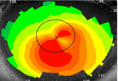
Computer-assisted videokeratoscopes, which generate color-coded maps and topographic indices, are currently the most widespread devices for confirming the diagnosis of keratoconus. With such devices, keratoconus appears as an area of increased surface power surrounded by concentric zones of decreasing surface power. Three features are common to keratoconus videokeratographs that use sagittal topography, a localized area of increased surface power, inferior-superior power asymmetry, and skewed steep radial axes above and below the horizontal meridian, depicting irregular astigmatism, the hallmark of keratoconus (2-4,7,8).
Figure 8. Typical features of keratoconus seen on sagittal topography, a localized area of increased surface power, inferior-superior power asymmetry, and skewed steep radial axes above and below the horizontal meridian (from our own database).
Placido-disk based computer videokeratoscopes, such as the TMS (Topographic Modeling System, Computed Anatomy, New York, NY) , have the combined features of both a keratometer and photokeratoscope, recording curvature changes in both the central and paracentral cornea, they are ideally suited for detecting subtle topographic changes present in early keratoconus and for documenting their progression by serial topographic analysis (40).
Several studies have been performed to characterize the topographic phenotype of clinically detectable keratoconus by videokeratography. The majority of patients have peripheral cones, with steepening extending into the periphery (Figure 8). The steepening in this group is usually confined to one or two quadrants. A smaller group of patients have central topographic alterations. Many central cones have a bow tie configuration similar to that found in naturally occurring astigmatism. In the keratoconus patients, however, the bow tie pattern is asymmetric, with the inferior loop being larger in most instances. In contrast to eyes having with-the-rule astigmatism, the steep radial axes above and below the horizontal meridian in keratoconus appear skewed, giving the bow tie a lazy-eight configuration.
Another pattern found in central cones is more symmetric steepening without a bow tie appearance (41, 42). The pattern is usually the same in both eyes, although it may be more advanced in one eye than in the other (3). Figure 9 depicts the typical topographic patterns of keratoconus.
Fig. 9. Classification scheme of normal videokeratographs in the absolute scale devised as a baseline to monitor topographic progression to keratoconus (41).
Top: round, oval, symmetric bow tie and symmetric bow tie (AB) with skewed radial axes.
Middle: asymmetric bow tie with superior steepening, asymmetric bow tie with inferior steepening (IS), asymmetric bow tie with skewed radial axes and superior steepening.
Bottom: inferior steepening, irregular and bow tie with skewed radial axes (AB/SRAX). Lower right figure is a schematic illustration of how to determine whether a pattern is AB/IS or AB/SRAX. A line is drawn to bisect the upper and lower lobes of the asymmetric bow tie (see solid lines), if there is no significant deviation from the vertical meridian (i.e., no skewing), the pattern is designated as AB/IS (as in bottom A); if the lines bisecting the two lobes appear skewed by more than 30° from the vertical meridian (i.e., 150° from one another), it is labeled as AB/SRAX.
Possible sources of confusion in the diagnosis of keratoconus are videokeratography patterns simulating keratoconus (videokeratographic pseudokeratoconus) (3). The most common culprit is contact lens wear (both hard and soft), which induces patterns of inferior steepening that may be very difficult to distinguish from keratoconus (43). These patterns,
however, disappear with time (1-2 month) after contact lens wear is discontinued.
Videokeratographic pseudokeratoconus may also result from technical errors during videocapturing, such as inferior eyeball compression, misalignment of the eye with inferior or superior rotation of the globe, and incomplete digitization of mires, causing formation of dry spots, which simulates inferior steepening (2,3).
Developing quantitative descriptors of videokeratography patterns in keratoconus would allow for easier recognition of patterns and enable us to develop a quantitative phenotype that could be universally used to formulate minimal topographic criteria for diagnosing keratoconus. The Smolek-Klyce method results in the keratoconus severity index (KSI), which is obtained with a combination of neural network models and decision tree analysis.36 The KSI increases in a more or less linear fashion with the progression of keratoconus. Thus, this variable can be used to track the natural history of the disease. It is also possible to detect asymptomatic keratoconus when KSI reaches 15%; a KSI of 30% or higher indicates clinical keratoconus (44). The network method accurately classified keratoconus suspect cases and was found to be significandy sensitive and specific for these cases. The severity network established statistically validated numeric thresholds which can be useful to clinically monitor and document cone development (45).
Rabinowitz et al. (46) have developed three indices that distinguished eyes with keratoconus from normals: central K (descriptive of central steepening); I-S values (inferior- superior dioptric asymmetry); and R versus L (difference between right and left central corneal power). These abnormalities were similar to, but less severe than, those found in the patients with keratoconus. It is possible that these indices are descriptive of the earliest stages of keratoconus in normal eyes before they progress to keratoconus, and these abnormalities might represent variable expression of a keratoconus gene in these families. A new index has also been developed that is more specific to keratoconus and that quantifies the irregular astigmatism that typifies the keratoconus videokeratograph, the SRAX index. Corneal topography is considered suspect of keratoconus when it meets the following criteria: central keratometry value >47.2 diopters (D) or inferior minus superior (I–S) average keratometry
>1.4 D or KISA% > 60%. (46-47).
Disease detection, even at early stages, has become increasingly important particularly in an attempt to prevent iatrogenic ecstasia formation – the lost of corneal shape – which has been widely documented in patients with subclinical forms of keratoconus who have undergone refractive surgery procedures. For this reason, several, above not mentioned index-based classification methods build on corneal topography systems for grading the severity of keratoconus have been developed.
1.5.3. Elevation based topography
Measurement of Placido-disk based corneal topography and central corneal thickness are widely used methods in the diagnosis of keratoconus, however they are of limited use.
Placido-disk based corneal topography only examines the anterior surface of the cornea and alteration in the reference point or viewing angle may result in inaccuracy of curvature measurement (48-51). Ultrasound pachymetry, which is widely used for the measurement of central corneal thickness, is a contact device and precise measurement depends on correct probe alignment and centration (52).
With the advent of Orbscan slit-scanning topography (Bausch & Lomb, Orbtek Inc., Salt Lake City, UT) and the Pentacam Comprehensive Eye Scanner (Oculus, Wetzlar, Germany) anterior and posterior corneal surface elevation data measurement and pachymetry map detection have become possible. Height data give a more accurate representation of the true shape of the corneal surface because they are independent of axis, orientation and position (7,8,48,49). Both the Orbscan and the Pentacam have the advantage of being non- contact methods.
1.5.3.1. Orbscan
Another technique, slit-scanning topography/tomography, scans the entire surface of the cornea; it measures elevation and pachymetric parameters, the ACD, and the anterior chamber angle of the eye. It is based on the principle of measuring the dimensions of a slit‐scanning beam projected on the cornea. Orbscan II and newer versions have a Placido- disc attachment in order to obtain curvature measurements directly. The latest hardware upgrade, Orbscan IIz, can be integrated with a Shack Hartmann aberrometer in the Zyoptix workstation. This integrated system offers total wavefront analysis through the 5th order and identifies the total aberrations of the eye. The Orbscan IIz scans the entire surface of the cornea and acquires over 9000 data points in 1.5 seconds. The curvature of the anterior and posterior surfaces of the cornea can be assessed along with the anterior surface of the lens and the iris. Mapping of the iris in conjunction with posterior surface corneal topography allows an estimation of the iridocorneal angle of the eye. Some studies demonstrated, that Orbscan seemed to underestimate corneal thickness readings in eyes with keratoconus and in eyes after excimer laser keratorefractive surgery (52-57).
1.5.3.2. Scheimpflug camera
Pentacam Scheimpflug uses the Scheimpflug principle in order to obtain images of the anterior segment. The Scheimpflug principle describes the optical properties involved in the photography of objects when their plane is not parallel to the film of the camera. It requires that the plane containing the slit beam and the image plane intersect at one point, with the corresponding angles equal. It has a rotating Scheimpflug camera that takes up to 50 slit images of the anterior segment in less than 2 seconds. Software is then used to construct a three dimensional image. A second camera captures eye movements and makes appropriate corrections. It calculates data for corneal topography (anterior and posterior corneal surface) and thickness, anterior chamber depth (ACD), lens opacification and lens thickness. It allows the measurement of local elevation points by fitting the corneal shape to a best fit sphere reference surface with variable diameters or to an ellipsoid surface. Examination of the posterior corneal surface is important in the early diagnosis of keratoconus as epithelial compensation can mask the presence of an underlying cone on the anterior surface (Figure 10) (56). It also provides data on corneal wavefront of the anterior and posterior corneal surface using Zernike polynomials. A newer version has recently become available, the Pentacam HR. In addition to a higher resolution camera, it has phakic intraocular lens (IOL) software that simulates the position of the proposed lens. The quality of the lens data depends on the pupil size, as only part of the lens can be examined through the pupillary aperture (53,55,58- 60). Several studies have evaluated the reproducibility and repeatability of Scheimpflug imaging devices, which have been shown to give reliable corneal thickness measurements in eyes with keratoconus (52,57).
There are few studies of other devices useful for the detection of keratoconus. The most common method for measuring central corneal thickness (CCT) is ultrasound biometry;
however, this requires a contact device and precise measurement depends on correct probe alignment and centration. Although US biomicroscopy allows detailed measurement of the cornea and anterior chamber, it also requires contact with the eye (52). Several other methods, including optical coherence tomography, specular microscopy and partial coherence interferometry were recently developed to measure the cornea and anterior chamber (54,57).
Figure 10. Typical features of keratoconus seen on an anterior elevation map of the Pentacam, an increased positive elevation (65µm) can be seen at the apex of the cone.
Keratometric and pachymetric data are depicted at the left side of the image (from our own database).
1.5.4. Wavefront aberrometry
Measurement of Placido-disk based corneal topography and central corneal thickness are currently the most widely used methods in the diagnosis of keratoconus. In more advanced cases this is not complicated, because of the typical biomicroscopic and topographic findings, but the detection of subclinical or forme fruste cases with corneal topography alone may impose difficulty. Understanding the characteristics of ocular higher-order aberrations due to keratoconus might be very useful to differentiate early cases from simple myopia or myopic astigmatism (61,62). Corneal higher-order aberrations in keratoconus have been investigated by several authors. This latter is mathematically calculated from corneal topographic elevation data and does not take into account the internal ocular aberrations. The advantage of measuring corneal aberrations is its ability to analyze most of the anterior corneal surface, it allows a better understanding of the optical behavior of the cornea (63-66).
Aberrometry uses wavefront sensing, which is a technique of measuring the complete refractive status, including irregular astigmatism, of an optical system. Light is defined differently in geometrical and physical optics. In geometrical optics, the rays from a point source of light radiate out in all directions. Light coming from infinity is considered to be
linear bundles of light rays. In physical optics, on the other hand, light is expressed as a wave, and the light waves spread in all directions as a spherical wave. The wavefront is the shape of the light waves that are all in-phase. Light coming from infinity is expressed as proceeding as a plane wavefront. A wavefront aberration is defined as the deviation of the wavefront that originates from the measured optical system from reference wavefront that comes from an ideal optic system. The unit for wavefront aberrations is microns or fractions of wavelengths and is expressed as the root mean square or RMS. The purpose of wavefront analyses of the eye is to evaluate the optical quality of the eye by measuring the shape of its wavefront as wavefront aberrations. For this, an aberrometer or wavefront sensor is used, and for measuring the corneal wavefront aberrations, a corneal topographer is used.
The shape of the wavefront can be analysed by expanding it into sets of Zernike polynomials. The Zernike polynomials are a combination of independent trigonometric functions that are appropriate for describing the wavefront aberrations because of their orthogonality. The first to sixth orders Zernike polynomials are shown graphically in Figure 11. The zero order has one term that represents a constant. The first order has two terms that represent tilt for the x and y axes. The second order includes three terms that represents defocus and regular astigmatism in the two direction. The third order has four terms that represent coma and trefoil, and similarly, the fourth order has five terms that represent tetrafoil, secondary astigmatism and spherical aberration.
Figure 11. The first to sixth orders Zernike polynomials shown graphically (67).
The polynomials can be expanded up to any arbitrary order if a sufficient number of measurements are made for the calculations. Spectacles can correct for only the second order aberrations, and not the third- and higher-orders that represent irregular astigmatism.
Monochromatic aberrations can be evaluated quantitatively using the Zernike coefficients for each term.
Although the total HOAs can be used to estimate the severity of deterioration of optical quality of the eye as the diagnostic purposes, it will be essential for the surgical treatments to quantify the details of wavefront of the eye using Zernike expansion or Fourier expansion.Wavefront aberrations caused by the anterior and/or posterior corneal surfaces can be calculated using the height data of the corneal topographers such as videokeratoscopes or slit-scanning corneal topographers (67).
1.6. Treatment of Keratoconus
1.6.1. Contact lenses
As irregular astigmatism can not be corrected with spectacles, contact lens is the most widly used optical correction method of keratoconus. Although contact lenses for keratoconus are manufactured with hydrogel, silicone hydrogel, gas permeable and hybrid (i.e., rigid centre and soft skirt) materials, gas permeable contact lenses remain the most commonly used contact lens type, as high levels of irregular astigmatism cannot normally be corrected with other contact lens types. Piggy back systems, consisting on the fitting a gas permeable on top of a soft contact lens, have also been used for keratoconus management. The soft contact lens is used to improve wearing comfort and provide a more regular area for the gas permeable contact lenses to sit, whereas the gas permeable contact lens is primarily used for providing adequate visual acuity. The use of high oxygen permeability soft (i.e., silicone hydrogel) and gas permeable contact lenses is highly recommended for keratoconus management as these corneas are well known to be compromised (2, 71-73).
1.6.2. Corneal crosslinking
Crosslinking is a widespread method in the polymer industry to harden materials and also in bioengineering to stabilize tissue. Using UVA at 370 nm and the photosensitizer riboflavin the photosensitizer is excited into its triplet state generating so-called reactive oxygen species (ROS) being mainly singlet oxygen and to a much lesser degree superoxide anion radicals. The ROS can react further with various molecules inducing chemical covalent bonds bridging amino groups of collagen fibrils. The wave length of 370 nm was chosen because of an absorption peak of riboflavin at this wavelength (74,75).
The first clinical study on the crosslinking treatment of keratoconus was performed by Wollensak. In this 3-year study, 22 patients with progressive keratoconus were treated with riboflavin and UVA. In all treated eyes, the progression of keratoconus was at least stopped (‗freezing‘). In 16 there was also a slight reversal and flattening of the keratoconus by two diopters. Best corrected visual acuity improved slightly in 15 eyes (74).
Crosslinking treatment of keratoconus is a very promising new method of treating keratoconus. At the present stage of knowledge, the treatment should only be performed in patients with documented progression of keratoconus in the preoperative months. With more long-term experience, prophylactic treatment of keratoconus at an early stage might become possible. Additional refractive corrections can also be considered if necessary. In case a recurrence of keratoconus progression should occur in the long run, which has not been observed so far, a second crosslinking procedure might be a choice.
However safe and promising technik CXL is, the proper determination of inclusion criteria may significantly reduce the complications and failures. A preoperative maximum K reading less than 58.00 diopters may reduce the failure rate to less than 3%, and restricting patient age to younger than 35 years may reduce the complication rate to 1% (76). As postopertiv complication stromal infiltrations and moderate anterior chamber inflammation has been described and diffuse lamellar keratitis (DLK) after myopic laser in situ keratomileusis. Herpes simplex keratitis and iritis recurrence has been also associated with the intervention (76).
1.6.3.1. Surgical interventions: ICRS
Intrastromal corneal ring segments (ICRS) were initially developed for the correction of mild to moderate myopia and are now considered in the management of keratoconus and
other ectatic disorders, such as ectasia after laser in situ keratomileusis and pellucid marginal degeneration. The ICRS acts as a passive element that flattens the central cornea by an arc- shortening effect on the corneal lamellae structure (Figure 12). Although ICRS implantation is an effective tool in managing corneal ectatic disease, several complications have been described. These include incomplete tunnel creation, anterior or posterior corneal perforation, epithelial defects, segment extrusion, and induced astigmatism by central migration of the ICRS along the horizontal corneal diameter. In most of these cases, ICRS explantation is mandatory and it is possible that a more appropriate ICRS can be implanted later. A main advantage of ICRS implantation is its potential reversibility because the technique does not require tissue removal; this has been shown in eyes with low to moderate myopia.
The outcomes of corneal remodeling by ICRS are mainly dependent on the biomechanical properties of the corneal tissue. The corneal response depends on the magnitude of the force and on the velocity of the application of the force. A specific ICRS inducing a specific force on the cornea will generate different changes in the ectatic corneal profile, depending on the underlying corneal biomechanical status. Theoretically, the cornea should have the ability to return to its original state after removal of the application of force.
In addition, it should be able to be remodeled when new forces are applied (ie, after implantation of a new ICRS). (77,78).
Figure 12. Clinical picture of an after ICRS implantation (79).
1.6.3.2. Surgical interventions: keratoplasty
The refractive error caused by the ectatic cornea is initially managed with either spectacles or contact lenses. When ectasia progresses to the point where contact lenses no longer provide useful vision, then surgical intervention may be considered. Penetrating keratoplasty is the most commonly performed surgical procedure for ectatic corneas, but is associated with complications including graft rejection, induced astigmatism, complications
of intraocular surgery such as glaucoma, cataract formation, retinal detachment, cystoid macular edema, endophthalmitis, and expulsive hemorrhage. To avoid these complications, new methods such as lamellar keratoplasty (LKP) have evolved. LKP has the advantages of being extraocular and reversible if tissue complications occur. Another advantage includes the ability to replace only selected areas of diseased corneal tissue with healthy donor tissue. LKP results, however, may be limited by vision-reducing graft-host interface problems and the technical nature of the surgical procedure (80,81). In keratoconus, conventional penetrating keratoplasty has long been associated with good long-term outcomes with graft survival of over 90% up to 13.8 years postoperatively. Visual outcomes have also been good with 73.2%
achieving BCVA over 20/40 (82,83).
Replacing weakened corneal tissue by corneal transplantation provides a permanent means of substituting, or augmenting of weakened corneal tissue, and the various forms of keratoplasty for ectatic corneal disease may be largely divided into penetrating or lamellar keratoplasty procedures, and central or peripheral keratoplasty procedures according to the precise site of tectonic weakness of the cornea. Recent innovations in lamellar keratoplasty address issues in the management of corneal ectasia. Nevertheless, corneal endothelial replacement is generally unnecessary in keratoconus, which is primarily a corneal stromal disorder, and as the major causes of long-term graft failure and attrition are endothelial in nature, an anterior lamellar keratoplasty approach to keratoconus which avoids unnecessary replacement of normal recipient endothelium is ideal. Penetrating keratoplasty has in the past been associated with better postoperative vision, due in part to the problem of interface haze and irregularities in lamellar keratoplasty (84).
New automated surgical technologies in anterior segment surgery, which have largely been developed for corneal refractive surgery, are now being utilized in keratoplasty surgery, and are able to partially substitute for conventional manual surgical procedures, providing greater precision and reproducibility in lamellar corneal dissection. These include an automated microkeratome-assisted lamellar keratoplasty device and femtosecond laser- assisted lamellar keratoplasty (85).
1.7. Other diseases affecting biomechanical properties of the cornea
1.7.1. Pellucid marginal degeneration
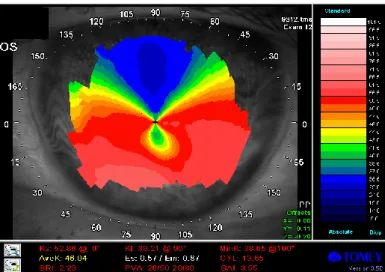
Pellucid marginal degeneration (PMD) is characterized by a peripheral band of thinning of the inferior cornea from the 4 to the 8 o‘clock position. There is 1–2-mm uninvolved area between the thinning and the limbus. The corneal protrusion is most marked above the area of thinning, and the thickness of the central cornea is usually normal. Like keratoconus, pellucid marginal degeneration is a progressive disorder affecting both eyes, although eyes may be asymmetrically affected. In moderate cases it can easily be differentiated from keratoconus by slit-lamp evaluation because of the classical location of the thinning. In early cases the cornea may look relatively normal, and in advanced cases it may be difficult to distinguish from keratoconus because the thinning may involve most if not all of the inferior cornea. In both instances videokeratography is very useful to make the distinction. The videokeratograph has a classical ―butterfly‖ appearance (Figure 13), demonstrating large amounts of against-the-rule astigmatism (2-4).
Figure 13. Corneal topography of a patient with PMD. The classical “butterfly” appearance can be clearly seen on the axial map (from our own database).
Pellucid marginal degeneration can be differentiated from other peripheral corneal thinning disorders, such as Terrien‘s marginal degeneration, because the area of thinning is always epithelialized, clear, avascular, and without lipid deposition. Terrien‘s corneal degeneration affects a similar age group and also causes high astigmatism; however, it may affect both the superior and inferior cornea and is accompanied by lipid deposition and
vascular invasion. Videokeratography can also be used to differentiate these two disorders because they have distinctly different topographic patterns (86).
Because of the large amounts of against-the-rule astigmatism, patients with pellucid marginal degeneration are much more difficult to fit with contact lenses than patients with keratoconus, although spherical or aspheric contact lenses with large overall diameter should initially be attempted in early-to-moderate cases. Surgery may be considered for patients whose vision is not adequately corrected by contact lenses or in patients who are contact lens- intolerant. Patients with pellucid marginal degeneration, however, are typically poor candidates for penetrating keratoplasty for two reasons. First, thinning occurs so near the limbus that the donor cornea must be placed very close to the corneal limbus, thus increasing the chances of graft rejection. Second, because of the extreme thinning and the location of the thinning, penetrating keratoplasty typically induces large amounts of postoperative astigmatism, which may be extremely difficult to correct because of disparity in graft-host thickness.
Crescentic lamellar keratoplasty is a useful initial surgical procedure in patients with pellucid marginal degeneration. This procedure involves removing a crescentic inferior layer of ectatic tissue by lamellar dissection and replacing it with a thicker lamellar donor graft.
This will, in most cases, eliminate large amounts of against-the-rule astigmatism. In some cases, the patient may become contact lens-tolerant, thus obviating a full-thickness procedure (87).
1.7.2. Keratoglobus
Keratoglobus is a rare disorder in which the entire cornea is thinned most markedly near the corneal limbus, in contrast to the localized thinning centrally or paracentrally in keratoconus. The cornea may be thinned to as little as 20% of normal thickness, and it assumes a globular shape. In advanced keratoconus, the entire cornea can also be thinned and globular-shaped, making it difficult to distinguish these two entities. However, even in very advanced keratoconus there may be a small area of uninvolved cornea superiorly that approaches normal corneal thickness (2-4).
Keratoglobus is bilateral, but it is usually present from birth and tends to be nonprogressive. It can be distinguished from megalocornea and congenital glaucoma because the cornea is usually of normal diameter. It is a recessive disorder and is often associated with blue sclerae and other systemic features, in contrast to keratoconus, which is most commonly an isolated disorder. In contrast to keratoconus, the corneas in keratoglobus are prone to
corneal rupture from even minimal trauma. Thus, hard contact lenses are contraindicated and protective spectacles should be strongly encouraged. If the cornea is extremely thin, a tectonic limbus-to-limbus lamellar keratoplasty should be considered to strengthen the cornea. A subsequent central penetrating keratoplasty may be considered if adequate visual rehabilitation cannot be achieved with glasses (88).
1.7.3. Post-LASIK ectasias
Corneal ectasia after LASIK is a progressive corneal steepening, usually inferiorly, with an increase in myopia and astigmatism, loss of uncorrected visual acuity, and often loss of best-corrected visual acuity that can present days to years after LASIK. The actual incidence remains undetermined, and no good data support firm predictions; previous estimates, however, have ranged from 0.04% to 0.6%. Among members of the International Society of Refractive Surgery (ISRS) of the American Academy of Ophthalmology (AAO) responding to the practice patterns survey in 2004, more than 50% had at least one case of ectasia develop in their practice. Approximately 50% of cases present within the first 12 months, but late onset ectasia can also occur (89).
Corneal refractive surgery alters the effective shape, thickness, curvature, and tensile strength of the cornea. The specific mechanisms resulting in post-LASIK ectasia remain undetermined, although complex biomechanical modeling that takes into account factors such as corneal plasticity and viscoelasticity and corneal parameters such as Young's modulus, Poisson's ratio, and curvature radius, among others, may provide insight in the future (90).
LASIK inevitably reduces the tensile strength of the cornea. Among the first four reported cases, all had greater than 10 diopters of myopia preoperatively and less than 250 µm residual stromal bed thickness (RSB) postoperatively, and two patients had forme fruste keratoconus. According to recommendation a RSB of at least 250–300 µm be maintained to prevent corneal ectasia after myopic keratomileusis (91).
Most authors recognize preoperative topographic abnormalities as uniquely indicative of increased risk for post-LASIK ectasia; the definition of ‗abnormal‘, however, remains a source of great debate. Forme fruste keratoconus as defined by the Rabinowitz criteria is a risk factor for post-LASIK ectasia. Pellucid marginal corneal degeneration suspects are also at increased risk. Based on extensive review of the literature, the members of the American Society of Cataract and Refractive Surgery (ASCRS) joint committee now recommend avoiding Lasik in patients with asymmetric inferior corneal steepening or asymmetric bowtie patterns with skewed steep radial axes above and below the horizontal meridian (92).
2. Purposes
1. Currently the diagnosis of keratoconus is based on biomicroscopic findings, corneal topography and ultrasound pachymetry. Placido-disk based corneal topography only examines the anterior surface of the cornea and alteration in the reference point or viewing angle may result in inaccuracy of curvature measurement. Height data give a more accurate representation of the true shape of the corneal surface because they are independent of axis, orientation and position. The Pentacam Comprehensive Eye Scanner uses a rotating Scheimpflug camera and measures both anterior and posterior corneal surfaces by an elevation based system. It allows the measurement of local elevation points by fitting the corneal shape to a best fit sphere reference surface with variable diameters or to an ellipsoid surface. Examination of the posterior corneal surface is important in the early diagnosis of keratoconus as epithelial compensation can mask the presence of an underlying cone on the anterior surface. The purpose of our first study (I) was to evaluate the discriminating ability of pachymetric and elevation data obtained by Pentacam and to rank them according to their usefulness in the differential diagnosis of keratoconus.
2. The purpose of the second study (II) was to find the anterior chamber characteristics of mild keratoconus with the aid of the rotating Scheimpflug imaging and to evaluate trends in corneal protrusion progression. We also aimed to demonstrate that morphological alterations of the cornea are accompanied by localized anterior chamber changes.
3. Understanding the characteristics of ocular higher order aberrations owing to keratoconus might be very useful to differentiate early cases from simple myopia or myopic astigmatism. It is particularly important to detect the disease among refractive surgery candidates, because keratorefractive procedures may worsen their condition. Corneal higher order aberrations are calculated from corneal topographic elevation data and do not take into account the internal ocular aberrations. The advantage of measuring corneal aberrations relates to their ability to analyze most of the anterior corneal surface, which allows a better understanding of the optical behavior of the cornea. The limitation of this method is that it neglects the pupil area and position and it is not aligned to the line of sight (LoS), which is the most relevant reference axis for defining retinal image quality at the point of fixation. The purpose of our third study (III) was to prove our hypothesis that keratoconus causes a shift in the LoS, which acts as a compensating mechanism for increased corneal higher order aberrations. To test our hypothesis, we examined location of the LoS, its position relative to the pupil center, and their impact on ocular higher order aberrations in keratoconic and control
eyes. Taking into account the changes of the LoS allows a better understanding of the pathologic optics of keratoconus, and may have a beneficial effect on increasing the precision of correcting higher order aberrations. According to our knowledge, no such comparisons were performed in previous articles.
3. Methods
3.1. Demographic data
In the first and second studies (I, II) forty-one eyes of 24 patients with mild to moderate keratoconus were included. In the control group, 70 eyes of 41 refractive surgery candidates were included. Among the forty-one eyes with keratoconus 18 patients had binocular keratoconus, 2 patients had unilateral disease, in 3 patients the fellow eye has already undergone penetrating keratoplasty. The control group consisted of 41 normal individuals, of 29 patients both eyes could be evaluated, of 12 patients only one eye could be included in the study, because of previous refractive surgery or corneal disease of the fellow eye. Table 1.A. shows the patients‘ demographic data. There were no statistically significant differences between the keratoconus and the control groups in age or sex distribution (p>0.05).
Table 1.A. Demographic features of the patients (I-II), difference statistically significant (p<0.05)
Subjects Eyes (n) Age (y)
mean±SD
Gender (M/F)
Keratoconus 24 41 38.7±14.1 15/9
Controls 41 68 40.2±15.1 24/17
p 0.61 0.21
Table 1.B. Demographic features of the patients (III), difference statistically significant (p<0.05)
Subjects Eyes (n) Age (y)
mean±SD
Gender (M/F)
Keratoconus 30 50 31.5±8.2 13/17
Controls 50 100 30.3±10.9 22/28
p >0.05 >0.05
In the third study (III) (Table 1.B.) fifty-five eyes of 30 patients with mild to moderate keratoconus and 100 eyes of 50 refractive surgery candidates with normal corneas were included. Among keratoconus patients, 25 had binocular keratoconus, 2 patients had unilateral disease, in 3 patients the fellow eye has already undergone penetrating keratoplasty. The control group consisted of 100 healthy eyes of 50 refractive surgery candidates. There were
no statistically significant differences between the keratoconus (mean age: 31.5±8.2 years, 13 men, 17 women) and the control groups by Mann–Whitney nonparametric test (mean age:
30.3±10.9 years, 22men, 28 women) in age or sex distribution (p=0.1).
3.2. Patient inclusion criteria
Both eyes of every patient have undergone a complete ophthalmologic evaluation including slit-lamp biomicroscopy, keratometry, retinoscopy, ophthalmoscopy and Placido- disk based videokeratography. Keratoconic patients included in the study were either mild or forme fruste cases. The criteria for diagnosing keratoconus were defined as the existence of central thinning of the cornea with Fleischer ring, Vogt‘s striae or both by slit-lamp examination. Forme fruste keratoconus was diagnosed when an abnormal, localized steepening was observed by corneal topography without any slit-lamp findings.
Both eyes of every subject were used in the study, except for those with previous ocular surgery, trauma or other pathology. Control subjects were age matched and had a refractive error of less than ± 5 spheric diopters, and/ or astigmatism less than 3 diopters.
Patients who wear rigid contact lenses, were asked to stop using them for 4 weeks, and soft contact lenses were ceased for at least one week before assesment.
From all patients, informed consent was obtained after the characteristics of the study was explained. A review by the local ethics committee was not required. For all study procedures, the tenets of the declaration of Helsinki were followed.
3.3. Patient exclusion criteria
Subjects with corneal scarring, previous ocular surgery, trauma or any other ocular diseases except for keratoconus and refractive errors were excluded from the study.
Severe cases were excluded, because of potential stromal haze or scar formation, which may alter the optical transparency of the cornea and image acquisition of the Pentacam.
3.4. Corneal topographic measurements
Corneal topographic measurement were taken with the Tomey corneal topographer (Tomey Topographic Modeling System software version-4, Tomey Corp, Nagoya, Japan).
Both topographic and wavefront aberration images were taken at 5 to 10 seconds after a complete blink, when the tear film layer is the most stable (93).
3.5. Pentacam measurements
All eyes were examined with the Pentacam HR (Oculus Inc.), used by three trained examiners. The readings were taken as recommended in the instruction manual. Briefly, the patients were instructed to keep both eyes open and fixate on the black target, in the center of the blue fixation beam. After attaining perfect alignment, the instrument automatically took 25 Scheimpflug images within 2 seconds. For each eye one high quality image was recorded.
In the first study (I) the following data were then exported to a Microsoft Excel spreadsheet:
keratometry values in the flat (K1) and steep (K2) meridian, corneal astigmatism (cylinder), corneal thickness at the center (central pachymetry) and at the thinnest point of the cornea (minimal pachymetry), the distance of the thinnest point of the cornea (apex of the cone in keratoconic patients) from the center of the cornea in the vertical meridian (y) and local elevation (anterior elevation, posterior elevation) values. For height data measurement, the best fit sphere (BFS) was used as a reference body, using the float option over a 9 mm fit. The float map means, that the reference body has no fixed center, the distance between the cornea and the sphere surface is optimized to be as small as possible. Elevation maps show the difference in height between cornea and reference body, their value is positive, when the measured point of the cornea is above the reference body, and negative, when it lies below.
Anterior and posterior elevation data were read as the maximum values above the BFS in the central 5 mm of the cornea.
In the second study (II) the following data were exported for further analysis: the ACD measured from the posterior corneal surface centrally, the ACD at the thinnest point of the cornea, and the ACD 1.0 mm, 2.0 mm, and 3.0 mm inferior–paracentral. For local posterior
elevation measurements, a toric ellipsoid was used as a reference body. The elevation maps from the device show the difference in height between the cornea and reference body; the value is positive when the measured point of the cornea is above the reference body and negative when it is below. Posterior elevation data were read as the maximum values above the toric ellipsoid surface in the central 5.0 mm of the cornea, which in the keratoconus eyes in this study was at the thinnest point of the cornea. The mean keratometry values and the smaller of the 2 chamber angles in the horizontal section calculated from the 3-dimensional model were also determined and processed for analysis.
3.6. Hartmann-Shack aberrometry measurements
All eyes were examined with the WASCA Hartmann-Shack wavefront sensor (Carl Zeiss Meditec AG, Jena, Germany) used by three trained examiners (III). The readings were taken as recommended in the instruction manual. Briefly, the patient positions his or her head on a chin rest and fixates on the center of a circular grid, which is optically fogged. The fixation target and the probe beam were coaxial, located at infinity in order that the retinal image of the narrow probe beam will remain centered on the retinal image of the fixation target (94). The operator manually aligns a reference box on a video monitor with the pupil.
This comprises a set of six symmetrical infrared diodes, which are seen as reflections off the corneal surface on the control monitor. The anterior-posterior focus point of measurement is determined by bringing into focus these corneal reflexions. This is achieved by first moving the instrument towards the patient and slowly drawing it back until the optimal focus is found.
Measurements are taken until the criteria of measurement selection are met. These are: 1, the CCD spot array is perfectly centered within the reference box both horizontally and vertically.
2, to ensure that there is no, or as little as possible, missing data within the analysis zone (95).
Wavefront aberration measurements were standardized to a 4.5 mm pupil. We used no dilating drop, as the aberration profile of a pharmacologically dilated pupil is less relevant to the natural pupil view normally experienced by the patient and in order to avoid instrument myopia (96). Wavefront measurements were performed in a dark room. We used the right- hand coordinate reference frame and the double-index convention for naming the Zernike coefficients and polynomials recommended by the OSA/VSIA Standards Taskforce (97,98).
The signs of bilaterally asymmetrical Zernike coefficients of the left eyes affected by enantiomorphism were reversed in order to allow comparison between right and left eyes (99).