Evaluation of changes in corneal morphology and sensory functions in patients with keratoconus
PhD thesis
Lóránt Dienes, MD
Semmelweis University
Doctoral School of Clinical Medicine
Consultant: Illés Kovács, MD, PhD
Official reviewers: Nóra Szentmáry, MD, PhD Katalin Gombos, MD, PhD
Head of the final examination comittee: László Tamás, MD, PhD Members of the final examination committee: Balázs Varsányi, MD, PhD
Ákos Lukáts, MD, PhD
Budapest
2017
1 Table of contents:
1. The list of Abbreviations ... 3
2. Introduction ... 5
2.1 Corneal layers and innervation ... 6
2.1.1. Corneal sensory nerves and receptors ... 9
2.2. Corneal degenerations and ectasia ... 12
2.3. Keratoconus ... 12
2.3.1. Prevalence ... 13
2.3.2. Etiology and genetics ... 13
2.3.3. Subjective and clinical signs of keratoconus ... 14
2.3.4. Patomechanism and pathology of keratoconus ... 15
2.3.5. Diagnostics of keratoconus ... 18
2.3.5.1. Slit lamp ... 18
2.3.5.2. Corneal topography ... 18
2.3.5.3. Scheimpflug imaging ... 19
2.3.5.4. Anterior segment optical coherence tomography (AS-OCT) ... 20
2.3.6. Keratoconus staging and classification systems ... 21
2.3.6.1. Rabinowitz classification ... 22
2.3.6.2. Amsler-Krumelich classification ... 25
2.3.6.3. Classification based on corneal topography and tomography imaging ... 26
2.3.7. Treatment options of keratoconus ... 35
2.3.7.1. Spectacles ... 36
2.3.7.2. Contact lenses ... 36
2.3.7.3. Radial keratotomy ... 37
2.3.7.4. Intra stromal corneal ring segments ... 38
2.3.7.5. Phakic intra ocular lenses ... 39
2.3.7.6. Photorefractive keratectomy ... 39
2.3.7.7. Anterior lamellar keratoplasty/deep anterior lamellar keratoplasty .... 40
2.3.7.8. Penetrating keratoplasty ... 41
2
2.3.7.9 Collagen cross linking treatment ... 42
2.3.8. Importance of keratoconus diagnostics before refractive surgery ... 43
2.3.9. Keratoconus and corneal nerves... 44
3. Objectives ... 46
4. Methods ... 47
4.1.1. Patients ... 47
4.1.2. Scheimpflug imaging in evaluation of intereye corneal asymmetry ... 48
4.1.3. Statistical analysis in evaluation of intereye corneal asymmetry ... 49
4.2.1. Corneal esthesiometry ... 49
4.2.2. Assessment of dry eyesymptoms with OSDI score ... 51
4.2.3. Measuring non-invasive tear film breakup time (NI-BUT) ... 51
4.2.4. Schirmer test... 52
4.2.5. Statistical analysis ... 52
5. Results ... 53
5.1. Between eye corneal asymmetry in normal subjects and in keratoconus patients ... 53
5.2. Corneal sensitivity esthesiometry and dry eye symptoms in keratoconus patients. ... 57
6. Discussion ... 65
7. Conclusions ... 71
8. Summary/Összefoglalás ... 73
9. Bibliography ... 75
10. Bibliography of the candidate’s publications ... 91
11. Acknowledgements ... 92
3 1.The list of Abbrevations
ALK-Anterior Lamellar Keratoplasty
AS-OCT-Anterior Segment Optical Coherence Tomography AST-Keratometric Astigmatism
BAD-Belin/Ambrosio Enhanced Ectasia Display BCVA-Best Corrected Visual Acuity
BFS- Best Fit Sphere
CAST-Calpastatin-Calcium-Dependent Cysteine Protease Inhibitor COL5A1-Collagen Type V Alpha1
COL4A3-Type IV Collagen Alpha3 COL4A4-Type IV Collagen Alpha4 CTSP-Corneal Thickness Spatial Profile CXL-Corneal/collagen Cross-Linking, dDALK-Descemetic Deep ALK
Dk-P = Dk = Diffusion (D) * Oxygen Solubility (k) DOCK9-Dedicator of Cytokinesis 9
FNDC3B-Fibronectin Type III Domain Containing 3B FOXO-Forkhead Box O1
HGF-Hepatocyte Growth Factor ICRS-Intra Corneal Ring Segment ICL-Implantable Contact Lens IL1A-Interleukin 1 Alpha IL1B-Interleukin 1 Beta
IL1RN-Interleukin 1 Receptor Antagonist IOL-Intraocular Lens
KC-Keratoconus
KCI-Keratoconus Index
KISA- keratometry, I-S, skew percentage, astigmatism KPI- keratoconus prediction index
KSI-keratoconus severity index LOX-Lysyl Oxidase
4
MPDZ-NF1B-Multiple PDZ Domain Crumbs Cell Polarity Complex Component NI-BUT-Non Invasive- Break Up Time
OSDI-Ocular Surface Disease Index PK-Penetrating Keratoplasty
PRK-Photorefractive Keratectomy
PTI- Percentage Thickness Increase from Thinnest Point
RAB3GAP1-RAB3 GTPase Activating Protein Catalytic Subunit RGP-Rigid Gas Permeable Lenses
RSB-Residual Stromal Bed
SLC4A11-Solute Carrier Family 4 Member 11 SOD1-Superoxide Dismutase 1
SRAX- Relative Skewing of the Steepest Radial Axes TGFBI-TGF Beta-Induced
UCVA-Uncorrected Visual Acuity USA-United States of America VSX1-Visual System Homeobox 1
WNT10A-Wingless-type MMTV Integration Site Family Member 10A ZEB1-Zinc Finger E-box Binding Homeobox 1
ZNF469-Zinc Finger Protein 469
5 2. Introduction
Keratoconus has been recognized for more than 150 years by ophthalmologists as part of the group called corneal „thinning disorder” or „corneal ectatic disease”. The name keratoconus comes from Greek word (kerato: Cornea; konos: Cone). Exact definition of the disease is not easy, but key findings for diagnosis are bilateral clinical non- inflammatory posterior ectasia with abnormal corneal thickness distribution which involves the central two-thirds of the cornea [1, 2]. Modern and more precise diagnostic tools such as corneal tomography, has increased the ability of ophthalmologist to recognise keratoconus and corneal ectasia at a much earlier stage than previously possible [3]. Regarding the increasing diagnostic potential previously established prevalence of keratoconus 50/100 000 in the general population has changed to a much higher prevalence rate 50-230/100 000 [4, 5, 6].
Global prevalence of refractive errors are increasing. Solely myopia will affect an estimated 4758 million people globally (and moreover 938 million with high myopia) by 2050 [7]. Hyperopia (8.4 % of the USA population of age 40 and older) and corneal astigmatism (1 in 3 people in the USA) also affect a significant population worldwide [8, 9].
In everyday life and during work one have to face a high amount of information. There is a need that people could process and respond to stimuli very fast during our accelerated life pace. Most of the stimuli comes through the visual system. These high standards and the spread of refractive laser procedures generate the need for perfect vision. An estimated 8,4 million people in the USA from 1995 to 2013 had undergone refractive surgery (including all types of refractive procedures) [10, 11]. Only in 2010 in the USA 800 000 refractive surgical procedures were performed [8, 12]. The most feared post-operative complication for laser refractive surgery is corneal ectasia after treatment [13, 14]. The pre-operative risk factors for post treatment corneal ectasia are high myopia, low preoperative corneal thickness, residual stromal bed (RSB) thickness less than 250 μm, younger age and keratoconus (especially forme fruste keratoconus) [15-18]. Despite the reasons detailed above at present time there is no precise and ultimate diagnostic system for early keratoconus [2].
6
Corneal nerves plays an important role in maintaining the integritiy of the human cornea. The vast majority of corneal nerves are sensory types, and their main function is to protect the ocular surface against harmful impacts. Changes in the keratoconic cornea impact all layers, and also influence the corneal nerves and their functions [1, 2, 3, 6].
Nerve dysfunction is well known for decades in keratoconus, but the exact origin and the correlation with the disease severity is unclear. Weather sensory disfunction is a cause or a consequence is still unknown [1, 2, 6]. Corneal esthesiometry could give exact and comparable information about the different type of sensory nerve functions, and could present additional information during decision making/screening.
Keratoconus screening and early diagnosis is mandatory when laser refractive surgery candidates are selected. The recognition of keratoconus plays an important role in pre- and post-operative surgery candidate management.
2.1 Corneal layers and innervation
The cornea has five definitive layers. The normal cornea is dome shaped, but more precisely it’s surface steeper in the center and flatter in the periphery. The average central corneal thickness (CCT) is approximately 550 µm, the thinnest site on the entire cornea is located approximately 0.9 mm from the visual axis, most commonly in the infero-temporal quadrant. The healthy cornea is avascular with oxygen coming mainly from the tear film and metabolic supply from the aqueous humor (Figure 1.).
7
Figure 1.: The structure of the human cornea.
(http://www.hybridcornea.org/aboutcornea.htm)
Epithelium: The outermost layer of cells that cover the outer surface of the cornea. It has a thickness of about 50 to 60 µm or 4 to 5 cell layers in thickness. These layers consist of a superficial layer of flattened cells, an intermediate layer of polyhedral cells called wing cells, and a basal germinal layer. The superficial layer cells peel off constantly and are replaced by the cells generated by multiplication in the basal layer, this cycle last about 7 days. The basal layer connected with a collagen-enriched basement membrane to Bowman’s layer. The epithelium is filled with thousands of demyelniated nerve endings that make the cornea extremely sensitive to various external (enviromental and noxious) stimuli. The primary functions of the epithelium are to provide a barrier for external materials (dust, water etc.) and bacteria; provide a smooth surface of the eye; to anchor the tear film. An injury at this level can heal without scar formation. [19, 20]
Bowman’s Layer: A thin, homogeneous, acellular, non-regenerating and transparent layer. This layer is located between the basal epithelium and the stroma and about 15 µm thick. Composed of compact collagen lamellae, these fibers are tightly connected with the stroma. The primary function is unclear, but acts as a physical barrier to protect the deeper corneal structures, and to orientate the subbasal nerve plexus. If the injury hits the level of this layer, scar formation is present. [21-23]
8
Stroma: The stroma is the thickest layer of the cornea. It represents 90% of total corneal thickness. It consists primarily of water (78%), collagen (16%), glycosaminoglycans and some keratinocytes between fibrils. Glycosaminoglycans are considered to be the “glue” of the cornea, responsible for providing plasticity and the structural support needed for successful corneal function. Along with other molecules, glycosaminoglycans form the solid portion of the cornea (22%), they provide corneal hydration, structural integrity, transparency and thickness. In normal conditions this layer is avascular, and transparent. About up to 300 regularly arranged (Type I) collagen lamellae and fibrils run parallel and extend across the entire cornea. This strict conformation of collagen lamellae and fibrils is necessary to keep the light-conductivity transparency, as well as the relatively dehydrated state. The stroma is not renewable if injured [24, 26].
Dua’s Layer: This newly discovered (in May 2013) sixth layer of the cornea, located just below the stroma. Harminder Dua and his research group were performing experiments with corneal transplants, and during corneal layer air dissection (with air bubbles) some corneas showed other type of dissection than others. Dua’s layer is very thin, only 15 microns thick. This layer could play a role in earlier unexplained corneal diseases, and could explain some earlier described pathologies but careful further research is needed. The literature is controversial about the existence of this layer. [27- 29].
Descemet’s Membrane: It’s composed of collagen fibers (Type IV.) and produced by the endothelial cells and is a true basement membrane. The layer is firm and highly elastic, but only about 10 to 12 µm thick. A tough layer, which is resistant to enzymatic degradation by phagocytes and toxins, and serves as a protective barrier against infection and injuries [30-32].
Endothelium: The thin (4 μm), innermost confluent monolayer of the cornea which cells have a polygonal shape. These cells are responsible for keeping the cornea (mainly the stroma) clear by dehydrating it, and serve as a barrier to fluid movement into the cornea. The corneal endothelium actively transports water from the stroma with active and passive ion exchangers. Critical to this energy-driven process is the role of Na+/K+ATPase and carbonic anhydrase. Bicarbonate ions formed by the action of carbonic anhydrase are translocated across the cell membrane, allowing water to
9
passively follow. The main goal is to keep the stroma ~3.5 mg H2O/mg dry or less, the stroma is highly-ransparent at these values. If this layer damaged or diseased, these cells will not regenerate and won’t multiply, and the stroma becomes edematous and hazy, at the end ultimately opaque. Cell density is about 3.500 cells/mm2 at birth and decrease gradually throughout life at about 0.6% per year and with about 10% loss per intraocular surgery. To maintain healthy stoma dehydration about of 700 cells/mm2 is required for endothelial functions and metabolism. This layer also allows nutrients and other molecules to enter the stroma, to feed the avascular corneal tissue inner part [33- 39].
The human cornea is one of the most richly innervated tissues in the body and the sensory nerves are derived from the ophthalmic division of the trigeminal nerve [40].
These nerve trunks enter the corneal stroma radially at the periphery next to the limbus.
The stromal nerve bundles contain mainly nociceptive Aδ and C fibers. Stromal nerve trunks are comprised of approximately 900–1200 myelinated and unmyelinated axons [41]. One millimeter after the limbus the myelinated fibers lose their myelin sheath, and both types of nerves are surrounded solely by Schwann cells [40, 42]. Stromal nerves are organized paralell to the corneal collagen lamellae network. Nerve density increases while nerve diameter thins as the stormal axons progress anteriorly to the superficial stroma. Some axons terminate as free nerve endings, others directly innervate keratinocytes [43, 44]. Superficial stromal axons penetrate the Bowman layer into the epithelium predominantly at the peripheral cornea, and form the subepithelial plexus.
These axons form a whorl-like pattern approximately 1 – 2.5 mm inferonasal to the corneal apex. The subbasal plexus run parallel to the corneal surface, and only beaded unmyelinated C fibers travel for a short before turning upward and terminating perpendicularly just beneath the epithelial surface as free nerve endings [45-48].
2.1.1. Corneal sensory nerves and receptors
As mentioned above, the cornea has rich sensory nerve fiber supply. Autonomic nerve fiber axons are also present, but they represent a minority and the exact function is not well understood. These nerves consist of sympathetic fibers that are derived from the superior cervical ganglion and parasympathetic fibers that originate from the ciliary
10
ganglion [49-52]. Sensory nerves mainly derived from the ophthalmic division of the trigeminal nerve. They have a variety of sensory and efferent functions, sensations result from the activation of sensory nerve afferents, which are the peripheral branches of various types of trigeminal nociceptive neurons. Corneal nerve stimulation produce predominantly a sensation of pain in humans but it is thought to depend on the modality of stimulus acting on the cornea [53-55]. These axons ensure and maintain the ocular (corneal) surface integrity, perceive irritation and pain, mediate midbrain reflexes, regulate tearing and blinking, corneal nerves are responsible for ocular surface sensations and play an important role in wound healing and tear production and thus, contribute to maintaining ocular surface integrity [40].
The distribution of corneal sensory nerves is as follows, about 70% are polymodal nociceptors, 15-20% are mechano-nociceptors and about 10%-15% are cold-sensitive thermal receptors [56]. The detection of stimuli by corneal receptor terminals are the same as in sensory receptors of other tissues of the body. It depends on membrane signaling proteins which convert the external/internal stimuli into a conformational change, which lead to an alteration in ionic permeability and finally cause an electrical depolarization at the membrane of the nerve endings. The electrical potential change (depolarization) at the peripheral nerve endings generates nerve impulses centripetally to the brain. Most transduction molecules are ion channels that are directly opened by the external stimulus or gated by internal molecules or membrane proteins [53, 55, 56].
The receptors at the sensory nerve endings are part of the TRP (Transient Receptor Potential) channel superfamily. The TRP superfamily is evolutionally conserved from nematodes to mammals [57]. These receptors could be divided into five sub-groups.
The common point at the TRP family is the six-transmembrane domain unit with a non- selective cation-permeable pore between domains 5 and 6 [58]. Four of these units could form a TRP channel. The main difference between the channels is the intracellular part and cation selectivity. In human corneal nerve endings TRPV1, TRPV4, TRPA1 and TRPM8 receptors expressed mainly [59, 60, 61].
The activation mechanisms of ion channels are unique in that there are a diverse host of stimuli that can activate TRP channels and exhibit sharp differences in stimulatory modes even within each TRP channel subfamily. This means, that with different kind of excitation one can investigate different ion channels/sensory nerve endings. In other
11
words different sensation modalities linked to different ion channels and indirectly to different sensory nerve types with some overlap [54, 57, 58].
Polymodal nociceptors: TRPV1 ion channels representing mainly this sensory ending.
This receptor type activated by noxious exogen and endogen stimuli, and is likely the origin of unpleasant sensations evoked by near-noxious and injurious chemical, thermal, and mechanical stimuli acting on the cornea [56]. Temperature under 29°C and over 40°C, hyperosmolarity, acidity (pH below 6), near-noxious mechanical energy, proinflammatory cytokines (IL-6, IL-8) are activators for TRPV1 containing nerve endings [62]. TRPV4 receptors seems to be an osmosensor for a hypoosmolar challenge [63] as well. Polymodal nociceptors respond to their natural stimuli with a continuous, irregular discharge of nerve impulses that present as long as the stimulus exists. The firing frequency of the nerves roughly proportional to the intensity of the stimulating noxa. So these sensory endings not only signal the presence of unpleasant noxas, but also encodes its intensity and duration in a certain degree [56, 62-64].
Mechanonociceptors: Stretch-activated receptors were described in corneal nerve endings and in other tissues of the body. These fibers fire with low frequency in response to brief or sustained indentations of the corneal surface and, also when the stimulus is larger. They have a very low threshold force for activation, even far below of in skin of the same kind of receptors. Mechanonociceptor function is to transfer very low mechanical sensations and to protect the corneal surface by starting the blinking reflex. These receptors are probably responsible for the acute, sharp sensation of pain produced by touching the corneal surface. Henceforward presumably polymodal nociceptors (TRPV1, TRPA1) are responsible for sustained chronic pain after mechanical impacts [55, 56, 58].
Cold-sensitive thermal receptors: TRMP8 channels have been described as cold sensors in cold thermoreceptor corneal nerve endings. Thermal sensory nerves at the cornea have an ongoing spontaneous firing activity at normal conditions. The normal corneal surface temperature is about 33 °C. These nerves have an increased firing rate when the normal temperature drop below 33°C, and decreased at warming. They react to different type of cooling modalities, and increase firing rate when evaporation at the corneal surface is present or when cold solution is applied on the cornea, blowing cold
12
air at the corneal surface is also a stimulating factor. Cold receptor fibers are able to detect small temperature variations of 0.1 °C or less. They also encode cold stimuli by changing in impulse frequency, and by this method the perception of non-noxious temperature drop could be a conscious sensation. TRMP8 receptors are probably the main modulators of basal tearing rate by percepting changes in the corneal surface temperature due to evaporation of the tear film [56, 59-61, 65, 66,].
2.2. Corneal degenerations and ectasia
Corneal degenerations are defined progressive deterioration of a tissue or an organ that was previously normal. This deterioration often accompanied by loss of functional activity. Degenerations usually characterized by the deposition of material, vascularization and tissue thinning [2, 67, 68].
The definition of „Ectasia” strictly means as a dilation or distention of a tubular structure. But in ophthalmology this term refer to conditions associated with changes in corneal shape [2].
Under the definition of „corneal ectatic disease” several entities should be characterized including keratoconus, pellucid marginal degeneration (PMD), keratoglobus, and postrefractive surgery progressive corneal ectasia. These conditions could be distinguished by the thinning location and pattern [2]. Corneal ectasias are associated with decreased uncorrected visual acuity (UCVA), an increase in ocular aberrations, and often a loss of best-corrected distance visual acuity (BCVA). To characterize keratoconus as true corneal dystrophy is controversial, because the lack of strict inherited mechanism. Recently there has been find link with some genes, but sporadic cases are also present in a large scale [2, 69, 70].
2.3. Keratoconus
Keratoconus (KC) is a non-inflammatory bilateral corneal ectatic disease, involving all layers of the cornea. In most cases KC present at different stages in each eye of the patient, in other words the disease is asymmetric. Recent findings, and definitions declare that true unilateral keratoconus does not exists [1, 2]. This bilateral disease defined as a progressive thinning of the corneal layers, which involves especially the central two-thirds of the cornea. The progressive thinning causes a decrease in
13
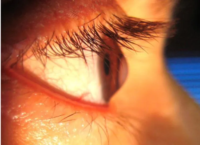
corneal/stromal biomechanical strength which leads to abnormal posterior ectasia and corneal protrusion causing abnormal corneal thickness distribution (Figure 2.) [1, 2].
Changes in the corneal curvature are responsible for myopic shift, irregular corneal astigmatism and visual disturbances at KC patients. The disease in most cases starts at the second decade of life about puberty, and rarely progress after the age of forty [73].
Mandatory findings for keratoconus diagnosis are abnormal posterior elevation, abnormal corneal thickness distribution and characteristic changes in corneal topography [2].
Figure 2.: Keratoconic eye.
(http://eyeworld.org/article-linking-keratoconus-and-floppy-eyelid-syndrome-to-sleep-apnea)
2.3.1. Prevalence
The disease is relatively common, affecting 50-230/100 000 people world wide [4, 5, 6].
It affects only in the United States approximately 300,000 patients [70-72].
2.3.2. Etiology and genetics
The exact etiology of KC is still unknown. The recent opinion about KC is that KC is a multifactorial disease caused mainly by environmental factors but it has a strong underlining genetic susceptibility [70]. Keratoconus has a very complex and not well understood nature regarding to its etiology. As described in earlier studies three entities could be distinguished:
I. The majority of KC cases reported by clinicians are isolated KC with no associations with other conditions [70, 72, 74, 75].
14
II. Increasing evidence show genetic predisposition to KC. Positive family history linked to higher odds ratio in family members for the diagnosis of keratoconus [72, 76]. GWLS (Genome-wide linkage study) and GWAS (Genome-wide association study) have made significant progress in identifying genetic variation that is strongly correlated with keratoconus.
SNPs (Single nucleotide polymorphisms ) associated with the following genes have been implicated: LOX, CAST, DOCK9, IL1RN, SLC4A11, HGF, RAB3GAP1, TGFBI, ZNF469, ZEB1, VSX1, COL5A1, COL4A3, COL4A4, FNDC3B, FOXO1, MPDZ-NF1B, WNT10A, SOD1, IL1B, IL1A, in addition to the microRNA MIR184. Notably, not all analyses of each of these genes completely confirm their role in KC [70].
III. Other conditions could be associated with KC such as Down-syndrome [70, 77], inflammatory bowel disease (IBD) [70, 78], atopic disease including vernal kerato-conjunctivitis, and atopic dermatitis. Higher incidence of KC was also reported in patients with connective tissue disorders (Marfan syndrome, Ehlers-Danlos syndrome) [70, 79, 80]. There is a reverse relationship between diabetes mellitus and KC [70, 81]. Diabetes in some circumstances could play a protective role in the progression of KC by increasing the number of corneal collagen cross-links and by altering the biomechanical properties of the cornea [82, 83].
Briefly most keratoconus cases appear spontaneously, although approximately 14% of cases present with evidence of some genetic transmission [84].
2.3.3. Subjective and clinical signs of keratoconus
Patients with KC often report eye itching, photophobia, distorted vision, glares and halos, progressive visual blur and distortion. Multiple unsatisfactory attempts to obtain optimum spectacle correction or progression from soft contact lenses to toric or astigmatism correcting contact lenses are also common warning signs. The progressively poor vision hardly corrected with spectacles is the most common complaint. These symptoms are secondary to the progressive myopia and irregular astigmatism [1, 2] due to the changes in corneal curvature.
15
Early signs including scissors reflex during retinoscopy, Rizzutti's sign (a conical reflection on the nasal cornea when light is shone temporally), and asymmetric refractive error with high or progressive astigmatism. Keratometry showing high astigmatism and irregularity are also early signs. A Fleischer ring, or iron deposits within the epithelial layer, might be found near the base of the cone. Fine and almost parallel vertical lines seen in the stroma called Vogt strias are secondary to stromal stress. In later stages of KC corneal protrusion may cause angulation of the lower lid on downgaze (Munson's sign). Corneal hydrops could cause spontaneous tears in the Descemet's membrane, and corneal scarring [1, 2].
2.3.4. Patomechanism and pathology of keratoconus
The exact etiology and trigger factors are still unknown for keratoconus. There are several studies and hypothesis exists parallel in the literature. At present time the most plausible is multifactorial etiology, with the interplay of possible genetic predisposition and a second hit by environmental/risk factors. Experts agreed some risk factors are frequent and could be linked to keratoconus.
Mechanical factors: Eye rubbing is a commonly mentioned risk factor.
According to several studies eye rubbing could cause direct micro trauma to the cornea, which activates the wound healing signaling pathway in the epithelium.
This mechanism accompanied with the activation of keratocytes and increased hydrostatic pressure in the cornea layers. This assumption explains the higher incidence of keratoconus in atopic patients (ocular allergy) or in contact lens wearers, where eye rubbing and epithelial micro trauma is common. In this group floppy eyelid syndrome and connective tissue disorders (Marfan syndrome etc.), Ehler-Danlos syndrome are also occur [1, 2, 85].
Oxidative stress: There are studies indicating an abnormal processing of the superoxide radicals in keratoconic corneas. Due to this change corneal self- repair mechanisms are not working properly or lack. Genomic deletion in the superoxide dismutase 1 (SOD1) gene is also often present [86]. An increased rate of free radicals (reactive oxygen species-ROS, reactive nitrogen species (RNS) in the corneal tissue causing direct collagen damage, consequence of this
16
collagen degradation biomechanical weakening and corneal thinning is a logical final result.
Hormonal causes: Keratoconus usually starts with puberty around in the second decade of life, and accelerated progression often seen in keratoconic patients during pregnancy. Both puberty and pregnancy accompanied by fundamental hormonal changes. This theory is controversial and has not been proven [87, 88].
Inflammation: Although keratoconus definition contains the non-inflammatory nature of the disease, recent studies show that some kind of inflammation may play a role in the pathogenesis of KC. According to studies significantly elevated levels of interleukin-6 (IL-6), tumor necrosis factor-α (TNF-α), and matrix metalloproteinase (MMP)-9 were found in the tear fluid of patients with KC [89, 90, 91]. Although this inflammation does not meet all the classic criteria for an inflammatory disease, the lack of inflammation is questionable.
Genetic associations have been already explained earlier in this work.
Briefly when keratoconus is present, all layers of the cornea are involved.
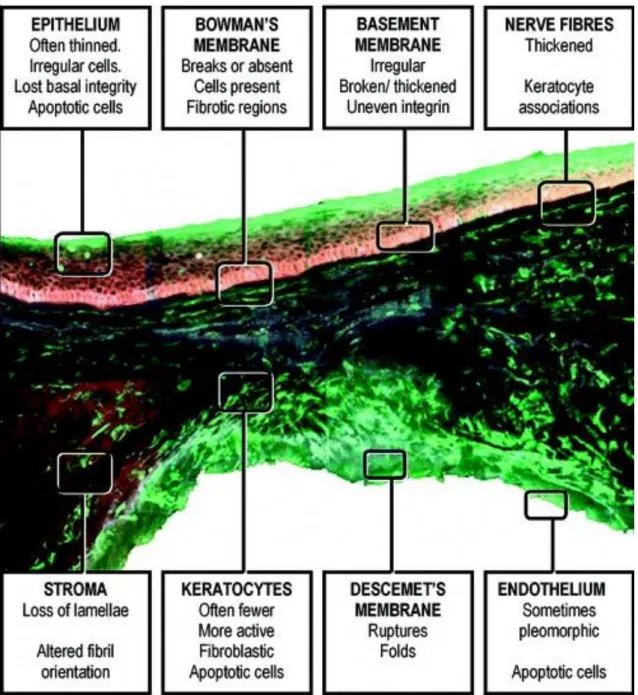
Histopathological findings are as follows: Corneal epithelial cells usually enlarge and elongate. After involvement of the basal epithelial cells disruption of the basement membrane are frequent. In later stages this degradation could be accompanied with epithelial ingrowth and collagen herniation trough the Bowman's layer forming typical Z-shaped interruptions or breaks in Bowman's layer. Bowman's layer and anterior segment scarring are also seen parallel with collagen fragmentation, fibrillation and increased fibroblastic activity. The stromal collagen has normal size, but the decreased number of collagen lamellae causing stromal thinning. Endothelial cells are also involved, and pleomorphism with polymegathism could also be manifested. Nerve fibers are also thickened, this will be explained in detail later (Figure 10). The severity of changes increase with disease duration and showing a higher grade at the apex of the cone than at the base [1, 2, 85].
Regarding to the discrepancies in studies it is hard to distinguish between association, cause and effect in keratoconus pathology.
17
Figure 10.: An anteroposterior section of the central 1 mm of a keratoconic cone from penetrating keratoplasty surgery. The tissue has been labelled with CellTracker Green (Molecular Probes) to mark viable cells and then counter-stained with antibodies to integrin (red) and fibronectin (blue). The cross-section shows some of the classical features of keratoconic pathology. Areas of the cornea are highlighted to show position and type of pathological features in keratoconus. [Morphological changes in keratoconus: Pathology or pathogenesis. Available from:
https://www.researchgate.net/publication/8634066_Morphological_changes_in_keratoconus_Pathology_
or_pathogenesis [accessed Sep 1, 2016]]
18 2.3.5. Diagnostics of keratoconus
Diagnosis sometimes could be very difficult, and need a lot of clinical experience in problematic cases. Briefly, diagnosis can be made based on history of changing refraction, poor best spectacle corrected vision, abnormalities in keratometry, corneal topography and tomography findings, in association with abnormal corneal thinning pattern. In advanced cases characteristic slit lamp findings and other signs can support prompt diagnosis of KC. In early stages of keratoconus corneal tomography (Scheimpflug imaging etc.) and the comparison of results to the other eye of the same patient as a reference (rather than artificial numbers or reference curves) are gaining popularity [1, 2, 92, 93, 94].
2.3.5.1. Slit lamp
Slit lamp biomicroscopy is a basic but necessary diagnostic tool. With the evaluation of the anterior segment KC signs could be find, including corneal thinning, Vogt strias, Fleischer ring (more easily with a cobalt blue filter) at the basis of the protrusion, and Descement tears or corneal scarring [1, 2] in more advanced forms.
2.3.5.2. Corneal topography
Most videokeratography systems used in clinical practice are based on placido disk principles. The instrument captures the projected placido disk images reflected from the corneal surface (precorneal tear film). The machine uses a central camera to capture the images from a standard point and digitizing computer software convert data to a color- coded dioptric map of the anterior cornea. The warmer colors (reds, oranges) represent steeper cornea with higher refractive power, the cooler colors (violets and blues) represent flatter cornea with lower dioptric power and greens and yellows represent colors found in normal cornea [95]. Changing the steps in color codes can cause a different look of the same cornea. The smaller steps increase the sensitivity to pick up early keratoconus, but can falsely diagnose a normal cornea as keratoconic, whereas larger steps can miss out on the early changes [95]. Different topographers use different steps of colors, making it difficult to compare two different devices.
19
Elevation is not measured directly by placido based topographers, but certain assumptions allow the construction of elevation maps for example by Orbscan.
Elevation of a point on the corneal surface displays the height of the point (in micron) on the corneal surface relative to a reference surface [95].
For a good quality and reliable scan the patient should have a stable precorneal tear film, and image acquisition requires good patient fixation and compliance to avoid eye lids covering the cornea. Videokeratography is a very useful diagnostic tool for both keratoconus screening, and KC progression follow-up, but it is incapable of capturing early KC changes of the posterior surface (posterior elevation changes).
2.3.5.3. Scheimpflug imaging
The cornea has a conic shape, therefore without using the Scheimpflug principle imaging of this tissue could lead to false results. The name of this imaging method came from Theodore Scheimpflug who worked on correcting ariel distortion in perspective photographs. Briefly, this method could give solution to a problem, when the plane of the prospective image and the plane of the object are not parallel. In this situation it will be impossible to focus all the image on a plane parallel to image plane. Thus this may lead to image distortion. But using the Scheimpflug principle when a planar subject is not parallel to the image plane, an oblique tangent can be drawn from the image, object and lens planes, and the point of intersection is called Schiempflug intersection (Figure 3). Careful manipulation of the planes (image and lens) could lead to a sharp and focused image of the non-parallel object [96, 97].
Using rotating Scheimpflug camera (Pentacam HR, Oculus Optikgerate, Wetzlar, Germany) offers significant advantages over placido based curvature analysis. This method allows for the creation of a three-dimensional reconstruction of the anterior segment by measuring not only the both surfaces of the cornea but the lens surfaces as well. Both posterior corneal elevation and corneal thickness map are significantly earlier indicators of KC and ectatic diseases than only anterior curvature and ultrasound pachymetry [98]. With this diagnostic tool ophthalmologist could have the possibility to recognize KC in a far earlier stage with less false positive or negative errors.
Scheimpflug imaging also covers significantly more of the cornea than was possible with placido based devices giving the opportunity to a more accurate diagnosis [98]. In
20
other words devices using Scheimpflug imaging become essential tools in the correct diagnosis and follow up of keratoconus. Placido-based topography analyzes the central anterior corneal surface, whereas tomography (Scheimpflug and/or optical coherence tomography) analyzes the anterior and posterior cornea and produces a near full corneal thickness map [2].
Figure 3.: Scheipflug imaging. Illustration shows Sheimpflug camera working principles, this method of image acquisition enhances the depth of focus (left) [98].
2.3.5.4. Anterior segment optical coherence tomography (AS-OCT)
Anterior segment optical coherence tomography (AS-OCT) is a noncontact imaging modality of the cornea and the anterior segment of the eye with a high resolution which can accurately map corneal thickness. This high resolution cross-sectional imaging modality first used for screening the back of the eye (the retina). [99]. A variety of high speed OCT scanners are now available that can image and measure the corneal thickness. Fourier domain technology provides the advantage of faster scan acquisition with greater axial resolution [99, 100]. This method provide a non-contact corneal pachymetric map (not just spot pachymetric data such as ultrasound pachymeters), with
21
full coverage of the cornea. AS-OCT has several benefits over placido disc based videokeratography.
Corneal thinning is a key pathologic feature of keratoconus, therefore a KC diagnosis based on corneal thickness measurement may offer additional information not available on topography [101, 102, 103, 104]. Last but not least, AS-OCT imaging provides a fast full view of the corneal surfaces. Recently, epithelial thickness profile maps using Fourier domain OCT have been shown to be useful in detecting subtle epithelial changes, which could be a sign of early keratoconus [105, 106].
2.3.6. Keratoconus staging and classification systems
At present time there is a lack of adequate classification/grading system of keratoconus [2]. Several classification systems co-exist in the literature based on different indicators.
These systems usually based on morphology, ocular signs or disease evolution. Index- based systems are also available. Experts agreed that some systems have only historical relevance at this time [1, 2, 98-101].
There is an explosion in the field of ophthalmology devices using different type of diagnostic principles (OCT, Scheimpflug imaging etc.). Currently there is no grading system that could integrate the potential of new imaging modalities into a universal and widely used system.
The prevalence of all kind of ammetropias is rising worldwide, hence the number of corneal refractive procedures is also increasing [7-12]. Before any type of laser refractive surgery, screening the candidates for the presence of KC is one of the most important task to avoid post-operative ectasia [15-18]. Therefore, there is an emerging need for an ultimate and adequate diagnostic system/method for KC.
Experts of the field agreed that new diagnostic systems should take posterior corneal elevation abnormalities into account rather than focusing solely on central pachymetry for diagnosing keratoconus [1, 2].
22
As I see in the literature functional changes like corneal sensitivity are out of focus in the diagnosis of keratoconus. But probably these functional changes prelude other signs of KC. This become from two important data reported in studies. With corneal in vivo confocal microscopy/imaging several findings shows microstructural alterations of the corneal tissue in KC, briefly derangement in the morphologic and morphometric features of central sub-basal and stromal nerves [107-112]. On the other hand there is a significant correlation reported between central corneal sensation and severity of keratoconus [113, 114]. Investigations on corneal functions (like corneal sensitivity) could forecast KC in an earlier stage than morphometrical changes.
In the followings, I describe some of the popular and widely used KC classification systems.
2.3.6.1. Rabinowitz classification
A) Rabinowitz investigated keratoconus intensively [1, 74, 115]. He described a grading system based on videokeratography findings. During the years as KC diagnostics had an evolution he and his colleagues made some refinement.
At the beginning four videokeratographic indices were described to help clinicians in discriminating normal corneas from KC:
- central corneal power >47.2 D
- inferior-superior dioptric asymmetry over 1.4 D - Sim-K astigmatism >1.5 D
- skewed radial axes >21°
B) After refinement Rabinowitz et al. made an index called KISA (keratometry, I-S, skew percentage, astigmatism). This index give a % for clinicians to discriminate KC from normal corneas more precisely [1, 74, 115, 116, 117].
The KISA% index is derived from the product of four indices: The K-value, an expression of central corneal steepening; the I-S value, an expression of the inferior- superior dioptric asymmetry; the (corneal astigmatism index), which quantifies the degree of regular corneal astigmatism (Sim K1-Sim K2); the skewed radial axis (SRAX) index, an expression of irregular astigmatism occurring in keratoconus [1, 74, 115, 116, 117]:
23
KISA%= (K) x (I - S) x (AST) x (SRAX) x 1/3
KISA% meaning:
-60%-100% are KC suspects with <0.5% chance of overlap with normal population.
-100% or higher without any other ocular pathologies is likely to have clinically detectable KC.
KISA index could support ophthalmologist in decision making when screening refractive surgery candidates.
C) The Rabinowitz keratoconus percentage index (KISA) and pachymetry/asymmetry index (PA/I-S) combines information from videokeratography and AS-OCT pachymetry measurements. With this refinement ophthalmologist could differ more precisely subclinical KC from normal corneas [114-116]. With this method indices are as follows:
K value quantifies the central corneal steepening. A value of 47.20 D or grater is suggestive of keratoconus.
I-S value quantifies the inferior-superior corneal dioptric asymmetry which is grater in KC corneas than in normal. A value of 1.4 D or greater is suggestive of keratoconus.
KISA% incorporates the K and I-S values with a measure quantifying regular and irregular astigmatism into one index. This index is highly sensitive and specific in separating normal from keratoconic corneas. See cut off values above.
PA/I-S index is the minimum pachymetry value measured with AS- OCT divided by the I-S value. The PA/I-S index allows a more sensitive detection of forme fruste and keratoconic suspects than KISA %.
24 Grading with this method:
Normal: No clinical signs of KC and no asymmetric bowtie (AB) with a skewed radial axis (SRAX) (ie, AB/SRAX) pattern on videokeratography. 95% of normals have a PA/I-S index of more than 106.
Keratoconus suspect: The fellow eye of a patient with keratoconus with mild inferior steepening on topography, no clinical signs. The average K reading is less than 47 D and PA/I-S index would have a value of less than 105.
Forme fruste keratoconus: The fellow eye of an individual with keratoconus, with AB/SRAX videokeratography pattern, and without clinical signs of keratoconus. The PA/I-S index would have a value less than 100.
Early keratoconus: No aberration connected to KC on slit-lamp examination.
Scissoring sign on retinoscopy and an AB/SRAX pattern on videokeratography.
Average K reading < 47 D, early keratoconus had a PA/I-S value between 10 and 57.
Keratoconus: Stromal corneal thinning accompanied by clinical signs of KC on slit lamp biomicroscopy.
D) According to Rabinowitz works Maeda and Klyce also created indexes to help decision making, and to gain accuracy in the diagnosis of KC. They used eight indices from topographic measurements [1, 104, 118]. In this classifier KPI (keratoconus prediction index) derived from eight quantitative videokeratography indexes. KCI%
(keratoconus index) is derived from KPI and other four indexes.
-KPI >0.23 is indicative of keratoconus.
-KCI% >0 is indicative of keratoconus
Briefly several topographic indices have been used for the interpretation of keratoconus.
Sedghipour et al. compared the sensitivity and specificity most of the topographic indices used above. They explored that while the K value and AST demonstrated >80%
sensitivity and the SRAX demonstrated >90% specificity, SRAX and AST indices had
25
the lowest sensitivity and specificity, respectively. KISA% was the only index with specificity and sensitivity >90%. Furthermore in their study KISA% was the only index demonstrating positive and negative predictive values >95% [119]. This means that KISA index is very useful detecting early/suspect KC cases, but further research is required to confirm this conclusion [119], in short there is a need for an ultimate index or cut off value to discriminate early KC with great precision.
2.3.6.2. Amsler-Krumelich classification
This grading system was one of the commonly used decision making tool in the past. It used central corneal thickness (CCT) value measured with ultrasonic pachymetry, keratometric readings, and the degree of myopia. The Amsler-Krumeich grading system (Table 1) utilized easily measured parameters and the staging followed closely the treatment decision tree [98]:
Table 1.: Amsler-Krumelich classification for keratoconus [98].
26
This method has some limitation on videokeratography and on newer devices.
Ultrasonic central pachymetry only measured one point on the cornea, which was typically not the thinnest point, and this technic did not reflect to the full thickness profile of the cornea [98]. According to experts central corneal pachymetric value is the least reliable factor in the detecting of KC [1, 2]. This system did not take posterior corneal surface (i.e. posterior elevation) into account, and did not give a picture of the properties of the anterior corneal surface witch is also a key finding in detecting KC [1, 2]. Nowadays this method has only limited value in the era of new imaging technics (videokertography, AS-OCT, Scheipflug imaging etc.).
2.3.6.3. Classification based on corneal topography and tomography imaging First we have to clear the difference between the two words topography and tomography. Topography means studying of the shape of the corneal surface (like videokeratographers- mentioned earlier in this work).
The emerging number of new corneal investigating devices using different principles (Orbscan, Pentacam, Oculyzer, Galilei, Sirius, AS-OCT-Visante etc.) led the term
"corneal tomography" used in the field of ophthalmology. This is because the images generated by new imaging devices are rather a cross section of the cornea (with elevation data analyzed further) than in contrast to enface images of concentric rings from the placido-based devices. Corneal tomography should be used for the examination of the front and back surfaces of the cornea, along with pachymetric mapping producing a three-dimensional cross section of the anterior segment of the eye [120].
Rabinowitz has described KISA % and topography devices became part of the everyday used evaluating methods between ophthalmologists. The magnitude of his work was to give a topography-based index which was derived from easily measurable and calculable topographic parameters from the corneal surface. The index based on these values express corneal surface asymmetry. With this index screening of KC was more precise and gave the opportunity to recognize it in an early stage than before.
Since then, new diagnostic techniques for the cornea like corneal tomography, wavefront analysis and biomechanical analyses have been expanded. These technics enable eye care professionals to identify keratoconus earlier than Rabinowitz would
27
probably have imagined in 1998. [121]. With these new diagnostic methods keratoconus can now be identified on a subclinical level, that is before topographic changes occur.
To analyze changes on a subclinical level, it is essential to differentiate properly between ‘normal’ eyes and those with early keratoconus stages. Another important thing is that there are certain fundamental differences in the videokeratoscopes and the Scheimpflug devices, thus the fact that their data is non-interchangeable. These devices work on totally different principles and have different methods of data acquisition, presentation and analysis [122]. Even data from devices using the same principles (placido-disc based, scanning slit beam or Scheimpflug imaging), created by different manufacturers are also not directly comparable [122].
The topographic/tomographic patterns of the two corneas of a healthy individual often show mirror-image symmetry with small variations in patterns are unique for the individual. This phenomenon is called enantiomorphism [123].
A) Classification based on corneal topography (videokeratography):
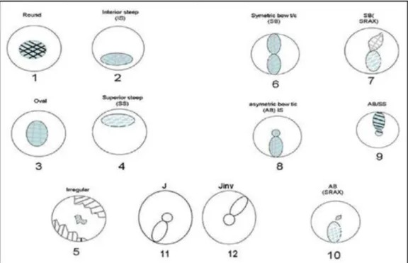
Normal cornea and corneal pathologies could be characterized by their pattern seen on videokeratographic records. With the several different indexes mentioned above ophthalmologist has the ability to distinguish between healthy and suspicious/non- healthy corneas. The distribution of keratographic patterns in healthy patients includes the following (Figure 4-5): round (23%), oval (21%), symmetric bow tie typical for regular astigmatism (18%), asymmetric bow tie (32%), and irregular (7%) [123].
Individuals with keratoconus has different types of pattern seen on topographic maps like (Figure 6): global cone, inferior cone, asymmetric bowtie, central cone, temporal cone, oblique bowtie, infero-temporal cone, nasal cone, superior cone [98, 104].
28
Figure 4.: Normal topgraphy map patterns distibution. Patterns can be classified into circular, oval, steepening (superior or inferior), bowtie (symmetric and asymmetric), and with or without skewing of the radial axes, J and the inverted J as shown in the template. The symmetrical bowtie, round, and the oval are considered normal, the asymmetric bowtie, skewed axes, inferior steepening, and J and inverted J pattern, and their various permutations as suspicious. The Pellucid (crab claw), butterfly, and the keratoconus (D) patterns are examples of abnormal patterns [121].
29
Figure 5.: Patterns of normal eyes seen on videokeratography.
(http://www.ejournalofophthalmology.com/ejo/ejo27c.html. 2016.08.23. 22:10)
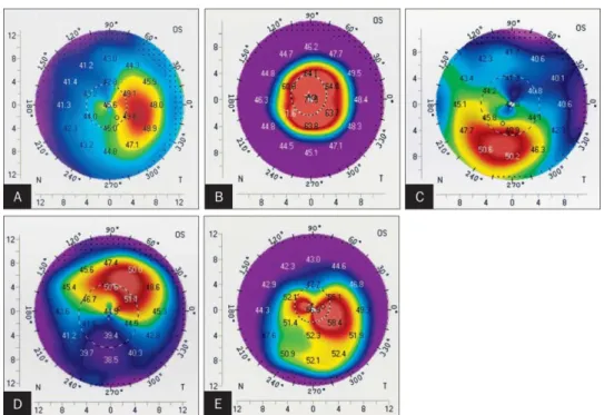
Figure 6.: Types of keratoconus based on topographic patterns A: temporal cone; B: central cone; C: infero-nasal cone; D: superior cone; E: oblique cone (Ertan A, Kamburoglu G, Colin J. Location of Steepest Corneal Area of Cone in Keratoconus Stratified by Age Using Pentacam. J Refract Surg. 2009; 25: 1012-1016. doi: 10.3928/1081597X-20091016-07 ).
30
B) Classification based on tomography (Scheimpflug imaging):
As mentioned above posterior elevation and the white to white corneal pachymetry map are the precious additions as compared to the videokeratographic (placido-based) devices. Changes in the posterior corneal surface like asymmetry, curvature and elevation differences have been reported in keratoconic eyes by several studies [124, 125, 126, 127]. Briefly these works find greater posterior astigmatism, posterior elevation, and prolacity in suspect eyes when compared to healthy [128]. The consensus about exact values in discriminating KC eyes from normal is lacking. Values extracted from devices using the same imaging principle are also non-comparable, which makes the whole decision making more complicated [2, 122].
Important definitions briefly before reading this part of the work:
-Best fit surface: It is that surface that is used for generating elevation maps and can be manually or automatically fitted to the surface in question using different algorithms like float or apex fit.
-Best fit sphere: It is a spherical reference surface that best fits the measured surface by the different fitting algorithms.
-Float: It is an algorithm to fit the reference surface to the surface in question using minimum square difference.
(Posterior) Elevation and Best Fit Sphere:
To determinate the elevation of a certain point or surface one need to have a reference (surface). Like in terrain topography, the surface elevation is studied in reference to sea level which is fixed. Localized corneal elevations (like in keratoconus) are usually relatively small compared to the whole cornea itself, to uncover these local abnormalities the global corneal curvature must be excluded likewise to pattern standard deviation in computer perimetry. This could be reached by fitting a surface onto the cornea with similar features, this called reference surface. This surface has different shapes like: sphere, ellipsoid, toric aspheroid, etc. When calculating elevation map
31
reference surface selection could affect the final output/image significantly [127].
Although the most commonly used reference surface is spherical, more precise unmasking could be achieved by using toric-ellipsoid as reference in detecting subtle changes in the cornea then with best fit sphere (BFS) [127]. Scheimpflug devices as opposed to attempting to generate elevation data from curvature (integral), the calculation of curvature from elevation data provides a unique solution (differential).
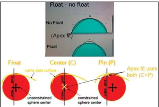
Floating is the most common method to fit the reference surface to the cornea (Figure 7). In short this method basically fits the reference surface to the surface in question with minimum square difference [127, 128]. The fitting type should always be kept in mind while analyzing maps/images captured with different instruments, because it significantly influences the final output.
Figure 7.: Fitting methods for a reference surface. Apex fit/center + pinned - Center of reference object is constrained on the view axis and it intersects data surface on the view axis. This flattens the central hill as it centers on it, Float - Center is unconstrained. Reference fits the corneal surface with minimum square difference. Almost all devices use this method as it has the least error [98].
Raw elevation data alone from normal eyes look very similar to the raw elevation data from abnormal eyes, and makes decision making/screening impossible. So to give a qualitative definition to the elevation data the machine using the above concept of
32
elevation and float, identifies the dimensions of a selected reference shape that can best fit to the examined surface for each eye tested depending on its individual characteristics. This calculated reference shape varies in dimensions for each eye and its shape and curvatures are indicated on the printout [127, 128]. In the nomenclature of tomography this is called as the best fit reference surface. Another characteristic of BFS is the pre-defined “Fit zone” which is 8 mm in diameter in most cases. Different device software has its own specific reference surface setting (e. g., the Belin Ambrosio display (BAD) has the BFS). For further evaluation one can use different reference settings depending on individual preferred practice and experience.
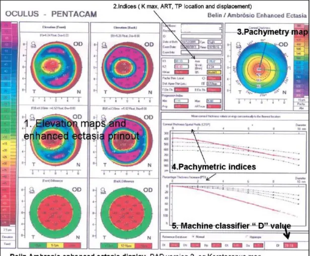
Belin/Ambrosio Enhanced Ectasia Display III (BAD III):
With this method a comprehensive refractive screening display (Belin/Ambrosio Enhanced Ectasia Display III- (BAD III)) is possible, and it is integrated into the Pentacam software. It combines nine different tomographic parameters into a unified screening tool. The display uses the parameters in a regression analysis to aid the ophthalmologists identifying patients with potential risk for corneal ectatic disease [98]:
Anterior elevation at the thinnest point
Posterior elevation at the thinnest point
Change in anterior elevation
Change in posterior elevation
Corneal thickness at thinnest point
Location of thinnest point
Pachymetric progression
Ambrósio relational thickness
Kmax
The BAD III displays each parameter and individually reports them as a standard deviation and then reports a final overall reading that is based on a regression analysis
33
to maximize the discrimination of normal corneas from those with keratoconus (Figure 8) [98].
Figure 8.: Keratoconus screening with BAD III. method. This case is a moderately advance keratoconus where all the analyzed parameters measured in the BAD III. analysis are highly abnormal [98].
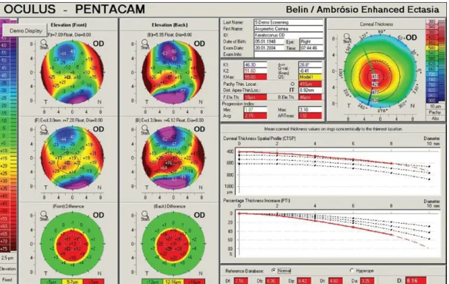
Keratoconus accompanied with corneal thinning, studies show difference in pachymetric variations between normal and KC corneas regarding to limbus to the thinnest point [129]. BAD III in the Pentacam also incorporates novel parameters as percentage thickness increase (PTI) from thinnest point and the corneal thickness spatial profile (CTSP). The software has the capability to enhance the cone location, by subtracting the 4mm area around the thinnest point and calculating the new BFS for the rest of the cornea (which would be flatter if the cone is located in the excluded area)[129]. As a result when the excluded area is compared with the flatter "new" BFS, it stands out if abnormal in the "enhanced map" that is also shown at the printout for both surfaces. In addition to the features above, the display in its current version (BAD III) incorporates the K max, maximum front, and back elevation in microns, a
34
pachymetry map, thin point location, displacement of the thin point from apex, and a pachymetry-based classifier the ART max (Figure 9). Besides this machine classifier, the main classifier, the "D" value, incorporates 9 parameters for its calculation and has been independently validated in a retest population [129].
Figure 9.: Belin Ambrosio enhanced ectasia display (BAD) version III. Keratoconus map
[98].
To summarize the knowledge, several topographic and tomographic parameters/indices are available to help decision making when corneal ectatic disease screened. It is important to know the advantages and limitations of the method/device being used.
Parallel to the careful evaluation of the cornea with such device mentioned above, proper slit lamp examination and clinical findings/signs should also take into account when decision is made. Briefly the findings below made patients suspect for keratoconus [128]:
35 Axial map abnormalities [128]:
1. K greater than 48 D.
2. SRAX greater than 21 degrees.
3. I-S greater than 1.42D.
4. Corneal astigmatism on anterior or posterior surface greater than 6 D.
5. Against the rule astigmatism.
6. S-I difference at the 5-mm zone >2.5 D.
On elevation map [128]:
1. Isolated island or tongue-like extension on either surface (BFS mode).
2. Elevation values greater than 12 microns on the anterior elevation map in the central 5 mm (BFTE mode).
3. Elevation values greater than 15 microns on the posterior elevation map (BFTE mode).
4.
Pachymetry/corneal thickness map (Scheimpflug devices) [121]:
1. Thinnest location less than 470 microns.
2. Displacement of the thinnest point >500 microns from the center.
3. Pachymetry difference asymmetry in two eyes at thinnest point >30 microns.
4. S-I difference at the 5 mm circle >30 microns.
5. Cone-like pattern on the thickness map.
2.3.7. Treatment options of keratoconus
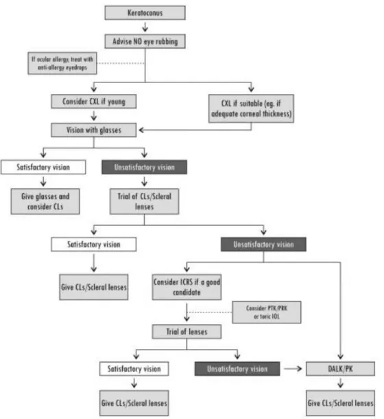
Several methods have been used to help patients with keratoconus since the discovery of the disease. Treatment options have a wide spectrum from the correction of refractive errors to surgical procedures. The procedures must be adjusted first to the patient (age, disease severity etc.), than to the doctor’s experience in the treatment modalities (Figure 11). The two main goals are visual rehabilitation and to halt disease progression [2]. All stages of the disease especially in earlier stages verbal guidance is the most important thing. To explain patients the risk factors, like the importance of not rubbing one’s eyes. Therefore the use of topical antiallergic medication in patients with allergy,
36
and use of topical lubricants (in case of ocular irritation) to decrease the impulse to rub one’s eyes is one of the first steps in disease management beside the others. Treatment modalities can be divided into surgical and non-surgical options [1, 2].
Figure 11.:Keratoconus treatment flowchart. CLs- contact lenses; CXL-corneal cross-linking;
PTK-phototherapeutic keratectomy [2].
2.3.7.1. Spectacles
Impaired visual acuity in consequence of keratoconus is initially managed with spectacles. Progressive addition glasses are not contraindicated during the disease, but
37
they are rarely successful, and often very expensive. Hence the vast majority of practicing ophthalmologists does not prescribe multifocal glasses in KC [1, 2].
2.3.7.2. Contact lenses
When doctors/optometrists failed to correct visual disturbances in patients with KC, the next step is the use of contact lenses. Contact lenses usually provide better vision than glasses by masking irregular astigmatism (higher-order aberrations). In mild cases the use of soft contact lenses are often enough for vision correction. More advanced cases may require the use of soft toric or custom soft toric contact lenses. The further step in correcting severe corneal irregularities are rigid gas permeable lenses (RGP). They mask higher-order aberrations with higher success rate. Special contact lenses designed for KC patients are exist on the market, such as Super Cone, and Rose K etc.. These special lenses has high oxygen permeability and a more comfortable fit by having a steep central posterior curve to arc over the cone and flatter peripheral curves to approach the more normal peripheral curvature. An alternative to RGP is a hybrid contact lens (containing: rigid center, soft skirt). This type of lens could provide stable vision by preventing toric rotation-with the soft skirt- accompanied with each blink.
One of the widely used lens is SynergEyes-KC (SynergEyes Inc., Carlsbad, CA, USA).
The last option for highly irregular corneas is the piggyback contact lens. This name means a soft contact lens which is fitted to the cornea and an RGP lens is placed on top of it [1, 2, 130]. When all other contact lenses fail newly designed scleral lenses made of material with high Dk (Oxygen permeability of a contact lens material; P = Dk = diffusion (D) * oxygen solubility (k)) are available. These lenses could be divided to corneo-scleral, mini-scleral and semi-scleral lenses regarding to the size and coverage of the bulbus [130].
2.3.7.3. Radial keratotomy
When non-surgical therapies fail the next step are invasive methods.
The procedure was first described by Sato (Sato et al., 1953) and popularized by Fyodorov (Fyodorov and Durnev, 1979) in 1974. During this surgery the surgeon place four to 16 tiny incision in the mid-periphery (out of the visual axis) of the cornea with a diamond-edged knife at 95% depth of the corneal thickness [131]. This method could
38
correct myopia and/or keratoconus. The theory is that keratotomy produces a hyperopic effect due to steepening of central cornea. In keratoconus management this surgery was found to be a reasonable option for the rehabilitation of a selected group of keratoconus patients in the early or moderate stages according to some studies [132-134]. To perform operation, KC patient should have 400 micron or greater central corneal thickness without apical scarring [131-134]. Nowadays this method has only historical meaning. Practicing ophthalmologist could meet patients treated earlier with radial keratotomy, but present time manual radial keratotomy is a rarely performed procedure.
2.3.7.4. Intra stromal corneal ring segments
This is another option for correcting myopia and irregular astigmatism due to keratoconus. This method also needs a clear central cornea. ICRS (Intra Corneal Ring Segment) segments are made of polymethyl methacrylate and have a crescent-shaped arc length of 150°. The inner diameter is 6.8 mm and the outer diameter is 8.1 mm when placed in the cornea. Intacs thickness ranges from 0.25 to 0.45 mm, in 0.05 mm increments. Practitioners insert the segments into corneal stromal tunnels. The tunnels could be made by mechanical and femtosecond laser-assisted [1].
Briefly when tunnels made mechanically, the surgeon perform radial incisions about 1.8 mm in length with a diamond edged knife approximately 70% of the mean corneal thickness depth. Special pocketing hooks are used to create corneal pockets on each side of the incision. Then the ring segments inserted into the pockets. In the femtosecond laser-assisted way a continuous circular stromal tunnel is created approximately 80% of the corneal thickness with the laser system [1, 134, 135].
Several type and modified Intacs segments exist on the market. For example flexible (sometimes full ring) Intac segment, which could be adjusted after implanted into the corneal pocket is a newly used. ICRS with elliptical cross-section called Intacs SK, Severe Kertaoconus (Addition Technologies Inc.), is also a variant with a smaller 6mm optical zone to provide correction of higher astigmatism/myopia like in keratoconus, and to minimize glare. The Ferrara ring (Keravision Inc., Fremont, CA, USA) is another option in correcting keratoconus. The segments vary in thickness (0.15, 0.20, 0.30 and 0.35 mm) and have a triangular cross-section and the base for every thickness is 0.60
![Figure 3.: Scheipflug imaging . Illustration shows Sheimpflug camera working principles, this method of image acquisition enhances the depth of focus (left) [98]](https://thumb-eu.123doks.com/thumbv2/9dokorg/1365386.111465/21.892.181.710.311.694/figure-scheipflug-imaging-illustration-sheimpflug-principles-acquisition-enhances.webp)