Original Contribution
Enhanced hydrogen peroxide generation accompanies the bene fi cial bioenergetic effects of methylene blue in isolated brain mitochondria
L. Tretter, G. Horvath, A. Hölgyesi, F. Essek, V. Adam-Vizi
nMTA-SE Laboratory for Neurobiochemistry, Department of Medical Biochemistry, Semmelweis University, Budapest H-1094, Hungary
a r t i c l e i n f o
Article history:
Received 12 May 2014 Received in revised form 2 September 2014 Accepted 18 September 2014 Available online 30 September 2014 Keywords:
Methylene blue Brain mitochondria Reactive oxygen species H2O2generation
Mitochondrial membrane potential Respiration
Free radicals
a b s t r a c t
The redox dye methylene blue (MB) is proven to have beneficial effects in various models of neurodegenerative diseases. Here we investigated the effects of MB (100 nM, 300 nM, and 1μM) on key bioenergetic parameters and on H2O2 production/elimination in isolated guinea pig brain mitochondria under normal as well as respiration-impaired conditions. As measured by high- resolution Oxygraph the rate of resting oxygen consumption was increased, but the ADP-stimulated respiration was unaffected by MB with any of the substrates (glutamate malate, succinate, or α-glycerophosphate) used for supporting mitochondrial respiration. In mitochondria treated with inhibitors of complex I or complex III MB moderately but significantly increased the rate of ATP production, restoredΔΨm, and increased the rate of Ca2þuptake. The effects of MB are consistent with transferring electrons from upstream components of the electron transport chain to cytochrome c, which is energetically favorable when theflow of electrons in the respiratory chain is compromised. On the other hand, MB significantly increased the production of H2O2 measured by Amplex UltraRed fluorimetry under all conditions, in resting, ATP-synthesizing, and respiration-impaired mitochondria, with each substrate combination supporting respiration. Furthermore, it also decreased the elimination of H2O2. Generation of H2O2 without superoxide formation, observed in the presence of MB, is interpreted as a result of reduction of molecular oxygen to H2O2by the reduced MB. The elevated generation and impaired elimination of H2O2should be considered for the overall oxidative state of mitochondria treated with MB.
&2014 The Authors. Published by Elsevier Inc. This is an open access article under the CC BY-NC-SA
license (http://creativecommons.org/licenses/by-nc-sa/3.0/).
Methylene blue (MB)1has been used with multiple therapeutical purposes in the treatment of malaria[3,30,41], carbon monoxide or cyanide poisoning[23], methemoglobinemia[47], and neurodegen- erative diseases [16,21,25,66,72,73,87,96] and in photodynamic therapy[58]. MB is able to take electrons on its aromatic thiazine ring to be reduced to leukomethylene blue (MBH2) and transfer electrons to other compounds depending on the redox states and the concentration of MB (see[65]). It is a lipophilic compound that accumulates in the mitochondria driven by the mitochondrial membrane potential (ΔΨm), and its major cellular target associated with the neuroprotective and cognitive-enhancing effect is assumed to be the mitochondrial metabolism ([8–10,93]; for review see[65]).
Owing to its redox-cycling property MB in mitochondria can be reduced to MBH2primarily by NADH at complex I of the respiratory chain and shuttle electrons to cytochrome cand oxygen to form
water (see [65]), therefore increasing oxygen consumption, which has been described in various cell types [10,44,93]. It has been described that MB depolarizes mitochondria in HeLa cells[58], but is protective against glutamate-induced mitochondrial depolarization in HT-22 cells[60].
Data concerning the effects of MB on reactive oxygen species (ROS) homeostasis are somewhat controversial. Pro-oxidant effects of MB (in 41μM) have been demonstrated by 20,70-dichlorofluor- escin oxidation[10,58], but MB proved to be protective against H2O2- induced cell death [10,87] and exhibited antioxidant effects in various systems[5,60,93].
Considering that mitochondrial mechanisms are assumed to underlie the beneficial effects of MB, studies addressing specific properties reflecting the bioenergetic competence of mitochondria treated with MB are scarce[88,89,93]. In this work we studied in detail the effects of MB on the mitochondrial bioenergetics and H2O2
release in isolated brain mitochondria. In most of the previous studies with MB, ROS formation was detected with dichlorofluor- escin diacetate (DCF-DA), which has the disadvantage of reacting with ROS slowly and with uncharacterized stoichiometry, lacking correct calibration, and reacting also with non-ROS compounds[37].
Here we used the Amplex UltraRedfluorescent dye, which enables a Contents lists available atScienceDirect
journal homepage:www.elsevier.com/locate/freeradbiomed
Free Radical Biology and Medicine
http://dx.doi.org/10.1016/j.freeradbiomed.2014.09.024
0891-5849/&2014 The Authors. Published by Elsevier Inc. This is an open access article under the CC BY-NC-SA license (http://creativecommons.org/licenses/by-nc-sa/3.0/).
Abbreviations:ΔΨμ, mitochondrial membrane potential; PTP, permeability tran- sition pore; ROS, reactive oxygen species; RET, reverse electron transport;α-GPDH, α-glycerophosphate dehydrogenase;α-GP,α-glycerophosphate; MB, methylene blue.
nCorresponding author. Fax:þ36 1 267 0031.
E-mail address:adam.veronika@med.semmelweis-univ.hu(V. Adam-Vizi).
fast, sensitive, and well-calibrated detection of H2O2released from mitochondria to the medium [59,71,91]. To identify the specific effects of MB on mitochondrial bioenergetics respiration, ATP syn- thesis,ΔΨm, and Ca2þuptake were investigated in both normal and respiration-impaired isolated brain mitochondria energized by var- ious respiratory substrates.
Materials and methods Isolation of mitochondria
Mitochondria of synaptic and nonsynaptic origin were isolated from guinea pig brain using a discontinuous Percoll gradient as detailed earlier[67,81]. Animals were decapitated by a process in accordance with the International Guiding Principles for Biome- dical Research Involving Animals and Guidelines for Animal Experiments at Semmelweis University. Brains were quickly removed and homogenized in buffer A (in mM: 225 mannitol, 75 sucrose, 5 Hepes, 1 EGTA, pH 7.4 (KOH)) and then centrifuged for 3 min at 1300g. The supernatant was centrifuged for 10 min at 20,000g, and then the pellet was suspended in 15% Percoll and layered on a discontinuous gradient consisting of 40 and 23%
Percoll layers, which was then centrifuged for 8 min at 30,700g.
After resuspension of the lowermost fraction in buffer A, it was centrifuged at 16,600g for 10 min, and then the pellet was resuspended in buffer A and centrifuged again at 6300g for 10 min. After the supernatant was discharged, the pellet was resuspended in buffer B (in mM: 225 mannitol, 75 sucrose, 5 Hepes, pH 7.4 (KOH)).
Incubation medium
Experiments were carried out in a standard assay medium containing (in mM) 125 KCl, 20 Hepes, 2 K2HPO4, 1 MgCl2, 0.1 EGTA, pH 7.0 (KOH), supplemented with 0.025% fatty-acid- free bovine serum albumin.
Mitochondrial respiration
Mitochondrial oxygen consumption assays were performed using the high-resolution respirometry system Oxygraph-2K (Oro- boros Instruments, Innsbruck, Austria) [28] at 371C in 2-ml chambers. Data were digitally recorded and analyzed; oxygenflux was calculated as the negative time derivative of the oxygen concentration, cO2(t). Oxygen sensors were calibrated routinely at air saturation and in oxygen-depleted medium. Mitochondria were energized with glutamate plus malate (5 mM each) or succinate (5 mM) orα-glycerophosphate (α-GP) (20 mM).
Measurement of mitochondrial ATP synthesis
Synthesis of ATP was measured by a coupled enzymatic assay [94]. Standard assay medium was supplemented with NADPþ (1.5 mM), hexokinase (2 U/ml), glucose 6-phosphate dehydrogen- ase (3.84 U/ml), 2.5 mM glucose, and 50μM P1,P5-di(adenosine-50) pentaphosphate (inhibitor of adenylate kinase). In the medium ATP phosphorylated glucose to glucose 6-phosphate in the pre- sence of hexokinase, then glucose 6-phosphate was converted by glucose-6-phosphate dehydrogenase to 6-phosphogluconate with the concomitant reduction of NADPþ to NADPH. Absorbance of NADPH was measured at 340 nm using a GBC-UV double-beam spectrophotometer. Measurements were calibrated with known amounts of ATP.
Measurement of membrane potential
ΔΨmwas determined using the cationic dye safranin O, which is accumulated and quenched in energized mitochondria[4]. The dye concentration was 2mM. The excitation and emission wavelengths were 495 and 586 nm, respectively, as described previously [40].
Measurements were performed at 371C with 0.1 mg/ml mitochon- drial protein using a Hitachi F-4500 spectrofluorimeter (Hitachi High Technologies, Maidenhead, UK).
Measurement of Ca2þuptake
Mitochondria (0.1 mg/ml) were added to the incubation med- ium in the presence of ADP and glutamate plus malate (5 mM each) and then Ca2þpulses were given in 100-s intervals. The free Ca2þ concentration at each added concentration of Ca2þ was calculated and measured. Ca2þ uptake by mitochondria was followed by measuring Calcium Green-5N (100 nM)fluorescence at 505 nm excitation and 535 nm emission wavelengths at 371C using a Hitachi F-4500 spectrofluorimeter.
Detection of H2O2formation
The assay is based on the detection of H2O2in the medium using the Amplex Redfluorescent dye[54]. Horseradish peroxidase (5 U per 2 ml) and Amplex UltraRed reagent (1mM), and then mitochon- dria, were added to the incubation medium. H2O2 formation was initiated by the addition of glutamate plus malate or α-GP or succinate, in the concentrations indicated, and fluorescence was detected at 371C in a PTI Deltascanfluorescence spectrophotometer (Photon Technology International, Lawrenceville, NJ, USA). The exci- tation wavelength was 550 nm and thefluorescence emission was detected at 585 nm. A calibration signal was generated with known quantities of H2O2at the end of each experiment.
Measurement of NAD(P)H steady state
The matrix NAD(P)H autofluorescence was measured in parallel with the Amplex assay using the double-excitation and double- emission mode of the PTI Deltascanfluorescence spectrophotometer.
Mitochondria were incubated at 371C as described above and the fluorescence was measured using 344 nm excitation and 460 nm emission wavelengths. Changes in the NAD(P)H level were expressed in photon count 103.
Measurement of H2O2elimination
H2O2 elimination was detected with an H2O2-sensitive elec- trode (World Precision Instruments) [24]. Electrode signal was calibrated with additions of known concentrations of H2O2. In the presence of glutamate plus malate (5 mM), 300 s after addition of ADP (2 mM) and H2O2(5μM), a 0.1 mg/ml mitochondrial suspen- sion was obtained and the amount of H2O2was detected. At the end of each measurement catalase (33 IU/ml) was added. H2O2
elimination was characterized by the half-time (τ) of H2O2 dis- appearance from the medium.
Chemicals
Standard laboratory chemicals were obtained from Sigma (St.
Louis, MO, USA). The Amplex UltraRed reagent and Calcium Green- 5N were from Molecular Probes (Eugene, OR, USA).
Statistics
Statistical differences were evaluated with ANOVA (Sigmastat) for multiple comparisons. Values of Po0.05 were considered statistically significant.
Results
The effects of MB on the respiration of mitochondria supported by three different substrates
Given the hormetic pharmacological effect of MB, meaning that it could have opposite effects at high and low doses[65], we used MB in a concentration range (100 nM–1μΜ) that is relevant to cellular studies, in particular to those reporting neuroprotective effects of MB[10,60,93].
When mitochondria are respiring on glutamate plus malate, electrons enter the respiratory chain from NADH via complex I, whereas with succinate orα-GP electrons from succinate dehydro- genase orα-glycerophosphate dehydrogenase (α-GPDH), respectively, reduce CoQ, bypassing complex I. The rate of State 4 respiration of isolated brain mitochondria supported by glutamate plus malate in the absence of added ADP was stimulated from 375720.6 to 618.3736.2 pmol/s/mg protein upon addition of 1mM MB (Fig. 1);
the effect of MB was significant already at the 300 nM concentration (463724.6 pmol/s/mg protein). The ADP-stimulated (State 3) respiration was unaffected by MB, but the stimulation of the State 4-like respiration was again evident after the addition of carboxya- tractylozide (CAT), which inhibits the adenine nucleotide translocator (ANT), preventing the effect of ADP; therefore mitochondria behave as if in the absence of ADP (Fig. 1).
The rate of respiration, as expected, was reduced by rotenone and addition of ADP was without an effect, but MB was capable of stimulating respiration also under this condition (Fig. 1, bars on the right).
Essentially similar results were obtained with mitochondria respiring on succinate orα-GP (Table 1). It is well documented that
with succinate in the absence of ADP, high ΔΨm is generated, allowing theflux of electrons from coenzyme Q (CoQ) in reverse via complex I, a phenomenon called reverse electron transport (RET) [11,34], resulting in the reduction of NADþ to NADH by complex I. NADH might reduce MB, which could then shortcut electrons to cytochromec, contributing to the MB-induced stimu- lation of respiration. RET and the related NADH generation in succinate-supported mitochondria are prevented by the complex I inhibitor rotenone [20,32,42,90], but this did not alter the MB- evoked stimulation of respiration (Table 1). Resting oxygen con- sumption was higher with succinate (1094769 pmol/s/mg pro- tein) than with glutamate plus malate and the effect of MB was less spectacular, but significant both in the absence and in the presence of rotenone. It is noteworthy that independent of RET, NADH could also be generated during the oxidation of the carbon skeleton of succinate subsequent to the succinate dehydrogenase reaction in the Krebs cycle. This ambiguity is lacking whenα-GP is
Fig. 1.The effect of MB on the rate of oxygen consumption in glutamate plus malate-supported mitochondria. Mitochondria (0.05 mg/ml) were incubated in the standard medium as described under Materials and methods. MB (100 nM, 300 nM, and 1μMfinal concentrations), ADP (2 mM), and then carboxyatractylozide (CAT; 2μM) were given as indicated in the inset (gray trace; no MB addition for black trace). The effects of MB (gray bars) are compared to controls (white bars; no MB, only buffer added) measured for each individual experiment. Data from similar experiments performed in the presence of rotenone (0.5μM) are shown separated on the right. Results are expressed as mean oxygen consumption in pmol/s/mg protein7SEM (n44) and written on the bars. *Significant difference (Po0.05) from the corresponding MB-free controls.
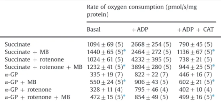
Table 1
The effect of methylene blue on the rate of oxygen consumption in mitochondria supported by succinate orα-GP in the absence or presence of rotenone.
Rate of oxygen consumption (pmol/s/mg protein)
Basal þADP þADPþCAT
Succinate 1094769 (5) 26687254 (5) 790745 (5)
SuccinateþMB 1440765 (5)n 24647272 (5) 1136767 (5)n Succinateþrotenone 1024761 (5) 42327395 (5) 738721 (5) SuccinateþrotenoneþMB 1232741 (5)n 38947280 (5) 944725 (5)n
α-GP 335719 (7) 822722 (7) 446716 (7)
α-GPþMB 550724 (5)n 906743 (5) 602721 (5)n
α-GPþrotenone 328711 (4) 795746 (4) 402710 (4)
α-GPþrotenoneþMB 472715 (5)n 854749 (5) 499716 (5)n MB, methylene blue;α-GP,α-glycerophosphate. Experiments were performed as for Fig. 1except that mitochondria were supported with either 5 mM succinate or 20 mMα-GP. Concentrations: MB, 1mM; rotenone, 5mM; ADP, 2 mM; carboxya- tractylozide (CAT), 2mM.
nPo0.05; significant difference from the corresponding data obtained under MB-free conditions.
used as a substrate, which is oxidized on the outer surface of the mitochondrial inner membrane byα-GPDH transferring the elec- trons to CoQ and in 20 mM concentration generating sufficient proton-motive force to drive RET [31,84]. Like in succinate- supported mitochondria, NADH generation by RET is prevented by rotenone, but unlike with succinate, no additional NADH generation should be considered with α-GP as a substrate.
Table 1 demonstrates that in mitochondria energized with 20 mMα-GP addition of MB stimulated basal respiration by 64%
(from 335719 to 550724 pmol/s/mg protein), which was also observed in the presence of CAT (446716 versus 602721 pmol/s/
mg protein), but no significant effect of MB was seen in ADP- stimulated mitochondria. In the presence of rotenone (Table 1) the
effects of MB were similar, both qualitatively and quantitatively, to those observed in the absence of complex I inhibition.
These results indicate that only the basal mitochondrial resp- iration is stimulated but the accelerated oxygen consumption in ATP-synthesizing mitochondria is unaffected by MB. Likewise, reduced respiration in mitochondria lacking complex I function is improved by MB. In addition, experiments with different respiratory substrates show that no matter whether electrons enter the respiratory chain via complex I or downstream at CoQ, MB remains a powerful stimulant of basal respiration, strongly suggesting that MB is able to accept electrons not only from NADH but also from theflavin group of succinate dehydrogenase orα- GPDH.
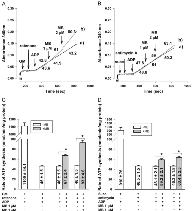
Fig. 2.The effects of MB on the rate of ATP production in respiration-impaired mitochondria. (A, C) In glutamate plus malate-supported mitochondria rotenone (0.5μM) was used to inhibit complex I; (B, D) in succinate-supported mitochondria complex III was inhibited by antimycin A (0.1mM). Original traces are shown in (A) and (B) with numbers calculated as the rate of ATP synthesis in nmol/min/mg in these particular experiments; additions were as indicated (MB 1 and 2μMfinal concentrations). For traces (a) no MB was given. In (C) and (D) bars represent the average ATP production rates in nmol/min/mg protein7SEM from at least three experiments. *Significant difference (Po0.05) from the corresponding controls measured in the absence of MB.
ATP production is partially restored in respiration-impaired mitochondria
Stimulation of respiration itself is not informative as to the bioenergetic competence of mitochondria. To address this, ATP production in mitochondria respiring on glutamate plus malate or succinate was measured. In fully functional mitochondria ATP generation was unaffected by MB applied in 2μM concentration (data not shown); however, significant effects were observed in respiration-compromised mitochondria. As expected, the rate of ATP generation initiated by addition of ADP in glutamate plus malate-supported mitochondria was drastically decreased by rotenone (from 1100744 to 4671.5 nmol/min/mg protein). The slow rate of ATP generation was significantly increased by 1 or 2mM MB (6772.4 or 9374.6 nmol/min/mg protein, respectively;
Figs. 2A and C). In succinate-supported mitochondria the rate of ATP generation was reduced by the complex III inhibitor, anti- mycin A, from 910776 to 46.571.5 nmol/min/mg protein. ATP generation from this low level was slightly but significantly stimulated by 1 or 2mM MB (to 59.272.1 or 63.472.5 nmol/
min/mg protein, respectively;Figs. 2B and D).
The effect of MB onΔΨmin resting and respiration-compromised mitochondria
ΔΨmis another key parameter that is essential for the bioener- getic performance of mitochondria. In highly energized mitochon- dria in the presence of glutamate plus malate (5 mM each)ΔΨm
was unchanged by MB as detected by safraninfluorescence (Fig. 3, traces a and b). With insufficient amount of substrates, in the presence of 50 or 10μM glutamate plus malate, mitochondria cannot be fully energized because of the limited availability of NADH as indicated byΔΨmset at a depolarized value (Fig. 3, traces c and e, respectively). Diversion of electrons from NADH to MB under these conditions is probably responsible for the evident further loss inΔΨm(traces d and f), which is not apparent when substrates are abundant (traces a and b). Similar results were obtained with mitochondria respiring on succinate (not shown).
In glutamate plus malate-supported mitochondria rotenone induced an abrupt drop inΔΨm, which recovered after addition of 1mM MB, and then these mitochondria, similar to competent ATP-synthesizing mitochondria, reacted with depolarization to the addition of ADP followed by recovery ofΔΨmin response to the ANT inhibitor, CAT (Fig. 4A, trace b).ΔΨmwas not rescued by MB when complex IV was inhibited by KCN (Fig. 4A, trace c). ATP
synthesis can also be blocked by oligomycin, instead of CAT, given after ADP (Fig. 4B, trace b), but with this, smaller repolarization was observed than with CAT (compare traces b inFig. 4A versus Fig. 4B). In mitochondria without MB addition, oligomycin given after ADP resulted in a small further depolarization (Fig. 4B, trace a), whereas CAT slightly repolarized ΔΨm (Fig. 4A, trace a). The explanation lies in the different reversal potentials of the adenine nucleotide translocase and the ATP synthase. At low ΔΨm ATP synthase can operate in reverse, but ANT still works in the forward mode. Under this condition application of oligomycin depolarizes ΔΨm because it stops proton pumping, but inhibition of ANT would hyperpolarize mitochondria[18].
Similarly, rescue of ΔΨmby MB, though to a smaller degree, was observed with succinate (Fig. 4C, trace b) orα-GP (Fig. 4D, trace b) as a substrate, when respiration was inhibited andΔΨm
was highly depolarized with the complex III inhibitor, antimycin.
In contrast to this, in mitochondria poisoned by cyanide, MB was unable to rescueΔΨm(Figs. 4A, C, and D, traces c), indicating that MB donates electrons to the respiratory chain proximal to cyto- chrome oxidase. These results indicate that MB supports the maintenance of ΔΨm in mitochondria subjected to inhibitors of complex I or complex III.
Ca2þ uptake capacity is increased by MB in energetically compromised mitochondria
Ca2þ uptake is also a key function of mitochondria and it is well established that mitochondrial Ca2þuptake is an energy- and ΔΨm-dependent process [17,64]. The effect of MB on the mito- chondrial Ca2þ uptake was investigated in resting and energeti- cally compromised mitochondria. The rate of Ca2þ uptake was decreased in the presence of 1μM MB in resting mitochondria energized with glutamate plus malate (Fig. 5A, upper trace).
In contrast to this, Ca2þ uptake reduced in rotenone-treated mitochondria was stimulated by MB (Fig. 5B, lower trace), con- sistent with the partially restored ΔΨm observed under similar conditions.
Enhanced H2O2generation by MB in both energized and respiration- impaired mitochondria
Given the controversy in the available data as to the pro- oxidant versus antioxidant response of various cells to treatment with MB[5,10,26,27,29,35,93], we compared the H2O2generation in brain mitochondria in the absence and presence of MB. The use of various substrates to support respiration in these experiments is justified by the fact that, owing to the different sites of entry of electrons into the respiratory chain, ROS are generated by distinct mechanisms. H2O2 formation was measured using the Amplex UltraRed horseradish peroxidase (HRP) fluorescence system as described under Materials and methods.
H2O2formation in mitochondria energized with glutamate plus malate
In mitochondria fueled with NADH-linked substrates, complex I, the major site of entry of electrons into the respiratory chain[7], and/orα-ketoglutarate dehydrogenase[6,61,77,80]appears to be the predominant site of ROS generation. Complex III in these mitochondria contributes to ROS production mainly when the respiratory chain is blocked at a downstream site[2].
As demonstrated in Fig. 6, MB caused a remarkably large stimulation of H2O2production in the presence of glutamate plus malate. The stimulation of H2O2 formation by MB was evident both in resting mitochondria (in the absence of ADP), in which the rate of H2O2 generation was increased from 378712 to Fig. 3.The effects of MB on safraninfluorescence indicatingΔΨmin mitochondria
respiring on glutamate plus malate (G-M; 5 mM for traces a and b, 50μM for traces c and d, and 10μM for traces e and f). MB (1μM) was added as indicated for traces b, d, and f. Traces are representative of three parallel experiments.
1792753 pmol/min/mg protein by 1mM MB (not shown), and in ATP-synthesizing mitochondria (in the presence of ADP, in which the rate of H2O2 generation is smaller than that without ADP owing to depolarization ofΔΨm;Figs. 6A and C). The rate of H2O2
formation under the latter condition was significantly increased by as low as 100 nM MB from 147.8721 to 660716 pmol/min/mg protein and further increased to 1516747 pmol/min/mg by 1mM MB. NAD(P)Hfluorescence was decreased by MB in parallel to the stimulation of H2O2 generation (Fig. 6D, trace c); an effect less apparent in the presence of ADP, when the level of NAD(P)H was already low (Fig. 6D, trace b, compared to trace a, for which no MB was given). To address H2O2formation in energetically impaired mitochondria, rotenone (0.5mM) was given after ADP, followed by addition of MB (100 nM, 300 nM, or 1μM). Rotenone, as docu- mented earlier[2,32,78,86,90] resulted in a huge stimulation of H2O2formation (from 13878 to 1268718 pmol/min/mg protein) and 1mM MB was able to induce a significant further increase in the rate of H2O2 generation to 2069728 pmol/min/mg protein (Fig. 6B).
H2O2formation in mitochondria energized with succinate
In succinate-supported mitochondria succinate dehydrogenase [62]and complex III could contribute to the overall ROS generation (see[1]) and when sufficient proton-motive force is generated, RET is considered a major ROS-forming mechanism [7,32]. The RET- related high rate of H2O2 generation observed in fully polarized mitochondria (in the presence of 5 mM succinate)[7,31,43,45]in the present experiment was 22197180 pmol/min/mg protein (Fig. 7A).
Addition of 1μM MB induced an additional increase in the rate of H2O2formation to 33217113 pmol/min/mg protein. When RET was prevented by rotenone the rate of H2O2generation was dropped to 451715.9 pmol/min/mg protein, but MB also under this condition resulted in a large stimulation of H2O2 generation (to 18877 83 pmol/min/mg protein). In parallel experiments the level of NAD (P)H was moderately decreased by MB (Fig. 7C, trace b). The sharp drop in the NAD(P)H fluorescence by rotenone (Fig. 7C, trace b) reflects the elimination of RET and NADH generation by complex I and was parallel with a decrease in H2O2formation as demonstrated in Fig. 7A. At low succinate concentration (50μM), which is Fig. 4.The effect of MB (1mM) onΔΨmin respiration-impaired mitochondria supported by (A, B) glutamate plus malate (5 mM each), (C) succinate (5 mM), or (D)α-GP (20 mM). Additions are indicated by arrows and the order of additions for traces a, b, and c is also shown in the frame. For traces a, no MB was given. For each trace mitochondria (m) and then respiratory substrate were given, then the order of additions was as follows: traces a, rotenone (0.5μM, for (A) and (B)) or antimycin (0.1μM, for (C) and (D)), ADP (2 mM), CAT (2μM), carbonyl cyanide 4-(trifluoromethoxy)phenylhydrazone (FCCP; 250 nM); traces b, rotenone ((A) and (B)) or antimycin ((C) and (D)), MB (2μM), ADP, CAT ((A), (C), and (D)) or oligomycin (2μM, (B)), FCCP; traces c, KCN (2 mM), MB, ADP, CAT, FCCP. Each trace represents an average7SEM of at least three independent experiments.
insufficient to support RET, the rate of ROS formation was low but addition of 1μM MB produced a sixfold elevation in the rate of H2O2
generation (from 41.374 to 274721 pmol/min/mg protein, which was further stimulated by rotenone (Fig. 7B). There was only a small and slow NAD(P)H formation here (Fig. 7C, traces c and d), unrelated to RET, as addition of rotenone significantly increased NAD(P)H level (Fig. 7C, trace c), suggesting that NADH generation due to succinate oxidation in the Krebs cycle is reflected in the NAD(P) signal under this condition.
To demonstrate that with Amplex UltraRedfluorescence H2O2 is specifically detected, we added catalase to the incubation medium containing MB. The MB-induced rise in thefluorescence signal was abruptly halted by catalase, which then reacted to the addition of HRP. This shows that the elevated signal in the presence of MB indeed reflects H2O2 generation (Fig. 7D). It is noteworthy that in this experiment lower activity of horseradish peroxidase was used to allow an efficient competition between HRP and catalase for scavenging H2O2, explaining the smaller rate of H2O2 formation found in this particular experiment. H2O2
generation in the experiments with MB in mitochondria respiring on glutamate plus malate orα-GP was also verified with catalase (not shown).
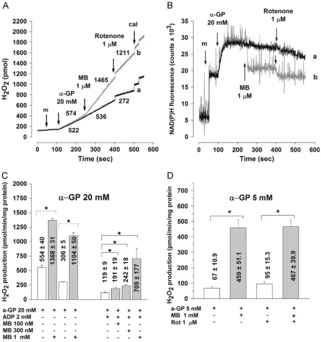
H2O2generation inα-GP-supported mitochondria
As previously characterized [53,82,84,85] ROS generation at high α-GP concentration (20 mM) can be attributed mainly to complex I receiving electrons from the oxidation ofα-GP via RET [31,82,84]. Oxidation ofα-GP at a lowα-GP concentration (5 mM) is unable to generate sufficient proton-motive force for RET;
electrons for H2O2generation under this condition were suggested to originate largely fromα-GPDH or, in the case of a blockage of electrons at complex III, they could be provided by CoQ or complex III[52,59,84].
In mitochondria respiring on 20 mMα-GP, MB induced a large stimulation of H2O2generation (Fig. 8A); the rate of H2O2forma- tion was increased from 554740 to 1368731 pmol/min/mg protein (Fig. 8C). Consistent with the involvement of RET and RET-related NADH generation in this effect, both rotenone and ADP, which decreased α-GP-supported ROS generation by
preventing RET[84,85], decreased the MB-induced H2O2 forma- tion (Fig. 8C). H2O2generation stimulated by MB was paralleled by a drop in the NAD(P)Hfluorescence (Fig. 8B, trace b), suggesting that for H2O2formation MB takes electrons from NAD(P)H gener- ated by RET. MB, however, remained capable of enhancing H2O2
formation in the total absence of RET (Fig. 8C). This is evident not only because MB increased H2O2 formation in 20 mM α-GP- supported mitochondria in the presence of rotenone or ADP, but also because mitochondria respiring on 5 mMα-GP, when no RET is possible, also responded with a huge increase in H2O2genera- tion to a treatment with MB (from 67710.9 to 459751 pmol/
min/mg;Fig. 8D). This shows that for MB-mediated H2O2genera- tion electrons could be provided byα-GPDH as well.
The rate of H2O2elimination in mitochondria is decreased by MB Mitochondria not only produce H2O2but also participate in the elimination/detoxification of H2O2[24,69,75,98], and we investi- gated whether this function is influenced by MB. Elimination of 5μM H2O2 from the medium was followed in the presence of mitochondria supported with glutamate plus malate and stimu- lated by ADP (Fig. 9). Half-life of H2O2 (τ) in the medium was 54.575.4 s, which increased to 139.2722.5 s when MB (1μM) was also present in the medium. MB at 300 nM concentra- tion was already effective at increasing the half-life of H2O2in the medium (not shown). MB also decreased the rate of H2O2 elimination in mitochondria treated with rotenone (0.5μM) (not shown).
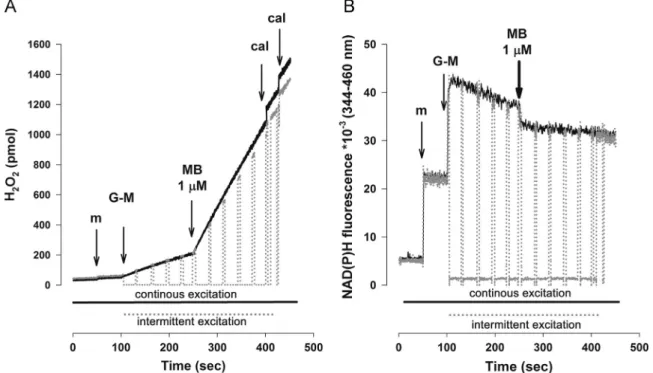
Enhanced H2O2signal in the presence of MB is unrelated to illumination during the experiment
MB is used in photodynamic therapy and MB-mediated photo- toxicity was suggested to be, at least partially, ROS-dependent [27,51,58]. It is important to demonstrate that the enhanced H2O2
signal in the presence of MB presented above is not due to the illumination during excitation in our experiments. Thus, to exclude false positive results with MB, key experiments were repeated without using continuous illumination of the samples. For this, the excitation light of the PTI Deltascan fluorescence Fig. 5.The effect of MB on the Ca2þuptake (A) in energized and (B) in rotenone-treated mitochondria respiring on glutamate plus malate. Ca2þpulses (150 nmol each for (A) except thefirst addition, which was 200 nmol; 50 nmol each for (B) except thefirst addition, which was 200 nmol) were given every 100 s in the absence (lower trace in (A); upper trace in (B)) or in the presence of 1μM MB (upper trace (gray line) in (A); lower trace (gray line) in (B)). Lower Ca2þconcentrations in the presence of rotenone were used to avoid permeability transition pore opening under this condition. At the end of the experiments Ca2þwas added at saturating concentrations. Numbers on the traces represent average rates of changes in Ca2þconcentration in the medium (nM/min). Traces are averages7SEM from at least four independent experiments.
spectrophotometer was switched on and off using a shutter. When the illumination time was reduced to 1/8 of the control, the Amplex UltraRedfluorescence signal in response to MB decreased only by 9% and there was no difference in the mitochondrial NAD (P)H fluorescence, either (Fig. 10). We can safely conclude that more than 90% of H2O2production observed in the presence of MB is independent of the illumination of the samples. Similarly, measurements on mitochondrial oxidation in the presence of MB were also repeated in the dark, providing results essentially similar to those presented above (not shown).
Discussion
Addressing several key bioenergetic parameters in this study, we aimed to dissect the mitochondrial effects of MB, which could determine its beneficial effects found in several pathological conditions [5,10,14,15,21,26,60]. In addition, release as well as elimination of H2O2 was addressed to reveal the pro-oxidant/
antioxidant effect of MB that accompanies the mitochondrial bioenergetic alterations.
(i) We established that MB stimulated respiration in isolated brain mitochondria and, by this, confirmed several earlier
reports demonstrating MB-stimulated oxygen consumption in different mitochondria and cellular models[10,44,88,89,93].
Stimulation of respiration by MB has been assigned to its ability to shunt electrons in the respiratory chain. It wasfirst described in the 1960s and 1970s that MB could be reduced in isolated mitochondria by electrons from NADH and transfer them to cytochromec, bypassing coenzyme Q in the respira- tory chain[70,92]. Although reduction of MB byflavoenzymes such as xanthine oxidase, NADH cytochromecreductase, and NADPH cytochrome creductase has been reported [39,50], generally NADH has been considered a major electron donor for MB in mitochondria [10,93,60]; recently it has also been found in isolated rat heart mitochondria that cytochrome c reduction was increased with NADH as an electron donor but not with succinate[44,60]. It is evident from our study that respiration was improved by MB not only with NADH-linked substrates but also with succinate or α-GP, suggesting that electrons from succinate dehydrogenase orα-GPDH could also reduce MB. This conclusion is supported by data obtained with diphenyleneiodonium (DPI), which was shown to inhibit complex I by keeping the flavin groups reduced[46]; MB in DPI-treated succinate-supported mitochondria significantly increased (by 10%) the State 3 respiration of mitochondria (not shown). Importantly, our study shows that in functional Fig. 6.The effects of MB on (A, B, C) H2O2production and (D) NAD(P)H steady state in glutamate plus malate-supported mitochondria. Mitochondria (m; 0.1 mg/ml), ADP (2 mM), MB, and 0.5mM rotenone (for (B)) were given where indicated. Representative experiments with original traces are shown in (C) and (D). Data on H2O2generation in pmol/min/mg protein in (A) and (B) represent the average7SEM from at least four experiments. *Significant difference.
mitochondria only the resting oxygen consumption is stimu- lated; the ADP-stimulated respiration is unaffected by MB.
Because mitochondria in vivo are functioning in the presence of ADP, this finding allows the conclusion that in vivo MB might not influence the respiration of normal ATP-synthesizing mitochondria. However, the significant stimulation of oxygen consumption in respiration-impaired mitochondria supports the suggestion that MB could partially restore respiration when theflux of electrons in the respiratory chain is inhibited either at complex I or at complex III, evident in this study with NADH-linked substrates in the presence of rotenone (Fig. 1) or with succinate in the presence of antimycin (not shown).
(ii) Accelerated respiration alone would not imply an improved bioenergetic performance of mitochondria, but only when this would result in an increased ATP generation. We report here, as a newfinding, that in respiration-impaired mitochon- dria, in the presence of complex I or complex III inhibitors, the severely inhibited ATP synthesis was increased by MB (Fig. 2).
The rate of ATP synthesis in rotenone-inhibited mitochondria was doubled in the presence of 2μM MB, but was still modest compared to that observed in mitochondria not treated with rotenone. Importantly, ATP synthesis in normally respiring isolated brain mitochondria was unchanged by MB. Thisfinding seems to be contradictory to that by Wen et al.[93]showing an increased cellular ATP level in HT-22 cells. However, in this report the ATP level in the transformed HT-22 cells was only moderately decreased by strong mitochondrial drugs such as the uncoupler carbonyl cyanide 4-(trifluoromethoxy)phenylhydra- zone or the complex IV inhibitor cyanide [93], implicating a largely nonmitochondrial ATP generation.
(iii) It is also revealed in this study that MB, although not influencing ΔΨm in fully respiring functional mitochondria, supports the maintenance ofΔΨmwhen complex I or complex III is inhibited (Fig. 4). The significance of thisfinding is that with a partially maintainedΔΨmmitochondria become more resistant against PTP induction [13,56,57,63]; therefore, the chances for survival of these mitochondria are highly Fig. 7.The effects of MB on succinate-supported (A, B) H2O2production and (C) NAD(P)H level. Succinate (5 mM for (A) and for traces a and b in (C); 50mM for (B) and for traces c and d in (C)) was given to mitochondria, then 300 s later 1μM MB was added where indicated, followed by addition of rotenone (0.5mM) after 300 s, where indicated. Numbers on bars in (A) and (B) indicate the average rate of H2O2production in pmol/min/mg7SEM from at least four independent experiments. (D) The effect of catalase on the MB-induced Amplex UltraRedfluorescence. Horseradish peroxidase activity at the beginning of these experiments was 20 times lower than in other Amplex experiments. Numbers indicate the rate offluorescence changes per second. Traces are representative of three similar measurements. *Significant difference.
increased. This suggestion is supported by data from Ca2þ- uptake measurements in this study (Fig. 5) showing an improved Ca2þ uptake capacity in MB-treated mitochondria.
RescuedΔΨmby MB in rotenone-treated mitochondria could contribute to a decreased rotenone-induced neurodegenera- tion observed in HT-22 cells[60,93]and retinal ganglion cells [96]. The finding that ADP depolarized ΔΨm in rotenone- treated mitochondria in the presence of MB (Figs. 4A and B) indicates that the rescuedΔΨmis able to drive ATP synthesis.
Depolarization of HT-22 cells by a neurotoxic amount of glutamate was also attenuated by MB [60], but HeLa cells were depolarized in the presence of MB[58]. Depolarization in our study was seen only when substrates were used in suboptimal concentrations (Fig. 3), decreasing the amount of NADH available for complex I, and under this condition diversion of electrons from NADH by MB could critically decrease proton pumping.
Our results show that mitochondria modestly but significantly benefit from the ability of MB to transfer electrons between NADH and cytochromecor betweenα-GPDH or succinate dehydrogen- ase and cytochromec, bypassing blocks in the respiratory chain at either complex I or complex III. The beneficial effects of MB are consistent with the modestly improved ATP synthesis and main- tainedΔΨmin respiration-impaired mitochondria shown in this report. The polarizing action of CAT in rotenone- and MB-treated mitochondria (Fig. 4) indicates that MB prevents ANT from functioning in reverse, which could be critical to save the glycolytic ATP from entering mitochondria and being hydrolyzed by ANT. The reversal of ANT is dependent upon ΔΨmand the concentrations of ADP and ATP on both sides of the inner membrane [18]. These data extend our understanding of the bioenergetic consequences of stimulated respiration by MB reported earlier by many studies[10,15,16,88,93,96], which alone would be insufficient to suggest improved mitochondrial function.
Fig. 8.The effect of MB on (A, C, D) H2O2production and (B) NAD(P)H level inα-GP-supported mitochondria. Mitochondria (m), MB (1μM; for traces b), or rotenone was given as indicated. Original traces from one experiment are shown in (A) and (B). Experiments with rotenone were as shown for (A), but in the experiments with ADP, this was given 150 s afterα-GP followed by MB addition 150 s later. Quantitative data from at least four experiments observed in the presence of 20 mM (C) or 5 mMα-GP (D) in pmol/min/mg7SEM are shown. *Significant difference.
(iv) We found in this study a remarkable increase in the rate of H2O2 release from mitochondria by MB. The extent of stimulation of H2O2 release by MB was unusually large; a greater than fourfold increase was induced already at 100 nM MB in ATP-synthesizing glutamate plus malate-supported mitochondria (Fig. 6A). The enhanced H2O2 generation was observed in respiring, as well as in respiration-impaired mitochondria, under resting as well as ATP-synthesizing conditions, and with each substrate combination. These observations were somewhat surprising considering literature reports on the antioxidant effects of MB[10,12,22,33,60,93].
MB, in the presence of cytochromec, decreased the paraquat- induced superoxide production [39] and suppressed the
superoxide generation in the xanthine oxidase reaction[68].
These effects were clearly related to a competition between MB and oxygen for electrons resulting with the two-electron reduction of MB to MBH2, rather than the one-electron reduction of O2to O2. As an explanation for the suppressed superoxide generation in the xanthine oxidase reaction by MB the possibility of reduction of superoxide to H2O2by MBH2 has been raised[68]. Similarly, Poteet et al.[60]assumed that MBH2might directly scavenge superoxide, contributing to the decrease in the glutamate-evoked ROS generation observed with the nonselectivefluorescent ROS indicator H2DCF-DA in HT-22 cells, and then H2O2could be eliminated by catalase or peroxidases, without the accumulation of harmful reactive oxygen species. However, the actual H2O2 generation or accumulation in the presence of MB has never been addressed in a biological system, in particular in mitochondria, where MB exerts its major cellular effects. Furthermore it has not been considered that H2O2 is one of the reactive oxygen species having its own damaging effect due to interaction with iron–sulfur centers and protein SH groups, though with less reactivity than superoxide (see[95]). H2O2has sensitive targets within mitochondria [19,36,55,76,79,83,97] and because it is a membrane-permeative ROS its effect in vivo is likely to extend beyond mitochondria.
For the ROS measurements in cellular studies suggesting antiox- idant effects of MB, DCF-DA[60,93], a nonselective ROS sensor, or MitoSOX[93], detecting superoxide, has been used. With these dyes H2O2 formation is not detected or could be masked by a reduced superoxide generation in the presence of MB. In fact, when we used MitoSOX to repeat crucial experiments done with Amplex red no increase in superoxide signal was observed in the presence of MB (not shown). H2O2measurements with Amplexfluorescence in mitochon- dria are generally done with the understanding that it reflects primarily generation of superoxide, which is dismutated to H2O2. In the particular case with MB, however, H2O2 could be generated without superoxide formation. Our results are in line with the effect
Fig. 10.The effect of intermittent excitation light on MB-induced changes in (A) H2O2production and (B) NAD(P)H level. Under control conditions (black continuous line) mitochondrial samples in the cuvette were exposed to continuous excitation light (550 nm for (A) and 340 nm for (B)). In the period from 100 to 400 s (gray, dotted line) samples were subjected to cycles of 35 s of dark and 5 s of illumination by closing and opening the shutter for excitation light. Additions were as indicated on the graphs.
Traces are representative of three experiments.
Fig. 9.Removal of exogenous H2O2in the presence and absence of MB (1μM) as measured by H2O2-sensitive electrode. H2O2(5μM) was added to the medium containing glutamate plus malate (5 mM each) and ADP (2 mM) with (trace b) or without (trace a) MB. 300 s later 0.1 mg/ml mitochondria were applied as shown.
Average half-life (sec) of H2O2is given as mean7SEM (n¼6).
of MB as an alternative electron acceptor in mitochondria taking electrons from complex I[10], but also from complex II andα-GPDH, as shown in this study, resulting in the formation of MBH2. We suggest that MBH2would reduce not only cytochromecbut also O2, generating H2O2 and recycling back to MB. This interpretation is consistent with the standard redox potential of MB - MBH2
(E0oþ10 mV) favoring electron donation for the½O2-H2O2reaction (E0oþ300 mV) under standard conditions. The remarkably large H2O2
generation implies that a significant number of electrons from MBH2
reduce O2instead of cytochromec, decreasing the efficiency of the improvement in the bioenergetic competence of MB-treated mito- chondria. This could explain the relatively modest increase in the ATP synthesis in MB-treated respiration-impaired mitochondria (1.5-fold increase;Fig. 2C), whereas the respiration under the same conditions is stimulated 3-fold (Fig. 1). In addition to stimulating the generation of H2O2MB also attenuates the elimination of H2O2(Fig. 9). This could also result, at least partly, from the electron-acceptor property of MB oxidizing glutathione directly (E0o 2GSH - GSSG 230 mV) as demonstrated in red blood cells[38]. Furthermore, the decrease in NAD(P)H level by MB could impair the glutathione peroxidase and thioredoxin system, the major mechanisms responsible for H2O2
elimination. In a mitochondrion-free experiment we could indeed demonstrate an MB-dependent oxidation of NADPH, but no direct inhibition of glutathione reductase by MB (data not shown).
The huge H2O2release from mitochondria in the presence of MB observed in our study clearly indicates an oxidative burden, which should be balanced when MB is applied in vivo. The need for an increased resistance to H2O2 may be reflected in the enhanced expression of thioredoxin reductase measured in HepG2 cells treated with MB[10].
In MB-treated piglets increased transcription of antioxidant enzymes in the brain was also found in vitro [49]. Likewise in a recent study MB improved among others the oxidative damage in P301S mice and upregulated the prosurvival Nrf2/ARE genes [74].
As Nrf2 is known to be upregulated by H2O2[48,74]it is likely that the stimulated H2O2production demonstrated in the present study contributes to the upregulation of Nrf2/ARE genes.
In summary, our results demonstrate significant bioenergetic improvement by MB in isolated respiration-impaired mitochondria, in particular a modest, but significant increase in ATP synthesis and a restoration ofΔΨm, which could be important in the beneficial in vivo neuroprotective and cognitive-enhancing action of MB. However, the highly elevated H2O2generation observed in our in vitro study has to be considered in the estimation of the overall oxidative state in vivo in mitochondria under treatment with MB.
Acknowledgments
This work was supported by OTKA (NK 81983), Hungarian Academy of Sciences MTA TKI 02001, and Hungarian Brain Research Program Grant No. KTIA_13_NAP-A-III/6.
References
[1]Adam-Vizi, V. Production of reactive oxygen species in brain mitochondria:
contribution by electron transport chain and non-electron transport chain sources.Antioxid. Redox Signaling7:1140–1149; 2005.
[2]Adam-Vizi, V.; Chinopoulos, C. Bioenergetics and the formation of mitochon- drial reactive oxygen species.Trends Pharmacol. Sci.27:639–645; 2006.
[3]Adjalley, S. H.; Johnston, G. L.; Li, T.; Eastman, R. T.; Ekland, E. H.; Eappen, A. G.;
Richman, A.; Sim, B. K.; Lee, M. C.; Hoffman, S. L.; Fidock, D. A. Quantitative assessment of Plasmodium falciparum sexual development reveals potent transmission-blocking activity by methylene blue.Proc. Natl. Acad. Sci. USA 108:E1214–E1223; 2011.
[4]Akerman, K. E.; Wikstrom, M. K. Safranine as a probe of the mitochondrial membrane potential.FEBS Lett.68:191–197; 1976.
[5]Aksu, B.; Umit, H.; Kanter, M.; Guzel, A.; Aktas, C.; Civelek, S.; Uzun, H. Effects of methylene blue in reducing cholestatic oxidative stress and hepatic damage after bile-duct ligation in rats.Acta Histochem.112:259–269; 2010.
[6]Ambrus, A.; Tretter, L.; Adam-Vizi, V. Inhibition of the alpha-ketoglutarate dehydrogenase-mediated reactive oxygen species generation by lipoic acid.J.
Neurochem. 109(Suppl. 1):222–229; 2009.
[7]Andreyev, A. Y.; Kushnareva, Y. E.; Starkov, A. A. Mitochondrial metabolism of reactive oxygen species.Biochemistry (Moscow)70:200–214; 2005.
[8]Atamna, H.; Kumar, R. Protective role of methylene blue in Alzheimer's disease via mitochondria and cytochrome c oxidase.J. Alzheimers Dis 20(Suppl. 2):
S439–S452; 2010.
[9]Atamna, H.; Mackey, J.; Dhahbi, J. M. Mitochondrial pharmacology: electron transport chain bypass as strategies to treat mitochondrial dysfunction.
Biofactors38:158–166; 2012.
[10]Atamna, H.; Nguyen, A.; Schultz, C.; Boyle, K.; Newberry, J.; Kato, H.; Ames, B.
N. Methylene blue delays cellular senescence and enhances key mitochondrial biochemical pathways.FASEB J.22:703–712; 2008.
[11]Azzone, G. F.; Ernster, L.; Weinbach, E. C. Succinate-linked acetoacetate reduction. I. Endergonic reduction of acetoacetate by succinate in liver mitochondria.J. Biol. Chem.238:1825–1833; 1963.
[12]Bardakci, H.; Kaplan, S.; Karadeniz, U.; Ozer, C.; Bardakci, Y.; Ozogul, C.;
Birincioglu, C. L.; Cobanoglu, A. Methylene blue decreases ischemia–reperfu- sion (I/R)-induced spinal cord injury: an in vivo study in an I/R rabbit model.
Eur. Surg. Res.38:482–488; 2006.
[13]Bernardi, P.; Rasola, A. Calcium and cell death: the mitochondrial connection.
Subcell. Biochem.45:481–506; 2007.
[14]Bittner, G. D.; Keating, C. P.; Kane, J. R.; Britt, J. M.; Spaeth, C. S.; Fan, J. D.;
Zuzek, A.; Wilcott, R. W.; Thayer, W. P.; Winograd, J. M.; Gonzalez-Lima, F.;
Schallert, T. Rapid, effective, and long-lasting behavioral recovery produced by microsutures, methylene blue, and polyethylene glycol after completely cutting rat sciatic nerves.J. Neurosci. Res.90:967–980; 2012.
[15]Callaway, N. L.; Riha, P. D.; Bruchey, A. K.; Munshi, Z.; Gonzalez-Lima, F.
Methylene blue improves brain oxidative metabolism and memory retention in rats.Pharmacol. Biochem. Behav.77:175–181; 2004.
[16]Callaway, N. L.; Riha, P. D.; Wrubel, K. M.; McCollum, D.; Gonzalez-Lima, F.
Methylene blue restores spatial memory retention impaired by an inhibitor of cytochrome oxidase in rats.Neurosci. Lett.332:83–86; 2002.
[17]Carafoli, E. Mitochondrial uptake of calcium ions and the regulation of cell function.Biochem. Soc. Symp.39:89–109; 1974.
[18]Chinopoulos, C.; Gerencser, A. A.; Mandi, M.; Mathe, K.; Torocsik, B.; Doczi, J.;
Turiak, L.; Kiss, G.; Konrad, C.; Vajda, S.; Vereczki, V.; Oh, R. J.; Adam-Vizi, V.
Forward operation of adenine nucleotide translocase during F0F1-ATPase reversal: critical role of matrix substrate-level phosphorylation. FASEB J.
24:2405–2416; 2010.
[19]Chinopoulos, C.; Tretter, L.; Adam-Vizi, V. Depolarization of in situ mitochon- dria due to hydrogen peroxide-induced oxidative stress in nerve terminals:
inhibition of alpha-ketoglutarate dehydrogenase.J. Neurochem.73:220–228;
1999.
[20]Cino, M.; Del Maestro, R. F. Generation of hydrogen peroxide by brain mitochondria: the effect of reoxygenation following postdecapitative ische- mia.Arch. Biochem. Biophys.269:623–638; 1989.
[21]Daudt III D. R.; Mueller, B.; Park, Y. H.; Wen, Y.; Yorio, T. Methylene blue protects primary rat retinal ganglion cells from cellular senescence.Invest.
Ophthalmol. Visual Sci.53:4657–4667; 2012.
[22]Demirbilek, S.; Sizanli, E.; Karadag, N.; Karaman, A.; Bayraktar, N.; Turkmen, E.; Ersoy, M. O. The effects of methylene blue on lung injury in septic rats.Eur.
Surg. Res.38:35–41; 2006.
[23]Draize, J. H. Sodium tetrathionate and methylene blue in cyanide and carbon monoxide poisoning.Science78:145; 1933.
[24]Drechsel, D. A.; Patel, M. Respiration-dependent H2O2 removal in brain mitochondria via the thioredoxin/peroxiredoxin system. J. Biol. Chem.
285:27850–27858; 2010.
[25]Eckert, G. P.; Renner, K.; Eckert, S. H.; Eckmann, J.; Hagl, S.; Abdel-Kader, R. M.;
Kurz, C.; Leuner, K.; Muller, W. E. Mitochondrial dysfunction—a pharmacolo- gical target in Alzheimer's disease.Mol. Neurobiol.46:136–150; 2012.
[26]Furian, A. F.; Fighera, M. R.; Oliveira, M. S.; Ferreira, A. P.; Fiorenza, N. G.; de Carvalho, M. J.; Petry, J. C.; Coelho, R. C.; Mello, C. F.; Royes, L. F. Methylene blue prevents methylmalonate-induced seizures and oxidative damage in rat striatum.Neurochem. Int.50:164–171; 2007.
[27]Gabrielli, D.; Belisle, E.; Severino, D.; Kowaltowski, A. J.; Baptista, M. S. Binding, aggregation and photochemical properties of methylene blue in mitochondrial suspensions.Photochem. Photobiol.79:227–232; 2004.
[28]Gnaiger, E. Bioenergetics at low oxygen: dependence of respiration and phosphorylation on oxygen and adenosine diphosphate supply.Respir. Physiol.
128:277–297; 2001.
[29]Grellier, P.; Sarlauskas, J.; Anusevicius, Z.; Maroziene, A.; Houee-Levin, C.;
Schrevel, J.; Cenas, N. Antiplasmodial activity of nitroaromatic and quinoidal compounds: redox potential vs. inhibition of erythrocyte glutathione reduc- tase.Arch. Biochem. Biophys.393:199–206; 2001.
[30]Guttmann, P.; Ehrlich, P. Über die Wirkung des Methylenblau bei Malar.Berl.
Klin. Wochenschr.28:953–956; 1891.
[31]Gyulkhandanyan, A. V.; Pennefather, P. S. Shift in the localization of sites of hydrogen peroxide production in brain mitochondria by mitochondrial stress.
J. Neurochem.90:405–421; 2004.
[32]Hansford, R. G.; Hogue, B. A.; Mildaziene, V. Dependence of H2O2formation by rat heart mitochondria on substrate availability and donor age.J. Bioenerg.
Biomembr.29:89–95; 1997.
[33]Heydrick, S. J.; Reed, K. L.; Cohen, P. A.; Aarons, C. B.; Gower, A. C.; Becker, J. M.;
Stucchi, A. F. Intraperitoneal administration of methylene blue attenuates