RESEARCH ARTICLE
Alternative methanogenesis - Methanogenic potential of organosulfur administration
Petra Varga1, Noe´mi Vida1, Petra HartmannID1, Anna Szabo´2, A´ rpa´d Moha´csi2, Ga´bor Szabo´2, Miha´ly Boros1, Eszter Tuboly1¤*
1 Institute of Surgical Research, University of Szeged, Szeged, Hungary, 2 MTA-SZTE Research Group on Photoacoustic Spectroscopy, University of Szeged, Szeged, Hungary
¤ Current address: Department of Pharmacology and Therapeutics, University College Cork (UCC), Cork, Ireland
*eszter.tuboly@ucc.ie
Introduction
Mammalian methanogenesis is principally linked to carbohydrate fermentation by the anaero- bic intestinal microflora [1], but alternative pathways for non-bacterial methane (CH4) genera- tion have also been described in aerobic living systems [2,3]. In this respect it has been shown that aerobic CH4can be readily formed from organosulfur compounds at ambient atmo- spheric pressure and temperaturein vitro[4]. When using iron (II/III), hydrogen peroxide and ascorbic acid as reagents, the S-methyl groups of methionine and other organosulfur com- pounds are efficiently converted into CH4[4]. To-datein vitroandin vivodata have estab- lished the possibility of biotic, non-archaeal CH4generation under various hypoxia-linked conditions as well [5–7].
An important novel aspect of this process is CH4bioactivity [6]. While CH4is convention- ally believed to be physiologically inert, anti-inflammatory effects were described for exoge- nous CH4in several experimental hypoxia-reoxygenation conditions [8]. Therefore, these and other data collectively raised the possibility that non-bacterial CH4emissions can be part of an adaptive response to oxido-reductive stress in eukaryotes [6,7].
Based on this background we hypothesized that the metabolic supply of molecules that con- tain the sulfhydryl (SH) side chain can influence the biotic methanogenesis. A promising can- didate appeared to be allyl isothiocyanate, a naturally occurring biothiol compound,
particularly abundant in mustard seed [9]. Biothiols can easily be internalized via dietary intake [10], and theoretically, this route might influence the process of CH4formation. There- fore, the major aim of our study was to investigate the methanogenic potential of organosulfur compounds in model experimental systems of oxido-reductive stress. In this line, the impact of increased biothiol intake (organosulfur-enriched or SH-diet) was examined in association with established markers of redox imbalance in a rodent model of hepatic dysfunction induced by high oral doses of ethanol.
Materials and methods
Methane-producing capacity of organosulfur compounds in the hydroxyl radical-generating Udenfriend reaction
These experiments were carried out in 20-ml gastight Supelco vials connected to disposable 20 gauge needles through the septum of the cap. The total volume of the Udenfriend-reaction mixture was 1 ml and it contained the following components: 1 mM H2O2, 0.5 mM L-ascorbic acid (ASC), 1 mM test compound, 10μM FeCl2and 6μM EDTA in 5 mM K-phosphate buffer.
a1111111111 a1111111111 a1111111111 a1111111111 a1111111111
OPEN ACCESS
Citation: Varga P, Vida N, Hartmann P, Szabo´ A, Moha´csi A´, Szabo´ G, et al. (2020) Alternative methanogenesis - Methanogenic potential of organosulfur administration. PLoS ONE 15(7):
e0236578.https://doi.org/10.1371/journal.
pone.0236578
Editor: James E. Wells, United States Department of Agriculture, Agricultural Research Service, UNITED STATES
Received: January 6, 2020 Accepted: July 8, 2020 Published: July 30, 2020
Copyright:©2020 Varga et al. This is an open access article distributed under the terms of the Creative Commons Attribution License, which permits unrestricted use, distribution, and reproduction in any medium, provided the original author and source are credited.
Data Availability Statement: All relevant data are within the manuscript and its Supporting Information files.
Funding: The study was supported by the following National Research Development and Innovation Office grants: NKFI K120232. It was further supported by the Szeged Scientist Academy Program (EMMI, TSZ:13725-2/2018/INTFIN) and the -New National Excellence Program of the Ministry of Human Capacities UNKP-17-4 and the
Sulfur-containing test compounds included dimethyl-thiourea (DMTU), 2-mercaptoethanol- sulfonic acid (MES), aqueous solutions of white mustard seed (Brassica hirta) and coriander seed (Coriandrum sativum) extracts and dimethyl-sulfoxide (DMSO) as positive control [4,5].
All chemicals were purchased from Sigma-Aldrich (Merck, Budapest, Hungary), commercial plant seed products (200–200 mg) from Kotanyi Ltd. were extracted with water (10 mL). The reaction mixture was adjusted to pH 7.4, the tubes were then closed the reaction was initiated by the addition of H2O2with a Hamilton syringe through the septum of the cap.
The emanating gas was detected in real-time by photoacoustic spectroscopy (PAS) for 10 min as described previously [11]. Briefly, PAS is a special mode of spectroscopy which mea- sures optical absorption indirectly via the conversion of absorbed light energy into acoustic waves. The amplitude of the generated sound is directly proportional to the concentration of the absorbing gas component. The light source of the system is a near-infrared diode laser that emits around the CH4absorption line at 1650.9 nm with an output power of 15 mW (NTT Electronics, Tokyo, Japan). Cross-sensitivity for common components of breath and ambient air were repeatedly examined, and no measurable instrument response was found for several vol % of CO2or H2O vapor. The narrow line width of the diode laser provides high selectivity;
the absorbance of CH4is several orders of magnitude greater than that of H2O, CO2or CO at 1.65μm, the wavelength we used. The device was previously calibrated with various gas mix- tures prepared by dilution of 100 ppm CH4in synthetic air (Messer, Budapest, Hungary), and it has a dynamic range of 4 orders of magnitude; the minimum detectable concentration of the sensor was found to be 0.25 ppm (3σ), with an integration time of 12 s.
In vivo
experiments
The animal experiments were performed on a total of 49 male SKH-1 hairless mice (weighing 30–36 g) in accordance with the National Institutes of Health guidelines on the handling and care of experimental animals and EU Directive 2010/63 for the protection of animals used for scientific purposes. The study protocol was reviewed by the National Scientific Ethical Com- mittee on Animal Experimentation (National Competent Authority of Hungary) and was approved by the Animal Welfare Committee of the University of Szeged (approval No. V/148/
2013 and I-74-16/2018).
Experimental protocols
Whole-body CH4measurements were carried out (see details later) and animals with non-pro- ducer CH4status (less than 0.2 ppm CH4changes in comparison to the ambient air) were included. After this pre-screening, the non-CH4producer animals were randomly allocated into 7 experimental groups. Group 1 (n = 7) served as control, these mice were fed with stan- dard laboratory chow (Innovo Kft., Isaszeg, Hungary) for 7 days. In Groups 2 and 3 (n = 7, each) the animals received standard laboratory chow enriched with 10% mustard-seed for 7 days or 14 days, respectively.
Large amounts of oral ethanol induce methanogenesis in non-CH4producer humans and rodents [7], thus in the second part of thein vivostudy the effect of SH-diet combined with ethanol challenge was monitored. These mice were fed with standard diet for 7 days (Groups 4 and 5; n = 7) or 10% mustard-seed containing chow for 7 or 14 days (Groups 6 and 7; n = 7, each). After this period the animals were provided drinking water (Group 4, n = 7) or water containing 12% ethanol (Groups 5–7) for further 7 days while fed with standard laboratory chow.Ad libitumsupply of the food and water, or ethanol solution was provided and the daily consumption was recorded.
Higher Education Institutional Excellence Program TUDFO/47138-1/2019-ITM.The funders had no role in study design, data collection and analysis, decision to publish, or preparation of the manuscript.
Competing interests: The authors have declared that no competing interests exist.
The whole-body CH4emission of mice was measured using the PAS system in a specially designed closed glass sampling chamber with an internal volume of 180 cm3as reported previ- ously [11]. Prior to the experiments the CH4concentration in the chamber was determined and used as baseline value. The mouse was placed into the chamber which was then sealed and a sample of the chamber gas was analyzed exactly 20 min later. The mouse was then removed, and the measurements were repeated on the subsequent days. The chamber was thoroughly flushed with room air before the next animal was placed in. The whole-body CH4emission was calculated as the difference in the baseline CH4concentration (using 50 measuring point area under the curve) and the cumulative CH4released during the first 5 min of the 20-min sample collection period (using 50 measuring point area under the curve) and then referred to the body weight.
Determination of the total thiols and non-protein bound thiols in liver samples
After the last gas measurements, the animals were anaesthetized (90 mg/kg ketamine + 25 mg/kg xylazine ip), and tissue biopsies were subsequently taken from the liver. At the end of sample taking, the animals were euthanized by overdosing the anesthetics (300 mg/kg keta- mine + 30 mg/kg xylazine ip). After measuring the wet weight, total sulfhydryl groups (TSH) in tissue homogenates (applying Potter homogenizer in PBS-based homogenization buffer containing 0.25 M sucrose, 10 mM Tris, 0.5 mM EDTA at pH 7.4) were spectrophoto- metrically determined (UV-1601 spectrophotometer, Shimadzu, Kyoto, Japan) by the method of Ellman [12] as well as reduced glutathione content (GSH) in using the method and calculation of Sedlak and Lindsay [13] and the concentrations were referred to tissue wet weight.
Determination of GSH/GSSG ratio in liver tissue samples
The GSH/GSSG ratio was detected using a Sigma-Aldrich (Merck, Budapest, Hungary, cata- logue No. 38185-1KT) kit according to the manufacturers instruction at 420 nm using a plate- reader (Fluorostar Omega, BMG Labtech, Ortenberg, Germany). Calculation was made by using the standard calibration curve and values were normalized to wet tissue weight.
Determination of NADPH oxidase (NOX) activity in liver biopsies
The liver samples were homogenized then 50μl of re-suspended homogenate was added in Dulbecco’s solution containing lucigenin (10 mM), EGTA (10 mM) and sucrose (900 mM).The NADPH oxidase activity was determined via the NADPH-dependent increase in lumines- cence elicited by adding 1 mM NADPH (in 20μl), measured with an FB12 Single Tube Lumin- ometer (Berthold Detection Systems GmbH, Bad Wildbad, Germany). Samples incubated in the presence of nitroblue-tetrazolium served as controls. The protein content of the samples was determined with Lowry’s method. The measurements were performed in triplicates and were normalized for protein content.
Statistical analysis
Data analysis was performed with the statistical software package GraphPad Prism 5.01 for Windows (GraphPad Software, La Jolla, California, USA). For time-dependent differences within groups, repeated measures Kruskal-Wallis test with Dunn’s multiple comparison test, and for time-dependent differences of multiple groups repeated measures two-way ANOVA on ranks, followed by Bonferroni post-hoc test was applied. Kruskal-Wallis test
with Bonferroni post-hoc test was applied to compare multiple groups. In the figures, mean values and standard deviation (SD) are given, p values<0.05 were considered significant.
Results
In vitro
CH
4emission
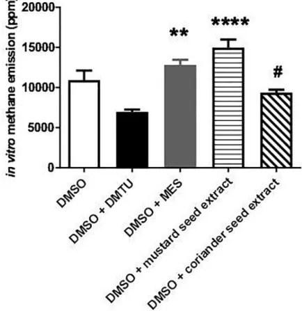
The relative effectiveness of different organosulfur compounds in terms of methanogenesis was detected in a chemical model reaction (Fig 1). DMSO is a potent CH4donor molecule in the presence of reactive oxygen species (ROS) generation [4,5] Methanogenesisin vitrowas promptly induced in the DMSO-containing tubes (10925±3139 ppm) while lower CH4emis- sion was detected in the DMSO+DMTU-containing tubes (6954±784 ppm; p = 0.164 vs DMSO). MES, however, augmented the methanogenic capacity of DMSO (12833±1666 ppm;
p =>0.9999 vs DMSO). A significant increase was detected after the administration of mus-
tard seed extract (14979±2812 ppm; p =<0.0001 vs DMSO+DMTU), while the released CH4
amount was lower in the coriander seed extract-containing tubes as compared to mustard seed
Fig 1. Methane formation in the ROS-generating Udenfriend reaction. Empty bar represents the positive control group, black bar represents the combination of DMSO + DMTU-containing group, grey bar relates to the combination of DMSO + MES-containing group, while white bar filled with light black stripes relates to the combination of DMSO + mustard seed extract-containing group and white bar filled with black diagonal lines to the DMSO + mustard seed extract-containing group. Mean values and standard deviation (SD) are given;��p<0.01 and����p<0.0001 vs. DMTU +DMSO; #p<0.01 vs. DMSO + mustard seed extract. Statistics: Kruskal-Wallis test with Dunn’s multiple comparison test.
https://doi.org/10.1371/journal.pone.0236578.g001
extract-induced generation (9364±872 ppm; p = 0.0294). Specifically, the mustard-seed extract kept up the process of methanogenesis during the 10-min observation period.
CH
4release in healthy, unexposed mice
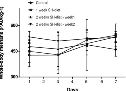
Based on thein vitrofindings, we used the mustard seed powder-containing SH-diet for fur- therin vivoexperiments. The whole-body CH4profile of healthy animals remained steadily low (Fig 2) during the observation period in all experimental groups (Group 1: day 7: 460±39 PAU/kg-1vs day 1: 427±67 PAU/kg-1; Group 2: day 7: 454±66 PAU/kg-1vs day 1: 449±63 PAU/kg-1; Group 3: day 14: 529±24 PAU/kg-1vs day 1: 476±42 PAU/kg-1).
CH
4release in mice exposed to ethanol challenge
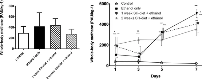
The baseline CH4values did not differ significantly and did not exceed 1547 PAU/kg-1in any of the animals on the night before the ethanol feeding protocol was started (Fig 3A). The die- tary ethanol challenge led to a significant CH4release in all groups in comparison to control (Fig 3B). 24h of ethanol consumption was already resulted in slightly elevated CH4output in the ethanol-fed group compared to the control group (day 1: Group 5: 1070±645 PAU/kg-1vs 397±276PAU/kg-1) and the whole-body CH4was significantly higher in those groups which were fed with the SH-diet (day 1: Group 6: 1864±1079 PAU/kg-1; Group 7: 2042±798PAU/kg-
1). This pattern was sustained until the 5th day of the ethanol-feeding protocol. Thereafter the highest CH4emanation was detected in animals with no antecedent SH-diet (day 5: Group 5:
3242±693PAU/kg-1vs Group 7: 2492±556 PAU/kg-1) and this was sustained thereafter (day 7:
Group 5: 5073±406 PAU/kg-1; Group 6: 4089±833PAU/kg-1; Group 7: 3540±654 PAU/kg-1).
No changes were observed in the untreated control group during the observation period (day 7: 223±107 PAU/kg-1) as compared to those were measured on day 1.
Fig 2. Whole-body CH4profile of the healthy animals during the SH-diet period on days 1, 3, 5, and 7. Black circles with continuous line relate to control group, dark grey square with continuous line to the 1-week SH-diet group. For the sake of simplicity, an individual curve with black, up-pointing triangles with continuous represents for the first half of the 2-weeks SH-diet and another individual curve with black, down-pointing triangles with continuous represents for the second half. Mean values and standard deviation (SD) are given. Statistics: Repeated measures Two- way ANOVA on ranks, Bonferroni post-hoc test.
https://doi.org/10.1371/journal.pone.0236578.g002
Total thiols and non-protein bound thiols in liver samples of the healthy, unexposed mice
The liver GSH content did not change in the SH-diet groups as compared to the control group ((Fig 4A); Group 2: 8.83±1.49μmol/mL/100 mg tissue vs Group 3: 10.3±1.62μmol/mL/100 mg tissue vs Group 1: 11.61±4.51μmol/mL/100 mg tissue). Nevertheless, the overall thiol con- centration in the liver significantly increased by the SH-diet in both groups ((Fig 4B); Group 2:
66.32±13.47μmol/mL/100 mg tissue vs Group 3: 69.24±10.07μmol/mL/100 mg tissue vs.
Group 1: 48.86±7.924μmol/mL/100 mg tissue). In line with these data, the total protein con- centration of the samples in Groups 2 and 3 also increased (control vs 2-weeks SH-diet p = 0.0441,data not shown) which might also reflect alterations in the SH-containing amino acid metabolism caused by the diet.
GSH/GSSG ratio in liver samples after ethanol challenge
The ethanol challenge led to a significant increase in the GSH/GSSGratio as compared to the control group (Group 5: 3.777±0.256μmol/L/100 mg tissue vs Group 4: 2.809±0.611μmol/L/
100 mg tissue). The 1-week or 2-weeks-SH-diet attenuated this effect significantly and kept the GSH/GSSG ratio at the control level (Group 6: 3.258±0.789μmol/L/100 mg tissue vs Group 4;
Group 7: 3.09±0.317μmol/L/100 mg tissue vs Group 4) (Fig 5).
NOX activity of liver samples
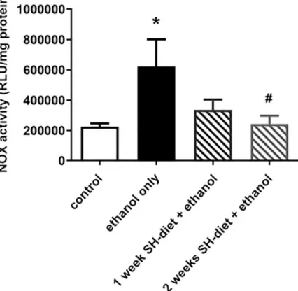
NOX is a family of membrane-bound oxidoreductase complexes whose main function is the catal- ysis of the reduction of oxygen (2NADPH +2O2—>2NADP++ 2H++ 2O2-
—>2NADP++ H2O2). Due to ethanol consumption, an elevation in NOX activity was detected as compared to control (Group 5: 622069±474047μmol/ml/mg protein vs Group 4: 225707±51216μmol/ml/mg
Fig 3. A) Baseline whole-body CH4emission in mice on the day before the ethanol-feeding was started. Mean values and standard deviation (SD) are given, significant differences were not observed. Empty bar represents the untreated control group, black bar represents the ethanol-treated group with no antecedent SH-diet, white bar filled with dark grey diagonal lines relates to the 1 week-SH- diet + ethanol treated group, while white bar filled with light grey diagonal lines to the 2 weeks-SH-diet + ethanol treated group.
Statistics: Kruskal-Wallis test with Dunn’s multiple comparison test. B) Whole body CH4production during the ethanol-feeding protocol on days 1, 3, 5, and 7. Empty circles with continuous line relate to control group, black square with continuous line to the 12%
ethanol-treated group with no antecedent SH-diet, dark grey triangles with dashed line to the 1 week-SH-diet + ethanol treated group, while light grey triangles with dashed line to the 2 weeks-SH-diet + ethanol treated group. On the day of measurements all the rats were already given 12% ethanol for 24 hrs, except for those in the control group. Mean values and standard deviation (SD) are given;���/��/� p<0.001/0.01/0.05 vs. control; ###/##/# p<0.001/0.01/0.05 vs. ethanol only. Statistics: Repeated measures Two-way ANOVA on ranks, Bonferroni post-hoc test.
https://doi.org/10.1371/journal.pone.0236578.g003
Fig 4. A) Total GSH content of liver samples of healthy animals. Empty bar indicates values for the control group, black bar represents data after 1-week SH-diet, while grey bar represents data after 2-weeks SH-diet. Mean values and standard deviation (SD) are given. Statistics: Kruskal-Wallis test with Dunn’s multiple comparison test. B) Total amount of SH-group-containing molecules in liver samples of healthy animals. Empty bar indicates values for the control group, black bar represents data after 1-week SH-diet, while grey bar represents data after 2-weeks SH-diet.
Mean values and standard deviation (SD) are given,�p<0.05 vs. control. Statistics: Kruskal-Wallis with Dunn’s multiple comparison test.
https://doi.org/10.1371/journal.pone.0236578.g004
Fig 5. GSH/GSSG ratio in liver samples. Empty bar represents the untreated control group, black bar represents the ethanol-treated group with no antecedent SH-diet, white bar filled with dark grey diagonal lines relates to the 1 week- SH-diet + ethanol-treated group, while white bar filled with light grey diagonal lines to the 2 weeks-SH-diet + ethanol- treated group. Mean values and standard deviation (SD) are given,�p<0.05 vs. control. Statistics: Kruskal-Wallis with Dunn’s multiple comparison test.
https://doi.org/10.1371/journal.pone.0236578.g005
protein, p = 0.0174). This increase was remarkably moderated by the 2-weeks SH-diet as the con- trol level was reached in this group (Group 7: 243191±143215μmol/ml/mg protein; p = 0.079, p = 0.04 vs Group 5) (Fig 6).
Discussion
SH-containing amino acids and their metabolites can be involved in the process ofin vitro, aerobic methanogenesis [4]. Taking these findings into consideration we hypothesized that mustard seed extract could act as a potential source of non-microbial CH4formation. After confirming this possibility,in vivoexperiments were performed with dietary SH-enrichment in non-CH4producer rodents with or without ethanol stress. Changes in liver thiol concentra- tions and representative oxido-reductive parameters were detected in order to shed light on the mechanistic aspects of the methanogenic reaction. On the one hand, the concentration of thiol-containing molecules in the liver was significantly increased, suggesting an influence on biothiol tissue storage. On the other hand, the SH-dietper sedid not influence the net CH4
output which might suggested that the onset of oxido-reductive imbalance is needed for an amplified CH4release. As expected, when these animals were involved in the high-dose etha- nol-feeding protocol, methanogenesis was induced. The SH-diet remarkably increased the CH4release after 24h of ethanol consumption, and this effect was not observed in animals kept on standard feeding. What is more, the CH4production declined in the SH-enriched diet groups by the end of the ethanol protocol possibly due to pertaining tissue protective events.
Fig 6. NOX activity in the liver. Empty bar represents the untreated control group, black bar represents the ethanol- treated group with no antecedent SH-diet, white bar filled with dark grey diagonal lines relates to the 1 week-SH-diet + ethanol-treated group, while white bar filled with light grey diagonal lines to the 2 weeks-SH-diet + ethanol-treated group. Mean values and standard deviation (SD) are given.�p<0.05 vs control; #p<0.05 vs. ethanol only. Statistics:
Kruskal-Wallis with Dunn’s multiple comparison test.
https://doi.org/10.1371/journal.pone.0236578.g006
In contrast, in groups with no extra biothiol feeding, the highest CH4production was reached by the later time point. The time course of the methanogenic reaction was somewhat different to that observed in a previousin vivostudy [7] but this can be explained by interspecies differ- ences to ethanol-induced adaptation as demonstrated recently by a large-scale work of Adkins et al. on cross-species comparisons of ethanol-responsive genes [14].
One of the consequences of alcohol consumption is NADH+H+production which leads to reductive stress and a critical disruption of the cellular redox balance [15]. Althoff and co- workers demonstrated that non-microbial methanogenesis can take place in a biphasic oxida- tion/sulfoxidation of methyl sulfides, plausible only in highly reductive environment [4]. As sulfoxidation of methyl sulfides is ubiquitous in the environment in the presence of high level of ROS, this chemical route is most likely to facilitate CH4release in living aerobic organisms.
This concept can explain the ethanol-induced CH4generation, but further experimental data are clearly needed to shed more light on the underlying organosulfur biochemistry. It should be added that NOX is the major source of ROS and play predominant roles in the pathogenesis of early alcohol-induced hepatic injury [16]. More importantly, the liver NOX activity was sig- nificantly elevated after the ethanol challenge; however, it was moderated in the SH-diet groups.
To-date many studies have examined the effects of high intakes of SH amino acids (i.e.
methionine and cysteine) on growth and life cycle in rodents [17]. A limited number of studies have also been conducted on the impact on inflammatory responses [18] and thein vitro effects of metabolic products of SH-amino acids on human immune cells [19]. Cellular con- centrations of biothiols and GSH have been linked to T-cell numbers [20]; the tissue biothiol content responds to SH-amino acid intake by a broad range of intra/intercellular signaling pathways involving metabolization of the essential cofactors leading to GSH production (e.g.
vitamin B6, riboflavin, and folic acid). Administration of thiol-containing GSH precursors as n-acetyl cysteine (NAC) or L-2-oxothiazolidine-4-carboxylate (OTC), are of interest in nutri- tion research [21]. It has been shown that a low-protein diet would suppress GSH synthesis, a situation that is reversed by cysteine or methionine [22]. Despite these supportive data, the impact of a high SH- amino acid diet on established inflammatory responses has not been investigated in any depth in humans or experimental animals.
The anti-inflammatory potential of organosulfur-containing plants such as mustard, horse- radish or wasabi is usually linked to the AITC content of these herbs [9,23] and it has been demonstrated that the nuclear factor erythroid 2–related factor 2 (Nrf2) and NF-κB routes are involved in the signaling pathways [23]. Several lines of evidence indicate that Nrf2 plays a key role in the regulation of cellular SH-amino acid/GSH turnover and controlled GSH/GSSG ratio as well.In vitro, Nrf2 regulates the cysteine/glutamate exchange transporter that main- tains intracellular GSH levels by allowing cysteine influx [24]. Besides, Nrf2 regulates glutathi- one peroxidase (GPX2) and glutathione-S-transferase (GST) in human alveolar epithelial cells [25]. In addition, higher levels of GSSG was detected inNrf2−/−alveolar macrophages than in wild-type Nrf-2 cells in response to oxido-reductive stress stimuli [26]. In the current study, we demonstrated a significant contribution of SH-diet on liver biothiol storage as the quantity of molecules with SH residues elevated significantly after 7 days of feeding. Interestingly, the tissue non-protein-bound thiol content (which is represented in 95% by GSH) did not alter in a same manner although the total protein concentration correlated positively with the changes of TSH, indicating that the liver antioxidant capacity was embedded to its proteome, rather than in free GSH. The SH-diet contained a relatively high concentration (10%) of crashed mustard-seed, thoroughly mixed and enriched into the standard laboratory chow which enabledad libitumconsumption. Others have already reported on the use of 5 to 10% mus- tard-seed enriched diet with anti-inflammatory and anti-carcinogenic effects in mice [27].
Upon the emerging reductive stress of the ethanol challenge, these biochemical routes were possibly activated to potentiate the metabolization of GSH from its amino-acid precursors.
This possibility is supported by the elevated GSH/GSSG ratio in the liver samples in the con- ventional diet-fed group after the ethanol protocol, as a higher ratio of GSH to GSSG suggests a reductive environment [28]. This elevation was not present in the mustard-seed diet groups and the GSH/GSSG ratios remained at the control levels. The accumulation of GSH can be linked to ethanol-evoked over-expression of Nrf2 as reported in similar stress conditions [29].
Nevertheless, further studies are necessary to investigate the direct interactions between the inflammatory trigger, non-microbial methane generation, AITC concentration and Nfr2 and GST/GPX2 expression patterns.
Conclusion
In summary, the present study providesin vivoevidence for the possibility of an SH-enriched diet to trigger non-microbial methanogenesis during ethanol feeding. The potential benefit of this approach is to influence a condition associated with redox imbalance, it might be a reason- able choice in cases where tissue hypoxia-related complications are predicted.
Supporting information
S1 Fig. Protocols for thein vivoexperiments. In Phase I, the effects of SH-diet (laboratory chow enriched with 10% mustard-seed) without ethanol feeding was monitored. The animals in control Group 1 were fed with standard laboratory chow for 7 days. In Groups 2 and 3, the animals received SH-diet for 7 or 14 days, respectively. In Phase II, the effect of SH-diet com- bined with ethanol challenge was investigated. These mice were fed with standard laboratory chaw for 7 days (Groups 4 and 5) or SH-diet for 7 or 14 days (Groups 6 and 7). After this period, the animals were provided drinking water (Group 4) or water containing 12% ethanol (Groups 5–7) for further 7 days while fed with standard laboratory chow.
(TIF)
S2 Fig. Summary of findings. Administration of plant extracts containing organosulfur (SH) moieties can increase the thiol content of the liver without influencing baseline methanogen- esis. Ethanol consumption induces hepatic oxido-reductive-stress condition, which is linked to increased methanogenesis, most probably a potentially tissue-protective mechanism. Pre- ceding oral SH feeding can effectively reduce the ethanol-induced hepatic injury.
(TIFF)
Acknowledgments
The authors are grateful to Nikolett Beretka, Csilla Mester, Ka´roly To´th and Pe´ter Sa´rka´ny for their skillful assistance.
Author Contributions Conceptualization: Eszter Tuboly.
Data curation: Petra Varga, Noe´mi Vida, Eszter Tuboly.
Formal analysis: Petra Varga, Noe´mi Vida, Eszter Tuboly.
Funding acquisition: Petra Hartmann, Miha´ly Boros, Eszter Tuboly.
Investigation: Petra Varga, Noe´mi Vida, Eszter Tuboly.
Methodology: Petra Varga, Noe´mi Vida, Anna Szabo´, A´ rpa´d Moha´csi, Ga´bor Szabo´, Eszter Tuboly.
Software: Anna Szabo´, A´ rpa´d Moha´csi, Ga´bor Szabo´.
Supervision: Petra Hartmann, A´ rpa´d Moha´csi, Ga´bor Szabo´, Miha´ly Boros.
Validation: A´ rpa´d Moha´csi.
Writing – original draft: Petra Varga, Eszter Tuboly.
Writing – review & editing: Petra Hartmann, Miha´ly Boros.
References
1. Roccarina D, Lauritano EC, Gabrielli M, Franceschi F, Ojetti V, Gasbarrini A. The role of methane in intestinal diseases. Am J Gastroenterol. 2010; 6, 1250–1256.https://doi.org/10.1038/ajg.2009.744 PMID:20216536
2. Keppler F, Hamilton JT, Brass M, Rockmann T. Methane emissions from terrestrial plants under aerobic conditions. Nature. 2006; 187–191.https://doi.org/10.1038/nature04420PMID:16407949
3. Tuboly E, Szabo´ A, Garab D, Bartha G, Janovszky A´ , Erős G, et al. Methane biogenesis during sodium azide-induced chemical hypoxia in rats. Am J Physiol Cell Physiol. 2013; C207–214.https://doi.org/10.
1152/ajpcell.00300.2012PMID:23174561
4. Althoff F, Benzing K, Comba P, McRoberts C, Boyd DR, Greiner S, et al. Abiotic methanogenesis from organosulfur compounds under ambient conditions. Nat Commun. 2014; 5, 4205.https://doi.org/10.
1038/ncomms5205PMID:24957135
5. Ghyczy M, Torday C, Boros M. Simultaneous generation of methane, carbon dioxide, and carbon mon- oxide from choline and ascorbic acid: a defensive mechanism against reductive stress? FASEB J. 17, 2003; 1124–1126.https://doi.org/10.1096/fj.02-0918fjePMID:12692080
6. Boros M, Tuboly E, Me´ sza´ros A, Amann A. The role of methane in mammalian physiology-is it a gaso- transmitter? J Breath Res. 1. 2015; 014001.https://doi.org/10.1088/1752-7155/9/1/014001PMID:
25624411
7. Tuboly E, Molna´r R, Tőke´s T, Tura´nyi RN, Hartmann P, Me´sza´ros AT, et al. Excessive alcohol con- sumption induces methane production in humans and rats. Sci Rep. 2017; 1, 7329.https://doi.org/10.
1038/s41598-017-07637-3PMID:28779149
8. Boros M, Ghyczy M, E´ rces D, Varga G, Tőke´s T, Kupai K. The anti-inflammatory effects of methane.
Crit Care Med. 2012; 4, 1269–1278.https://doi.org/10.3892/ol.2012.929
9. European Food Safety Authority. Scientific Opinion on the safety of allyl isothiocyanate for the proposed uses as a food additive. EFSA Journal. 2010; 12, 1943.https://doi.org/10.2903/j.efsa.2010.1943 10. Dermikol O, Adams C, Ercal N. Biologically Important Thiols in Various Vegetables and Fruits. J. Agric.
Food Chem. 2004; 52, 8151−8154.https://doi.org/10.1021/jf040266fPMID:15612810
11. Tuboly E, Szabo´ A, Erős G, Moha´ csi A, Szabo´ G, Tengo¨lics R, et al. Determination of endogenous methane formation by photoacoustic spectroscopy. J Breath Res. 2013; 7, 046004.https://doi.org/10.
1088/1752-7155/7/4/046004PMID:24185326
12. Ellman GL. Tissue sulfhydryl groups. Arch Biochem Biophys. 1959; 1, 70–77.https://doi.org/10.1016/
0003-9861(59)90090-6
13. Sedlak J, Lindsay RH. Estimation of total, protein-bound, and nonprotein sulfhydryl groups in tissue with Ellman’s reagent. Anal Biochem. 1968; 1, 192–205.https://doi.org/10.1016/0003-2697(68)90092-4 14. Adkins AE, Hack LM, Bigdeli TB, Williamson VS, McMichael GO, Mamdani M, et al. Genomewide Asso-
ciation Study of Alcohol Dependence Identifies Risk Loci Altering Ethanol-Response Behaviors in Model Organisms. Alcohol ClinExp Res. 2017; 5, 911–928.https://doi.org/10.1111/acer.13362PMID:
28226201
15. Houtkooper RH, Canto C, Wanders RJ, Auwerx J. The secret life of NAD + : an old metabolite control- ling new metabolic signaling pathways. Endocr Rev. 2010; 31, 194–223.https://doi.org/10.1210/er.
2009-0026PMID:20007326
16. Kono H, Rusyn I, Yin M, Ga¨bele E, Yamashina S, Dikalova A, et al. NADPH oxidase-derived free radi- cals are key oxidants in alcohol-induced liver disease. J Clin Invest. 2000; 7, 867–872.https://doi.org/
10.1172/JCI9020PMID:11018074
17. Harper AE, Benevenga NJ, Wohlheuter RM. Effects of ingestion of disproportionate amounts of amino acids. Physiol. Rev. 1970; 50, 428–558.https://doi.org/10.1152/physrev.1970.50.3.428PMID:
4912906
18. Grimble RF, Grimble GK. Immunonutrition: role of sulfur amino acids, related amino acids, and poly- amines. Nutrition. 1998; 14, 605–610.https://doi.org/10.1016/s0899-9007(98)80041-5PMID:9684264 19. Grimble RF. The Effects of Sulfur Amino Acid Intake on Immune Function in Humans. J Nutr. 2006; 6,
1660S–1665S.https://doi.org/10.1093/jn/136.6.1660SPMID:16702336
20. Kinscherf R, Fischbach T, Mihm S, Roth S, Hohen-Sievert E, Weiss C, et al. Effect of glutathione deple- tion and oral N-acetyl-cysteine treatment on CD4+ and CD8+ cells. FASEB J. 1994; 8, 448–451.
https://doi.org/10.1096/fasebj.8.6.7909525PMID:7909525
21. Ferreira LF, Gilliam LA, Reid MB. L-2-Oxothiazolidine-4-carboxylate reverses glutathione oxidation and delays fatigue of skeletal muscle in vitro. J ApplPhysiol. (1985). 2009; 1, 211–216.https://doi.org/10.
1152/japplphysiol.00001.2009PMID:19407260
22. Hunter EA, Grimble RF. Cysteine and methionine supplementation modulate the effect of tumor necro- sis factorαon protein synthesis, glutathione and zinc content of tissues in rats fed a low-protein diet. J Nutr. 1994; 124, 2319–2328.https://doi.org/10.1093/jn/124.12.319PMID:16856311
23. Ernst IM, Wagner AE, Schuemann C. Allyl-, butyl- and phenylethyl-isothiocyanate activate Nrf2 in cul- tured fibroblasts. Pharmacol Res. 2011; 63, 233–240.https://doi.org/10.1016/j.phrs.2010.11.005 PMID:21109004
24. Thimmulappa RK, Lee H, Rangasamy T, Reddy SP, Yamamoto M, Kensler TW, et al. Nrf2 is a critical regulator of the innate immune response and survival during experimental sepsis. J Clin Invest. 2006;
4, 984–995.https://doi.org/10.1172/JCI25790PMID:16585964
25. Sasaki H, Sato H, Kuriyama-Matsumura K, Sato K, Maebara K, Wang H, et al. Electrophile response element-mediated induction of the cystine/glutamate exchange transporter gene expression. J Biol Chem. 2002; 47, 44765–44771.6.https://doi.org/10.1074/jbc.M208704200PMID:12235164 26. Singh A, Rangasamy T, Thimmulappa RK, Lee H, Osburn WO, Brigelius-Flohe´ R, et al. Glutathione
peroxidase 2, the major cigarette smoke-inducible isoform of GPX in lungs, is regulated by Nrf2. Am J Respir Cell Mol Biol. 2006; 36, 639–650.https://doi.org/10.1165/rcmb.2005-0325OCPMID:16794261 27. Yuan H, Zhu M, Guo W, Jin L, Chen W, Brunk UT, et al. Mustard seeds (Sinapis Alba Linn) attenuate
azoxymethane-induced colon carcinogenesis. Redox Rep. 2011; 1, 38–44.https://doi.org/10.1179/
174329211X12968219310918PMID:21605497
28. Pe´rez-Torres I, Guarner-Lans V, Rubio-Ruiz ME. Reductive Stress in Inflammation-Associated Dis- eases and the Pro-Oxidant Effect of Antioxidant Agents. Int J Mol Sci. 2017; 5, 18(10).https://doi.org/
10.3390/ijms18102098PMID:28981461
29. Narasimhan M, Mahimainathan L, Rathinam ML, Riar AK, Henderson GI. Overexpression of Nrf2 pro- tects cerebral cortical neurons from ethanol-induced apoptotic death. Mol Pharmacol. 2011; 6, 988–
999.https://doi.org/10.1124/mol.111.073262PMID:21873460