Safety, Clinical Outcome, and Fracture Rate of Femoropopliteal Stenting Using a 4F Compatible Delivery System
H. Sarkadia, V. Bérczib, A. Kollárb, D. Kissb, P. Jakabfib, E.M. Végha, B. Nemesa, B. Merkely a, K. Hüttla, E. Dósaa,*
aHeart and Vascular Center, Semmelweis University, Városmajor Street 68, 1122 Budapest, Hungary
bDepartment of Radiology and Oncotherapy, Semmelweis University, Budapest, Hungary
WHAT THIS PAPER ADDS
Minimally invasive interventional procedures tend to be even less invasive. In this spirit, 4F sheath compatible femoropopliteal stents have been developed. The number of studies regarding the safety, clinical outcome, and fracture rate of 4F stents is limited. Access site complications occurred in 2% of the patients. The overall primary patency rate was 80% with 85% freedom from target lesion revascularization at 12 months. The incidence of stent fractures was 26% after an average follow up of 25 months. These results confirm the validity of 4F stents in patients with femoropopliteal occlusive disease.
Objective:To determine the safety, clinical outcome, and fracture rate of femoropopliteal interventions using 4F stents.
Methods:Between January 2010 and December 2011, 112 symptomatic patients were treated by stent implantation. Ten patients were lost to follow up; therefore, 102 patients (62 men; mean age 66.410.1 years) were retrospectively analyzed. The indication for femoropopliteal revascularization was severe claudication (RutherfordeBecker score¼3) in 63 (62%) patients and chronic critical limb ischemia (RutherfordeBecker score¼4e6) in 39 (38%). Follow up included palpation of peripheral pulses and measurement of ankle brachial index. In patients with suspected in-stent restenosis duplex ultrasonography was performed. In 2013, patients were asked to return for afluoroscopic examination of the stents.
Results:114 lesions (Trans-Atlantic InterSociety Consensus-C and D,n¼45) were treated with 119 stents (Astron Pulsar,n¼42; Pulsar-18,n¼77). Lesions were long (100 mm) in 49 cases and heavily calcified in 35. Stents were long (120 mm) in 46 cases. Ten stents were partially overlapped. The technical and clinical success rates were 100%. Two puncture related complications were noted, neither of which required surgical repair. Eleven patients died (myocardial infarction,n¼4; stroke,n¼2; cancer, n¼5) and nine patients underwent major amputation (above knee,n¼4). The primary patency rate was 83% at 6 months and 80% at 12 months. The primary assisted patency rate was 97% at 6 months and 94% at 12 months. The secondary patency rate was 86%
at 6 months and 85% at 12 months. The prevalence of fractures was 26% (type III and IV, 10%) after an average follow up of 25 months.
Conclusion:Femoropopliteal stenting using a 4F compatible delivery system can be accomplished with a low complication rate, acceptable fracture rate, and with similar 12 month patency and revascularization rates as their 6F counterparts.
Ó2014 European Society for Vascular Surgery. Published by Elsevier Ltd. All rights reserved.
Article history: Received 18 August 2014, Accepted 1 December 2014, Available online 8 January 2015 Keywords:4F stents, Femoropopliteal stenting, In-stent restenosis, Patency, Stent fracture
INTRODUCTION
Nowadays, femoropopliteal interventions are one of the most commonly performed endovascular procedures. If the stenosis is suitable for endosurgery, percutaneous trans- luminal angioplasty (PTA) should be the treatment offirst
choice. Stent placement is reserved for patients with unfa- vorable lesion morphology (heavily calcified lesions, total occlusions) and suboptimal PTA results (residual stenosis 30%,flow limiting dissection). Over the years, significant changes in the structure and design of vascular stents to improve their radial force,flexibility, and fracture resistance have been made. However, until a few years ago, femo- ropopliteal stenting has been performed using 6F sheaths.
Recently, self expanding stents that are compatible with 4F delivery devices have become available. However, with the exception of the 4-EVER (4F Endovascular Treatment Approach to Infrainguinal Disease) and PEACE I (Patency
* Corresponding author.
E-mail address:dosaedit@yahoo.com(E. Dósa).
1078-5884/Ó2014 European Society for Vascular Surgery. Published by Elsevier Ltd. All rights reserved.
http://dx.doi.org/10.1016/j.ejvs.2014.12.004
Evaluation After Implantation of the Pulsar-18 Self-Expand- ing Nitinol Stent in the Superficial Femoral and Popliteal Arteries) trials,1,2 no comprehensive data have been pub- lished on 4F stents in patients with femoropopliteal disease.
Therefore, the aim of this retrospective study was to determine the safety, clinical outcome, and fracture rate of femoropopliteal stenting using a 4F compatible delivery system.
PATIENTS AND METHODS
Patient selection
The number of patients having any form of endovascular intervention to the femoropopliteal segment between January 2010 and December 2011 was 441. In this retro- spective study only those 112 consecutive patients with severe claudication (RutherfordeBecker score ¼ 3) or chronic critical limb ischemia (CLI; Rutherford-Becker score ¼ 4e6) who underwent femoropopliteal stenting using a 4F compatible delivery system were analyzed.3The indication for stent placement was suboptimal PTA due to unfavorable lesion morphology (heavily calcified lesions, total occlusions) or failed angioplasty (residual stenosis 30%, flow limiting dissection). The procedures were per- formed at the Heart and Vascular Center and at the Department of Radiology and Oncotherapy of the Sem- melweis University (Budapest, Hungary).
Stenting protocol
The pre-procedural work up included collection of clinical data (symptoms, risk factors, medical history), palpation of femoropopliteal and foot pulses, measurement of ankle brachial index (ABI), and assessment of lower extremity arterial disease with duplex ultrasonography or computed tomography angiography.
The procedures were carried out through the femoral artery, via an ipsilateral antegrade approach in 79 cases and via a contralateral crossover approach in 31. In two patients an ipsilateral retrograde approach was necessary (one through the popliteal artery [PA] and one through the dorsalis pedis artery) because of failed recanalization through the femoral artery. A 4F introducer sheath was used in all cases.
After selective angiography, a 0.035 inch or 0.018 inch guidewire was advanced into the punctured artery to pass through the femoropopliteal stenosis or occlusion. Lesions were classified according to the Trans-Atlantic InterSociety Consensus (TASC II) document.4 Beside the TASC classifi- cation, lesions were considered long if their length was 100 mm, and were defined as heavily calcified if calcifi- cation was visible over>75% of the length of the stenotic or occluded segment byfluoroscopy.
The diameter of the patent segment and length of the lesion were measured on baseline angiograms for accurate sizing of the balloons and stents. The angioplasty balloons were Passeo-18 (4e640e200 mm; Biotronik AG, Bülach, Switzerland) in 82 patients and Fox SV (5e640e120 mm;
Abbott Vascular Inc., Santa Clara, CA, USA) in 32. Self- expanding nitinol stents (Astron Pulsar, n¼42; Pulsar-18, n¼77 [Biotronik AG]) were implanted in all cases. Stents were considered long if their length was120 mm. At the end of the procedure completion angiography was per- formed, the punctured artery was compressed manually, and an overnight pressure bandage was then applied. Pa- tients were discharged 1e2 days after the procedure.
Technical success was defined as<30% residual stenosis without dissection or extravasation, whereas clinical success meant improvement or resolution of the symptoms.
Follow up
Follow up visits were scheduled at 4 weeks, 3e6, and 12 months after stenting and annually thereafter. Follow up examinations included evaluation of symptoms, palpation of femoropopliteal and foot pulses, and measurement of ABI. In patients with worsened symptoms, impalpable popliteal pulse, and ABI0.5 significant in-stent restenosis (ISR) was suspected. The presence of ISR was verified with duplex ultrasonography.
Patency rates were expressed as primary (those remain- ing patent without further intervention), primary assisted (those patent with additional intervention to maintain patency), and secondary (those in which patency needed to be re-established by another intervention).5 Target lesion revascularization was defined as any repeat percutaneous intervention of the target lesions (including 5 mm proximal and distal to the stent) or surgical bypass of the target vessel performed for restenosis or other complication involving the target lesion, whereas target vessel revascu- larization meant any repeat percutaneous intervention or surgical bypass of any segment of the target vessel.6
In 2013, patients were asked to return for an additional follow up visit, whenfluoroscopic stent fracture evaluation was also performed in addition to the above mentioned examinations. The high magnification fluoroscopic exami- nations were done in the angiography suite (AXIOM Artis FA; Siemens Medical Solutions AG Corp., Erlangen, Ger- many) with the following parameters: 7.5 fps, 100e125 kV, and 550e800 mA (Heart and Vascular Center); or in an examination room equipped with an X-ray system using a digital flat panel (Carestream DRX-1 System; Carestream Health Inc., Rochester, NY, USA) with the following param- eters: 60e70 kV, 63 mA with grid (Department of Radiology and Oncotherapy). To visualize the implanted stents, three cine loops with a length of three cardiac cycles (Heart and Vascular Center) or digital X-rays (Department of Radiology and Oncotherapy) were recorded in postero-anterior, and right and left anterior oblique 30e45 projections. The post- processing was performed on a Leonardo Workstation (Syngo 2003; Siemens Medical Solutions) (Heart and Vascular Center) or on a PACS Workstation (IMPAX 6.5.2;
Agfa HealthCare NV Corp., Mortsel, Belgium) (Department of Radiology and Oncotherapy). The fluoroscopic images were analyzed by two experienced interventional radiolo- gists (E.D., B.N.) in consensus. Stent fractures were defined
according to a nitinol stent fracture classification that has been proposed by the Cardiovascular Institute of the South (Houma, LA, USA).7 Briefly, type I ¼single strut fracture, type II ¼ multiple strut fractures at different locations in the stent, type III ¼ multiple strut fractures resulting in complete transverse fracture of the stent, and type IV ¼ complete transverse fracture with subsequent stent separation. Thefluoroscopic examination of the implanted stents was conducted in accordance with the Declaration of Helsinki. This study was approved by the ethics committee of the Semmelweis University and informed consent was obtained from every patient.
Statistical analysis
Statistical analysis was performed with SPSS 21.0 software (IBM, Armonk, NY, USA). Categorical variables were expressed as numbers and percentages, and were compared between two groups using the chi-square test or the Fisher’s exact test. Continuous variables were expressed as meansSDs and were compared between two groups using the Studentttest or the ManneWhitneyUtest. The patency rates were analyzed using the KaplaneMeier technique. Univariate logistic regression model was used to identify independent predictors of ISR and stent fracture.
All analyses were two tailed, and values of p .05 were considered to be statistically significant.
RESULTS
Patient data
Ten patients were lost to follow up; therefore, they were excluded from the analysis. The mean age of the remaining 102 patients (40 women, 62 men) was 66.4 10.1 years (range 39e87 years). The indication for femoropopliteal revascularization was severe claudication in 63 (62%) patients and chronic CLI in 39 (38%). At the time of inter- vention, none of the patients had significant ipsilateral iliac or common femoral artery stenosis. Atherosclerotic risk factors included smoking in 89 (87%) patients, hypertension in 96 (94%), hyperlipidemia in 41 (40%), diabetes mellitus in 42 (41%), obesity in 44 (43%), and chronic kidney disease in 16 (16%). According to the medical history, 27 (26%) patients had coronary artery bypass grafting and/or percutaneous coronary intervention, 23 (23%) had supra- aortic surgical and/or endosurgical reconstruction, and 21 (21%) had lower extremity open and/or percutaneous revascularization.
Lesion and stent characteristics
One hundred and fourteen lesions were treated with 119 stents. The stented lesions were de novo stenosis in 87 (76%) cases and recurrent stenosis after previous PTA in 27 (24%). Sixty four (56%) of the treated lesions were stenoses and 50 (44%) were total occlusions. Lesions were long in 49 (43%) cases and heavily calcified in 35 (31%). The side dis- tribution of the lesions was almost equal (left,n¼58; right, n ¼ 56). Lesions were located in the superficial femoral
artery (SFA) in 36 (32%) cases, in the femoropopliteal transitional zone in 47 (41%), and in the PA in 31 (27%). In 17 (15%) patients crural interventions were also performed.
According to the TASC classification, the lesions were TASC- A, TASC-B, TASC-C, and TASC-D in 35 (31%), 34 (30%), 30 (26%), and 15 (13%) cases, respectively (Table 1). Forty six (39%) of the implanted stents were long and 10 (8%) stents were placed in a partially overlapping position (Table 2).
Early post-procedural period (within 30 days)
The technical success rate was 100%. A femoral pseudoa- neurysm developed at the femoral puncture site in two patients; both of them were treated with ultrasound guided injection of thrombin. The 30 day all cause mortality rate was zero. Five (5%) patients underwent minor amputation (interphalangeal,n¼2; ray,n¼2; transmetatarsal,n¼1).
All patients reported improvement or resolution of the pre- procedural symptoms.
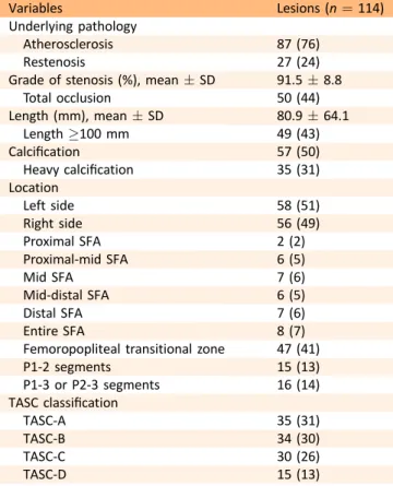
Table 1.Lesion characteristics.
Variables Lesions (n¼114)
Underlying pathology
Atherosclerosis 87 (76)
Restenosis 27 (24)
Grade of stenosis (%), meanSD 91.58.8
Total occlusion 50 (44)
Length (mm), meanSD 80.964.1
Length100 mm 49 (43)
Calcification 57 (50)
Heavy calcification 35 (31)
Location
Left side 58 (51)
Right side 56 (49)
Proximal SFA 2 (2)
Proximal-mid SFA 6 (5)
Mid SFA 7 (6)
Mid-distal SFA 6 (5)
Distal SFA 7 (6)
Entire SFA 8 (7)
Femoropopliteal transitional zone 47 (41)
P1-2 segments 15 (13)
P1-3 or P2-3 segments 16 (14)
TASC classification
TASC-A 35 (31)
TASC-B 34 (30)
TASC-C 30 (26)
TASC-D 15 (13)
Note. Values are given as n (%) unless otherwise indicated.
SFA ¼ Superficial femoral artery; TASC ¼ Trans-Atlantic InterSociety Consensus.
Table 2.Stent characteristics.
Variables Stents (n¼119)
Type
4F Astron Pulsar 42 (35)
4F Pulsar-18 77 (65)
Length (mm), meanSD 92.253.0
Length120 mm 46 (39)
Overlapping stents 10 (8)
Note. Values are given as n (%) unless otherwise indicated.
Follow up period
The mean follow up time was 25.36.2 months (range 3e39 months). Eleven (11%) patients died during the follow up period (acute myocardial infarction,n¼4; stroke,n ¼2;
cancer,n¼5). Significant ISR was observed in 21 (21%) pa- tients. Target lesion revascularization was carried out in 15 (15%) patients (PTA,n¼13; stenting,n¼2), while target vessel revascularization was carried out in six (6%) patients (PTA,n¼3; stenting,n¼1; surgery,n¼2). Major ampu- tation was necessary in nine (9%) patients (below knee,n¼5;
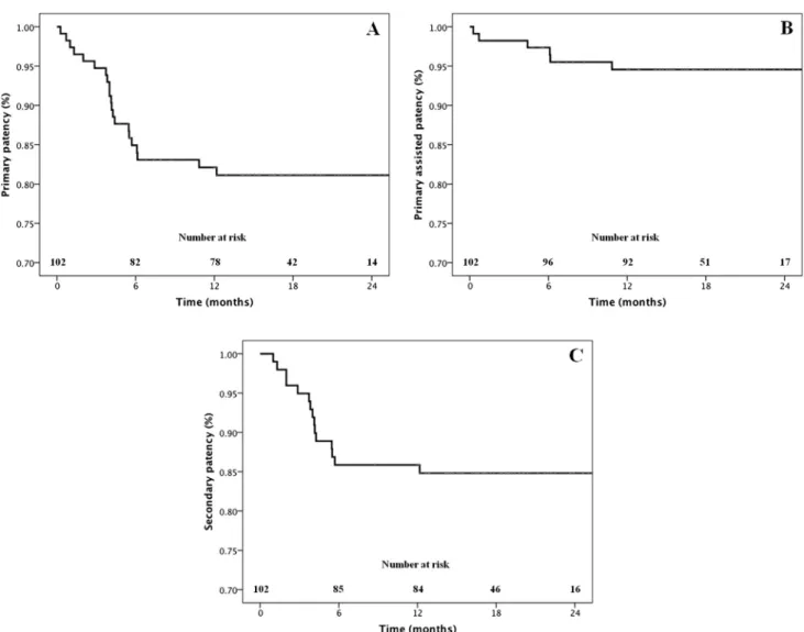
above knee,n¼4). Indications for amputation were acute ischemia in four cases and chronic ischemia infive. All stents were patent in the below knee amputation group, while all stents were blocked in the above knee amputation group. The primary patency rate was 83% at 6 months and 80% at 12 months. The primary assisted patency rate was 97% at 6 months and 94% at 12 months. The secondary patency rate was 86% at 6 months and 85% at 12 months (Fig. 1).The mean resting ABI improved from 0.50.1 before the procedure to 0.80.2 at the most recent follow up (p<.01). The mean RutherfordeBecker classification improved from 3.9 1.1 before the procedure to 2.11.4 at the most recent follow up (p<.01).
Stent fracture
Patients who died or underwent above knee amputation and who had stents that were placed during the follow up were excluded from the analysis of stent fractures. One hundred and four stents were examined. Twenty seven (26%) fractures were detected in 87 patients: fractures were type I in nine, type II in eight, type III infive, and type IV in five cases. Eighty three percent of the fractures were pre- dominantly in the middle portion of the stents. At the time of the fluoroscopic investigation, the number of patients with ISR (occurring at any time during the follow up) was significantly higher in the fractured than in the non- fractured group (n¼15 vs.n¼2;p<.01).
Predictors of ISR and stent fracture
The following variables were examined: female sex, age 70 years, smoking, hypertension, hyperlipidemia, diabetes mellitus, obesity, chronic kidney disease, total occlusion, long lesion, calcified lesion, heavily calcified lesion, TASC-A lesion, TASC-B lesion, TASC-C lesion, TASC-D lesion, prox- imal SFA stent, proximal-mid SFA stent, mid SFA stent, mid- distal SFA stent, distal SFA stent, femoropopliteal transi- tional zone stent, P1-2 stent, P1-3 or P2-3 stent, long stent,
Figure 1.(A) Primary, (B) primary assisted, and (C) secondary patency rates.
overlapping stents, Astron Pulsar stent, and Pulsar-18 stent.
Univariate analysis showed that stents placed in P1-2 location are associated with an increased incidence of ISR (odds ratio [OR] 3.83, 95% confidence interval [CI]
1.10e13.31, p¼.03). Calcified, especially heavily calcified lesions were found to be predictive of stent fracture (OR 19.64, 95% CI 4.31e89.47, p < .01; OR 116.07, 95% CI 22.57e597.03, p<.01, respectively).
DISCUSSION
Access site complications were found in 2% of the patients.
At 12 months, there was an 80% overall primary patency rate and 85% freedom from target lesion revascularization.
The incidence of stent fractures was 26% (after an average follow up of 25 months).
The number of peripheral vascular interventions is increasing annually. Besides major complications, there are additional arterial puncture related complications, such as hematoma, bleeding, arteriovenous fistula, pseudoaneur- ysm, arterial occlusion, femoral neuropathy, and infection, which, in turn, are associated with increased morbidity, mortality, and cost.8,9 Increased sheath size increases the risk of access site complications.9Up until a few years ago, femoropopliteal stenting was performed using 6F sheaths.
The reported incidence of puncture site complications during interventions through 6F sheaths varies between studies, but could be as high as 20%,9depending on the definition and criteria used. Recently, self expanding fem- oropopliteal stents that are deliverable through 4F sheaths have been introduced. Two large prospective multicenter trials (4-EVER and PEACE I) have been carried out to examine the safety and efficacy of implantation of 4F stents in patients with symptomatic femoropopliteal occlusive disease.1,2The access related complication rates were 3.3%
in the 4-EVER trial and 2.0% in the current study, and all of the complications could be managed by non-surgical treatments.1These rates are lower than most of the pub- lished rates on 6F devices.9
Regarding efficacy of the procedures, the overall 12 month primary patency rate was 81.4% in the 4-EVER trial, 79.5% in the PEACE I trial, and 80.0% in the current study, while the freedom from target lesion revascularization at 12 months was 89.3%, 81.0%, and 85.0%, respectively.1,2The slightly worse results in the PEACE I trial can be explained by the fact that the percentage of patients with TASC-D lesions and total occlusions was higher (32.2% and 56.7%, respectively) compared with the 4-EVER trial (0% and 20.8%, respectively) and the current study (13.0% and 44.0%, respectively).1,2Moreover, the treated lesions were longer in the PEACE I trial (111.5 71.4 mm) than they were in the 4-EVER trial (71.045.9 mm) and the current study (80.9 64.1 mm).1,2 Two other smaller 4F studies should be mentioned; in one of them only patients with TASC-D lesions were enrolled, while only those having long segment femoropopliteal stenosis (120 mm) were enrolled in the other.10,11 The 12 month patency and revascularization rates of these two studies are similar to
the rates of the PEACE I trial.2,10,11More importantly, these results are not worse than the reported 12 month outcomes of the 6F stents.12e18
Neointimal hyperplasia is the major cause of ISR. Patient specific characteristics (e.g., ubiquitous comorbidities, insufficient antiplatelet therapy), as well as many lesion, stent, and procedure related factors (e.g., stent type, stent design, stent configuration, stent diameter, stent length, incomplete stent apposition, incomplete stent expansion, overlapping stents) are known to precipitate ISR.19 Femo- ropopliteal ISR occurs with a frequency of 19e37% at 12 months in lesions that are<150 mm in length.14,20In the current study, the incidence of ISR was 21% and stents in P1-2 location were found to be associated with ISR. Early and Kelly have shown that certain dynamic forces, such as bending the artery during knee flexion, cause permanent deformation of the stent, which negatively affects the dynamics of blood flow in the vessel and contributes to ISR.21
Stent fractures have been extensively investigated in many vascular territories and were noted to be one of the most common in the femoropopliteal arteries, with a prevalence of 2e65%.22Here, a 26% fracture rate was re- ported after an average follow up of 25 months. A literature search for the fracture rates of 4F Astron Pulsar and Pulsar- 18 stents revealed only one study (the 4-EVER trial).1 Although, the fracture rate was 4.2% in this trial, the per- centage of patients with TASC-D lesions, total occlusions, calcified lesions, and long stents was significantly lower compared with the current study.1Femoropopliteal stents are almost continuously exposed to mechanical forces, which can themselves result in“stress”fractures, especially when stents are placed behind the knee. Patient, lesion, stent, and procedure related parameters may further in- crease the risk of fracture.23Calcified lesions were found to be predictive of fracture in the current study. Calcification due to changes in the regional wall stiffness and creating excessive focal pressure on certain parts of the stent has already been demonstrated to play a role in the develop- ment of stent fracture.22
Conclusion
Femoropopliteal stenting using a 4F sheath compatible delivery system can be accomplished with few puncture site complications and similar 12 month patency and revascu- larization rates as their 6F counterparts. The early results are promising, even if stent fractures are common, espe- cially in patients with calcified lesions. But the more important long-term outcomes are still awaited.
CONFLICT OF INTEREST None.
FUNDING
This paper was supported by the János Bolyai Research Scholarship of the Hungarian Academy of Sciences (E.D.).
REFERENCES
1 Bosiers M, Deloose K, Callaert J, Keirse K, Verbist J, Hendriks J, et al. 4French-compatible endovascular material is safe and effective in the treatment of femoropopliteal occlusive disease: results of the 4-EVER trial.J Endovasc Ther 2013;20:746e56.
2 Lichtenberg M, Kolks O, Hailer B, Stahlhoff WF, Tiefenbacher C, Nolte-Ernsting C, et al. PEACE I all-comers registry: patency evaluation after implantation of the 4french pulsar-18 self- expanding nitinol stent in femoropopliteal lesions.J Endovasc Ther2014;21:373e80.
3 Rutherford RB, Baker JD, Ernst C, Johnston KW, Porter JM, Ahn S, et al. Recommended standards for reports dealing with lower extremity ischemia: revised version.J Vasc Surg1997;26:
517e38.
4 Norgren L, Hiatt WR, Dormandy JA, Nehler MR, Harris KA, Fowkes FG. TASC II Working Group. Inter-Society Consensus for the Management of Peripheral Arterial Disease (TASC II).J Vasc Surg2007;45(Suppl.):S5e67.
5 Baum S, Pentecost MJ.Percutaneous aortoiliac intervention in vascular disease. abrams’angiography: interventional radiology.
2nd ed. Philadelphia: Lippincott Williams & Wilkins; 2005. p. 305.
6 Hicks KA, Hung HMJ, Mahaffey KW, Mehran R, Nissen SE, Stock- bridge NL, et al.Standardized definitions for end point events in cardiovascular trials. Available at: http://www.clinpage.com/
images/uploads/endpoint-defs_11-16-2010.pdf [last accessed 12.10.14].
7 Allie DE, Hebert CJ, Walker CM. Nitinol stent fractures in the SFA.Endovasc Today2004;1:22e34.
8 Koreny M, Riedmüller E, Nikfardjam M, Siostrzonek P, Müllner M.
Arterial puncture closing devices compared with standard manual compression after cardiac catheterization: systematic review and meta-analysis.J Am Med Assoc2004;291:350e7.
9 Merriweather N, Sulzbach-Hoke LM. Managing risk of compli- cations at femoral vascular access sites in percutaneous coro- nary intervention.Crit Care Nurse2012;32:16e29.
10 Lichtenberg M, Stahlhoff W, Boese D. Superficial femoral artery TASC D Registry: twelve-month effectiveness analysis of the Pulsar-18 SE nitinol stent in patients with critical limb ischemia.
J Cardiovasc Surg (Torino)2013;54:433e9.
11 Baumann F, Do DD, Willenberg T, Baumgartner I, Diehm N.
Treatment for long-segment femoro-popliteal obstructions:
initial experience with a 4F compatible self-expanding nitinol stent and review of the literature. J Cardiovasc Surg (Torino) 2012;53:475e80.
12 Bosiers M, Torsello G, Gissler HM, Ruef J, Müller-Hülsbeck S, Jahnke T, et al. Nitinol stent implantation in long superficial
femoral artery lesions: 12-month results of the DURABILITY I study.J Endovasc Ther2009;16:261e9.
13 Schulte KL, Müller-Hülsbeck S, Cao P, Becquemin JP, Langhoff R, Charalambous N, et al. MISAGO 1:first-in-man clinical trial with Misago nitinol stent.Eurointervention2010;5:687e91.
14 Laird JR, Katzen BT, Scheinert D, Lammer J, Carpenter J, Buchbinder M, et al. Resilient Investigators. Nitinol stent im- plantation versus balloon angioplasty for lesions in the super- ficial femoral artery and proximal popliteal artery: twelve- month results from the RESILIENT randomized trial. Circ Car- diovasc Interv2010;3:267e76.
15 Dake MD, Scheinert D, Tepe G, Tessarek J, Fanelli F, Bosiers M, et al. Zilver PTX Single-Arm Study Investigators. Nitinol stents with polymer-free paclitaxel coating for lesions in the superfi- cial femoral and popliteal arteries above the knee: twelve- month safety and effectiveness results from the Zilver PTX single-arm clinical study.J Endovasc Ther2011;18:613e23.
16 Bosiers M, Deloose K, Callaert J, Moreels N, Keirse K, Verbist J, et al. Results of the Protégé EverFlex 200-mm-long nitinol stent (ev3) in TASC C and D femoropopliteal lesions. J Vasc Surg 2011;54:1042e50.
17 Schulte KL, Kralj I, Gissler HM, Bagnaschino LA, Buschmann I, Pernès JM, et al. MISAGO 2: one-year outcomes after im- plantation of the Misago self-expanding nitinol stent in the superficial femoral and popliteal arteries of 744 patients.
J Endovasc Ther2012;19:774e84.
18 Matsumura JS, Yamanouchi D, Goldstein JA, Pollock CW, Bosiers M, Schultz GA, et al. The United States StuDy for EvalUating EndovasculaR TreAtments of Lesions in the Super- ficial Femoral Artery and Proximal Popliteal By usIng the Pro- tégé EverfLex NitInol STent SYstem II (DURABILITY II).J Vasc Surg2013;58. 73e83.e1.
19 Razzouk L, Aggarwal S, Gorgani F, Babaev A. In-stent restenosis in the superficial femoral artery.Ann Vasc Surg2013;27:510e24.
20 Schillinger M, Sabeti S, Loewe C, Dick P, Amighi J, Mlekusch W, et al. Balloon angioplasty versus implantation of nitinol stents in the superficial femoral artery.N Engl J Med2006;354:1879e88.
21 Early M, Kelly DJ. The consequences of the mechanical envi- ronment of peripheral arteries for nitinol stenting. Med Biol Eng Comput2011;49:1279e88.
22 Rits J, van Herwaarden JA, Jahrome AK, Krievins D, Moll FL. The incidence of arterial stent fractures with exclusion of coronary, aortic, and non-arterial settings. Eur J Vasc Endovasc Surg 2008;36:339e45.
23 Neil N. Stent fracture in the superficial femoral and proximal popliteal arteries: literature summary and economic impacts.
Perspect Vasc Surg Endovasc Ther2013;25:20e7.