---
Surgical Alternatives in the Therapy of Hypopharyngeal Tumours of Various Locations - 30 Years (1986-2018) of Experience at the Department of Otorhinolaryngology, Head, and
Neck Surgery, University of Szeged, Hungary
Iván László1*, Gholam Hossein Alim Marvasti1
1. Department of Otorhinolaryngology, Head, and Neck Surgery, University of Szeged, Szeged, Hungary.
Article Info Abstract
Article Note:
Received: February, 2019 Accepted: November, 2019 Publish Online: December, 2019
Corresponding Author:
Dr. Habil. Iván László Email:
ivan@orl.szote.u-szeged.hu
Keywords:
Econstruction;
Hypopharyngeal tumors;
Pharyngeal defect;
Radical neck dissection;
Total laryngectomy;
Background and Aim: The authors present advances in the surgical management of hypopharyngeal tumours in Hungary, specifically at the department of otorhinolaryngology and Head-Neck Surgery, Faculty of Medicine, University of Szeged. Resective and function-preserving surgical interventions performed via open operation are described separately.
Indications for external exploration and the endoscopic-microscopic approach are also detailed, and the surgical repertoire used in the management of hypopharyngeal tumours at the department is presented.
Results:At the department of Otorhinolaryngology, Head, and Neck Surgery, University of Szeged we have experienced a continuous increase in the number of hypopharyngeal tumours since the 1980s. It is not only the increased incidence that we have noted but the tumours are also getting more advanced.
When focusing on the surgical management of hypopharyngeal tumours, we aim to achieve radical removal as well as retain certain functions, such as the ability of speech, which is an important aspect of the quality of life.
Conclusion: Resection of hypopharyngeal tumors with laser increases the risk of recurrence, therefore it is only considered safe in some selected cases.
Laryngoscopic assess and the straight line of CO2 laser determines the direction of laser line, which makes it difficult for surgeon to evaluate the involvement of deeper tissues. However, if the indications are closely observed, laser surgery is a potential alternative to surgical intervention in the treatment of small hypopharyngeal tumors.
Conflicts of Interest: The authors declare no conflicts of interest.
Please cite this article as: László I, Alim Marvasti GH. Surgical Alternatives in the Therapy of Hypopharyngeal Tumours of Various Locations- 30 Years (1986-2018) of Experience at the Department of Otorhinolaryngology, Head, and Neck Surgery, University of Szeged, Hungary. J Otorhinolaryngol Facial Plast Surg. 2019;5(2):1-8.
https://doi.org/10.22037/ORLFPS.v5i2.27993
Introduction
At the Department of Oto-Rhino- Laryngology and Head-Neck Surgery, University of Szeged we have experienced a continuous increase in the number of hypopharyngeal tumours since the 1980s. It is not only the increased incidence that we have noted but the tumours are also getting more advanced. When focusing on the surgical management of hypopharyngeal tumours, we aim to achieve radical removal as well as
retain certain functions, such as the ability of speech, which is an important aspect of the quality of life.
The Table 1 shows the types of surgery we have developed and applied.
The hypopharynx is an anatomical region between the free edge of the epiglottis and the esophageal inlet surrounding the larynx – the lowest section of the pharynx. The abundant blood and lymph supply of the submucosal layer favours the unchecked spread of cancer cells (3). In addition, there are no histological
--- barriers that would hinder the spread of tumors
in this region.
Table 1. Operation techniques for hypopharyngeal tumor (1986-2018.) Types of operations: Number
Laryngectomia + res. part. Pharyngis 496
Laryngectomia + res. part. pharyngis + PM flap 34
Laryngopharyngectomia tot. + PM flap 11
Res. part. laryngis + res. part. Pharyngis 126
Res. part. laryngis + res. part. pharyngis + PM flap 34
Res. part. Pharyngis 78
Res. part. pharyngis + PM flap 19
Laser excisions 73
Total 871
Hypopharyngeal carcinomas have a poor prognosis: they are one of the most malignant types of ENT tumors (16).
Most hypopharyngeal tumors arise in the pyriform sinus (70%), followed by the posterior pharyngeal wall (15 to 20%) and the retrocricoid area (10 to 15%) (23).
The proven risk factors are smoking, alcohol consumption, poor dental condition and oral hygiene (16).
Histological examination almost always detects poorly differentiated squamous cell carcinoma (3).
As the hypopharyngeal region has an abundant lymph supply, several lymph vessels connect the area with the cervical lymph nodes, therefore malignant lesions arising from the hypopharynx readily metastasise to the neck (24). As a result of their relatively hidden location, hypopharyngeal cancers typically remain undetected until the enlargement of the cervical lymph nodes draws the attention to some pathological process, at which stage the tumour is usually advanced (11). This tendency to give rise to early metastases contributes to the poor prognosis of hypopharyngeal tumours.
As regards location, besides tumours arising from the pyriform sinus, posterior pharyngeal wall and the retrocricoid area, isolated lesions sometimes develop on the lateral pharyngeal wall, or rarely the tumour can spread from the
pyriform sinus and infiltrate the laryngeal inlet or margin through the pharyngoepiglottic fold.
The symptoms of the tumour depend on its location: in most cases patients complain of dysphagia and pain that radiates to the ears. In advanced cases, difficulty breathing, hoarseness or even haemoptysis may occur.
In this stage, cervical metastases are almost certainly palpable.
The diagnosis is based on a detailed history (covering smoking, alcohol consumption and oral hygiene) and further investigations:
Otolaryngological examinations (direct and indirect laryngoscopy, cervical palpation)
Diagnostic imaging: CAT scan, MRI, cervical ultrasound (to visualise the primary tumour, relationship of the metastases to the cervical vessels)
Histological examination
Staging (to look for distant metastases) Considering the above mentioned features of the tumour, treatment is a difficult and comprehensive task.
There is no international consensus on the treatment of hypopharyngeal tumours.
Depending on the extent of the lesion and the status of the cervical lymph nodes some centres prefer radiation therapy, sometimes combined with chemotherapy. Others support surgical interventions, endoscopic laser dissection or reduction combined with chemotherapy (9, 13).
--- Some authors claim that the best results can be
achieved by combined therapy (surgery plus postoperative radio- and/or chemotherapy, or induction chemotherapy plus surgery plus radiotherapy, or possibly radio-chemotherapy plus salvage surgery) (24).
At the Department of Oto-Rhino-Laryngology and Head-Neck Surgery in Szeged the first line therapy is surgery, which is mostly determined or justified by the patients’
condition. In many cases, we simply do not have the time to wait several weeks for chemotherapy or radiation, as the presence of advanced cervical metastases would make this option impossible. The metastasis may become fixed thereby getting irresectable, which further impairs the poor prognosis of the condition.
Historically, the surgical management of hypopharyngeal tumours invariably involved radical neck dissection (RND) with complete laryngectomy and resection of the pharynx.
This type of surgery was inevitably accompanied by the patient’s complete loss of speech and permanent tracheostomy. In extensive cases, the size of the resected area made primary closure impossible, which slowed down the development of surgical techniques. Some authors reported the use of tubed flaps in the replacement of pharyngeal tissues, but this solution did not become routine use (1, 2).
As surgical interventions often involved significant resection and defects, the operative management of hypopharyngeal tumours did not regain its popularity until the resurfacing of the pharyngeal-oesophageal defects became feasible. Since 1979 skin flaps originating from the pectoralis major (PM) and latissimus dorsi (LDMC) muscles or myocutaneous flaps have been successfully used in the reconstruction of head and neck defects (4-7).
Besides the application of myocutaneous flaps, defects of the upper digestive tract have been resurfaced with the use of „gastric pull up” or free jejunal loops (17, 19).
Another major step in the treatment of laryngeal cancers was the introduction of endoscopic-microscopic laser surgery.
Methods Open operations
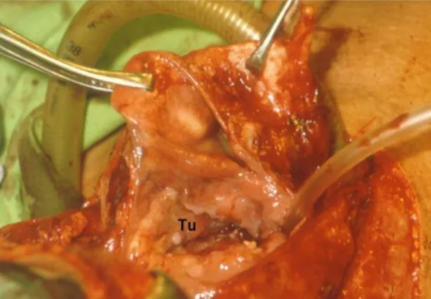
Without defect reconstruction (Figure. 1) Extensive, advanced, T3-T4 tumours that fill the pyriform sinus, infiltrate the lateral pharyngeal wall, spread to the retrocricoid area with hemilaryngeal fixation can only be treated with block dissection (RND/mRND), total laryngectomy and partial pharyngectomy If there is a sufficient amount of pharyngeal mucosa left, the pharyngo-oesophageal defect can be closed primarily. As a result of this radical intervention, patients will lose their ability to speak with original voice, they will need permanent tracheostomy, but their ability to swallow (and thus feed per os) will be retained.
Figure 1. Extended (T3-T4) tumor from piriform sinus. Tu: Hypopharyngeal tumor.
For voice rehabilitation, indwelling voice prostheses implanted into the tracheostoma are more popular nowadays than the previously used electrolarynx.
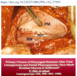
With defect reconstruction (Figure. 2)
If the tumor totally infiltrates the lateral pharyngeal wall and affects the posterior wall as well, the removal of the larynx has to be accompanied by extensive pharyngeal resection, and the resulting pharyngeal defect will make primary closure impossible.
--- Figure 2. Pharyngeal defect reconstruction with
PM flap after total laryngectomy and partial pharyngectomy. (PM: Pectoralis maior flap, NGT:
Nasogastric tube, RM: Remained pharyngeal mucose membrane).
Primary closure is not feasible if the difference between the widths of the residual relaxed (324 mm) and stretched (483 mm) pharyngeal mucosa is less than 16 mm (10).
In such cases, we tend to use pedicled myocutaneous PM flaps introduced into the pharyngeal defect via a tunnel between the clavicle and the cervical skin. Superiorly, the
pedicled flap is fixed to the base of the tongue, laterally to the residual pharyngeal wall, inferiorly to the proximal stump of the oesophagus, then it is turned inwards to surround the inserted nasogastric tube. If the laryngopharyngeal tumour already communicates with the outside preoperatively, the cervical cutaneous defect has to be reconstructed as well (Figure 3) (26).
The scientific literature reports several cases of pharyngocutaneous fistula development following the use of myocutaneous flaps (19);
however, we have never experienced this complication.
Function-preserving open operations
Depending on the location and extent of the hypopharyngeal lesion, in some cases it may be possible to spare the ability to speak and swallow. If the tumour is confined to the wall of the meso-hypopharynx, it can be resected with an electric surgical knife via a lateral approach with safe and adequate surgical margins. Extensive pharyngeal defects can be resurfaced by applying PM, lower arm or LDMC flaps.
Figure 3. Replacement of pharyngeal wall-, and skin defect with PM gemini flap.
Supracricoid hemipharyngolaryngectomy If the tumour arises from the pyriform sinus and spreads to the lateral pharyngeal wall and
the supracricoid region to the level of the cricoid cartilage, supracricoid hemip haryngolaryngectomy can spare laryngeal
--- function. After the separation of the
prelaryngeal muscles, we remove half of the hyoid bone together with the lateral third of the thyroid cartilage then processing laterally and superiorly in the cricothyroid ligament we remove the pyriform sinus as well. Finally, the residual pharyngeal mucosa is fixed to the perichondrium of the thyroid cartilage above the level of the nasogastric probe. If the resulting pharynx seems to be too narrow, one of the above mentioned flaps has to be used for the reconstruction. The function of speech can be retained in each case (14).
Supraglottic laryngeal resection and partial pharyngeal resection with skin flap reconstruction (Figure. 4)
This type of surgery was first performed by Iván and Czigner in Hungary (8).
This approach is used in the treatment of multiregional hypopharyngeal tumours that spread over the pharyngoepiglotttic fold to involve the marginal areas of the pharynx, the pyriform sinus, the lateral pharyngeal wall and the supraglottic region of pharynx.
As a result of the presence of regional metastases, the intervention has to be combined with cervical dissection.
Figure 4. Pharyngeal replacement with PM flap after horizontal supraglottic laryngeal-, and partial pharyngeal resection. (BT: Base of the tongue, NGT: Nasogastric tube, RM: Remained pharyngeal mucose membrane, PM:
Pectoralis maior flap)
Following the dissection, supra- and infrahyoid muscles are cut, half of the hyoid bone is removed and the top third of the thyroid cartilage is resected. In the next step, from a suprahyoid approach the tumorous pharyngeal wall is removed with an electric surgical knife together with the supraglottic part of the larynx. The level of the resection is above the plane of the vocal cords, in the sinus of Morgagni. The pharyngeal defect is reconstructed with a PM flap: superiorly, the pedicled flap is fixed to the base of the tongue, laterally, inferiorly and posteriorly to the residual pharyngeal mucosa, and then it is
turned inwards to create a new pharynx. This technique spares the ability to speak and swallow.
Endoscopic-microscopic tumour resection Endoscopic-microscopic laser resection is a minimally invasive surgical technique that can be performed transorally (13). It means minimal burden for the patient and requires only 1-2 days of hospitalisation. There is no need for temporary tracheotomy, no postoperative bleeding and oedema, and all functions are retained.
However, this technique is appropriate for the treatment of small hypopharyngeal tumors
--- only if their location meets the very limited
indication criteria (21, 22).
Small tumors located on the aryepiglottic fold can be readily removed with a 3-10 watt
CO2 laser while the patient is intubated or placed on jet ventilation). Patients can be discharged the following day (Figure 5).
Figure 5. Laryngo-pharyngeal tumor suitable for laser resection. (T: Tumor, C: Cured status).
Small tumors located on the free margin of the epiglottis or at the border of the epiglottis and the aryepiglottic fold can be resected with the same method in the setting of day-care surgery. It is also considered relatively safe to use the same method of resection if the tumour
is located in the epiglottic vallecula and it not bigger than the tip of a small finger or if the size of a lesion located in the pyriform sinus and spreading towards its medial wall does not exceed 15 mm (Figure 6).
Figure6. Tumor on the top of piriform sinus suitable for laser resection. (T: Tumor, C: Cured status)
However, the laser resection of tumours located on the lateral wall of the pyriform sinus is not recommended as we might accidentally penetrate the parapharyngeal space and damage cervical blood vessels or nerves (IJV, ICA, VA, IX, X, XI) (Figure 7)
It is absolutely contraindicated to attempt the laser removal of tumours located in the lower segment of the pyriform sinus or filling it completely since we may accidentally penetrate the mediastinum, which can have severe consequences.
--- Figure 7. Parapharyngeal space.
Isolated tumors located on the posterior pharyngeal wall (approx. 40x40 mm) can nevertheless be safely resected with laser and there is no need for reconstruction either, as
the surgical area will undergo spontaneous epithelialisation (Figure 8).
Figure 8. Hypopharyngeal tumor on the posterior wall.
Discussion
Hypopharyngeal tumors comprise about 7% of head and neck malignancies, and 90% of these tumors are squamous cell carcinoma. The carcinogenic role of smoking and spirits have been established by several sources (23).
If primary radiotherapy or the combination of radio and chemotherapy fails and salvage surgery is performed, it is usually associated with slower wound healing, and the development of postoperative complications, such as pharyngocutaneous fistulae, is more common as a result of tissue damage caused by irradiation. Consequently, the first line therapy at our department is normally surgical intervention.
In the treatment of small hypopharyngeal tumors endoscopic-microscopic laser resection
can serve as an alternative to surgical intervention providing that the indications are closely observed. The international scientific literature reports on transoral coblation (TOS) resection as well; however, we have no experience related to this technique (25).
Conclusion
Resection of hypopharyngeal tumors with laser increases the risk of recurrence, therefore it is only considered safe in some selected cases. Laryngoscopic assess and the straight line of CO2 laser determines the direction of laser line, which makes it difficult for surgeon to evaluate the involvement of deeper tissues. However, if the indications are closely observed, laser surgery is a potential alternative to surgical
--- intervention in the treatment of small
hypopharyngeal tumors. Nevertheless, at our department the vast majority hypopharyngeal cancers are removed via external exploration, and based on both the international Hungarian scientific literature we think that this is the appropriate choice.
Acknowledgments
We thank our colleagues and also from our patients who provided insight and expertise that greatly assisted the research.
Conflicts of Interest
The authors declare no conflicts of interest.
Financial Support
This research was supported by Department of Otorhinolaryngology, Head, and Neck Surgery, University of Szeged, Hungary.
References
1. Paul JA, MCA. Cochlear Implants: Auditory Prostheses And Electric Hearing. New York:
Springer; 2004.
2. Smeltzer S, B Brenda J, Janis L, G. K. Medical Surgical Nursing. 2 Ed. Tehran: Salemi- Jameanegar Publication; 2010.
3. Byung In Han, Ho Won Lee, Tae You Kim, Jun Seong Lim, Kyoung Sik Shin. Tinnitus:
Characteristics, Causes, Mechanisms, and Treatments. J Clin Neurol. 2009 Mar;5(1):11–9.
4. Madani S, K. M. Evaluation Tinnitus Characteristics Of Patients In Sarry Avicenna Hospital ENT Clinic 1377. Sci J Mazandaran Univ Med Sci 2001;11(32):15-77.
5. Heller AS. Classification And Epidemiology Of Tinnitus. Otolaryngol Clin North Am 2003;36(2):239-48.
6. Enrico P, Sirca D, M. M. Antioxidants, Minerals, Vitamins, And Herbal Remedies In Tinnitus Therapy. Tinnitus: Pathophysiol Treat 2007;166:
323-30.
7. Emami F, Farahani F, Behnood F, M. G.
Comparison Of Digital And Analogue Behind The Ear Hearing Aids Effectiveness In Treatment Of Tinnitus In Elderly Patients Of Otolaryngology Centers In Hamadan. Sci J Hamedan Univ Med Sci 2007;15(4).
8. Craig W. Martin. Tinnitus Retraining Therapy.
Tinnitus. 2004:556-8.
9. Gardiner P, Kemper KJ. Herbs in Pediatric and Adolescent Medicine. Pediatr Rev 2000;21 (2):44- 57.
10. Steele T; Rogers C J; Jacob SE. Herbal Remedies for Psoriasis: What Are Our Patients Taking?.
Dermatol Nurs 2007;19(5):457-63.
11. Newman CW, Jacobson GP, Spitzer JB.
Development Of The Tinnitus Handicap Inventory.
Arch Otolaryngol Head Neck Surg.
1996;122(2):143-8.
12. Afshin-Majd S, Yarmohamadi ME, Izadi P, Shahbeigi S, Davati A. A comparison of gabapentin and placebo in the treatment of subjective tinnitus.
Iran J Neurol. 2008;7(21):153-9.
13. Kim NK, Lee DH, Lee JH, Oh YL, Yoon IH, Seo ES, et al. Bojungikgitang and banhabaekchulchonmatang in adult patients with tinnitus, a randomized, double-blind, three-arm, placebo-controlled trial--study protocol. Trials 2010;28;11:34.
14. Rejali D, Sivakumar A, N. B. Ginkgo Biloba Does Not Benefit Patients With Tinnitus: A Randomized Placebo‐Controlled Double‐Blind Trial And Meta‐Analysis Of Randomized Trials. Clinical Otolaryngology & Allied Sciences. 2004;29(3):226- 31.
15. Okhovat A, Berjis N, Okhovat H, Malekpour A, H. A. Low-Level Laser For Treatment Of Tinnitus:
A Self-Controlled Clinical Trial. Journal Of Research In Medical Sciences: The Official Journal Of Isfahan University Of Medical Sciences.
2011;16(1):33.