R E S E A R C H A R T I C L E
The involvement of the canonical Wnt-signaling receptor LRP5 and LRP6 gene variants with ADHD and sexual dimorphism:
Association study and meta-analysis
Edna Grünblatt
1,2,3| Zsofia Nemoda
4,5| Anna Maria Werling
1| Alexander Roth
1| Nora Angyal
4| Zsanett Tarnok
6| Hauke Thomsen
7| Triinu Peters
8| Anke Hinney
8| Johannes Hebebrand
8| Klaus-Peter Lesch
9,10,11| Marcel Romanos
12| Susanne Walitza
1,2,31Department of Child and Adolescent Psychiatry and Psychotherapy, University Hospital of Psychiatry Zurich, University of Zurich, Zurich, Switzerland
2Neuroscience Center Zurich, University of Zurich and ETH Zurich, Zurich, Switzerland
3Zurich Center for Integrative Human Physiology, University of Zurich, Zurich, Switzerland
4Institute of Medical Chemistry, Molecular Biology and Pathobiochemistry, Semmelweis University, Budapest, Hungary
5Molecular Psychiatry Research Group, MTA-SE NAP-B, Hungarian Academy of Sciences, Budapest, Hungary
6Vadaskert Child and Adolescent Psychiatric Hospital, Budapest, Hungary
7Division of Molecular Genetic Epidemiology (C050), German Cancer Research Center (DKFZ), Heidelberg, Germany
8Department of Child and Adolescent Psychiatry, Psychosomatics and Psychotherapy, University of Duisburg-Essen, University Hospital Essen, Essen, Germany
9Division of Molecular Psychiatry, Center of Mental Health, University of Wuezburg, Wuerzburg, Germany
10Laboratory of Psychiatric Neurobiology, Institute of Molecular Medicine, I. M. Sechenov First Moscow State Medical University, Moscow, Russia
11Department of Neuroscience, School of Mental Health and Neuroscience, Maastricht University, Maastricht, The Netherlands
12Center of Mental Health, Department of Child and Adolescent Psychiatry, Psychosomatics and Psychotherapy, University Hospital of Wuerzburg, Wuerzburg, Germany
Correspondence
Prof. Dr. Edna Grünblatt; Department of Child and Adolescent Psychiatry and Psychotherapy, University Hospital of Psychiatry Zurich, University of Zurich, Wagistrasse 12, 8952 Schlieren, Switzerland.
Email: edna.gruenblatt@kjpd.uzh.ch Funding information
Hungarian Brain Research Program, Grant/
Award Number: NAP-B
KTIA_NAP_13-2014-0011; University of Zurich, Filling the Gap, Grant/Award Number:
Postdoctoral to Anna Maria Werling;
University of Zurich; Deutsche Forschungsgemeinschaft, Grant/Award Number: HE1446/9-1HI865/2-1KFO125
Wnt-signaling is one of the most abundant pathways involved in processes such as cell-prolifer- ation, -polarity, and -differentiation. Altered Wnt-signaling has been linked with several neuro- developmental disorders including attention-deficit/hyperactivity disorder (ADHD) as well as with cognitive functions, learning and memory. Particularly, lipoprotein receptor-related protein 5 (LRP5) or LRP6 coreceptors, responsible in the activation of the canonical Wnt-pathway, were associated with cognitive alterations in psychiatric disorders. Following the hypothesis of Wnt involvement in ADHD, we investigated the association of genetic variations inLRP5andLRP6 genes with three independent child and adolescent ADHD (cADHD) samples (total 2,917 partici- pants), followed by a meta-analysis including previously published data. As ADHD is more preva- lent in males, we stratified the analysis according to sex and compared the results with the recent ADHD Psychiatric Genomic Consortium (PGC) GWAS. Meta-analyzing our data including previously published cADHD studies, association ofLRP5intronic rs4988319 and rs3736228 (Ala1330Val) with cADHD was observed among girls (OR = 1.80 with 95% CI = 1.07–3.02, p= .0259; and OR = 2.08 with 95% CI = 1.01–4.46,p= .0026, respectively), whereas in boys association between LRP6 rs2302685 (Val1062Ile) and cADHD was present (OR = 1.66, CI = 1.20–2.31,p= .0024). In the PGC-ADHD dataset (using pooled data of cADHD and adults) tendency of associations were observed only among females with OR = 1.09 (1.02–1.17) for LRP5rs3736228 and OR = 1.18 (1.09–1.25) forLRP6rs2302685. Together, our findings suggest a potential sex-specific link of cADHD withLRP5andLRP6gene variants, which could contribute DOI: 10.1002/ajmg.b.32695
This is an open access article under the terms of the Creative Commons Attribution-NonCommercial-NoDerivs License, which permits use and distribution in any medium, provided the original work is properly cited, the use is non-commercial and no modifications or adaptations are made.
© 2018 The Authors.American Journal of Medical Genetics Part B: Neuropsychiatric Geneticspublished by Wiley Periodicals, Inc.
Am J Med Genet.2019;180B:365–376. wileyonlinelibrary.com/journal/ajmgb 365
to the differences in brain maturation alterations in ADHD affected boys and girls, and suggest possible therapy targets.
K E Y W O R D S
attention-deficit hyperactivity disorder, gender, genetics, polymorphism, SNP
1 | I N T R O D U C T I O N
Attention-deficit/hyperactivity disorder (ADHD), a neurodevelopmen- tal disorder, is one of the most common psychiatric and behavioral disorders in children and adolescents, with more than 5% of the pedi- atric population affected worldwide, often persisting into adulthood (Polanczyk, Willcutt, Salum, Kieling, & Rohde, 2014; Thomas, Sanders, Doust, Beller, & Glasziou, 2015). Although both the cause and patho- physiology of ADHD are largely unknown, there is a growing body of evidence supporting interactions of multiple genetic and environmen- tal factors during early development thus providing a neurobiological susceptibility to the disorder (Curatolo, D'Agati, & Moavero, 2010).
ADHD diagnosis in children and adolescents is more frequent in boys than girls, with males being 2–4 times more likely to meet diag- nostic criteria than females (Davies, 2014). Moreover, age-at-onset, severity, and comorbidities were found to be sex-dependent (Davies, 2014). Just recently, a fMRI study reported significant volume reduc- tion in putamen and thalamus of girls with ADHD, whereas a signifi- cant subcortical volume reduction was observed in ADHD patients independent of sex (Rosch et al., 2018). Subsequently, the authors dis- cuss the importance to study the trajectories of such neurodevelop- mental disorders together with the sex-dimorphic neuroanatomical development, as maturation timelines between boys and girls are sub- stantially different (Rosch et al., 2018). Differential susceptibility to pre- and postnatal stress has been discussed to affect brain develop- ment and maturation in a sex-dimorphic manner, which was hypothe- sized to add to the genetic load that already leads to the different ADHD frequencies between genders (Van den Bergh et al., 2017).
The aforementioned recent findings as examples, point to the impor- tance of studying genetic susceptibility for ADHD not only at the diagnostic level, but also at age-at-onset and sex differences.
Wnt-signaling pathways orchestrate cellular proliferation, polarity and differentiation; processes that are crucial for healthy tissue mor- phogenesis, especially in the embryonic stage (MacDonald, Tamai, &
He, 2009). Two most known Wnt-pathways exist: a canonical path- way and a noncanonical pathway. In the canonical pathway, the secreted Wnt glycoproteins bind to Frizzled receptors, as well as to either lipoprotein receptor-related protein 5 (LRP5) or LRP6 corecep- tors, to initiate a signaling cascade, in which downstreamβ-catenin is harnessed as a cotranscription factor in the nucleus (Tamai et al., 2000; Wehrli et al., 2000). Canonical Wnt-signaling has a pivotal role both in the developing and mature brain. During development the Wnt pathway regulates the balance between proliferation and differ- entiation of neuronal progenitor and precursor cells (Noelanders &
Vleminckx, 2016). Furthermore, Wnt-signaling also affects neuronal
stem cell proliferation and differentiation in the mature brain (Bengoa-Vergniory & Kypta, 2015), and it has a supportive role in the maturation of dendrites and spines (Hussaini et al., 2014).
Several neurodevelopmental psychiatric disorders have been shown to overlap not only at the behavioral levels, but also at the genetic levels, as it has been repeatedly shown among ADHD, autism spectrum disorder (ASD), intellectual disability (ID), bipolar disorder, and psychosis (Brainstorm Consortium et al., 2018; Bulik-Sullivan et al., 2015; Khanzada, Butler, & Manzardo, 2017; Polimanti & Gelern- ter, 2017; Taurines et al., 2012; van Hulzen et al., 2017; Zhao &
Nyholt, 2017). Interestingly, the Wnt-signaling pathway seems to be one of the overlapping pathways, showing pathological alterations in the above disorders (Kwan, Unda, & Singh, 2016; Mulligan & Chey- ette, 2017; Oron & Elliott, 2017; Zhao & Nyholt, 2017). For example, genome-wide association studies (GWAS) reported the 4p15.31 region to be nominally associated with ADHD (not reaching genome- wide p-value < 5×10⁻⁸), where the KCNIP4 (potassium voltage- gated channel interacting protein 4) gene is located (Lasky-Su et al., 2008; Lesch et al., 2008; Neale et al., 2008). KCINP4 is known to play a role in the negative feedback loop of the Wnt/β-catenin pathway. In a later candidate gene study, sixKCNIP4single markers and one hap- lotype block were found to associate with adult ADHD (aADHD;
Weissflog et al., 2013). Another GWAS analyzed a subpopulation of ADHD cases, which had concomitant oppositional defiant disorder, and found theβ-catenin-pathway to be highlighted in the enrichment analysis (Aebi et al., 2015). Moreover, Wnt-signaling has been also associated with learning and memory, especially with deficits in work- ing and spatial memory (Fortress, Schram, Tuscher, & Frick, 2013;
Maguschak & Ressler, 2011; Maguschak & Ressler, 2012). Epidemio- logical studies pointed to Wnt-involvement in behavioral problems including hyperactivity (Hussaini et al., 2014; Maguschak & Ressler, 2012). In ASD, highly comorbid disorder in ADHD, a wide range of evidence points to the involvement of the Wnt-pathway (Belinson et al., 2016; Caracci, Avila, & De Ferrari, 2016; Packer, 2016; Zhang, Yuan, Wang, & Li, 2014). Lastly, an indirect evidence of the Wnt- signaling involvement in ADHD could be demonstrated by our research group, showing that methylphenidate (a psychostimulant and one of the first line treatment in ADHD therapy) influences cell prolif- eration and differentiation supporting neuronal maturation (Grünblatt, Bartl, & Walitza, 2018). More specifically, we could demonstrate that methylphenidate activates Wnt-signaling, which was not due to the dopamine transporter inhibition—the main know therapeutic mecha- nism of this drug—as selective dopamine transporter inhibitor GBR- 12909 treatment demonstrated the opposite effects (Grünblatt et al., 2018).
As the two possible coreceptors LRP5 and LRP6 play major role in Wnt-pathway activation, we conducted literature search selecting func- tional gene variants affecting either receptor function, gene expression or epigenetic modulations, such as histone modifications or DNA methyl- ation levels (see Table 1 and Supporting Information Table S1). Published genetic associations with psychiatric, neurological, or metabolic diseases are also listed in Table 1. Based on the hypothesis of the involvement of Wnt-signaling in ADHD, we carried out genetic association analyses of the selected fourLRP5andLRP6gene variants with ADHD in three inde- pendent European samples (total 2,917 participants), and a meta-analysis with previously published data. Furthermore, due to sex-discrepancy in ADHD frequency, we stratified the analysis according to sex and com- pared our results to the recent Psychiatric Genomic Consortium (PGC) ADHD GWAS results (Demontis et al., 2017; Martin et al., 2018).
2 | M E T H O D S
2.1 | Study samples
2.1.1 | Zurich child and adolescent ADHD (cADHD) patients and parents including unrelated control-sample One hundred and ninety six Caucasian nuclear families (146 families with both parents and 50 families with one parent) were recruited and
the index patients (aged 6–21 years) were phenotypically character- ized in the outpatient units of the Department of Child and Adoles- cent Psychiatry and Psychotherapy, University Hospital of Psychiatry Zurich. Families were included if at least the index patient fulfilled the diagnostic criteria for ADHD (F90.0 or F90.1) according to ICD-10 (Dilling, Freyberger, & Stieglitz, 1996; World Health Organization, 2016). Accordingly, this resulted in 727 individuals (258 probands with ADHD (males = 179, females = 79) and 469 controls (males = 203, females = 266); total of 382 male and 345 female participants).
The ADHD diagnoses of the parents and siblings were reported by the parents. The psychiatric diagnostics of the index patient was assessed by a child and adolescent psychiatrist or psychologist under supervision of a senior psychiatrist in the clinic. The index patient was required to be≥6 years and to have an IQ over 75 as assessed with either the Wechsler Intelligence Scale for Children (WISC; Tewes, Rossmann, & Schallberger, 1999; Wechsler, 1991), the Kaufman Assessment Battery for Children (K-ABC; Kaufman & Kaufman, 1983;
Melchers & Preuss, 1994), the Culture Fair Test (CFT-20-R; Weiss, 2006), Snijders-Oomen Nonverbal Intelligence Test (SON-R; Tellegen, Winkel, & Laros, 2003) or Intelligence and Development Scales (IDS;
Grob, Meyer, & Hagmann-von Arx, 2009). Exclusion criteria were:
(a) potentially confounding and severe psychiatric diagnoses such as psychosis, any pervasive developmental disorder, primary mood or TABLE 1 Literature summary linking LRP5 and LRP6 gene variants with ADHD and other disorders
Gene SNPa
Functional effects in HEK293T cells
Functional effects in neuronal cells or animal
models Association studies
LRP5 Loci/gene NA Overexpression ofLRP5&LRP6in SH-SY5Y cells protected against oxidative stress and reduced tau phosphorylation (Zhang, Bahety, & Ee, 2015).
BMD (Estrada et al., 2012); schizophrenia and major depressive disorder (Zhao & Nyholt, 2017)
LRP5 rs4988319 NA NA BMD (Tran, Nguyen, Eisman, & Nguyen, 2008);
Wagner syndrome (Rothschild et al., 2013) LRP5 rs3736228 LRP5-V1330 demonstrated
reduced Wnt3a signaling compared to wild-type (Urano et al., 2009)
NA Males low diastolic blood pressure (Suwazono
et al., 2006); obesity and BMI (Guo et al., 2006);
male bone fractures (van Meurs et al., 2006);
BMD (Estrada et al., 2012; Tran et al., 2008);
risk of having metabolic syndrome (Yang et al., 2013)
LRP6 Loci/gene NA Neural tube defects in humans as well as in mice (Allache et al., 2014; Carter et al., 2005;
Kokubu et al., 2004; Lei et al., 2015; Pinson, Brennan, Monkley, Avery, & Skarnes, 2000);
Lrp6mutant mice (insertion mutation) demonstrate suboptimal development of brain regions (e.g., forebrain, midbrain, and hindbrain), and defected neurogenesis of dopaminergic neurons (Castelo-Branco et al., 2010; Pinson et al., 2000; Zhou, Zhao, &
Pleasure, 2004), as well as age-dependent synaptic loss and memory impairments in these mice (Liu et al., 2014)
AD, diabetes mellitus type 2, osteoporosis (Wang, Luo, Xu, Zhou, & Zhang, 2017)
LRP6 rs1012672 LRP6-variant demonstrated reduced Wnt signaling compared to wild-type (Xu et al., 2014)
NA AD (miR-141 miR-23a miR-23b; Mallick & Ghosh,
2011); AD (Alarcon et al., 2013; De Ferrari et al., 2007)
LRP6 rs2302685 LRP6-variant demonstrated reduced Wnt signaling compared to wild-type (De Ferrari et al., 2007; Xu et al., 2014)
NA Risk of ischemic stroke (Harriott et al., 2015); AD
(Alarcon et al., 2013; De Ferrari et al., 2007);
male bone fractures (van Meurs et al., 2006)
Abbreviations: AD = Alzheimer's disease; BMD = bone mineral density; BMI = body mass index; NA = not available.
aDetails on annotation, GTEx and epigenetic findings are presented in the Supporting Information Table S1.
anxiety disorder, and Tourette's disorder, (b) neurological disorders such as epilepsy, (c) a history of any acquired brain damage or evi- dence of the fetal alcohol syndrome, (d) premature deliveries (delivery before 37th gestational week), and/or (e) maternal reports of severe prenatal, perinatal or postnatal complications.
In the case–control setting, the 196 index patients (males = 148, females = 48) were compared to genetically independent 124 Cauca- sian healthy controls (males = 72, females = 52, aged between 5 and 18 years) who were recruited at the Departments of Child and Ado- lescent Psychiatry of the Universities of Würzburg and Zurich. The index patient in the case–control study was part of the family study and the inclusion and exclusion criteria were the same as described above. Informed written consent was obtained in all cases from the participants and their parents. The study was approved by the ethical commissions of all involved universities in accordance with the latest version of the Declaration of Helsinki, including an ethical permission granted by the Ethic Committees from Würzburg, and the Cantonal Ethic Committee of Zurich (Ref. Nr. KFO 140/03 and KEK-ZH-Nr.
2016–00101). Demographic characteristics of Zurich ADHD cases and controls are summarized in Supporting Information Table S2a and S2b.
2.1.2 | Replication samples (Hungarian and German cADHD)
The SNP association study was replicated in samples from Budapest, Hungary and Würzburg, Germany, which were described in detail pre- viously (Kereszturi et al., 2007; Walitza et al., 2005). The replication sample included child and adolescent patients with ADHD and their parents (Würzburg; Walitza et al., 2005), and ADHD patients, their available parents and unrelated controls (Budapest; Kereszturi et al., 2007) with the following characteristics: There were 171 Caucasian nuclear families (106 families with both parents and 65 families with one parent) with additional cADHD cases and healthy young adult controls from Budapest. In the family-based setting this yielded 181 probands with ADHD according to ICD-10 (males = 157, females = 24) and 312 controls (males = 132, females = 180), whereas the case–control study consisted of 206 cADHD patients (males = 180, females = 26, aged between 5 and 17 years) and 262 healthy controls (males = 160, females = 102, aged between 18 and 29 years). The Würzburg-trios and duos included 387 children and adolescents affected with ADHD also according to ICD-10 (males = 296, females = 91) and 540 controls (males = 275, females = 265).
The Ethics Committees of the respective universities approved the study and written informed consents were obtained from the par- ticipants and their parents after the study have been fully explained (see previous publications; Kereszturi et al., 2007; Walitza et al., 2005).
2.1.3 | Genotyping
The study samples were genotyped for rs4988319 and rs3736228 in LRP5, and rs1012672 and rs2302685 in LRP6, which were chosen based on previous genetic findings (described in the introduction) and the availability of validated or functionally tested TaqMan assays.
DNA was isolated either from whole blood collected in ethylenediami- netetraacetic acid tubes using QIAamp DNA Blood Maxi Kit (Qiagen), or from saliva collected in the Oragene DNA collection kit (DNA Gen- otek, Canada) and isolated as per manufacturer's protocol. DNA con- centrations, A260/A280, and A260/A230 ratios were measured using a spectrophotometer (NanoVue Plus, GE). The study population was genotyped with DNA (10 ng), TaqMan® Genotyping Master Mix (Applied Biosystems), and LRP5 or LRP6 SNP Genotyping Assays (Applied Biosystems—see Supporting Information Table S3) combined in a 384-well plate. Real-time PCR was performed in a C1000™ CFX384™Thermal cycler (Bio-Rad) using TaqMan®SNP Genotyping Assay PCR standard protocol. Genotypes were determined by the allelic discrimination program of Bio-Rad CFX Manager™Software version 2.1. Samples were run in duplicates to ensure reproducibility.
In case of ambiguity in duplicates, genotyping was repeated in a sepa- rate run to resolve the discrepancy. No-template controls were included in every run to exclude impurities.
2.1.4 | Statistical analysis
All association studies were run on the PLINK v1.7 (URL: http://pngu.
mgh.harvard.edu/purcell/plink/; Purcell et al., 2007). Each study group (case–control study) was tested for Hardy–Weinberg equilibrium (Supporting Information Table S4). For the case–control association study, the Fisher's Exact Test was conducted and significance was set atp< .00417 following multiple testing corrections (four SNPs, three groups = male, female, and all together). For the family association study, Mendel errors test (none were found) followed by the transmis- sion disequilibrium test was conducted as well as a parent-of-origin analysis. Power was calculated using the Genetic Association Study (GAS) calculator http://csg.sph.umich.edu/abecasis/cats/gas_power_
calculator/index.html.
2.1.5 | Meta-analysis
We conducted literature search to find any publications that described genetic association studies forLRP5andLRP6SNPs in con- nection to ADHD or GWAS in European (Caucasian) ADHD. No previ- ous GAS was available for these two genes in ADHD. However, we could find the GWAS by Hinney et al. (2011), and the GWAS results of the PGC (Demontis et al., 2017; Martin et al., 2018) containing the SNPs analyzed in the current study. A meta-analysis was conducted with the currently studied populations (Zurich, Budapest, and Würz- burg) together with the cADHD results from Hinney et al. (2011) using the MIX 2.0 Pro v.2.0.1.4 (BiostatXL, 2011. http://www.meta- analysis-made-easy.com). The PGC-ADHD results were not added into the meta-analysis, as the results from the PGC GWAS would out- weigh our three study samples, due to a larger sample size, as well as high heterogeneity since the PGC did not contain solely cADHD patients. Therefore, the results of the PGC-ADHD were used only as comparison to the meta-analysis results. Variability, due to between- study heterogeneity, was estimated byI2and funnel plots (Supporting Information Table S5), followed by Begg's and Egger's regression test (Begg & Mazumdar, 1994; Egger, Davey Smith, Schneider, & Minder, 1997; Supporting Information Table S6) to evaluate publication bias due to heterogeneity, and the quality of the studies was assessed
based on traditional epidemiological considerations as previously described in Liu et al. (2015) (Supporting Information Table S7 and S8). Following heterogeneity tests, fixed-effects model was used to conduct the mea-analysis whenI2demonstrated no significant hetero- geneity in the samples, whereas the random-effects model meta- analysis was run if heterogeneity was found (see type of test used for each test in Supporting Information Table S5).
2.1.6 | Gene expression patterns and functional findings To elaborate whether the four SNPs studied may have any potential functional effects on gene expression or epigenetic targets, the SNPnexus (http://www.snp-nexus.org/; Chelala, Khan, & Lemoine, 2009; Dayem Ullah, Lemoine, & Chelala, 2012; Dayem Ullah, Lemoine, & Chelala, 2013) and eQTLs for both genes according to GTEx (https://www.gtexportal.org/home/; Consortium, 2013) were run (see results Supporting Information Table S1). Furthermore, the gene-expression profiles in various brain regions compared to whole blood and nerve-tibial were extracted from GTEx data forLRP5and LRP6 stratified by sex. Welch two-sided t test was conducted between the two sexes for each of the tissue analyzed. Since ADHD is a neurodevelopmental disorder, and most likely the transcript pat- terns alter with age, we downloaded the expression profiles of the two genes from the BRAINSPAN consortium (http://www.brainspan.
org/) focusing on several brain regions (dorsolateral prefrontal cortex, orbital frontal cortex, hippocampus, amygdala, striatum, and cerebel- lum) to create age trajectories for the expression of the two genes.
3 | R E S U L T S
3.1 | LRP5 and LRP6 genetic associations in cADHD
Linkage disequilibrium (LD) values between the twoLRP5SNPs and theLRP6SNPs demonstrated weak linkage in all studied cohorts, simi- lar to the NIH LDlink data for European populations (extracted from https://analysistools.nci.nih.gov/LDlink/?tab=home; see Supporting Information Table S9). Therefore, we could conclude that the resulted association was independent of each other.
In the case–control study a nominal significant association between LRP5 rs3736228 and ADHD was observed in the Zurich sample (OR = 2.043, 95% CI 1.209–3.453,p= .0067, power = 0.944;
Supporting Information Table S10). This association was significant after stratification by sex only in ADHD females (OR = 3.614, 95% CI 1.519–8.6, p= .0024, power = 1.0). Nominal significant sex-specific association was also present at thisLRP5SNP in the Budapest case– control sample (OR = 2.549, 95% CI 1.218–5.334,p= .0109, power = 0.995). Moreover, LRP5 rs4988319 was nominally associated with ADHD in the female subgroup from Budapest (OR = 2.12, 95% CI 1.007–4.462,p= .0444, power = 0.923).
Although no significant association betweenLRP6rs1012672 or rs2302685 and ADHD was detected in any one of the three European samples, some tendencies could be observed (Supporting Information Table S10). However, following sex stratification, nominal significant association of the LRP6rs2302685 was observed among males using the case–control design at both the Zurich and Budapest
ADHD samples (OR = 1.971, 95% CI 1.13–3.438,p= .0159, power = 0.904; OR = 1.516, 95% CI 1.009–2.277,p= .0441, power = 0.519, respectively). Family based association analyses of the LRP6 SNPs yielded only tendency toward association in the Zurich sample (Supporting Information Table S10).
3.2 | Meta-analysis of LRP5 and LRP6 SNPs in children and adolescents with ADHD
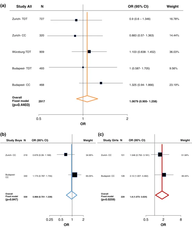
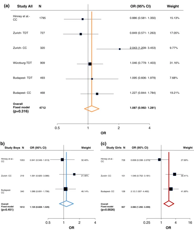
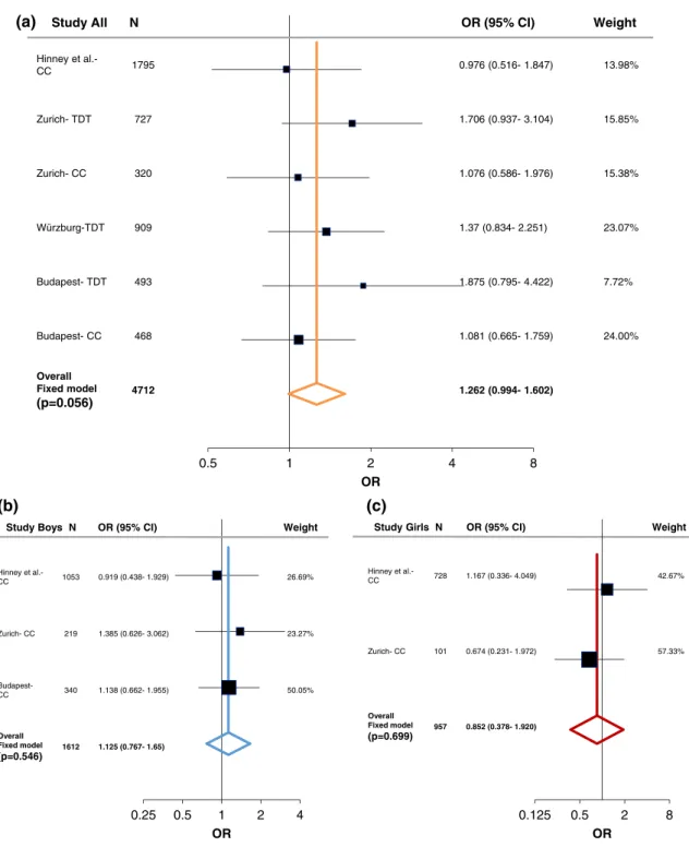
In order to evaluate the associations found with ADHD, we performed a meta-analysis including both the case–control and family studies (Zurich, Würzburg, and Budapest) with available published data from Hinney et al. (2011). As described in the methods section, the large PGC-ADHD GWAS (Demontis et al., 2017; Martin et al., 2018) was not added into the current meta-analysis and was only used as a com- parison study, because it consisted of both cADHD and aADHD, as well as due to its weight comparing to the other studies (Supporting Information Table S7-S8). Following heterogeneity analysis (Supporting Information Table S5 and Figure S1) a fixed-effect model analysis was conducted since no significant heterogeneity was detected. At theLRP5SNPs we could not find significant association with cADHD (Figures 1a and 2a). On the other hand, a tendency was observed atLRP6rs1012672 (totaln= 4,712; OR = 1.262,p= .0559, power = 0.884; Figure 3a), and a nominal significant association was detected at LRP6 rs2302685 with ADHD (total n= 2,917; OR =
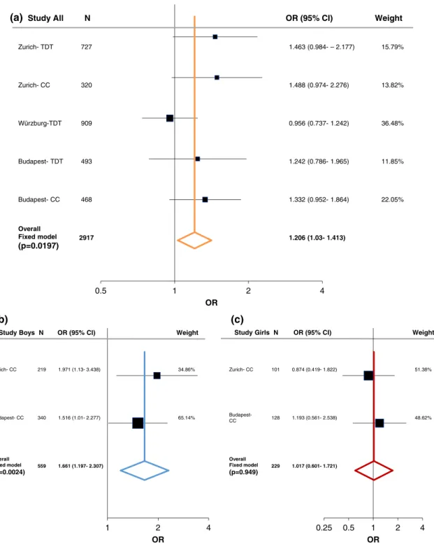
1.206, 95% CI 1.03–1.413;p= .0197; power = 0.794; Figure 4a).
Following sex stratification, a nominal significant association of LRP5rs4988319 and a significant association ofLRP5rs3736228 was observed with cADHD among females (totaln= 229; OR = 1.8, 95%
CI 1.073–3.024, p= .0259, power = 0.731; total n= 957; OR = 2.083, 95% CI 1.292–3.359,p= .0026, power = 0.998; respectively;
Figures 1c and 2c). On the other hand, atLRP6rs2303685 significant association with cADHD was present in males (totaln= 559; OR =
1.661, 95% CI 1.196–2.307,p= .0024, power = 0.846; Figure 4b).
To test the sex effect, we ran a regression analysis (generalized linear mixed model) looking at the sex and SNP dosage and their inter- action including the sites as a random effect in order to account for possible site-effects. The regression analysis showed significant asso- ciation between sex and ADHD but no significant SNP×sex interac- tion was detected (Supporting Information Table S11). Nevertheless, some suggestive sex effects were found supporting the meta-analysis results, as the probability to have ADHD increased with the number of risk allele atLRP5rs4988319 in females (Supporting Information Figure S2a). Similarly, a modest increase for having ADHD could be seen for those with the risk allele of LRP5rs3736228 (Supporting Information Figure S2b). Whereas atLRP6rs2302685 the probability to have ADHD increased with the number of risk allele only in males (Supporting Information Figure S2d).
3.3 | PGC-ADHD GWAS results for LRP5 and LRP6 SNPs
In order to compare the current findings that focused on Caucasian cADHD patients, we extracted the summary statistics of the newest (November 2017) available PGC data for European ancestry ADHD
patients (child and adolescent as well as adults) and then stratified data by sex (Demontis et al., 2017; Martin et al., 2018). Only a sugges- tive association was observed between ADHD andLRP5rs3736228 among females (OR = 1.0942, 95% CI 1.0197–1.1742, p= .2779).
While, LRP6rs2302685 showed association with ADHD only in the female subgroup of the PGC cohort (OR = 1.177, 95% CI 1.0858–1.2503,p= .0208). Therefore, none of the associations found in the meta-analysis could be confirmed in the PGC-ADHD data (Supporting Information Table S10). Nevertheless, observing all SNPs' association results of the PGC-ADHD dataset at the LRP5 gene
(Supporting Information Figure S3), a nominally significant signal could be seen in the male/female combined population at rs4988321 (p= .032), which is in moderate LD with rs3736228 (D0= 1.0, R2= 0.227 at CEU; D0= 0.967, R2= 0.266 at EUR) but not with rs4988319 (D0= 0.219,R2= 0.009 at CEU;D0 = 0.514,R2= 0.056 at EUR). This association was specifically stronger (p= .0086) at the female population in the sex stratified Manhattan plot (Supporting Information Figure S3c). Similarly, around the studied LRP6 SNPs, some nominally significant signals could be observed in the male/
female combined study population (Supporting Information 2
1 5
. 0
OR
0.25 0.5 1 2
OR
0.5 2 8
OR
% 8 7 . 6 1 )
6 4 3 . 1 – 6 . 0 ( 9 . 0 7
2 7 T D T - h c i r u Z
% 4 4 . 4 1 )
3 6 3 . 1 - 7 5 . 0 ( 3 8 8 . 0 0
2 3 C C - h c i r u Z
% 3 0 . 6 3 )
2 5 4 . 1 - 8 3 8 . 0 ( 3 0 1 . 1 9
0 9 T D T - g r u b z r ü W
% 6 5 . 9 )
5 0 7 . 1 - 7 8 5 . 0 ( 1 3
9 4 T D T - t s e p a d u B
% 9 1 . 3 2 )
6 6 8 . 1 - 4 9 . 0 ( 5 2 3 . 1 8
6 4 C C - t s e p a d u B
Overall Fixed model (p=0.4403)
) 8 5 2 . 1 - 5 0 9 . 0 ( 9 7 6 0 . 1 7
1 9 2
t h g i e W )
I C
% 5 9 ( R O N
l l A y d u t S
% 5 9 . 4 3 )
9 6 1 . 1 - 9 3 . 0 ( 6 7 6 . 0 9 1 2 C C - h c i r u Z
Budapest-
CC 340 1.175(0.787-1.755) 65.05%
Overall Fixed model (p=0.847)
559 0.968 (0.701- 1.339)
% 6 5 . 1 5 )
1 8 1 . 3 - 2 5 7 . 0 ( 6 4 5 . 1 1 0 1 C C - h c i r u Z
% 4 4 . 8 4 )
2 6 4 . 4 - 7 0 0 . 1 ( 2 1 . 2 8 2 1 C C - t s e p a d u B
Overall Fixed model (p=0.0259)
229 1.8 (1.073- 3.024) t
h g i e W )
I C
% 5 9 ( R O N s y o B y d u t
S StudyGirls N OR(95%CI) Weight
(b) (a)
(c)
FIGURE 1 Summary and meta-analysis of all cohorts and published association analyses of theLRP5(rs4988319) gene variation with attention- deficit hyperactivity disorder (ADHD) following sex stratification. (a) Forest plot for rs4988319 in male/female combined cohorts. (b) Forest plot for rs4988319 in males. (c) Forest plot for rs4988319 in females. Black whiskers in the forest plot represent 95% confidence intervals (CI) for odds ratio; the weight of the study is reflected in symbol size. Sample demographics, individual statistics, heterogeneity, literature bias statistics, quality assessments and scores, and type of tests was summarized in Supporting Information Tables S5–S9. Abbreviations: CC = case–control;
TDT = transmission disequilibrium test [Color figure can be viewed at wileyonlinelibrary.com]
Figure S4a), that were particularly enhanced in the stratified female ADHD population (Supporting Information Figure S4b).
3.4 | Sex and age dependent LRP5 and LRP6 gene expression patterns in brain tissue
According to GTEx database,LRP5transcript expression in various brain regions is rather low compared to the nerve-tibial, but higher than whole blood samples (Supporting Information Figure S5). Similar pattern was observed for LRP6transcript with slightly higher expression levels in
brain regions (Supporting Information Figure S6). LRP5 transcript was expressed significantly higher in male brain-spinal cord, whereas slightly higher expression in female could be observed in the cerebellum, nucleus accumbens, putamen, and substantia nigra, however in the last three not reaching significance.LRP6transcript was expressed significantly higher in male brain-cortex, and slightly more expressed in male caudate, spinal cord, and nerve-tibial, however not reaching significance. As the GTEx data represents aged population, age dependent gene-expression was extracted from the BRAINSPAN database (Supporting Information Figure S7). From the six brain regions extracted, LRP5 and LRP6
0.5 1 2 4
OR Hinney et al.-
CC 1795 0.886(0.581-1.350) 15.13%
% 5 0 . 7 1 )
3 6 2 . 1 - 1 7 5 . 0 ( 9 4 8 . 0 7
2 7 T D T - h c i r u Z
% 7 7 . 9 )
3 5 4 . 3 - 9 0 2 . 1 ( 3 4 0 . 2 0
2 3 C C - h c i r u Z
% 6 1 . 1 3 )
3 0 4 . 1 - 9 7 7 . 0 ( 6 4 0 . 1 9
0 9 T D T - g r u b z r ü W
% 8 6 . 7 )
9 7 9 . 1 - 6 0 6 . 0 ( 5 9 0 . 1 3
9 4 T D T - t s e p a d u B
% 1 2 . 9 1 )
4 8 7 . 1 - 4 4 8 . 0 ( 7 2 2 . 1 8
6 4 C C - t s e p a d u B
Overall Fixed model (p=0.316)
) 1 8 2 . 1 - 2 9 9 . 0 ( 7 8 0 . 1 2
1 7 4
t h g i e W )
I C
% 5 9 ( R O N
l l A y d u t S
0.5 1 2 4
OR
Hinney et al.-
CC 1053 0.941(0.549-1.613) 32.40%
% 6 4 . 1 2 )
6 8 0 . 3 - 0 2 8 . 0 ( 1 9 5 . 1 9 1 2 C C - h c i r u Z
Budapest-
CC 340 1.086(0.691-1.706) 46.14%
Overall Fixed model
(p=0.451) 1612 1.125 (0.828- 1.529)
Hinney et al.-
CC 728 0.838(0.338-2.079) 27.69%
% 1 4 . 0 3 )
1 8 1 . 3 - 2 5 7 . 0 ( 6 4 5 . 1 1 0 1 C C - h c i r u Z
% 0 9 . 1 4 )
2 6 4 . 4 - 7 0 0 . 1 ( 2 1 . 2 8 2 1 C C - t s e p a d u B
Overall Fixed model
(p=0.0026) 957 2.083 (1.292- 3.359) t
h g i e W )
I C
% 5 9 ( R O N s y o B y d u t
S StudyGirlsN OR(95%CI) Weight
0.25 1 4 16
OR
(a)
(b) (c)
FIGURE 2 Summary and meta-analysis of all cohorts and published association analyses of theLRP5(rs3736228) gene variation with attention- deficit hyperactivity disorder (ADHD) following sex stratification. (a) Forest plot for rs3736228 in male/female combined cohorts. (b) Forest plot for rs3736228 in males. (c) Forest plot for rs3736228 in females. Black whiskers in the forest plot represent 95% confidence intervals (CI) for odds ratio; the weight of the study is reflected in symbol size. Sample demographics, individual statistics, heterogeneity, literature bias statistics, quality assessments and scores, and type of tests was summarized in Supporting Information Tables S5–S9. Abbreviations: CC = case–control;
TDT = transmission disequilibrium test [Color figure can be viewed at wileyonlinelibrary.com]
expression show a strong age dependency with higher levels at embryo- nal and early postnatal stages compared to middle age subjects (up to 40 years of age), in which the expression becomes lower starting from around 19 years of age (corresponding to 1,000 weeks).
4 | D I S C U S S I O N
The involvement of Wnt-signaling in neurodevelopmental and neuro- degenerative disorders have been widely discussed, particularly in
ASD and AD with several studies pointing to its role in cognition and behavior (Kwan et al., 2016; Libro, Bramanti, & Mazzon, 2016; Mulli- gan & Cheyette, 2017; Rios, Cisternas, Arrese, Barja, & Inestrosa, 2014; Wang et al., 2017; Zhang et al., 2014). In the current study, we tested the hypothesis thatLRP5andLRP6gene variants, coding for essential receptors for the Wnt-pathway activation, associate with ADHD among children and adolescents, in a sex-specific manner.
Among the four studied gene variants, our meta-analysis showed sig- nificant association between LRP5 rs3736228 (Ala1330Val) and
0.5 1 2 4 8
OR Hinney et al.-
CC 1795 0.976(0.516-1.847) 13.98%
% 5 8 . 5 1 )
4 0 1 . 3 - 7 3 9 . 0 ( 6 0 7 . 1 7
2 7 T D T - h c i r u Z
% 8 3 . 5 1 )
6 7 9 . 1 - 6 8 5 . 0 ( 6 7 0 . 1 0
2 3 C C - h c i r u Z
% 7 0 . 3 2 )
1 5 2 . 2 - 4 3 8 . 0 ( 7 3 . 1 9
0 9 T D T - g r u b z r ü W
% 2 7 . 7 )
2 2 4 . 4 - 5 9 7 . 0 ( 5 7 8 . 1 3
9 4 T D T - t s e p a d u B
% 0 0 . 4 2 )
9 5 7 . 1 - 5 6 6 . 0 ( 1 8 0 . 1 8
6 4 C C - t s e p a d u B
Overall Fixed model (p=0.056)
) 2 0 6 . 1 - 4 9 9 . 0 ( 2 6 2 . 1 2
1 7 4
t h g i e W )
I C
% 5 9 ( R O N
l l A y d u t S
0.25 0.5 1 2 4
OR
Hinney et al.-
CC 1053 0.919(0.438-1.929) 26.69%
% 7 2 . 3 2 )
2 6 0 . 3 - 6 2 6 . 0 ( 5 8 3 . 1 9 1 2 C C - h c i r u Z
Budapest-
CC 340 1.138(0.662-1.955) 50.05%
Overall Fixed model
(p=0.546) 1612 1.125 (0.767- 1.65)
Hinney et al.-
CC 728 1.167(0.336-4.049) 42.67%
% 3 3 . 7 5 )
2 7 9 . 1 - 1 3 2 . 0 ( 4 7 6 . 0 1 0 1 C C - h c i r u Z
Overall Fixed model (p=0.699)
957 0.852 (0.378- 1.920) t
h g i e W )
I C
% 5 9 ( R O N s y o B y d u t
S StudyGirlsN OR(95%CI) Weight
0.125 0.5 2 8
OR
(a)
(b) (c)
FIGURE 3 Summary and meta-analysis of all cohorts and published association analyses of theLRP6(rs1012672) gene variation with attention- deficit hyperactivity disorder (ADHD) following sex stratification. (a) Forest plot for rs1012672 in male/female combined cohorts. (b) Forest plot for rs1012672 in males. (c) Forest plot for rs1012672 in females. Black whiskers in the forest plot represent 95% confidence intervals (CI) for odds ratio; the weight of the study is reflected in symbol size. Sample demographics, individual statistics, heterogeneity, literature bias statistics, quality assessments and scores, and type of tests was summarized in Supporting Information Tables S5–S9. Abbreviations: CC = case–control;
TDT = transmission disequilibrium test [Color figure can be viewed at wileyonlinelibrary.com]
cADHD in girls, whileLRP6rs2303685 (Val1062Ile) was associated with cADHD in boys. This phenomenon could also be observed in the more heterogeneous ADHD population studied in the PGC-ADHD, consist- ing of both cADHD and aADHD samples, in which nominally significant SNP signals on the Manhattan plots were found to be specific for females at LRP5, however no clear-cut result for LRP6gene variants emerged. This discrepancy between our results and the PGC-ADHD might be due to higher heterogeneity observed in the PGC-ADHD
sample, considering the age of onset that may play a major role with these gene variants. Indeed, BRAINSPAN dataset analysis (Supporting Information Figure S7) demonstrated age-dependent gene expression of bothLRP5andLRP6with higher transcript levels at brain develop- mental stages while lower in adulthood. Independent to age effects, in GTEx dataset some hint for higher expression ofLRP5in aged females and of LRP6 in aged males was observed (Supporting Information Figures S5 and S6, respectively), point to sex-dependent regulation.
0.5 1 2 4
OR
% 9 7 . 5 1 ) 7 7 1 . 2 – - 4 8 9 . 0 ( 3 6 4 . 1 7
2 7 T D T - h c i r u Z
% 2 8 . 3 1 )
6 7 2 . 2 - 4 7 9 . 0 ( 8 8 4 . 1 0
2 3 C C - h c i r u Z
% 8 4 . 6 3 )
2 4 2 . 1 - 7 3 7 . 0 ( 6 5 9 . 0 9
0 9 T D T - g r u b z r ü W
% 5 8 . 1 1 )
5 6 9 . 1 - 6 8 7 . 0 ( 2 4 2 . 1 3
9 4 T D T - t s e p a d u B
% 5 0 . 2 2 )
4 6 8 . 1 - 2 5 9 . 0 ( 2 3 3 . 1 8
6 4 C C - t s e p a d u B
Overall Fixed model (p=0.0197)
) 3 1 4 . 1 - 3 0 . 1 ( 6 0 2 . 1 7
1 9 2
t h g i e W )
I C
% 5 9 ( R O N
l l A y d u t S
1 2 4
OR
% 6 8 . 4 3 )
8 3 4 . 3 - 3 1 . 1 ( 1 7 9 . 1 9 1 2 C C - h c i r u Z
% 4 1 . 5 6 )
7 7 2 . 2 - 1 0 . 1 ( 6 1 5 . 1 0 4 3 C C - t s e p a d u B
Overall Fixed model
(p=0.0024) 559 1.661 (1.197- 2.307)
% 8 3 . 1 5 )
2 2 8 . 1 - 9 1 4 . 0 ( 4 7 8 . 0 1 0 1 C C - h c i r u Z
Budapest-
CC 128 1.193(0.561-2.538) 48.62%
Overall Fixed model
(p=0.949) 229 1.017 (0.601- 1.721) t
h g i e W )
I C
% 5 9 ( R O N s y o B y d u t
S StudyGirlsN OR(95%CI) Weight
0.25 0.5 1 2 4
OR
(a)
(b) (c)
FIGURE 4 Summary and meta-analysis of all cohorts and published association analyses of theLRP6(rs2302685) gene variation with attention- deficit hyperactivity disorder (ADHD) following sex stratification. (a) Forest plot for rs2302685 in male/female combined cohorts. (b) Forest plot for rs2302685 in males. (c) Forest plot for rs2302685 in females. Black whiskers in the forest plot represent 95% confidence intervals (CI) for odds ratio; the weight of the study is reflected in symbol size. Sample demographics, individual statistics, heterogeneity, literature bias statistics, quality assessments and scores, and type of tests was summarized in Supporting Information Tables S5–S9. Abbreviations: CC = case–control;
TDT = transmission disequilibrium test [Color figure can be viewed at wileyonlinelibrary.com]