Chapter 7 Studies of Zinc Metabolism
R. M . FORBES
Division of Nutritional Biochemistry Department of Animal Science University of Illinois
Urbana, Illinois
I. Introduction 339 II. Methods of Analysis 340 III. Zinc in Tissues 341
IV. Biochemically Recognized Functions 344 V. Accumulation and Turnover of Zinc-65 in Animal Tissues 347
VI. Zinc-Hormone Interrelations 350 VII. The Zinc-Deficiency Syndrome 351 VIII. Zinc Requirements and Dietary Factors Influencing Utilization . 352
IX. Clinical Aspects 356 X. Toxicity 357
References 358
I. INTRODUCTION
The techniques employed in studies of zinc metabolism range from those of animal husbandry to those employing the most modern methods of enzyme kinetics and studies of "reaction sites." Since much of this material has been reviewed recently (1, 2), it is the purpose of the present chapter to collate as far as possible developments in this area since 1960, referring to earlier work when necessary to give proper per- spective to current views of the problem of zinc metabolism and nutriture in animals.
Current interest in the metabolic role of zinc stems from the work of Tucker and Salmon (3) who demonstrated for the first time the practi- cal necessity for zinc supplementation of diets for swine raised by usual husbandry procedures, from investigations such as those of Nason et al. (4) and of Keilin and Mann (5) and Vallee (6) demonstrating the nature of the enzymatic role of this trace element, and from clinical evidence of Prasad and co-workers showing an abnormality of zinc me- tabolism in humans (7-9). As Underwood has pointed out (10), the biochemical lesions responsible for the clinical syndrome of zinc deficiency are as yet unknown despite the known roles of zinc in a
339
340
number of enzymes and enzyme-catalyzed reactions. Thus the search must go on.
II. METHODS OF ANALYSIS
A wide variety of techniques has been employed in quantitative analysis for zinc in biological materials. In most instances some means of separation of zinc from interfering materials must precede the actual determination. Whatever methods are used, scrupulous care to avoid contamination is required since zinc is so common an element in the environment. This aspect of the problem has been treated in detail by Thiers (11). Of particular interest with respect to zinc is Thiers' warning that care must be taken in dry ashing procedures to prevent the sample from becoming heated above 450°C in order to avoid loss through vola
tilization. In spite of this, Thiers favors controlled dry ashing over wet ashing because of the contamination hazards inherent in the latter pro
cedure. A survey of methods available for determination of zinc in biological materials was published by Malmstrom (12) in 1954, and specifics of the widely used dithizone procedure are given. A more recent and detailed study of the separations prior to color development with dithizone is that of Margerum and Santacana (13). In their hands the use of diethanoldithiocarbamate according to the procedure of Butts et al. (14) gave the most satisfactory separations of zinc. We have employed this procedure as well as that of Vallee and Gibson (15) using the mixed colorimetric method for the determination of the zinc dithizonate. Both methods were satisfactory, but the procedure of Butts was simpler in terms of manipulations and in our opinion is the method of choice among the dithizone procedures. Reference to other previously reviewed methods of zinc detection may be found in the article by Vallee
( i ) .
The advent of atomic absorption spectroscopy as a practical means of trace-element analysis has been reviewed by Robinson (16), and this principle has been rapidly applied to the determination of zinc in biologi
cal materials. In view of the sensitivity, accuracy, freedom from inter
ference, and simplicity of operation this is now the method of choice.
Application of the method to biological fluids is discussed by Fuwa et al. (17) who used a hydrogen-air flame. We have used an acetylene-air flame in making measurements of zinc content of animal tissues, excreta, and feedstuffs. Sample preparation involves ashing with appropriate care to prevent contamination and then dilution of the sample with HC1 to make a final concentration of 0.36 Ν HC1. A more general treatment of the applications of this method is that of Allen (18). Recent appli
cations of emission spectroscopy (19) and neutron-activation analysis (20) have also been published.
7. STUDIES OF ZINC METABOLISM 341
III. ZINC IN TISSUES
The quantitative and qualitative aspects of the zinc content of animal tissues have been treated in detail in previous reviews (1, 2). The zinc concentration in most of the soft tissues of the body approximates 25 ppm on the fresh basis and, except in the liver, is not appreciably changed by alteration of zinc intake (21-24). Considerably higher
(100-400 ppm) concentrations of zinc are found in bone, hair and wool, and portions of the prostate and the eye. Human prostatic fluid contains 10 times the concentration of zinc as is found in the whole gland (25), a finding in agreement with histochemical demonstration of zinc concen- tration in the glandular epithelium. In the young, growing animal the zinc concentration in bone ash is a sensitive reflection of zinc absorption, especially at suboptimal intake levels (26-29). It is apparent that zinc accumulates in cartilage at sites of calcification and, once deposited in the calcified tissue, is firmly bound (30-32). Analysis of human autopsy material did not reveal age changes in bone ash zinc (33), nor did an experiment in which rats were fed a constant diet over more than a year. It was found that the shaft of the rat femur had a lower zinc concentration (330 ppm of ash) than the head of the femur (430 ppm).
The major portion of the zinc in whole blood is in the erythrocytes where it occurs mainly as a constituent of carbonic anhydrase. Species differences are apparent in blood zinc, but the relative distribution among blood components is similar. More data are available for man than for other species, and average values indicate 7-8, 1.1-1.3, and 12-18
of zinc per milliliter of whole blood, plasma, and erythrocytes, respec- tively. According to Underwood (2) leukocytes contain 3 % of whole blood zinc, but each leukocyte contains 25 times as much zinc as each erythrocyte. Plasma zinc is in part protein bound (34, 35).
The occurrence of zinc in skin and other epidermal structures has been of interest because of its concentration in hair, wool, and nails (100-200 ppm) and because of the marked histological aberration in the skin of zinc-deficient animals. Actually, the skin itself does not con- tain remarkable concentrations of zinc [20-60 ppm of dry tissue (36, 37)]. The epidermis does, however, contain 3 times the concentration of zinc that the corium does (25 vs. 75 ppm). Recent investigations (38) indicate that the transitional zone of human epidermis concentrates zinc, and evidence points to protein-bound histidine as the binding agent.
This viewpoint is supported by the similarity in distribution of histidine and of zinc in epidermal structures, by the disappearance of histochemi- cally detectable histidine following zinc uptake, and by the absence of other structures that might have zinc-binding capabilities.
342
Only fragmentary data are available on the zinc content of skeletal muscle. Large variation in zinc content of individual muscles of swine has been reported recently (39). Porcine trapezius muscle had 3 times the concentration of zinc and of myoglobin that was present in longis- sumus dorsi. Interestingly, it also had a higher lactic dehydrogenase activity. The relationship between these findings and observed differences in post-mortem changes is not clear.
The previously reported intracellular distribution of zinc in rat liver (40) has been confirmed (41, 42). With the data reported in terms of micrograms of zinc per milligram of nitrogen, the following distribution was found: nuclei, 0.77; mitochondria, 0.42; microsomes, 0.65; super
natant, 2.0; and reconstituted whole liver, 1.05. These findings are con
sistent with the view (40) that the majority of the known zinc-containing enzymes are in the supernatant fraction of the liver cell. On a percentage basis 18-28, 7-9, 11-16, and 54-58% of the total liver zinc is present, respectively, in nuclei, mitochondria, microsomes, and supernatant. The intracellular distribution of a single dose of 6 5Zn in mouse liver is re
ported not to change within the time limits of 1 hour and 7 days (42).
The unusual concentration of zinc in certain portions of the eye has attracted much attention and has been carefully documented (1, 2).
The choroid of carnivora and particularly the tapetum lucidum contains higher concentrations of zinc than any other animal organ; this amounts in some species to as much as 13% of the dry tissue. A significant amount of the zinc seems to be bound in a zinc-cysteine complex, but its function is not known. The lower but significant amounts of zinc in the retina may be incorporated in part with retinene reductase which catalyzes the interconversion of vitamin A alcohol and aldehyde and may point to a vitamin Α-zinc interrelationship. Another reason to inves
tigate such a relationship is the general similarity of skin lesions in deficiencies of vitamin A and of zinc.
Previously reported investigations have indicated that species vari
ation in zinc content of milk is not large and that "mature" milk may be expected to contain 37.5 ppm whereas colostrum may contain 4 times this amount. In recent investigations mature cow's milk was found to contain 4.1 ppm (43) and sow's milk 7.3 ppm (44). Miller et al. (43) found supplementation of the cow's ration with 500, 1000, and 2000 ppm of zinc resulted in milk containing, respectively, 6.7, 8.0, and 8.4 ppm of zinc. Stevenson and Earle (44) also found an influence of dietary zinc on sow-milk zinc: the addition of 100 ppm of zinc to the ration raised the milk zinc from 7.3 to 10.4 ppm. Sow colostrum, however, was not affected by the zinc supplement and contained about 23 ppm of zinc, 2-3 times the value in the mature milk.
7. STUDIES OF ZINC METABOLISM 343 An area of great interest in zinc metabolism is related to the possible role of the large amounts of zinc found in portions of the male reproduc- tive tract and its secretions. The prostate gland in particular concentrates zinc, although there are wide variations between species in the distribu- tion of this element in the gland. In the rat, for example, the dorsal prostate may contain 180 ppm of zinc on the fresh basis whereas the ventral prostate contains less than one-tenth of this amount (45). On the other hand, in the human prostate no correlation could be found between zinc content and histologic features of the gland, although there was a 20-fold difference in zinc content of different areas of a given gland (46) and a general increase in zinc content as samples were taken further from the bladder. One gland in this series contained a localized carcinoma, and this area contained about one-third as much zinc as did unaffected areas of the same gland. This is roughly comparable to the earlier report (47) that five cancerous human prostates contained 190 ppm zinc on the dry basis whereas thirty-one normal prostates con- tained 682 ppm. As yet no functional role has been found for these significant concentrations of zinc. Although there is a general correlation between zinc content and carbonic anhydrase activity of prostatic tissue, the latter accounts for only a small portion of the total zinc present (48), and there is no correlation between zinc content and the activity of either acid or alkaline phosphatase. It is interesting that the necrotiz- ing effect of dithizone on the prostate of the rat and the dog is correlated with formation of a fine intracellular deposit of zinc dithizonate (49, 50). This indicates that a large portion of the zinc present is not so firmly bound to protein that it is unavailable for chelation.
The high zinc content of testes and of spermatazoa has been well documented (1). Recent investigations have shown that sperm obtained from vas deferens and epididymis contain as much zinc as those from the total ejaculate (51), which shows that they are not dependent on the zinc-rich prostatic fluid for their zinc content. The role of zinc in normal sperm function remains obscure since zinc deficiency produces testicular degeneration and aspermia, as well as markedly reducing the zinc concentration in the total male reproductive tract.
The zinc content of the pancreas and its endocrine and exocrine secre- tions has been of much interest since the finding that crystalline zinc- insulin has certain advantages in treatment of diabetes over metal-free insulin. This matter has been thoughtfully discussed by Vallee (1) who raises pertinent questions about some of the analytical methods used to detect zinc in the whole pancreas and in the insulin-secreting /?-cells of the islands of Langerhans. Whatever the facts of this portion of the story may be, it is clear that zinc-free insulin (and glucagen) are fully
active biologically, and the ultimate test of the necessity for zinc in their activity has yet to be devised.
I V . BIOCHEMICALLY RECOGNIZED FUNCTIONS
Specific information on the manner in which zinc is involved in metabolic reactions in the animal body dates from the original discovery that zinc is a constituent of bovine erythrocyte carbonic anhydrase (52).
Since that time a considerable body of research has appeared, revealing the roles in which zinc may participate in metabolism either as an acti
vator of enzyme systems, forming a metal-enzyme complex, or, more specifically, as a constituent of enzymes, forming metalloenzymes. While it is interesting to study the former situation—the activating effects of metals on specific enzyme systems in vitro—the results of such investi
gations tell little about the in vivo relationships of the metal and indeed, more often than not, merely demonstrate that the metal requirements for this sort of activation are nonspecific. It is typical of the metal- enzyme complex that it is readily and reversibly inactivated by removal of the metal from the system and that a number of different metals may activate a specific reaction. The case of the metalloenzymes is more specific; in such compounds the metal is firmly bound to the en
zyme. Removal of the metal irreversibly inactivates the enzyme, and in such enzymes, purified to the point of physicochemical homogeneity, the ratio of metal to enzyme becomes constant, and stoichiometry can be shown to exist between moles of metal and moles of coenzyme or prosthetic group involved in enzyme action. These examples represent classified extremes which are bridged by a continuous spectrum of activi
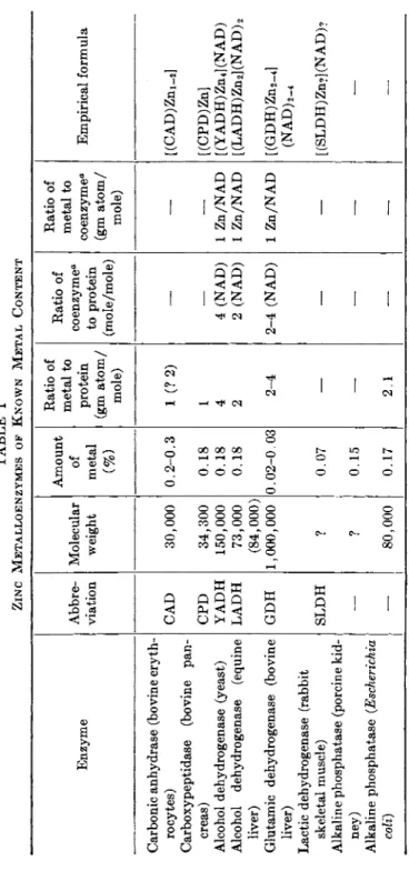
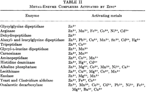
ties of the metal-enzyme complexes on the one hand and metalloenzymes on the other. These views have been discussed (53), and the application of these concepts to the study of zinc metabolism has been expertly reviewed (1, 53, 54). Tables I and II list the known zinc metalloenzymes and the metal-enzyme complexes activated by zinc as cited in the review by Vallee (1) who has been responsible for the major portion of the work done on zinc metalloenzymes.
In addition to the data of Tables I and II, more recent investigations have shown that D-glyceraldehyde-3-phosphate dehydrogenase, isolated from bovine and crayfish muscle and bakers yeast, is a zinc metallo- enzyme with 2 moles of zinc per mole of enzyme (55), apparently bound by means of cysteine and histidine (56). Malic dehydrogenase has also been reported to contain zinc in a 1:1 mole ratio with the enzyme (57) and to possess the qualities of a metalloenzyme.
The role of zinc in regulating the activity of alkaline phosphatase has been of continued interest since some preparations of this enzyme
TABLE I ZINC METALLOENZYMES OF KNOWN METAL CONTENT Enzyme Abbre viation Molecular weight
Amount of metal (%)
Ratio of metal to protein (gm atom/ mole) Ratio of coenzyme" to protein (mole/mole) Ratio of metal to coenzyme" (gm atom/ mole)
Empirical formula Carbonic anhydrase (bovine eryth rocytes) CAD 30,000 0.2-0.3 1 (?2) — — [(CAD)Zm-,] Carboxypeptidase (bovine pan creas) CPD 34,300 0.18 1 — — [(CPD)Zn] Alcohol dehydrogenase (yeast) YADH 150,000 0.18 4 4 (NAD) 1 Zn/NAD [ (Y AD Η) Zn 4 ] (N A D) Alcohol dehydrogenase (equine LADH 73,000 0.18 2 2 (NAD) 1 Zn/NAD [ (LADH) Zn2] (NAD) 2 liver) (84,000) Glutamic dehydrogenase (bovine GDH 1,000,000 0.02-0.03 2-4 2-4 (NAD) 1 Zn/NAD [(GDH)Zn2_4] liver) (NAD) 2-4 Lactic dehydrogenase (rabbit skeletal muscle) SLDH ? 0.07 — — — [ (SLDH) Zn?] (NAD)? Alkaline phosphatase (porcine kid ney) — ? 0.15 — — — — Alkaline phosphatase (Escherichia coli) — 80,000 0.17 2.1 — — — β NAD, previously known as DPN.
7. STUDIES OF ZINC METABOLISM 345
346
have been reported to constitute zinc metalloenzymes and also to exhibit properties of metal-enzyme complexes. Reports of animal feeding studies have shown that low levels of serum alkaline phosphatase accompany zinc deficiency (58-61), although lack of such correlation has also been reported (62, 63). The possible role of reduced feed intake in contributing to this effect has not been extensively investigated, and in fact has ordinarily not been considered. An exception to this is the report by Starcher and Kratzer (64) who investigated effects of zinc deficiency on bone alkaline phosphatase in turkey poults. In this experiment it was shown that restriction of intake by zinc-adequate birds to that
TABLE II
METAL-ENZYME COMPLEXES ACTIVATED BY ZINC*
Enzyme Activating metals
Glycylglycine dipeptidase Z n2+
Arginase Z n2 +, M n2 +, F e2 +, C o2 +, N i2 +, C d2+
Dehydropeptidase Z n2+
Alanyl- and leucylglycine dipeptidase Z n2 +, P b2 +, C u2 +, M n2 +, S n2 +, C d2 +, H g2+
Tripeptidase Z n2 +, C o2+
Glycyl-L-leucine dipeptidase Z n2 +, M n2+
Carnosinase Z n2 +, M n2+
Aminopeptidase Z n2 +, C o2 +, M n2+
Histidine deaminase Zn2+, H g2 +, C d2+
Alkaline phosphatase Z n2 +, M g2 +, C o2 +, M n2 +, Ni2+, C a2+
Lecithinase Z n2 +, C u2 +, M g2 +, C o2 +, M n2+
Enolase Z n2 +, M g2 +, M n2+
Yeast and Clostridium aldolase Z n2 +, F e2 +, C o2+
Oxalacetic decarboxylase Z n2 +, M n2 +, C o2 +, C d2 +, P b2 +, N i2 +, Fe2+ M g2 +, B a2 +, C u2+
a Other metals which increase the observed activity are also listed.
of severely zinc-deficient birds lowered bone alkaline phosphatase below that of the ad libitum positive controls, but the values were higher than those of the zinc-deficient birds. Thus it appears that both feed intake and zinc status play a role in activity of bone alkaline phos
phatase and might well do so also for other phosphatases.
Since the original discovery that carboxypeptidase from bovine pan
creas is a zinc metalloenzyme (65), Vallee and his associates have con
ducted a long series of meticulous experiments whose objective has been to learn more about the nature of the active binding site in this enzyme and others. These findings have been summarized (66, 67) in detail, and the reader is referred to them for study of the application of pres-
7. STUDIES OF ZINC METABOLISM 347 ent-day techniques in biochemistry to the problems of enzyme function and specificity. With reference to alcohol dehydrogenase Vallee states (66) that, "The available data suggest that the metal is bound as a dimercaptide between two adjacent subunits of the molecule while it maintains the molecular configuration which is crucial to the binding of DPN [NAD1] by a three-point attachment: one each of two adjacent subunits and the third to zinc" With reference to carboxypeptidase he summarizes that, "The catalytically active zinc atom is bound to the sulphur atom of the sole cysteine residue and to the N-terminal aspara
gine residue in the single peptide chain of γ- and δ-carboxypeptidase.
Replacement of zinc by other metal atoms results in enzymatically active metallocarboxypeptidases which differ from the native enzyme both in specific activity and in substrate specificity." These concise statements illustrate the current status and future direction of progress of our under
standing of the ultimate mechanism of function of many trace metals in general and of zinc in particular.
In addition to following enzymatic activity and correlating this with zinc content, other metalloproteins may be investigated without regard to their biological activity. Identification of metals in stoichiometric amount in highly purified proteins has revealed the presence of a num
ber of metals including copper, cadmium, and zinc (68, 69). This ap
proach, though laborious, holds promise of expanding our knowledge of metabolic roles of zinc and other trace elements as we become more familiar with the function of these newly identified metalloproteins.
A wide variety of metals including zinc has been found in ribonucleic acids from diverse sources (70), and it is suggested that they play a role in maintaining configuration of the ribonucleic acid (RNA) molecule and thus have a functional relationship to protein synthesis and the transmission of genetic information. In Rhizopus nigricans cultures, addi
tion of zinc to deficient cultures results in an immediate increase in RNA content, followed by an increase in protein content (71); these results verify the importance of zinc in RNA metabolism.
V. ACCUMULATION AND TURNOVER OF ZINC-65 IN ANIMAL TISSUES
Employment of 6 5Zn as a tool to study the metabolism of zinc in animal bodies and tissues has been a fruitful venture for investigators interested in rates of deposition and removal, sites of concentration, and various factors affecting these parameters of zinc utilization.
Representative of early studies on uptake and removal is the report by Gilbert and Taylor (32) in which it was shown that, after intravenous
1 N A D (nicotinamide-adenine dinucleotide), also known as D P N (diphospho- pyridine dinucleotide).
348
administration to rats of a single dose of 6 5Zn, 11-15% of the dose was present in the skeleton after 3 days and was still present when the animals were sacrificed after 259 days. The 6 5Zn deposited in hair amounted to a maximum of 12% of the dose at 9 days and then decreased slowly. In other tissues the evidence indicated equilibration with the dose at about 3 days with an exponential decrease thereafter. Of the total zinc administered, 73% was excreted by way of the intestine, indi
cating the importance of this pathway, since 12% was retained in the bone and 9% appeared in the hair and was probably lost by shedding.
Another early report in this field (72) showed that 18% of the dose was in liver of rats 24 hours after an injection of 6 5Zn and that 2.6%
was in pancreas of normal rats, but only 0.3% in pancreas of alloxan- diabetic rats. Rubini et al. (73) in a study of absorption, deposition, and excretion of 6 5Zn in mice, rats, and dogs found that injected 6 5Zn accumulated first in pancreas, liver, and spleen. Subsequently a large portion was transferred to bone. Similar findings covering the preferred sites of initial deposition were reported by McKenney et al. (74) using pregnant ewes as experimental subjects. They also reported a limited transmission of 6 5Zn across the placenta, the fetuses containing 0.1-0.5 times the concentration of 6 5Zn found in comparable tissues of the ewes.
Other evidence for transfer of 6 5Zn across the placenta has been obtained with the rabbit (75) and the mouse (76); in the latter instance the pres
ence of 6 5Zn in the milk was suggested since mice previously unexposed
to 6 5Zn accumulated this element while suckling mothers which had been
injected with 6 5Zn. Stand et al. (77) have also cited pancreas and liver as two of the sites of early accumulation of 6 5Zn in mice. They also found that chelated 6 5Zn was more rapidly excreted and accumulated to a lesser degree in the body than was ionic zinc. In the human, Rosoff et al. (78) found liver, kidney, spleen, and pancreas to be preferred sites of concentration of a single intravenous dose of 6 5Zn.
The effect of status of zinc nutriture on uptake of 6 5Zn by tissues of chickens has been recently reported by two groups of investigators.
Zeigler et al. (79) demonstrated that zinc deficiency altered total zinc concentration only of bone, liver, and duodenum but that all tissues in zinc-deficient chicks accumulated higher concentrations of intramuscu
larly administered 6 5Zn than did those of normal chicks. It was also observed that 6 5Zn is rapidly removed from blood plasma as it accumulates in other tissues. The data of Turk (80), obtained with laying hens, are in good agreement with those obtained with the chick. There was little 6 5Zn incorporated in the egg shell.
The rapid removal of zinc from the blood has been verified by Dennes et al. (81), who employed rabbits and made more detailed studies on
7. S T U D I E S OF ZINC METABOLISM 349 the binding of zinc to plasma proteins. They found, after injection of
a 6 5Zn glycine complex, that 90% of the intravenously injected zinc
left the bloodstream within 3 hours. During the period of 3-24 hours after injection essentially all of the zinc in the plasma was bound, about half of it to α-globulin. They also found that leukocytes concentrated zinc 30-70-fold more than did erythrocytes, and that, whereas there was erythrocyte-plasma exchange of 6 5Zn, the leukocytes retained their
6 5Zn and did not exchange it back to plasma. They thus consider red cell zinc as a source of mobilizable or "available" zinc in the animal body. The exchange of 6 5Zn between red cells and plasma has been veri
fied by Herrick et al. (82) with rat red cells and plasma by means of in vitro incubation systems.
The occurrence of high concentrations of zinc in the male reproductive system has led to studies of 6 5Zn movement in these organs. Gunn et al. (83) have demonstrated in the rat that the dorsolateral prostate concentrates a single dose of 6 5Zn 15-25 times as much as other tissues, and that this accumulation is maximum at 1-2 days and rapidly reduces by 4 days to a fairly constant value. They have also reported (84) on the movement of 6 5Zn in testes and epididymes, finding maximum concentration in the testes at 5 days, with a fall thereafter. As 6 5Zn decreases in testes it accumulates in epididymes. They have reported that destruction of testicular tubules by cadmium results in a maximum
6 5Zn uptake by testes only 30% of normal. They thus suggest that a portion of the zinc in testes moves out with spermatazoa and that another portion is associated with the nontubular elements and leaves the testes by plasma exchange. Other recent investigations of the distribution of
6 5Zn in the prostate may be consulted for further details (85-88).
It has been shown (89) that hepatic injury by CC14 does not influence accumulation of 6 5Zn by the liver or other organs, although it does de
crease deposition of 5 4Mn in the liver. It is suggested that this difference in behavior is the result of a difference in intracellular distribution.
Manganese is normally concentrated in mitochondria and microsomes, which were damaged by CC14, whereas zinc is mainly in nuclei and cell walls, which were little affected by CC14.
Whole body counting of 6 5Zn has been employed in studies of the overall turnover of zinc. Cotzias et al. (90) administered 6 5Zn by injec
tion to mice and followed the retention of the element for 10-12 days as influenced by the administration of zinc citrate or sodium citrate.
They showed that the increased zinc load increased the rate of removal of the injected 6 5Zn but sodium did not. It was further noted that the loss of zinc from the body was via the feces. The same technique has also been used (91, 92) to study the effects of a number of dietary
350
variables on zinc absorption and turnover. These studies have shown that increasing the dietary zinc lowers 6 5Zn absorption and increases turnover, as does increased dietary calcium. Purified diets allowed better
6 5Zn absorption than did natural (phytate-containing) diets of similar calcium, phosphorus, and zinc content.
V I . ZINC-HORMONE INTERRELATIONS
Previously reviewed investigations have indicated that gonadotropins and testosterone increase not only the size but the zinc content of and rate of 6 5Zn uptake by the rat prostate (93, 94), and that androgen and estrogen variously affect zinc uptake by the prostate, depending on the presence or absence of functional testes (95). More recent investi
gations (96) have shown that gonadotropins and testosterone will stimu
late growth in size of male accessory sex organs of zinc-deficient rats but will not prevent the lowered zinc content of these organs or the testicular atrophy that accompanies zinc deficiency. Thus, the atrophy is probably specifically due to the lack of sufficient zinc to maintain spermatogenesis and the survival of the germinal epithelium. The reduc
tion in size of the accessory organs is probably a result of a depressed gonadotropin output. In a study differentiating the effects of the follicle- stimulating hormone (FSH) and interstitial-cell-stimulating hormone
(ICSH) components of pituitary gonadotropin (97) it was found that ICSH would prevent the fall of 6 5Zn uptake by testes in hypophysec- tomized rats; FSH was much less effective, and growth hormone and pro
lactin were ineffective. The same investigators (98) had previously sug
gested that 6 5Zn uptake by the dorsolateral prostate of hypophysecto- mized rats might be used as a specific bioassay for ICSH.
Further evidence of sex hormone influence on zinc metabolism has been reported, with chickens used as subjects (99). Six-week-old chicks were either normal, testosterone treated, or given dienestrol diacetate, and retention of intravenous 6 5Zn uptake at 24 hours was determined.
Testosterone given to females decreased femur 6 5Zn uptake, but this was not true of males. The untreated males had lower 6 5Zn uptake than untreated females. Dienestrol diacetate had the opposite effect; it in
creased femur zinc uptake of males but not that of females.
Adrenalectomy also lowers zinc content and 6 5Zn uptake of testes and the dorsolateral prostate of the rat. This effect can be reversed by cortisone given to adrenalectomized animals (100) and by adrenocor
ticotropic hormone (ACTH) given to hypophysectomized animals (101);
these results point to a relationship between, adrenal activity and zinc metabolism.
The relationship of zinc to pancreas function has been well reviewed
7. STUDIES OF ZINC METABOLISM 351 (1) and need not be detailed here except to point out that a control of zinc over endogenous insulin function or a control of insulin on zinc metabolism has yet to be found.
V I I . THE ZINC-DEFICIENCY SYNDROME
The physiological changes observed in zinc-deficient animals have been described many times since the initial experimental production of this condition in the rat by Todd et al. (102) who noted particularly a reduced growth rate and the appearance of alopecia. The initial his- tological studies of zinc deficiency were made on the rat by Follis et al. (103) who noted the hyperkeratinization of the skin and esophagus.
Since these early investigations, zinc deficiency has been described fur- ther in rats (104-106), mice (107, 108), pigs (59, 109-112), dogs (113), chickens (63, 114, 115-119), turkeys (120, 121), pheasant (122), quail
(28, 34), cattle (123-125), sheep (126), goats (127), and man (7).
So far as animals other than man are concerned, young of all species exhibit a sharp decrease in feed intake and weight gain when placed on zinc-deficient diets. This is noticeable within 1-2 weeks after initiating the low-zinc regimen. The development of parakeratotic skin lesions is also common to these animals, although there seem to be some species differences in relative severity and distribution of the lesions. In the pig in particular the lesions thicken and crack, and the fissures thus developed provide ready access for secondary infection. Hair follicles disappear in the affected areas so that alopecia develops. Much of the hair that does remain is coarse, oily, and matted, perhaps as a result of continued activity of the sebaceous glands which do not atrophy as do the hair follicles. In poultry the feather development is poor.
Skeletal development is retarded, and particularly in poultry the leg bones are not only shorter but also are thicker than normal. Decreased hatchability of eggs and production of deformed embryos in poultry are also reported. In male animals there is retarded development of the sexual organs and a lack of spermatogenesis due to testicular atrophy involving loss of function of the germinal epithelium. Except for testicu- lar atrophy, these symptoms are readily reversible by addition of zinc to the diet.
The time sequence of development of and recovery from zinc deficiency is well described by Beardsley (109). A group of 2-week-old pigs, placed on a zinc-deficient purified diet (10 ppm of zinc), exhibited, after 14 days, a decreased feed intake and rate of gain relative to pigs on the same diet with supplemental zinc (100 ppm). The first skin lesions appeared at 21 days in the form of reddish, rough patches on the scrotum.
The spread of the dermatitis was rapid, and within 1 week nearly all
352
areas of the body were involved, with greatest severity occurring on the legs, thighs, and ventral surfaces of the body and head. The reddish areas soon became encrusted and cracked. After 6 weeks on the deficient diet some of the animals were given a zinc supplement. A marked in
crease in appetite was noticeable in 2 days, and weight gain was stimu
lated correspondingly. The skin lesions had almost completely healed in 2 weeks, and the heavy crusts had sloughed off and were replaced by new, pliable skin.
Zinc deficiency in man has not been produced experimentally but apparently occurs in conjunction with parasitic infestation or dietary abnormalities such as geophagia which may reduce zinc absorption.
Dwarfism and hypogonadism occur in many of these cases together with low blood and urine zinc as well as plasma 6 5Zn disappearance curves suggestive of zinc deficiency (8, 9). Hair zinc has also been shown to be low in these individuals and to increase when oral zinc sulfate was ad
ministered (128).
In spite of the extensive knowledge concerning zinc-enzyme relation
ships, the metabolic abnormalities leading to the production of the clini
cally observable deficiency symptoms are as yet unknown. As previously cited, total zinc content of most tissues is not materially affected by zinc deficiency. Also changes in enzyme activity have not been revealed consistently in zinc-deficient animals except perhaps in studies showing a decrease in serum alkaline phosphatases, but these results are compli
cated by the coexistent decrease in feed intake (see Section IV). In any event, a relationship between alkaline phosphatase and maintenance, for example, of normal epithelia has not been demonstrated, and further search for the specific metabolic lesions in zinc-deficient animals is indicated.
VIII. ZINC REQUIREMENTS AND DIETARY FACTORS INFLUENCING UTILIZATION
The statement (129) that a zinc intake of 40 μg daily by young rats "entirely prohibited the production of a zinc deficiency" was the first expression of a quantitative zinc requirement. Liberally interpreted, the above figure implies a required zinc concentration in the diet of 4 ppm or less and was for many years the basis on which zinc was relegated by nutritionists to an unimportant role in practical animal nutrition.
In more recent investigations with the rat (108), pig (130), and chick (118, 131, 132) employing optimum conditions for zinc utilization and control of contamination from nonfeed zinc sources it is apparent that 8-10 ppm of absorbable zinc or 10-12 ppm of dietary zinc are
7. STUDIES OF ZINC METABOLISM 353 minimum values consistent with optimum weight gain of young animals and freedom from symptoms of zinc deficiency. The fact that such levels are not sufficient in many practical situations has been clearly demon- strated and well summarized (2) with the conclusion that, particularly in the presence of plant proteins, a level of 40-60 ppm of zinc constitutes a more realistic dietary allowance figure. The difference between these two sets of figures lies largely in the effects of chelating agents such as phytic acid on absorbability of dietary zinc, although it seems likely that other interfering materials may be present in specific feedstuffs such as sesame meal (133-135).
The absorption of zinc into the body occurs mainly through the walls of the small intestine, but few studies have been conducted on the mecha- nism or sites of absorption. Zinc has the reputation of being poorly absorbed from usual diets (2) but when fed at low levels in purified diets may be absorbed at a rate exceeding 80% of that fed (104, 136).
Increasing the level of zinc (104) decreases the percent of absorption, although the absolute amount absorbed increases. There is no evidence of active absorption or of an intestinal block to absorption, although evidence has been cited for a homeostatic feedback mechanism regulating zinc absorption and excretion (137). The major pathway of zinc excretion from the body is via the feces; primarily, zinc enters the intestinal lumen via pancreatic secretions and very little by way of the bile (138).
A recent study of the sites of absorption of zinc in cattle (139), employ-
ing 1 4 4Ce and C r203 as unabsorbed markers and determining the ratio
of 6 5Zn to marker, showed that 35% of the daily administered 6 5Zn
was absorbed in the abomasum. Secretion of zinc into the first sixth of the small intestine raised its concentration markedly, but in subse- quent sections net absorption occurred although there was no absorption through the cecum, colon, or rectum. Net absorption of 6 5Zn by baby calves, older calves, and mature cows was 55, 20, and 12%, respectively.
Under normal circumstances zinc excretion by the urine is a small fraction of the intake and is uninfluenced by the amount of zinc absorbed (104, 139). However, chelating agents administered other than by way of the gastrointestinal tract may markedly increase urinary zinc excre- tion (140, 141).
Since the original publication by Tucker and Salmon (3) implicated high calcium content of diets as a factor in production of zinc deficiency, many investigations have been conducted in order to verify the finding and to define the problem more clearly. The situation as it existed up to 1960 was reviewed (142) with the conclusion that the interference of calcium with zinc function was at the cellular level, rather than at the intestinal level. Subsequent work has contributed much toward clari-
354
fying the situation, and it now seems certain that the major effect of calcium is indeed to depress the absorption of dietary zinc through the intestinal wall (143), although in the presence of adequate zinc, calcium sometimes increases zinc utilization (144, 145). The breakthrough in this problem stems from the finding of OT)ell and Savage (146) that phytic acid, present in many plant protein sources, would reduce weight gain and produce zinc deficiency in chicks, and that this could be pre
vented by increasing the zinc content of the diet. It was hypothesized that a zinc-protein-phytic acid complex was formed which rendered zinc unavailable for absorption. The original finding of O'Dell has been con
firmed for the chick (59, 147-149), the rat (27), and the pig (59).
Specific evidence that phytic acid interferes with zinc absorption has been provided by balance studies with the rat (27) and the chick
(147). These studies with purified diets have also revealed that the effects of calcium and of phytic acid on zinc utilization are synergistic. In other words, in the absence of phytic acid calcium has only a slight, if any, effect on zinc utilization, and, conversely, at low levels of dietary calcium the phytic acid effect is minimal. Again, these facts have been demonstrated in terms of weight gain (59, 149, 150) and zinc absorption (27). Studies of solubility of phytate salts in aqueous solutions (150, 151) have shown that the mixed salts of calcium and zinc are much less soluble than either single salt, which appears to explain the above in vivo findings. These findings may also explain the reduction in parakeratosis in swine fed autoclaved soybean meal as compared to unautoclaved meal (152, 153) since phytic acid may be destroyed by autoclaving. It has also been shown that dietary protein is not a requisite for phytic acid interference with zinc utilization (149). In this study phytic acid interfered with zinc utilization by chicks, as measured by weight gain and concentration of zinc in bone ash, whether the diet contained casein or free amino acids as the sole nitrogen source.
Another important facet to our understanding of zinc utilization was initially revealed by the publication of Kratzer et al. (152) who showed that zinc utilization was improved by the addition of 200 ppm ethylene- diaminetetraacetic acid (EDTA) to soybean protein diets fed to turkey poults. The beneficial effects of EDTA have been verified in experiments with chicks (133, 150, 154-156), pigs (130, 157, 158), and rats (26) in which it has also been demonstrated that the decreased utilization of zinc accompanying addition of phytic acid to diets can be overcome by addition of EDTA. In an extensive screening process, Vohra and Kratzer (159) investigated the growth-promoting ability of twenty-three chelating compounds added to zinc-deficient soybean protein diets for turkey poults. Stability constants of the chelators for zinc ranged from
7. STUDIES OF ZINC METABOLISM 355 5.3 to 18.8. It was found that effective chelators possessed stability con
stants for zinc between 13 and 17.
Although the mechanism is not clear, it appears that the chelating ability of EDTA in some way either destroys or prevents the formation of calcium-zinc-phytate complexes, yet permits the zinc to be readily absorbed. Evidence for the absorption of the EDTA portion of the chelate indicates that orally administered EDTA is only slightly ab
sorbed (160, 161) and that 6 5Zn absorbed from EDTA-1 4C-6 5Zn is a greater percent of the dose than is the 1 4C (162).
In a series of experiments with weanling pigs (163) it was reported that replacement of corn by glucose in a corn-soybean meal diet appar
ently increased the zinc requirement, and there seemed to be differences in the availability of the zinc in corn from different sources. Although it may have no bearing on this problem, there is preliminary evidence
(164, 165) that, particularly in the presence of phytic acid, zinc utiliza
tion by rats is better when starch rather than glucose is the dietary carbohydrate. The mechanisms of these effects remain obscure.
As the evidence above indicates, studies of zinc utilization must take into account a number of factors other than the zinc content of the diet. In addition to interactions between zinc and organic dietary con
stituents, the levels of inorganic compounds must also be considered.
This viewpoint has been emphasized by Underwood (2) and by Hoekstra (166) not only with respect to zinc but to mineral utilization in general.
In addition to the calcium-zinc relationship already discussed, interac
tions of zinc with copper, molybdenum, iron, cadmium, and phosphorus have been investigated.
The partial alleviation of zinc deficiency of swine by increasing the dietary level of phosphorus (112, 167) might be a result of a more favorable Ca: Ρ ratio, but in a balance study with rats fed either zinc- deficient or adequate diets, zinc absorption was not influenced by addi
tion of phosphorus to change the calcium-phosphorous ratio from 1:1 to 1:2 or from 2:1 to 1:1 (136).
An unresolved controversy involves the effects of copper in preventing or alleviating zinc deficiency in swine. A positive effect has been reported in a series of three experiments (58, 168, 169) from one laboratory, but lack of this effect has also been reported (166, 170, 171) from two laboratories, and copper has been shown to reduce the growth rate of zinc-deficient chicks (172). A combined copper-zinc deficiency reported in cattle was not relieved by copper alone (173).
Cadmium appears to exert a definite antagonistic effect on zinc. Al
though cadmium loading of tissues does not affect zinc absorption (137), it does depress excretion of zinc via the intestine (174). Cadmium injury
356
to rat testes can be prevented by simultaneous administration of zinc (175), and more recent studies have shown (176) that cadmium depresses
6 5Zn uptake by testes and the dorsolateral prostate (a function under ICSH or androgen control) but not that of the ventral prostate (whose
6 5Zn uptake is not under ICSH control). Cadmium-induced testicular injury is the result of injury to the vascular supply (177), and both the seminiferous tubules and interstitial tissue undergo extensive necrosis.
Eventually the latter regenerates and interstitial-cell tumors develop (178). The vascular injury, necrosis, and tumor development may be prevented by zinc (179). The above studies have all been conducted on rats and mice. The same general type of injury together with skin lesions reminiscent of zinc deficiency have been produced by cadmium administered to dairy calves (180) and to turkey poults (181) and chicks
(182). In the latter cases evidence is clear that increasing the zinc intake is protective against the cadmium toxicity. Further aspects of zinc inter
actions with other elements will be considered under the heading of zinc toxicity.
I X . CLINICAL ASPECTS
Studies of zinc metabolism in clinical disease states have revealed certain aberrations of as yet unknown significance. It seems clear, for example, that chronic liver disease results in decreased zinc levels in the liver (1, 183), serum, and leukocytes (184, 185) as well as in an increased urinary zinc excretion (1, 186). In human subjects a single dose of ethanol did not affect urinary zinc (187), but ethanol adminis
tered daily for 7 days in the rat significantly lowered liver zinc (188).
These findings, coupled with the previously reported tendency for zinc administration to restore normal excretory patterns and liver function in postalcoholic cirrhosis, have been interpreted by Vallee to suggest conditioned zinc deficiency as a feature of this disease (1).
It was earlier pointed out (1) that the knowledge concerning relation
ships between zinc, insulin, and diabetes is in a state of confusion. The situation has changed little, and a more recent study involving sixty diabetic subjects failed to show a relationship between plasma zinc and insulin requirements (189).
As in the case of zinc and diabetes, relationships have been sought between zinc and cancer. Again, it has been pointed out (1) that the data are sketchy and contradictory. The earlier finding has been con
firmed that leukocyte zinc is markedly lower in chronic leukemia pa
tients and that an increase accompanies response to therapy (185).
Analyses of zinc content of the hair of carcinoma patients (190) have shown no change from normal.
7. S T U D I E S OF ZINC METABOLISM 357 Recent interest in the use of hair zinc levels as a tool for studying zinc metabolism and status of humans stems from earlier observations that hair zinc content varies widely and, indeed, shares with bone the characteristic of being influenced markedly by dietary zinc supply (128).
Two groups of investigators have reported that hair zinc levels in pa- tients with atherosclerosis are about 50% of "normal" values (128, 191).
Induction of abnormalities in zinc metabolism by alcohol (128) or by cadmium (180) does not, however, appear to affect hair zinc levels.
Provocative evidence of a lowering of hair zinc in burned patients and of concentration of zinc in healing wounds, together with animal studies showing that a zinc-methionine complex speeds healing of wounds (192), points to a role of zinc in epidermal repair which should be studied in detail. It is interesting to speculate on the effect of zinc in the preven- tion of cadmium-induced injury to the vascular supply of selected tissues (177), and the observation that zinc accumulation in healing wounds coincides with the development of vascularity of the healing area (193).
From the immediately preceding paragraphs and the important obser- vations of Prasad and co-workers relative to clinical evidence of zinc deficiency in humans (7-9) it appears that the role of zinc in human metabolism is a fascinating one and one likely to be of practical signi- ficance in a variety of clinical situations.
X . TOXICITY
Effects of excess zinc ingestion have been reviewed and discussed (1, 2) from the viewpoints of industrial toxicology, public health, and animal experimentation. In spite of widespread warnings about the use of galvanized containers in food handling, instances of zinc poisoning in humans continue to occur (194). There appear to be species differences in zinc tolerance. Rats can tolerate at least 2500 ppm of zinc and swine up to 4000 ppm (195) without exhibiting symptoms of toxicity. Poultry seem to be more sensitive, with 1500 ppm of zinc in the feed adversely affecting growth rate (196, 197). Calves (198) and sheep (199) exhibit reduced gain when fed 1000-1500 ppm of zinc.
The laboratory rat has continued to be the animal of choice (conve- nience) in studies of mineral interrelationships involved in zinc toxicity.
The previously reported studies showing anemia to be a consequence of zinc toxicity which can be avoided or cured by copper supplements (200) have been extended by studies including tissue analyses for copper and iron. For example, zinc toxicosis was shown (201, 202) to result in an increase in liver zinc and a sharp decrease in liver iron. In the latter study it was shown that iron but not copper supplements would increase liver iron of zinc-toxic rats but that both elements were needed
358
to provide for complete cure of the anemia. It was subsequently reported (203) that the iron loss in the liver was in the form of both ferritin and hemosiderin, the former loss being the greater. Interestingly, in spite of a reduction of liver xanthine oxidase activity, liver molybdenum levels were not affected. The original finding that molybdenum toxicity is exag
gerated by toxic levels of zinc (204) has been confirmed (205, 206), and a complex interrelationship between copper, zinc, and molybdenum has been revealed (206).
A further aspect of zinc toxicity, that is, the detrimental effect which normally innocuous concentrations of zinc may have on animals fed diets deficient in copper, has been revealed by Hill and co-workers (207, 208). These investigators have further developed the thesis that mineral biological relationships might be explainable on the basis of similarity of chemical parameters of the ions. They have demonstrated, in the chick, relationships between zinc, copper, cadmium, iron, mercury, and silver in agreement with this theory.
The source of protein has been reported to influence the production of zinc toxicity (209), soybean meal being distinctly protective in com
parison to casein, and evidence has been presented (210, 211) that factors other than iron and copper are responsible for the protective effect of a variety of natural materials such as a methanol extract of liver, other liver fractions, distillers' dried solubles and soybean meal.
An effect of zinc toxicity on the metabolism of calcium, phosphorus, and magnesium has been detected (212). The absorption of all of these elements was reduced markedly by inclusion of 0.75% zinc in rat diets.
Although the percent calcium and phosphorus in dried femurs was lowered, that of magnesium was not. The effects on bone composition and the toxic symptom of decreased weight gain were alleviated by supplements of cal
cium and phosphorus. At the same time, bone zinc accumulation was reduced, presumably by interference with zinc absorption by the excess levels of calcium and phosphorus.
REFERENCES
1. B. L. Vallee, Physiol. Rev. 39, 443 (1959); in "Mineral Metabolism" (C. L.
Comar and F. Bronner, eds.), Vol. II, Pt. B, p. 443. Academic Press, New York, 1962.
2. E. J. Underwood, "Trace Elements in Human and Animal Nutrition." Academic Press, New York, 1962.
3. H. F. Tucker and W. D. Salmon, Proc. Soc. Exptl. Biol. Med. 88, 613 (1955).
4. A. Nason, N. O. Kaplan, and S. P. Colowick, J. Biol. Chem. 188, 397 (1951).
5. D. Keilin and T. Mann, Nature 153, 107 (1944).
6 . B. L. Vallee, Advan. Protein Chem. 10, 318 (1955).
7. A. S. Prasad, A. R. Schulert, A. Miale, Z. Farid, and Η. H. Sandstead, Am.
J. Clin. Nutr. 12, 437 (1963).
7. STUDIES OF ZINC METABOLISM 359 8. A. S. Prasad, A. Miale, Z. Farid, Η. H. Sandstead, and A. Schulert, J. Lab.
Clin. Med. 61, 537 (1963).
9. A. S. Prasad, Η. H. Sandstead, A. R. Schulert, and A. S. El Rooby, J. Lab.
Clin. Med. 62, 591 (1963).
10. E. J. Underwood, Proc. 6th Intern. Congr. Nutr., Edinburgh, Scotland, 1963, p. 289. Livingstone, Edinburgh and London, 1964.
11. R. E. Thiers, in "Trace Analysis" (J. H. Yoe and H. J. Koch, eds.), p. 637.
Wiley, New York, 1957.
12. B. G. Malmstrom, Methods Biochem. Anal. 3, 327 (1954).
13. D . W. Margerum and F. Santacana, Anal. Chem. 32, 1157 (1960).
14. P. G. Butts, H. Gahler, and M. G. Mellon, Metal Finishing 49, 50 (1951).
15. B. L. Vallee and J. B. Gibson, J. Biol. Chem. 176, 435 (1948).
16. J. W. Robinson, Anal. Chem. 32, 17A (1960).
17. K. Fuwa, P. Pulido, R. McKay, and B. L. Vallee, Anal. Chem. 36, 2407 (1964).
18. J. E. Allen, Analyst 86, 531 (1961).
19. L. S. Valberg, J. M. Holt, and J. Szivek, Anal. Chem. 36, 790 (1964).
20. R. M. Parr and D. M. Taylor, Biochem. J. 91, 424 (1964).
21. E. W. Kienholz, M. L. Sunde, and W. G. Hoekstra, Poidtry Sci. 43, 667 (1964).
22. H. G. Moses and Η. E. Parker, Federation Proc. 23, 132 (1964).
23. W. G. Hoekstra, P. K. Lewis, P. H. Phillips, and R. H. Grummer, J. Animal Sci. 15, 752 (1956).
24. D. E. Turk, Poultry Sci. 4 4 , 122 (1965).
25. A. R. Mackenzie, T. Hall, and W. F. Whitmore, Nature 193, 72 (1962).
26. R. M. Forbes, J. Nutr. 74, 194 (1961).
27. H. J. A. Likuski and R. M. Forbes, J. Nutr. 85, 230 (1965).
28. M. R. S. Fox and Β. N. Harrison, Proc. Soc. Exptl. Biol. Med. 116, 256 (1964).
29. L. S. Hurley, H. Swenerton, and J. T. Eichner, Federation Proc. 23, 292 (1964).
30. S. H a u m o n t ,H i s t o c h e m . Cytochem. 9, 141 (1961).
31. J. Vincent, Clin. Orthopaed. 26, 161 (1963); Chem. Abst. 62, 2081 (1965); see also Index Medicus 4 , A-610 (1963).
32. I. G. F. Gilbert and D. M. Taylor, Biochim. Biophys. Acta 21, 545 (1956).
33. G. V. Alexander and R. E. Nusbaum, Nature 195, 903 (1962).
34. M. R. S. Fox and Β. N. Harrison, J. Nutr. 86, 89 (1965).
35. W. Biirgi and K. Schmid, / . Biol. Chem. 236, 1066 (1961).
36. W. G. E. Eggleton, Chinese J. Physiol. 31, 399 (1938).
37. R. Hasegawa, Japan. J. Dermatol. Venereol. 71, 1307 (1961); Biol. Abstr.
39, 23142 (1962).
38. E. P. Reaven and A. J. Cox, / . Histochem. Cytochem. 11, 782 (1963).
39. R. G. Cassens, W. G. Hoekstra, and E. J. Briskey, Food Technol. 17, 127 (1963).
40. R. E. Thiers and B. L. Vallee, J. Biol. Chem. 226, 911 (1957).
41. C. Edwards, Κ. B. Olson, G. Heggen, and J. Glenn, Proc. Soc. Exptl. Biol.
Med. 107, 94 (1961).
42. G. C. Cotzias and P. S. Papavasiliou, Am. J. Physiol. 206, 787 (1964).
43. W. J. Miller, C. M. Clifton, and P. R. Fowler, / . Animal Sci. 23, 885 (1964).
44. J. W. Stevenson and I. P. Earle, / . Animal Sci. 23, 300 (1964).
45. C. A. Mawson and Μ. I. Fischer, Nature 167, 859 (1951).
360
46. W. Κ. Kerr, A. G. Keresteci, and H. Mayoh, Cancer 1 3 , 550 (1960).
47. C. A. Mawson and Μ. I. Fischer, Can. J. Med. Sci. 3 0 , 336 (1952).
48. Μ. I. Fischer, A. 0 . Tikkala, and C. A. Mawson, Can. J. Biochem. Physiol.
3 3 , 181 (1955).
49. J. Logothetopoulos, Am. J. Pathol. 3 7 , 357 (1960).
50. A. R. Mackenzie, T. Hall, and W. F. Whitmore, J. Urol. 87, 923 (1962).
51. D. Birnbaum, T. Hall, and R. Lee, Proc. Soc. Exptl. Biol. Med. 1 0 8 , 321 (1961).
52. D. Keilin and T. Mann, Biochem. J. 3 4 , 1163 (1940).
53. B. L. Vallee, in "The Enzymes" (P. D. Boyer, H. Lardy, and M. Myrback, eds.), Vol. 3, p. 225. Academic Press, New York, 1960.
54. F. L. Hoch and B. L. Vallee, in "Trace Elements" (C. A. Lamb, O. G. Bentley, and J. M. Beattie, eds.), p. 337. Academic Press, New York, 1958.
55. T. Keleti, S. Gyorgyi, M. Telegdi, and H. Zaluska, Acta Physiol. Acad. Sci.
Hung. 2 2 , 11 (1962); Biol. Abstr. 4 3 , 8965 (1963).
56. T. Keleti, Biochim. Biophys. Acta 8 9 , 422 (1964).
57. J. H. Harrison, Federation Proc. 2 2 , 493 (1963).
58. J. A. Hoefer, E. R. Miller, D. E. Ullrey, H. D. Ritchie, and R. W. Luecke, J. Animal Sci. 1 9 , 249 (1960).
59. D. Oberleas, Μ. E. Muhrer, and B. L. O'Dell, J. Animal Sci. 2 1 , 57 (1962).
60. H. R. Roberts, W. G. Hoekstra, and R. H. Grummer, J. Animal Sci. 2 1 , 1011 (1962).
61. C. A. Cabell and I. P. Earle, J. Animal Sci. 2 3 , 869 (1964).
62. J. W. Stevenson and I. P. Earle, J. Animal Sci. 1 5 , 1036 (1956).
63. A. B. Morrison and H. P. Sarett, J. Nutr. 6 5 , 267 (1958).
64. B. Starcher and F. H. Kratzer, J. Nutr. 7 9 , 18 (1963).
65. B. L. Vallee and H. Neurath, J. Am. Chem. Soc. 7 6 , 5006 (1954).
66. B. L. Vallee, Federation Proc. 2 0 , Suppl. 10, 71 (1961).
67. B. L. Vallee, Proc. 6th Intern. Congr. Nutr., Edinburgh, Scotland, 1963 p. 270.
Livingstone, Edinburgh and London, 1964.
68. J. H. R. Kagi and B. L. Vallee, Λ Biol. Chem. 2 3 5 , 3460 (1960).
69. J. H. R. Kagi and B. L. Vallee, / . Biol. Chem. 2 3 6 , 2434 (1961).
70. W. E. C. Wacker and B. L. Vallee, J. Biol. Chem. 2 3 4 , 3257 (1959).
71. W. S. Wegner and A. H. Romano, Science 1 4 2 , 1669 (1963).
72. J. R. Lowry, R. R. Baldwin, and R. V. Harrington, Science 1 1 9 , 210 (1954).
73. Μ. E. Rubini, G. Montalvo, C. P. Lockhart, and C. R. Johnson, Am. J.
Physiol. 2 0 0 , 1345 (1961).
74. J. R. McKenney, R. O. McClellan, R. C. Persing, J. E. West, Μ. E. Kerr, and L. K. Bustad, U. S. At. Energy Comm. Res. Develop. Dept. H W - 6 9 5 0 0 , 46 (1961).
75. C. W. Terry, Β. E. Terry, and J. Davies, Am. J. Physiol. 1 9 8 , 303 (1960).
76. S. A. Gunn, T. C. Gould, and W. A. D . Anderson, Radiation Res. 2 0 , 504 (1963).
77. F. Stand, B. Rosoff, G. L. Williams, and H. Spencer, J. Pharmacol. Exptl.
Therap. 1 3 8 , 399 (1962).
78. B. Rosoff, F. Stand, and H. Spencer, Federation Proc. 22, 306 (1963).
79. T. R. Zeigler, R. M. Leach, M. L. Scott, F. Huegin, R. K. McEvoy, and W. H. Strain, J. Nutr. 82, 489 (1964).
80. D. E. Turk, Poultry Sci. 4 2 , 1314 (1963).
81. E. Dennes, R. Tupper, and A. Wormall, Biochem. J. 8 2 , 466 (1962).