Pharmacological Research 176 (2022) 106045
Available online 28 December 2021
1043-6618/© 2022 The Authors. Published by Elsevier Ltd. This is an open access article under the CC BY license (http://creativecommons.org/licenses/by/4.0/).
The dualistic role of the purinergic P2Y12-receptor in an in vivo model of Parkinson ’ s disease: Signalling pathway and novel therapeutic targets
Andr ´ as Iring
a, Adri ´ an T oth ´
a,b,c, M ´ aria Baranyi
a, Lilla Otrokocsi
a, L ´ aszl ´ o V. M ´ odis
d,
Fl ´ ora G ol ¨ ¨ oncs ´ er
a, Bernadett Varga
a,b, Tibor Hortob ´ agyi
d,e,f,g, D aniel Bereczki ´
c, Ad ´ ´ am D ´ enes
h, Be ´ ata Sperl ´ agh
a,b,*aLaboratory of Molecular Pharmacology, Institute of Experimental Medicine, 1083 Budapest, Hungary
bJ´anos Szentagothai School of Neurosciences, Semmelweis University School of Ph.D. Studies, 1085 Budapest, Hungary ´
cDepartment of Neurology, Faculty of Medicine, Semmelweis University, 1083 Budapest, Hungary
dMTA-DE Cerebrovascular and Neurodegenerative Research Group, Department of Neurology, University of Debrecen, 4032 Debrecen, Hungary
eInstitute of Pathology, Faculty of Medicine, University of Szeged, 6725 Szeged, Hungary
fDepartment of Old Age Psychiatry, Institute of Psychiatry Psychology and Neuroscience, King’s College London, London SE5 8AF, UK
gCentre for Age-Related Medicine, SESAM, Stavanger University Hospital, 4011 Stavanger, Norway
hMomentum Laboratory of Neuroimmunology, Institute of Experimental Medicine, 1083 Budapest, Hungary
A R T I C L E I N F O Keywords:
P2Y12-receptor
NeurodegenerationNeuroinflammation G-protein coupled receptor Microglia
Parkinson’s disease
A B S T R A C T
Parkinson’s disease (PD) is a chronic, progressive neurodegenerative condition; characterized with the degen- eration of the nigrostriatal dopaminergic pathway and neuroinflammation. During PD progression, microglia, the resident immune cells in the central nervous system (CNS) display altered activity, but their role in maintaining PD development has remained unclear to date. The purinergic P2Y12-receptor (P2Y12R), which is expressed on the microglia in the CNS has been shown to regulate microglial activity and responses; however, the function of the P2Y12R in PD is unknown. Here we show that MPTP-induced PD symptoms in mice are associated with marked neuroinflammatory changes and P2Y12R contribute to the activation of microglia and progression of the disease. Surprisingly, while pharmacological or genetic targeting of the P2Y12R augments acute mortality in MPTP-treated mice, these interventions protect against the neurodegenerative cell loss and the development of neuroinflammation in vivo. Pharmacological inhibition of receptors during disease development reverses the symptoms of PD and halts disease progression. We found that P2Y12R regulates ROCK and p38 MAPK activity and control cytokine production. Our principal finding is that the receptor has a dualistic role in PD: functional P2Y12Rs are essential to initiate a protective inflammatory response, since the lack of the receptor leads to reduced survival; however, at later stages of neurodegeneration, P2Y12Rs are apparently responsible for main- taining the activated state of microglia and stimulating pro-inflammatory cytokine response. Understanding protective and detrimental P2Y12R-mediated actions in the CNS may reveal novel approaches to control neu- roinflammation and modify disease progression in PD.
1. Introduction
Parkinson’s disease (PD) is a chronic, progressive neurodegenerative
disease [1]. The incidence of new Parkinson’s cases ranges from 5 to 15 in 100,000 annually [2], most prominently during the sixth to ninth decades of life. PD patients suffer debilitating symptoms that reduce the
Abbreviations: ADP, adenosine diphosphate; ANOVA, Analysis of variance; cAMP, Cyclic adenosine monophosphate; CASP3, Caspase-3; CNS, Central nervous system; DA, Dopamine; DAMPs, Damage-associated molecular patterns; DOPAC, 3,4-Dihydroxyphenylacetic acid; HVA, Homovanillic acid; Iba1, Ionized calcium- binding adapter molecule 1; IL-10, Interleukin 10; IL-1β, Interleukin 1 beta; IL-6, Interleukin 6; LPS, Lipopolysaccharide; MAP2, Microtubule-associated protein 2; MPTP, 1-methyl-4-phenyl-1,2,3,6-tetrahydropyridine; NA, Noradrenaline; P2Y12R, P2Y12 receptor; p38 MAPK, p38 mitogen-activated protein kinase; PBS, Phosphate buffered saline; PD, Parkinson’s disease; PI3K, Phosphoinositde-3-kinase; PKA, Protein kinase A; RhoA, Small GTPase Ras homolog family member A;
ROCK, Rho-kinase; SEM, Standard error of mean; TH, Tyrosine hydroxylase; TNFα, tumor necrosis factor α; WT, Wild-type.
* Corresponding author.
E-mail address: sperlagh@koki.hu (B. Sperl´agh).
Contents lists available at ScienceDirect
Pharmacological Research
journal homepage: www.elsevier.com/locate/yphrs
https://doi.org/10.1016/j.phrs.2021.106045
Received 25 October 2021; Received in revised form 9 December 2021; Accepted 23 December 2021
quality of life and present increased mortality risks [3,4]; symptoms that include motor function impairments, such as asymmetric tremor, cogwheel rigidity, bradykinesia, as well as non-motor features, including constipation, depression and sleep disorder [2]. Characteristic pathological feature of PD is the selective degeneration of dopaminergic neurons in the substantia nigra pars compacta and deposition of insoluble α-synuclein. While both genetic and environmental factors influence disease development, they converge on common pathways to induce PD.
These pathways include mitochondrial dysfunction, oxidative stress, impaired autophagy, protein aggregation and neuroinflammation [5].
The ADP-sensitive P2Y12-receptors (P2Y12R) belong to the G-protein coupled subfamily of nucleotide receptors, and are the molecular target site of widely used antithrombotic drugs preventing stroke and myocardial infarction [6]. Whilst the main location of P2Y12Rs in the periphery are platelets, in the central nervous system (CNS) they are primarily expressed by the microglia, allowing the discrimination of microglia from other cells of the myeloid lineage [7,8]. Our previous work implicated P2Y12R in the regulation of different pain modalities, where receptor inhibition decreased pro-inflammatory IL-1β, TNFα and CXCL1 levels [9]. Recently, we have identified novel microglia-neuron somatic interaction sites, where P2Y12R regulates neuronal activation and pathology [10]. Microglia isolated from P2Y12R-deficient mice show normal baseline motility, but have impaired polarization, migra- tion or branch extension towards sites of injury [11]. While the involvement of P2Y12R has been explored in certain pathological con- ditions, the function of the receptor in Parkinson’s disease has not been studied yet. Neurodegeneration during PD occurs either through “cel- l-autonomous” mechanisms or via indirect degeneration caused by interaction with resident glial cells (astrocytes, microglia). Microglia are the primary innate immune cells in the CNS, continually monitoring synaptic activity, clearing apoptotic cells, and reacting to pathological events through complex inflammatory responses [12]. Microglial acti- vation has been implicated in the pathomechanism of PD in humans [13]; however, it is unclear whether microglia are involved in the initiation of dopaminergic cell death in PD or increased microglial ac- tivity merely responds to the production of cytokines or damage-associated molecules from degraded neurons. The functional differences between monocyte-derived macrophages and microglia regarding immunoregulatory, cell migratory and phagocytic function [14,15] imply that microglia contribute differently from other myeloid cells to CNS injury and repair.
Presently available treatment options for Parkinson’s disease are based on the supplementation of levodopa (3,4-dihydroxy-L-phenylala- nine), an intermediate in the dopamine synthesis pathway, and augmented with drugs that enhance the efficiency of levodopa [16]. The shortcoming of these therapies is that the treatment is symptomatic, improving mainly the motor impairments, moreover, has no effect on disease progression. Here we have investigated the involvement of P2Y12-receptors in the acute and subchronic experimental models of Parkinsonism. Blockade of P2Y12R acutely augments mortality after MPTP administration, but apparently protects against the neurodegen- erative cell loss and hinders neuroinflammation at later phases of neu- rodegeneration. Pharmacological inhibition of the receptor during disease progression alleviates the PD symptoms by modulating Rho-kinase (ROCK) and p38 MAPK activity and reducing pro- and anti-inflammatory cytokine production. Interestingly, while ROCK-inhibition via fasudil has an apparent protective effect during experimental PD, it also conveys a wide-range of undesirable side-effects that are detrimental during disease resolution. Our findings implicate a pivotal role for P2Y12R in experimentally-induced PD, and we propose the receptor as a promising highly selective and specific pharmacolog- ical target to impede disease progression in Parkinson’s Disease.
2. Materials and methods 2.1. Animals and treatment
All procedures involving animal care and use in this study were performed in accordance with the Institutional Ethical Codex and the Hungarian Act of Animal Care and Experimentation guidelines (40/
2013, II.14), which are in accordance with the European Communities Council Directive of September 22, 2010 (2010/63/EU). The Animal Care and Experimentation Committee of the Institute of Experimental Medicine and the Animal Health and Food Control Station, Budapest, have also approved all experiments (PEI/001/776–6/2015). Experi- mental animals were treated humanely, all efforts were made to mini- mise animal suffering and reduce numbers of experimental animals.
Animal studies are reported in compliance with the ARRIVE guidelines [17,18]. Animals were housed under a 12-hour light- dark cycle in a temperature- (23 ±2 ◦C) and humidity-controlled room (60 ±10%) and had access to food and water ad libitum. All studies in vivo were carried out during the light phase of the cycle. Experiments were performed using wild-type (C57/Bl6N) and P2ry12 gene-deficient (P2ry12–/–) male mice aged 8–14 weeks, with an average weight of 27 ±1.4 gramms. The original breeding pairs of P2ry12–/– knockout mice, B6;129-P2ry12tm1Dgen/H were obtained from Deltagen Inc. (San Matteo, CA, USA). Cloning and breeding strategy, and genotyping protocol has been described previously [19].
All mice were backcrossed onto a C57BL/6N background at least 8–10 times, and experiments were performed with littermates as controls.
2.2. Treatment protocol
Two different experimental approach was used.
2.2.1. in vivo acute MPTP model
The acute induction of Parkinson’s disease was described in detail elsewhere [20]; briefly, animals were randomly assigned into experi- mental groups. All animals received either intrathecal injection of PSB 0739 (0.3 mg / kg) or saline 18 h prior to MPTP treatment. Intrathecal administration enables the direct administration of small molecules that are otherwise unable to cross the blood-brain barrier to the central nervous system without damaging the spinal cord [21]. Previous studies showed that PSB 0739 can hardly permeate the blood brain barrier due to its chemical character [22], therefore this route of drug administra- tion allowed for the selective targeting of the centrally expressed P2Y12R (ie. expressed on microglia), without influencing the peripherally expressed receptors. MPTP was injected (4 ×20 mg / kg intraperito- neally) 2 h apart; behavioral tests were performed 6 and 24 h after the first MPTP injection; while animals were sacrificed either 6 and 24 h for the assessment of microglia activation, cell apoptosis and neuron viability; 24 h after the first MPTP injection for the assessment of cytokine levels or 72 h after the initial MPTP-treatment for measurement of biogenic amine content determined by HPLC-EC analysis and for histochemistry. Schematic of the experimental protocol is included in the Supporting Data section (Supplementary Fig. 1A).
2.2.2. in vivo subchronic MPTP model
Animals were treated daily dose of MPTP (20 mg / kg, intraperito- neally) or saline for five consecutive days; in previous studies we have verified that the majority of MPTP is converted to the toxic metabolite MPP+by this time point [20]. Subsequently, animals were randomly divided into two groups, receiving daily intrathecal injections of PSB 0739 (0.3 mg / kg) or saline for four consecutive days [20]. Alterna- tively, animals were treated with daily dose of MPTP (20 mg / kg, intraperitoneally) or saline for five consecutive days, followed by replacing the drinking water containing either fasudil (50 mg / kg body weight per day) or its vehicle for three weeks. Fasudil has the advantage
over Y-27632 (a distinct Rho-kinase inhibitor) that it allows for oral delivery and does not require continuous parenteral administration.
Behavioral tests were performed before MPTP administration and 21 days after treatment with PSB 0739, fasudil or its vehicle. Mice were sacrificed 21 days after MPTP treatment complemented with PSB 0739, fasudil or its vehicle treatment, and samples were taken for histo- chemistry and biogenic amine contents analysis. In some cases, the experiment was terminated after the last MPTP treatment and tissues were collected for histochemistry or for the assessment of microglia activation. Schematic of the experimental protocol is included in the Supporting Data section (Supplementary figure 1B and C).
2.3. Behavioral analyses
Rotarod test was performed to assess motor coordination on the IITC (Woodland Hills, CA, USA) Rotarod Apparatus. The modified protocol used was described by Shiotsuki [23]. Briefly, the tests are performed on an 8 cm diameter rotating rod 25 cm above the base of the apparatus rotating with fixed speed (10 rpm) in order to obtain a steep learning curve, consequently this protocol is superior in the assessment of motor skill learning rather than maximal gait performance. Motor coordination of animals was tested for 180 s. Acclimatization to the device was per- formed for 2 consecutive days before the start of the experiment.
Regarding the acute MPTP treatment model, baseline latencies to fall were determined 1 h before drug administration, followed by either PSB 0739 (0.3 mg / kg) treatment or its vehicle. The falling latency was measured at 6 and 24 h after the final MPTP treatment. Regarding the subchronic MPTP model, baseline values were obtained 1 h prior to the start of MPTP treatment or its vehicle on day 1. The latency time to fall was again measured 21 days after the last MPTP administration.
2.4. Tyrosine hydroxylase (TH) immunohistochemistry
Mice were euthanized with gradual filling of CO2 inhalation and the chest cavity was opened for perfusion with 4 ◦C phosphate buffered saline (PBS) followed by fixation with 4% PFA at room temperature for 20 min. After fixation brain was carefully removed and cut in the cor- onal plane with a brain matrix slicer at the region of +0.74 mm from Bregma [24], followed by 24 h post-fixation in 4% PFA at 4 ◦C. Subse- quently, the sample was sectioned with a sliding microtome (Leica Microsystems Inc., Richmond Hill, ON) at 40 µm thickness starting at 2.46 mm from Bregma and ending at 4.04 mm from Bregma and four series of coronal midbrain sections were collected. A single series of sections were used for immunostaining, where sections were processed together and the same batch of reagents were used. Endogenous peroxidase activity was blocked by 0.3% H2O2 in methanol for 20 min.
To reduce non-specific binding, Vector blocking solution (2.5% normal horse serum) was applied for 2 h at room temperature. Sections were incubated with anti-tyrosine hydroxylase antibody (Sigma-Aldrich GmbH, Merck KGaA, Darmstadt, Germany, Cat. No. AB152) overnight at 4 ◦C. Following, the secondary antibody (The ImmPRESS Universal Antibody Kit, anti-mouse/rabbit) and ImmPACT DAB (both purchased from Vector Laboratories, Burlingame, CA) was applied according to the manufacturer’s instructions. The sections were dried on glass slides and coverslipped with ProLong™ Gold Antifade Mountant (Thermo Fisher Scientific, Waltham, MA, USA).
2.5. Stereological estimation
Ten randomly selected sections across the entire antero-posterior extent of the substantia nigra, separated by 160 µm (1/4 series), were used for counting as described by Ip et al. [24]. TH-immunoreactive dopaminergic neuronal perikarya were identified by their rounded or ovoid shape and cell size. Parameters used for TH stereological counting were as follows; grid size, 300 µm ×300 µm; counting frame, 50 µm × 50 µm, and 3 µm guard zones. Mean tissue thickness was determined
from three randomly selected counting sites per section using the im- aging software (NIS-Elements, Nikon Instruments Inc., Melville, NY, USA). The estimated number of TH-positive cells per animal (N) were calculated using the formula [25]:
N = ΣQ− × t h × 1
asf × 1 ssf
where ƩQ- is the sum of all TH-positive neurons counted in all optical disectors of one brain section; h is the height of the optical disector; t is the mean tissue thickness of the section; asf is the area sampling fraction defined as the proportion of the area (A) of the optical disector (A optical disector) frame size within the square size of the grid (A x,y step) (A optical disector/A x,y step); ssf is the section sampling fraction defined as the proportion of sections of the whole serially cut brain. The final N values were only included if their coefficient of error (CE) were less than 0.10 [26]. The analysis was performed by a user blinded to group assignment. The n number displayed on the figures and marked in the figure legends signify the number of animals used for the indicated experimental condition.
2.6. Cresyl-violet (Nissl) histochemistry
Mice were euthanized with gradual filling of CO2 inhalation and the chest cavity was opened for perfusion with 4◦C phosphate buffered saline (PBS) followed by fixation with 4% PFA at room temperature for 20 min. After fixation brain was carefully removed and cut in the cor- onal plane with a brain matrix slicer at the region of +0.74 mm from Bregma [24], followed by 24 h post-fixation in 4% PFA at 4◦C. Subse- quently, the sample was sectioned with a vibratome at 40µm thickness starting at 2.46 mm from Bregma and ending at 4.04 mm from Bregma and all coronal midbrain sections were collected. Cresyl-violet staining was performed as described by Paul et al. [27,28]. Briefly, samples were mounted on Menzel SuperFrost Ultra Plus™ slides (Thermo Fisher Sci- entific) and allowed to dry for 24 h on room temperature. Slides were processed together and the same batch of reagents were used. Samples were demyelinated using increased ethanol containing solutions (2 min each) and Xylene (5 min), followed by the return to aqueous phase (30 s each step). Cresyl-violet staining was performed using 0.1% solution for 3 min. Clearing of the samples was carried out in 50% ethanol for 15 min. Slides were fixed in xylene and mounted overnight with DPX Mounting medium (Sigma-Aldrich GmbH).
2.7. Immunofluorescent labeling and confocal laser imaging
For animal experiments, mice were sacrificed and brain sections have been prepared as described previously. Samples were washed three times with PBS and incubated in PBS containing 5% BSA and 0.3%
Triton X-100 for 2 h. Samples were randomly divided into groups and were incubated overnight at 4◦C in the same buffer containing anti- CD68 antibody (1:250), anti-Iba1 antibody (1:500) together with anti- phospho-p38 MAPK (T180/Y182) antibody (1:250). Samples from the same animal were incubated overnight at 4◦C in the same buffer con- taining anti-Iba1 antibody (1:500) together with anti-p38 MAPK anti- body (1:250) to test whether treatment influences the basal expression of p38 MAPK. Next, a second set of experiments were performed to test the acute, time-dependent effect of MPTP administration, where sam- ples were incubated in the same buffer overnight at 4◦C containing anti- CD68 antibody (1:250), anti-Caspase-3 antibody (1:500) together with anti-MAP2 antibody (1:1000). A third set of experiments were per- formed to test P2Y12-receptor expression in the substantia nigra, where samples were incubated overnight at 4◦C in the same buffer containing anti-CD68 antibody (1:250) together with anti-P2Y12-receptor antibody (1:1000). To validate the anti-P2Y12-receptor antibody specificity, samples from wild-type control and P2Y12R-KO mice were harvested and prepared as described. Samples were incubated overnight at 4◦C in
the PBS containing 5% BSA and 0.3% Triton X-100 buffer supplemented with anti-Iba1 antibody (1:500) together with anti-P2Y12-receptor antibody (1:1000). After overnight incubation, samples were washed in PBS and incubated with the appropriate AlexaFluor-564 conjugated antibody and AlexaFluor-488 conjugated secondary antibody (Molecu- lar Probes, Thermo Fisher Scientific; 1:200) as well as with Hoechst 33342 (Thermo Fisher Scientific; 1:10000) for 1 h avoiding exposure to light. For cell culture experiments, BV-2 cells were cultured on cover slips placed in cell culture dishes. To visualize microglia activation after ADP treatment, an ectonucleotidase inhibitor, ARL 67156 (30µM), either combined with PSB 0739 (500 nM) or with its vehicle, was added to the extracellular solution 20 min before the experiment. Following, the medium was replaced, and ADP (100µM) was added to the solution for 0, 6 or 24 h. After the allotted time, the medium was removed, and the cells were washed with ice-cold PBS, thereafter fixed in 4% PFA at room temperature for 20 min. Following, cells were washed three times with PBS and incubated in PBS containing 5% BSA and 0.3% Triton X- 100 for 2 h. Samples were incubated overnight at 4◦C in the same buffer containing anti-CD68 antibody (1:250), anti-Iba1 antibody (1:500) together with anti-phospho p38 MAPK (T180/Y182) antibody (1:250).
After overnight incubation, samples were washed in PBS and incubated with the appropriate AlexaFluor-564 conjugated antibody and AlexaFluor-488 conjugated secondary antibody (Molecular Probes, Thermo Fisher Scientific; 1:200) as well as with Hoechst 33342 (Thermo Fisher Scientific; 1:10000) for 1 h avoiding exposure to light. Samples were mounted in ProLong™ Gold Antifade Mountant (Thermo Fisher Scientific) overnight. Immunofluorescent signal was analyzed using a Nikon Eclipse Ti-E inverted microscope (Nikon Instruments Europe B.V., Amsterdam, The Netherlands), and a C2 laser confocal system. Immu- nofluorescent signal intensity was quantified following the protocol of Shihan et al. [29]. Briefly, during image acquisition, identical laser settings for gain, offset and intensity parameters were used. The ac- quired pictures were processed in ImageJ Fiji, where initially the background was subtracted for all channels (rolling ball radius: 50 pixels), followed by the automated measurement of mean fluorescent intensity for each channel. Quantitative analysis of immunostaining was performed on at least three, randomly selected fields within the region of interest for each brain section. Three independent brain slices were analysed from each animal.
2.8. HPLC determination of nucleotides, catechol- and indoleamines content
Catechol- and indole amines, nucleotides (ATP, ADP, AMP) and adenosine, in extracts from brain tissue were determined using HPLC method. Mice were decapitated 3 or 21 days after the last MPTP or vehicle treatment, the striatum was dissected on ice and was snap frozen in liquid nitrogen. The tissue was ultrasonically homogenized with 200µl of ice-cold 0.01 M perchloric acid solution containing theophyl- line (as an internal standard) at a concentration of 10μM and 0.5 mM sodium metabisulfite (antioxidant of biogenic amines). The tissue extract was centrifuged at 3510 ×g for 10 min at 4◦C and the pellet was saved for protein measurement according to Lowry et al. [30].
Perchloric anion from the supernatant was precipitated by 1 M potas- sium hydroxide, the precipitate was then removed by centrifugation.
The sample extracts were kept at − 20◦C until analysis. Quantification of nucleotides and biogenic amines from tissue was performed by online column switching separation. ACE Ultra Core Super 5µm particle size packed columns from A.C.T.L. (Scotland) were used for analysis. The phenylhexyl packed (7.5 cm×2.1 mm ID) column was used for online solid phase extraction (SPE). Upon completion of sample enrichment and purification the separation was continued by connecting the analytical C-18 (150×2.1 mm) column. The flow rate of the mobile phases [“A” 10 mM potassium phosphate, 0.25 mM EDTA “B” with 0.45 mM octane sulphonyl acid sodium salt, 8% acetonitrile (v/v), 2%
methanol (v/v), pH 5.2] was 350 or 450µl / min, respectively in a step
gradient application [31]. The enrichment and stripping flow rate of buffer [10 mM potassium phosphate, pH 5.2] was during 4 min and the total runtime was 55 min. The HPLC system used was a Shimadzu LC-20 CE Analytical & Measuring Instruments System, with an Agilent 1100 Series Variable Wavelength Detector and BAS CC-4 amperometric de- tector in a cascade line. The detection of nucleotides and the internal standard (theophylline) was performed at 253 nm wavelengths by UV and biogenic amines were determined electrochemically at an oxidation potential of 0.73 V. Concentrations were calculated by a two-point calibration curve internal standard method: (Ai ×f ×B)/(C ×Di ×E) (Ai: Area of nucleotide or biogenic amine component; B: Sample volume;
C: Injection volume; Di: Response factor of 1 pmol biogenic amine and 1 nmol nucleotide standard; E: Protein content of sample; f: factor of In- ternal Standard (IS area in calibration / IS area in actual)). The data were expressed as pmol / mg protein, unless stated otherwise. Statistical analysis was performed using the TIBC Statistical Program to assess normal distribution of all continuous variables and the nonparametric Kolmogorov-Smirnov test was used to test statistical differences. Where the measured variables met the normality assumption, parametric tests were performed. The threshold for statistical significance was set at p<0.05.
2.9. Cytokine measurement
For experiments to measure cytokine levels from brain tissue, sam- ples were collected 24 h after the last intraperitoneal injection of MPTP (20 mg / kg) or saline. Mice were perfused transcardially with 0.1 M phosphate buffered saline (PBS), and substantia nigra and striatum were homogenized in RIPA lysis buffer (150 mM NaCl, 50 mM Tris-HCl (pH 7.4), 5 mM EDTA, 0.1% (w/v) SDS, 0.5% sodium deoxycholate and 1%
Triton X-100) supplemented with protease inhibitors [32,33]. After centrifugation (16000 × g for 20 min at 4◦C), supernatants were collected and protein concentration were measured using BCA Protein Assay Kit (Thermo Fisher Scientific, Pierce). For experiments to deter- mine cytokine concentration after ADP administration in cell culture dishes, the murine immortalized microglia cell line (BV-2) was used. In order to prevent rapid degradation of ADP, 30µM of ARL 67156, either combined with PSB 0739 (500 nM) or with its vehicle, was added to the extracellular solution 20 min before the experiment. Following, the medium was replaced, and ADP (100µM) was added to the solution for 0, 6 or 24 h. Following the allotted time period, the medium was collected and after a brief centrifugation (700 ×g for 5 min at 4◦C), supernatants were collected. The cells from the culture dishes were lysed in RIPA buffer supplemented with protease inhibitors, the insoluble cellular components were pelleted (16000 ×g for 20 min at 4◦C) and supernatants were transferred to a new Eppendorf tube. The protein concentration was measured using BCA Protein Assay Kit (Thermo Fisher Scientific, Pierce). Concentration of IL-1β, IL-6, IL-10 and TNFα were measured using BD Cytometric Bead Array Flex Sets (BD Bio- sciences, San Jose, CA, USA). Measurements were performed on a BD FACSVerse flow cytometer, and data were analyzed using the FCAP Array version 5 software (Soft Flow). Cytokine concentrations of brain tissue or cell culture supernatant were normalized to total protein levels measured. The cytokine levels are expressed as pg / mg total protein, unless stated otherwise.
2.10. Determination of [3H]dopamine release ([3H]DA)
Experiments were performed on young adult (2–3 months) male wild-type and P2Y12-receptor knockout (P2ry12–/–) mice. The [3H]DA release experiments were conducted using the method with slight modifications described in our previous papers [34]. Briefly, the mice were euthanized with gradual filling of CO2 inhalation, and subse- quently decapitated. The striatum was dissected in ice-cold Krebs solu- tion saturated with 95% O2 and 5% CO2, sectioned (400-µm-thick slices) using a McIlwain tissue chopper and incubated in 1 ml of modified Krebs
solution (113 mM NaCl, 4.7 mM KCl, 2.5 mM CaCl2, 1.2 mM KH2PO4, 1.2 mM MgSO4, 25 mM NaHCO3, and 11.5 mM glucose), pH 7.4, in the presence of 5 µCi/ml [3H]dopamine, (specific activity 60 Ci/mmol;
ARC, Saint Louis, MO, USA) for 45 min. The medium was bubbled with 95% O2 and 5% CO2 and maintained at 37◦C. After loading, the slices were continuously superfused with 95% O2 and 5% CO2-saturated modified Krebs solution (flow rate: 0.7 ml/min). After a 90 min washout period to remove excess radioactivity, perfusate samples were collected over 3 min periods and assayed for tritium content. The temperature was strictly kept at 37◦C. At 6 min after the start of the collection, the slices were subjected to a 3 min perfusion of the Na+-channel activator veratridine (5µM) and then changed to normal Krebs solution until the end of the collection period. In some experiments, two consecutive veratridine stimulus were applied 30 min apart (S1, S2) and the P2Y12R antagonist PSB 0739 (1µM) was perfused 15 min before the second veratridine stimulation (S2). The effect of PSB 0739 on the veratridine-evoked [3H]DA release was expressed as the ratio of S2 over S1 and compared with control S2/S1 values. The radioactivity released from the preparations was measured using a Packard 1900 Tricarb liquid scintillation spectrometer, using Ultima Gold Scintillation cock- tail. The release of tritium was expressed in Bq/g or as a percentage of the amount of radioactivity in the tissue at the sample collection time (fractional release). The tritium uptake in the tissue was determined as the sum of release +the tissue content after the experiment and expressed in Bq/g. For the evaluation of the basal tritium outflow, the fractional release measured in the first 3 min sample under drug free conditions were taken into account. The veratridine-induced [3H]DA efflux calculated as the net release in response to the respective stimulus by subtracting the release before the stimulation from the values measured after stimulation (S1, FRS1).
2.11. Cultured cells
Murine immortalized microglia cell line (BV-2) were cultured in Dulbecco’s modified Eagle’s medium supplemented with heat inacti- vated FBS (10%), insulin, non-essential amino acid, penicillin- streptomycin and gentamycin at pH 7.2–7.4 (ThermoFisher Scientific, Cat No. 11966025), and confluent cells at a passage of ten or less were used in experiments.
2.12. Western blotting
Cells were lysed in radioimmunoprecipitation assay (RIPA) buffer containing 150 mM NaCl, 50 mM Tris-HCl (pH 7.4), 5 mM EDTA, 0.1%
(w/v) SDS, 0.5% sodium deoxycholate and 1% Triton X-100 as well as protease inhibitors (10 mg / ml leupeptin, pepstatin A, 4-(2-aminoethyl) benzensulfonyl-fluorid and aprotinin) and phosphatase inhibitors (PhosSTOP™, Roche AG, Basel, Switzerland). Total cell lysates were separated by sodium dodecyl sulfate-polyacrylamide gel electropho- resis. Protein was then transferred onto nitrocellulose membranes, fol- lowed by overnight incubation with primary antibodies. Membranes were incubated with horseradish peroxidase-conjugated secondary an- tibodies (Cell Signaling Technology Inc., Danvers, MA, USA) for 1 h room temperature and were developed using the ECL detection system (Thermo Scientific Pierce, Life Technologies). In cases where protein phosphorylation was analyzed using phosphosite-specific antibodies, membranes were first developed with anti-phosphosite-specific anti- bodies. After evaluation, antibody dissociation from the membrane was induced using Restore™PLUS Western Blot Stripping Buffer (Thermo- Fisher Scientific) according to the manufacturer’s instructions, and membranes were then reprobed with antibodies recognizing the corre- sponding protein. Protein band intensities were analyzed by ImageJ software (NIH). Intensity values of bands representing phosphorylated sites of proteins were normalized to the intensity of the band repre- senting total protein.
2.13. Post-mortem human brain tissue
Human brain tissue was obtained from patients who died from causes linked to Parkinson’s disease (ethical approval ETT-TUKEB 62031/2015/EKU, 34/2016 and 31443/2011/EKU [518/PI/11]).
Informed consent was obtained for the use of brain tissue and for access to medical records for research purposes, and the use of tissue samples were in accordance with the Declaration of Helsinki. Brains of 2 patients with Parkinson’s disease dementia were removed within 3 days after death and immersion fixed in 4% paraformaldehyde. Brain dissection, macroscopic description, regional sampling, tissue processing and staining were done following standard protocols as described in earlier [35] including BrainNet Europe and Brains for Dementia Research UK.
Briefly, dissected and paraffin embedded samples from the middle frontal gyrus (Brodmann area 9) and striatum, respectively, were selected for this study, and 7µm thick sections were cut on a sledge microtome. Sections were routinely de-waxed, blocked for endogenic peroxidase activities in ethanol containing 3% (v/v) H2O2 for 30 min, and heat-treated in appropriate antigen retrieval buffer solutions using a household microwave oven (5 min at 800 W, 2× 5 min at 250 W).
Non-specific epitopes were blocked with 1% (v/v) bovine serum albu- min dissolved in Tris-buffered saline (TBS pH=7.4) for one hour. The sections were incubated with the primary antibodies in 1:500 concen- tration overnight at 4◦C. Detection was processed using biotin-free secondary antibodies (MACH 4 Universal HRP-Polymer, Biocare Medi- cal LLC, Pacheco, CA, USA; Cat. No. M4U534). For visualization, 3, 3′-diaminobenzidine tetrahydrochloride (DAB) reagent (Biocare Medi- cal LLC, Cat. No. DB801) was applied. Harris’ haematoxylin was used to perform nuclear counterstain. After dehydration the sections were covered with ScienCell medium and Mountex coverslip.
2.14. Reagents
1-methyl-4-phenyl-1,2,3,6-tetrahydropyridine (MPTP, Cat. No.
M0896), adenosine 5′-diphosphate (Cat. No. A2754), bovine serum al- bumin (Cat. No. A2153), lipopolysaccharide (Cat. No. L2880) and veratridine (Cat. No. V5754) were purchased from Sigma-Aldrich. Y- 27632 were obtained from Cayman Chemicals, Ann Arbor, Michigan, USA (Cat. No. 129830-38-2). ARL 67156 (Cat. No. 1283) and PSB 0739 were from Tocris Biosciences, Bristol, UK (Cat. No. 3983) and Fasudil- HCl were from LC Laboratories, Woburn, MA, USA (Cat. No. F-4660).
Antibodies directed against phosphorylated p38 MAPK (Cat. No. 9211;
Lot. No. 25) and p38 MAPK (Cat. No. 9212; Lot. No. 26) were purchased from Cell Signaling Technology, against tyrosine hydroxylase was from Sigma-Aldrich (Cat. No. AB152; Lot. No. 3256647), against P2Y12-re- ceptor was from AnaSpec, Fremont, CA, USA (Cat. No. AS-55043A; Lot.
No. UB1701), against Iba1 was from Synaptic System GmbH, Gottingen, ¨ Germany (Cat. No. 234004; Lot. No. 2–20), against CD68 was purchased from Bio-Rad Laboratories, Hercules, CA, USA (Cat. No. MCA1957).
Antibody against MAP2 (Cat. No. ab5392; Lot. No. AB_U77256) was from Abcam plc., Cambridge, UK, and directed against Caspase-3 was from Synaptic System (Cat. No. 236003).
2.15. Experimental design and statistical analyses
Sample size was calculated as described previously [36], and was estimated based on a pilot study of MPTP and vehicle treated mice.
Experimental animals were randomly assigned to experimental groups prior to the start of the experiment. Data acquisition and evaluation were performed by investigators blind to the experimental status of the subject. To adhere to the 3 R reduction strategies, experimental mice were used in multiple experiments to reduce the number of experimental animals to the greatest possible extent. Specifically, following the initial set of experiments, surviving mice were randomly assigned to the next set of experiments. The exact number and origin of distinct biological samples taken without applying preselection criteria from experimental
animals are indicated in their respective figures legend. In case of in vivo studies the dose selection was based on previous studies [9,20]. Statis- tical analysis was performed using the GraphPad Prism software v.6.07 from GraphPad Software Inc. (La Jolla, CA, USA). Values are presented as mean ±SEM; n represents the number of independent experiments.
Probability distribution of all continuous variables was performed;
nonparametric data were analyzed using Kolmogorov-Smirnov test, whereas in case of normally distributed data, statistical analysis between two groups were performed with an unpaired two-tailed Student’s t test, while multiple group comparisons were analyzed with one-way ANOVA followed by Tukey’s post-hoc test, unless stated otherwise, and com- parisons between multiple groups at different time points were per- formed using two-way ANOVA followed by Bonferroni’s post-hoc test. A p value of less than 0.05 was considered to be statistically significant.
3. Results
3.1. P2Y12-receptor invalidation promotes MPTP induced mortality and modulates the early phase of microglia activation
First, we investigated the role of P2Y12R in the development of Parkinson’s disease, utilizing mice with genetic deficiency for the P2yr12 gene (herein referred to as P2Y12R-KO) or using pharmacological blockade by PSB 0739, a specific P2Y12R inhibitor [37]. We have observed that genetic deletion or pharmacological blockade of the re- ceptor contributes to lower survival following MPTP-treatment (Fig. 1A and Supplementary Fig. 1A). While the survival rate in wild-type control and P2Y12R-KO mice receiving only PSB 0739 was indistinguishable from the vehicle treated control group, MPTP administration contrib- uted to a casualty rate of approximately 25% in wild-type control mice.
Remarkably, MPTP treatment combined with dysfunctional P2Y12R markedly increased mortality (approximately 41% in wild-type PSB
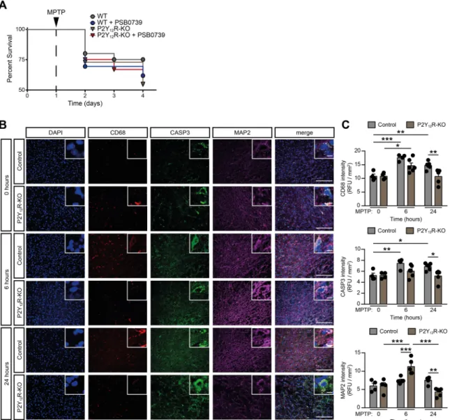
Fig. 1. P2Y12-receptor invalidation promotes MPTP induced mortality and modulates the early phase of microglia activation (A-C) WT or P2Y12-KO mice were pretreated with 0.3 mg / kg PSB 0739 or its vehicle and 4×20 mg / kg MPTP or its vehicle as indicated. Kaplan-Meier survival analysis performed on experimental groups receiving MPTP treatment (n=21–24) (A). Representative immuno-confocal microscopy images of brain slices during the acute phase of MPTP adminis- tration isolated at the indicated time from WT or P2Y12-KO mice, and stained with antibodies directed against CD68 (microsialin, red), Caspase-3 (CASP3, green), Microtubule-associated protein 2 (MAP2, purple), DAPI (blue) and overlay image (merge). Scale bar: 100µm, corresponds to 20µm inset (B) Quantification of CD68 fluorescence intensity (upper panel), CASP3 fluorescence intensity (middle panel) and MAP2 fluorescence intensity (lower panel) (n=4–6, upper panel; n=4–6, middle panel; n=4–6, lower panel) (C). Data represent the mean ±SEM; * , p≤0.05; * *, p≤0.01; * ** , p≤0.001 (two-way ANOVA, with Bonferroni’s post-hoc test (C)). (For interpretation of the references to colour in this figure legend, the reader is referred to the web version of this article.)
0739 group (P=0.0242 vs. WT control) and 45% in P2Y12R-KO groups (P=0.0052 vs. WT control). This observation suggests that the presence of functional P2Y12R is necessary to prevent the deterioration of acute neurotoxic events and to initiate disease resolution after MPTP treatment.
To further understand the acute effect of MPTP-administration and the role of dysfunctional P2Y12R during early stages of the treatment, we have investigated microglia activation, cell apoptosis and neuron viability in wild-type control and P2Y12R-KO mice before, at six hours and at twenty-four hours after the first MPTP injection. The myeloid specific lysosomal-associated membrane protein, CD68, was character- ized as a microglial activation marker previously [38,39]; the cysteine-aspartic acid protease 3 (Caspase-3, CASP3) is a well described common apoptotic marker [40] and is activated by both extrinsic mol- ecules (DAMPs) or intrinsic (mitochondrial) pathways [41]. The microtubule-associated protein 2 (MAP2) is known to be strongly and specifically expressed in the neuron perikarya and dendrites and its in- tensity can be used as an indirect marker of neuronal viability [42]. In our experiments, MPTP administration markedly increased CD68 and CASP3 fluorescence intensity in brain slices in the substantia nigra area both at six and twenty-four hours in wild-type control mice, compared to basal conditions (P=0.0001 and P=0.0069 for CD68 and P=0.0035 and P=0.0168 for CASP3; respectively), whereas MAP2 intensity was unchanged (P=0.6121 and P=0.5354; respectively) (Fig. 1B and C).
These findings indicate prompt microglia activation and increased apoptosis in this early time period. In P2Y12-receptor deficient animals, although MPTP-administration increased microglia activation at six hours compared to baseline conditions (P=0.008), but not at twenty-four hours (P=0.9980), and was significantly lower than in the wild-type control animals at this later point (P=0.0058 vs. WT at 24 h) (Fig. 1B and C). Likewise, Caspase-3 levels were lower, when compared to the wild-type control animals (P=0.0116 vs. WT at 24 h), and did not change significantly after MPTP-injection at six hours (P=0.2947) and at twenty-four hours (P=0.9817) compared to baseline conditions.
MAP2 fluorescent intensity was markedly increased at six hours after MPTP-treatment in the P2Y12R-KO animals (P=0.0001), whereas it returned to the baseline level at twenty-four hours (P=0.1390) (Fig. 1B and C). MAP2 intensity was significantly increased in the knock-out animals compared to the wild-type controls at six hours (P=0.0007 vs. WT at 6 h), and was significantly reduced at twenty-four hours (P=0.0018 vs. WT at 24 h) (Fig. 1C). These findings implicate that functional P2Y12Rs are required following MPTP treatment for early, protective microglia activation, such as being able to phagocytose apoptotic cells, and thereby to prevent the spill over of intracellular contents, and further forestalling an uncontrolled neuroinflammatory state.
3.2. P2Y12R gene deficiency and pharmacologic blockade is protective against MPTP-induced motor impairment, monoamine depletion, neurodegeneration and neuroinflammation
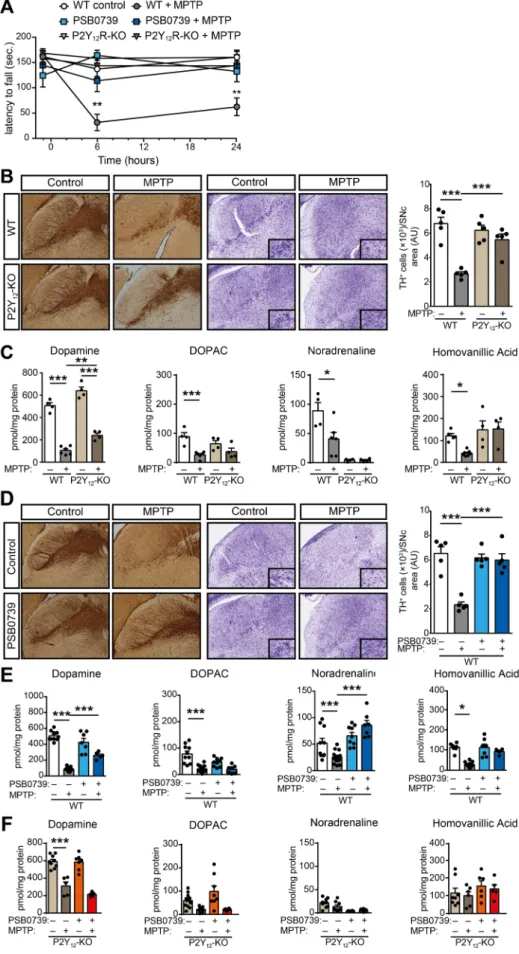
Next, we examined how genetic deletion or pharmacological blockade of P2Y12R influences MPTP-induced neurodegeneration and motor function impairment. Administration of MPTP to wild-type mice resulted in severe motor function deterioration, when the latency to fall from rotarod apparatus was measured (P=0.0026 and P=0.0034 vs.
WT control 6-hours and 24-hours post treatment, respectively) (Fig. 2A).
Surprisingly, inhibition of P2Y12R, either genetically or with pharma- cological blockade ameliorated motor function impairment in MPTP- treated mice (P < 0.0001 and P=0.039 vs. MPTP-treated 6-hours post treatment, respectively) (Fig. 2A). As expected, we found that adminis- tration of MPTP to wild-type mice induced selective degeneration of dopaminergic neurons in the substantia nigra pars compacta (Fig. 2B), demonstrated by reduced tyrosine-hydroxylase immunostaining and the presence of degenerating neurons with condensed nuclei, shrunken cytoplasm and prominent nuclear alterations (P < 0.0001 WT control
vs. WT MPTP-treatment). MPTP-induced neurodegeneration was asso- ciated with reduced levels of monoamines, specifically dopamine (P < 0.0001), DOPAC (P < 0.0001), homovanillic acid (P=0.0174) and noradrenaline (P=0.0005) (Fig. 2C Supplementary Table 1) and decreased ATP levels in the striatum indicating a state of an energy deficit, as reflected by lower ATP/ADP ratio (P=0.0007) (Supple- mentary Figure 2 A and B). Corroborating our findings on motor impairment, genetic deletion of the receptor protected from dopami- nergic neuronal cell death in the substantia nigra pars compacta following MPTP-treatment (P < 0.0012 WT MPTP-treatment vs. KO MPTP-treatment) (Fig. 2B), mitigated the decrease in ATP concentration (P=0.7707) and alleviated the reduction in MPTP-induced dopamine levels (P < 0.0031 WT MPTP-treatment vs. KO MPTP-treatment) in the striatum (Fig. 2C and Supplementary Figure 2 A). P2Y12R gene defi- ciency had no effect on striatal noradrenaline (P=0.0887 WT MPTP- treatment vs. KO MPTP-treatment), DOPAC (P>0.9999 WT MPTP-treat- ment vs. KO MPTP-treatment) and homovanillic acid concentration (P=0.1005 WT MPTP-treatment vs. KO MPTP-treatment) (Fig. 2C and Supplementary Fig. 2A).
Intrathecal administration of the selective P2Y12-receptor antagonist PSB 0739 (0.3 mg/kg i.t.) 18 h prior to MPTP largely replicated the ef- fect of P2Y12R gene deficiency on MPTP-induced Parkinsonism in wild- type mice (Fig. 2D and E). In vehicle treated animals, MPTP adminis- tration promoted the degradation of dopaminergic neurons in the sub- stantia nigra pars compacta (P < 0.0001 WT control vs. WT MPTP- treatment) (Fig. 2D), and reduced the concentration of monoamines, specifically dopamine (P < 0.0001), DOPAC (P < 0.0001), homo- vanillic acid (P=0.0270) and noradrenaline (P=0.0004) (Fig. 2E and Supplementary Table 1). PSB 0739 administration prevented the loss of TH-positive cells in the substantia nigra pars compacta (P < 0.0001) (Fig. 2D), and alleviated the depletion of dopamine (P=0.0031), and noradrenaline levels in the striatum (Fig. 2E). Furthermore, pharmaco- logical blockade of P2Y12R with PSB 0739 in the P2Y12R-KO mice had no further effect on monoamine concentration in the striatum following MPTP-treatment, when compared to vehicle treated littermate controls (P=0.2576 for dopamine; P=0.9490 for noradrenaline; P>0.9999 for DOPAC and P=0.9035 for homovanillic acid) (Fig. 2F). This finding in- dicates that PSB 0739 selectively act on P2Y12R, and its effect requires the presence of functional receptors.
Additionally, we investigated the influence of PSB 0739 on MPTP- induced neuroinflammation and microglia activation. Treatment with MPTP in vehicle treated mice was also associated with neuro- inflammation, characterized by increased levels of TNFα, IL-1β and IL-6 and the anti-inflammatory cytokine IL-10 in the striatum (P=0.0021;
P < 0.0001; P=0.0018 and P < 0.0001; respectively) (Fig. 3B). PSB 0739 pre-treatment abolished the increase in cytokine levels in the substantia nigra (P=0.0004 for TNFα; P=0.0005 for IL-1β; P=0.0007 for IL-6 and P=0.0688 for IL-10) and the striatum (P=0.0004 for TNFα; P < 0.0001 for IL-1β; P=0.0008 for IL-6 and P < 0.0001 for IL-10) (Fig. 3A and B, respectively).
These data indicate that P2Y12R in the central nervous system has a biphasic effect in the MPTP-induced Parkinson’s disease model.
Initially, functional P2Y12R are essential to reduce the effects of acute toxicity; however, with the progression of the disease and the develop- ment of neuronal loss, P2Y12R may further contribute to uncontrolled neuroinflammation. To further understand the mechanisms involved, we have also investigated the involvement of P2Y12R in the modulation on dopamine release from the striatum. Although we could observe significant change in veratridine evoked [3H]DA efflux in the slices of P2Y12R-KO mice (P=0.0018) indicating a potential neuromodulatory effect, this finding was not recapitulated by the P2Y12-receptor antag- onist PSB 0739 (no significant difference was observed) (Supplementary Figure 3).
Fig. 2. P2Y12R gene deficiency and pharmacolog- ical blockade is protective against MPTP-induced motor impairment, monoamine depletion and neurodegeneration (A-F) WT or P2Y12-KO mice were pretreated with 0.3 mg / kg PSB 0739 or its vehicle and 4×20 mg / kg MPTP or its vehicle as indicated. Effect of P2Y12R gene deficiency or 0.3 mg / kg PSB 0739 on the motor performance during MPTP induced PD measured on the rotarod test (n=5–8) (A). Immuno-DAB staining for TH or cresyl violet staining on representative tissue sec- tions after MPTP-treatment of WT or P2Y12-KO mice; immunoreactivity is seen in the cell body and processes of dopaminergic and noradrenergic neu- rons (B); bar diagram shows the quantification of TH positive cells in the substantia nigra pars com- pacta (n=5) (B, right panel). Concentration of dopamine, DOPAC, homovanillic acid and noradrenaline were determined from striatum samples of WT and P2Y12-KO mice 72 h after the initial MPTP administration (n=4–6) (C).
Immuno-DAB staining for TH or cresyl-violet staining on representative tissue sections after MPTP-treatment from PSB 0739 treated WT mice;
immunoreactivity is seen in the cell body and pro- cesses of dopaminergic and noradrenergic neurons (D); bar diagram demonstrates the quantification of TH-positive cells in the substantia nigra pars com- pacta (n=5) (D, right panel). Concentration of dopamine, DOPAC, homovanillic acid and noradrenaline were determined from striatum samples 72 h after the first MPTP administration (n=5–15) (E); or from striatum samples of P2Y12- KO mice pretreated with PSB 0739 or its vehicle, 72 h after the initial MPTP injection (n=5–12) (F).
Data represent the mean ±SEM; * , p≤0.05; * *, p≤0.01; * ** , p≤0.001 (two-way ANOVA, with Tukey’s post-hoc test (A-F)).
3.3. P2Y12-receptor blockade halts disease progression after subchronic MPTP administration
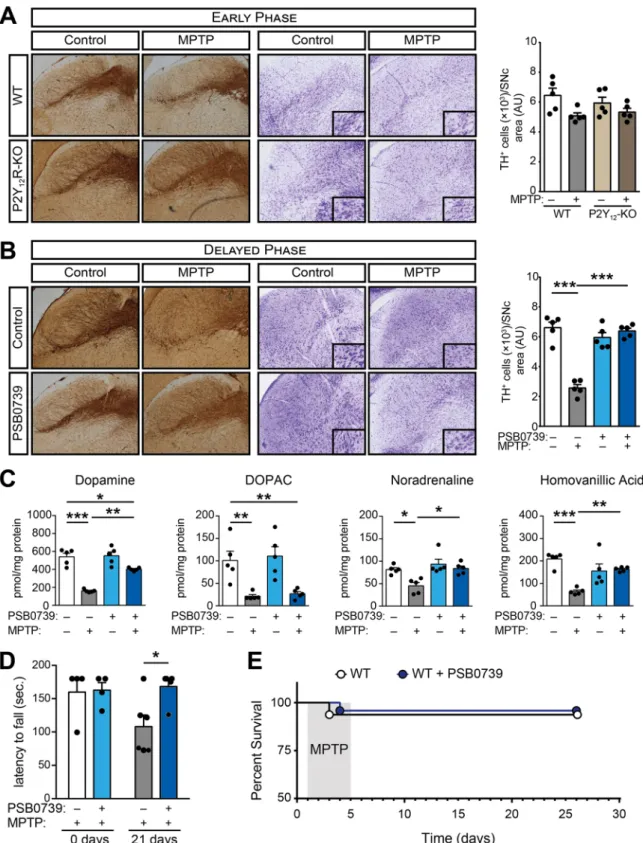
Since our data implicated a pivotal role for P2Y12R in neuro- inflammation, we further investigated the contribution of P2Y12R using an animal model of MPTP-induced subchronic neurodegeneration. To account for the markedly different survival rate of wild-type and P2Y12R deficient animals during the acute experimental PD model, an alterna- tive MPTP treatment protocol was used to explore P2Y12R actions during disease progression; rather than administering the P2Y12R-inhibitor prior to MPTP, a subchronic Parkinson’s disease model was generated by a single daily administration of MPTP over the course of five days, fol- lowed by receptor blockade for four consecutive days (Supplementary Figure 1B). During the early phase of the MPTP treatment, on day five to six, limited effect could be observed on dopaminergic neuron loss in wild-type and P2Y12R-KO mice (P=0.0576 and P=0.6108; respec- tively) (Fig. 4A). However, twenty-one days following MPTP adminis- tration, we detected significant loss of dopaminergic neurons in the substantia nigra pars compacta (P < 0.0001) (Fig. 4B), reduction in dopamine, DOPAC, noradrenaline and homovanillic acid levels (P < 0.0001; P=0.0082; P=0.0095 and P < 0.0001; respectively) (Fig. 4C), and markedly decreased motor functions (P=0.0206) (Fig. 4D). P2Y12R inhibition increased dopaminergic cell survival (P < 0.0001) (Fig. 4B), reduced the decrease in dopamine, noradrena- line and homovanillic acid levels (P=0.0002; P=0.0039 and P=0.0057; respectively) (Fig. 4C), and prevented deterioration in motor function (P=0.9950) (Fig. 4D) assessed three weeks after treatment.
Furthermore, using this model, there was no difference in the survival rate between the experimental groups (Fig. 4E).
3.4. P2Y12-receptor mediates microglia activation via Rho-kinase and p38 MAPK phosphorylation
To characterize the intracellular mechanism underlying the protec- tive effect of P2Y12R blockade, the relationship between P2Y12R stim- ulation and microglia activation was investigated. Initially, since the activation of p38 mitogen-activated protein kinase (p38 MAPK) can directly promote or indirectly stimulate cytokine production via MK2 or MSK1/2 in microglia [43–46], changes in the p38 MAPK activity was measured. Phosphorylation of p38 MAPK at Threonine 180 / Tyrosine 182 increases the kinase activity, thus promoting phosphorylation of
several transcriptional factors and further bolster inflammatory cytokine biosynthesis [47]. In initial experiments we confirmed that treatment with LPS (1µg / ml, 60 min) or the P2Y12R agonist ADP (10µM, 60 min) promotes p38 MAPK phosphorylation at T 180 / Y 182 in vitro (P=0.0002 and P < 0.0001; respectively) (Fig. 5A and B) using murine microglial cell line BV-2. The small GTPase, Ras homolog family mem- ber A (RhoA) and the downstream effector, Rho-associated, coiled-coil containing protein kinase 1 (ROCK1) has been previously shown to mediate p38 MAPK phosphorylation and activation [48]; we have tested the effect of the ROCK1 inhibitor, Y-27632, on LPS or ADP-induced p38 MAPK activation. Pretreatment with Y-27632 (10µM, 30 min) mark- edly decreased LPS or ADP-induced p38 MAPK phosphorylation (P=0.0044 and P=0.0013; respectively) (Fig. 5B); additionally, pre- treatment with the selective P2Y12R inhibitor, PSB 0739 (500 nM, 30 min) blocked ADP-induced, but not LPS-induced p38 MAPK activa- tion (P=0.0007 and P=0.9999; respectively) (Fig. 5A). These results indicate that under in vitro conditions, both P2Y12R and ROCK1 are necessary for ADP-mediated p38 MAPK phosphorylation. To confirm the influence of P2Y12R on p38 MAPK phosphorylation and microglia acti- vation, as well as on the cytokine levels produced by microglia in vitro, murine microglia cells were treated with the ectonucleotidase inhibitor ARL 67156 (30µM, 20 min) combined with either PSB 0739 (500 nM, 20 min) or with the solvent [49]. Cells were stimulated with ADP (100µM) for six or twenty-four hours, and microglia activation and p38 MAPK phosphorylation at Threonine 180 / Tyrosine 182 were analysed (Fig. 5C and D). ADP stimulation markedly increased CD68 fluorescence intensity at six and at twenty-four hours compared to basal conditions (P=0.0005 and P < 0.0001 at 6 and 24 h; respectively), whereas phos- phorylated p38 MAPK intensity was unchanged at six hours (P=0.4343), but was significantly increased at twenty-four hours (P < 0.0001) (Fig. 5C and D). In cells treated with P2Y12-receptor antagonist, ADP administration was without effect on microglia acti- vation and p38 MAPK phosphorylation compared to the baseline con- dition (P=0.3342 and P=0.9994 at 6 and 24 h for CD68; and P=0.7589 and P=0.3572 at 6 and 24 h for p-p38 MAPK; respectively), and was significantly different from the control cells (P=0.0138 and P < 0.0001 vs. Control at 6 and 24 h for CD68; and P < 0.0001 vs.
Control at 24 h for p-p38 MAPK; respectively) (Fig. 5C and D). Stimulation with ADP was also associated with increased production of TNFα (P=0.0191 and P < 0.0001 at 6 and 24 h; respectively), IL-1β at twenty-four hours (P=0.2340 and P < 0.0001 at 6 and 24 h;
Fig. 3. Pharmacological blockade of P2Y12R abrogates MPTP-induced pro-inflammatory cytokine production in the striatum (A-B) WT mice were pretreated with 0.3 mg / kg PSB 0739 or its vehicle and 4×20 mg / kg MPTP or its vehicle as indicated. TNFα, IL-1β, IL-6 and IL-10 concentration measured from substantia nigra (n=5) (A) or striatum samples (n=5) (B). Data represent the mean ±SEM; * , p≤0.05; * *, p≤0.01; *** , p≤0.001 (two- way ANOVA, with Bonferroni’s post-hoc test (A- B)).
Fig. 4.P2Y12R blockade halts disease progression (A-E) WT or P2Y12-KO mice were treated with 20 mg / kg MPTP daily for five consecutive days, followed by treatment with 0.3 mg / kg PSB 0739 or its vehicle. Immuno-DAB staining for TH and cresyl-violet staining on representative tissue sections five days after the initial MPTP administration; immunoreactivity is seen in the cell body and processes of dopaminergic and noradrenergic neurons (A); bar diagram shows the quantification of TH-positive cells in the substantia nigra pars compacta (n=5) (A, right panel). Immuno-DAB staining for TH and cresyl-violet staining on representative tissue sections 21 days after the last PSB 0739 treatment (B); bar diagram displays the quantification of TH-positive cells in the substantia nigra pars compacta (n=5) (B, right panel). Concentration of dopamine, DOPAC, homovanillic acid and noradrenaline were determined from striatum samples 21 days after last PSB 0739 administration (n=5) (C). Effect of PSB 0739 or its vehicle on the motor performance during MPTP-induced PD measured on the rotarod test before and 21 days after treatment (n=4–6) (D). Kaplan-Meier survival analysis performed on experimental groups receiving MPTP treatment (n=8–16) (E). Data represent the mean
±SEM; * , p≤0.05; **, p≤0.01; *** , p≤0.001 (two-way ANOVA, with Bonferroni’s post-hoc test (A-D)).
respectively) and IL-6 (P < 0.0001 and P < 0.0001 at 6 and 24 h;
respectively) but not the anti-inflammatory cytokine IL-10 (concentration of IL-10 was below the detection limit) (Fig. 5E). P2Y12-receptor blockade reduced TNFα (P=0.0179 and P < 0.0001 vs. Control at 6 and 24 h; respectively), IL-1β at twenty-four hours (P=0.1547 and P=0.0027 vs. Control at 6 and 24 h; respectively) and IL-6 levels at six hours (P=0.0013 and P=0.0866 vs. Control at 6 and 24 h; respectively) (Fig. 5E).
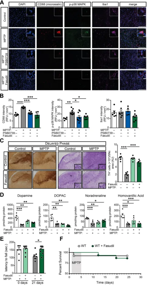
Next, the involvement of P2Y12R and ROCK1 blockade on p38 MAPK phosphorylation and microglia activation under in vivo conditions was tested. Apart from the myeloid specific lysosomal-associated membrane protein (CD68); the ionized calcium-binding adapter molecule 1 (Iba1), which is known to be strongly and specifically expressed in both se- nescent and activated microglia [50] was stained. In our experiments, MPTP administration markedly increased phospho-p38 MAPK T180/Y182 and CD68 fluorescence intensity in brain slices in the sub- stantia nigra area compared to saline treated control animals (P=0.0063 and P=0.0003; respectively), whereas Iba1 intensity was
unchanged (P=0.9931) 21 days after treatment (Fig. 6A and B).
Intrathecal administration of 0.3 mg / kg PSB 0739 alone was without effect. However, P2Y12-receptor blockade significantly reduced MPTP-induced p38 MAPK activity and microglia activation, measured by phospho-p38 MAPK T180/Y182 and CD68 positivity (P=0.0406 and P=0.0026; respectively) (Fig. 6A and B). To assess ROCK1 involvement, the specific ROCK1 inhibitor fasudil, which has the advantage over Y-27632 that it does not require parenteral administration, was orally delivered in the drinking water (50 mg / kg body weight per day) [27]
(Supplementary Figure 1 C). Fasudil-treatment alone had no effect on phospho-p38 MAPK T180/Y182 or CD68 fluorescence intensity. When fasudil was administered during MPTP-treatment, CD68 and phosphorylated-p38 MAPK fluorescence intensity was significantly reduced in comparison to MPTP-treatment alone (P=0.0181 and P=0.0034; respectively) (Fig. 6A and B). No changes could be observed in both p38 MAPK and Iba1 fluorescence intensity between control, MPTP treated or MPTP combined with PSB 0739 or fasudil-treated groups (P=0.1621; P=0.1371 and P=0.6200 for p38 MAPK; and Fig. 5. P2Y12R mediates ADP-induced microglia activation in vitro to control cytokine production via Rho-kinase-dependent p38 MAPK phosphorylation (A-B) Murine BV-2 microglia cells were pretreated with PSB 0739 (500 nM, 30 min) (A) or with Y-27632 (10µM, 30 min) (B) and were incubated with LPS (100 ng / ml, 60 min), ADP (10µM, 5 min) or solvent (control) and phosphorylation of p38 MAPK at Threonine 180 / Tyrosine 182 were determined by immunoblotting. Graphs show the densitometric evaluation (n=4). (C-E) Murine BV-2 microglia cells were pretreated with ARL 67156 (30µM, 20 min) combined with PSB 0739 (500 nM, 30 min) or the solvent (Control) as indicated; thereafter, ADP (100µM) was added to the extracellular solution for the indicated time periods to induce microglia activation and cytokine production. Shown are representative immuno-confocal microscopy images of cultured cells after ADP administration, and stained with antibodies directed against CD68 (microsialin, red), phosphorylated-p38 MAPK (green), DAPI (blue) and overlay image (merge). Scale bar: 100µm, corresponds to 20µm inset (C) Quantification of CD68 fluorescence intensity (left panel), and phosphorylated-p38 MAPK fluorescence intensity (right panel) (n=4–8, left panel;
n=3–7, right panel). Values were normalized to the average fluorescent intensity of Iba1 staining (not shown) (D). TNFα, IL-1β, IL-6 and IL-10 concentration measured from the cultured cell supernatant at 0, 6 and 24 h of ADP stimulation (n=3–5) (E). Data represent the mean ±SEM; * , p≤0.05; **, p≤0.01; *** , p≤0.001 (two-way ANOVA with Bonferroni’s post-hoc test (A, B, D, E)). (For interpretation of the references to colour in this figure legend, the reader is referred to the web version of this article.)
Fig. 6.Pharmacological P2Y12R or Rho-kinase blockade in vivo abolish microglia activation, interrupts disease pro- gression and alleviates MPTP-induced Parkinsonism (A-F) WT mice were treated with 20 mg / kg MPTP daily for five consecutive days, followed by treatment with 0.3 mg / kg PSB 0739, 50 mg / kg body weight per day fasudil or its vehicle. Shown are representative immuno-confocal mi- croscopy images of brain slices isolated from WT mice stained with antibodies directed against CD68 (microsialin, red), phosphorylated-p38 MAPK (green), Iba1 (purple), DAPI (blue) and overlay image (merge). Scale bar: 100µm, corresponds to 20µm inset (A) Quantification of CD68 fluorescence intensity (left panel), phosphorylated-p38 MAPK fluorescence intensity (middle panel) and Iba1 fluorescence intensity (right panel) (n=5–8, left panel;
n=5–9, middle panel; n=5–7, right panel) (B). Repre- sentative images of immuno-DAB staining for TH and cresyl-violet staining on tissue sections 21 days after the last treatment; immunoreactivity is seen in the cell body and processes of dopaminergic and noradrenergic neurons (C); bar diagram show quantification of TH-positive cells in the substantia nigra pars compacta (n=5) (C, right panel).
Concentration of dopamine, DOPAC, homovanillic acid and noradrenaline were determined from striatum samples 21 days after last treatment (n=5) (D). Effect of fasudil or its vehicle on the motor performance during MPTP-induced PD measured on the rotarod test before and 21 days after treatment (n=8) (E). Kaplan-Meier survival analysis per- formed on experimental groups receiving MPTP treatment (n=8–10) (F). Data represent the mean ±SEM; * , p≤0.05; **, p≤0.01; ***, p≤0.001 (two-way ANOVA, with Bonferroni’s post-hoc test (B-E)). (For interpretation of the references to colour in this figure legend, the reader is referred to the web version of this article.)