Role of microglia in neurotropic viral infections
Ph. D. thesis
Rebeka Fekete
Semmelweis University
János Szentágothai Doctoral School of Neurosciences
Supervisors: Dr. Dénes Ádám, Ph.D, Dr. Környei Zsuzsanna, Ph.D Official reviewers: Dr. Kékesi Adrienna Katalin, Ph.D
Dr. Jakus Zoltán, Ph.D
Head of the Final Examination Comittee: Dr. Falus András, Ph.D, D.sc Members of the Final Examination Comittee: Dr. Kardos József, Ph.D
Dr. Alpár Alán, Ph.D, D.sc
Budapest
2019
1
Table of contents
TABLE OF CONTENTS ... 1
LIST OF ABBREVIATIONS ... 3
1. INTRODUCTION ... 7
1.1. BARRIERS IN THE BRAIN AND REGULATION OF IMMUNE CELL TRAFFICKING ... 7
1.2. THE NEUROVASCULAR UNIT ... 10
1.3. ORIGIN OF MYELOID CELLS IN THE CNS ... 12
1.4. THE ROLE OF MICROGLIA DURING DEVELOPMENT AND IN NORMAL BRAIN FUNCTION ... 13
1.4.1. Developmental role of microglia ... 13
1.4.2. The role of microglia in adulthood ... 14
1.5. THE ROLE OF MICROGLIA IN BRAIN INFLAMMATION AND INJURY ... 18
1.6. NEUROTROPIC VIRUSES ... 19
1.6.1. Pseudorabies virus ... 22
1.7. INFECTION-INDUCED INFLAMMATION AND ANTI-VIRAL IMMUNITY IN THE BRAIN ... 25
1.8. THE ROLE OF MICROGLIA IN NEUROTROP VIRAL INFECTIONS OF THE CNS ... 28
2. OBJECTIVES ... 30
3. MATERIALS AND METHODS ... 31
3.1. PROCESSING OF HUMAN SAMPLES ... 31
3.2. IN VIVO MOUSE EXPREIMENTS ... 34
3.3. IN VITRO MOUSE EXPERIMENTS ... 40
4. RESULTS ... 48
4.1. MICROGLIA ARE ESSENTIAL FOR ANTI-VIRAL IMMUNITY IN THE CENTRAL NERVOUS SYSTEM ... 48
4.1.1. Microglia control the spread of viral infection in the brain ... 48
4.1.2. Selective elimination of microglia results in uncontrolled viral spread and neurobehavioral pathologies ... 49
4.1.3. Microglia phagocytose infected neurons but are resistant to productive viral infection ... 52
4.2. MICROGLIA RECRUITMENT IS INITIATED RAPIDLY TO VIRUS-INFECTED NEURONS IN THE BRAIN 54 4.2.1. Microglia are recruited to infected neurons within hours as suggested by differential expression of viral immediate-early and structural proteins ... 54
4.2.2. Imaging microglia recruitment with in vivo two-photon microscopy in real-time ... 55
4.2.3. Neurons are directly contacted by microglia at the early phase of virus infection ... 58
4.3. VIRUS INFECTION TRIGGERS THE RECRUITMENT AND PHAGOCYTIC ACTIVITY OF MICROGLIA IN VITRO 60 4.3.1. Microglial responses to infection in vitro ... 60
4.3.2. Microglia phagocytose infected cells in vitro ... 61
4.4. NUCLEOTIDES RELEASED FROM INFECTED CELLS TRIGGER MICROGLIA RECRUITMENT AND PHAGOCYTOSIS VIA MICROGLIAL P2Y12 RECEPTORS ... 62
4.4.1. Neurotropic virus infection induces the production of inflammatory mediators ... 62
4.4.2. Release of purinergic nucleotides triggers rapid microglia activation ... 64
4.4.3. Microglia recruitment to infected neurons is mediated by P2Y12 receptors in vitro ... 65
4.5. MICROGLIAL P2Y12 RECEPTORS MEDIATE RECRUITMENT OF MICROGLIA AND ELIMINATION OF VIRUS INFECTED CELLS IN VIVO ... 67
2
4.5.1. Microglial P2Y12 receptors form clusters when contacting the cell membranes of infected
neurons ... 67
4.5.2. Microglia recruitment is impaired in the absence of P2Y12 receptors in vivo, but neurological symptoms are not augmented by P2Y12R deficiency ... 69
4.6. MICROGLIA RECRUIT LEUKOCYTES INTO THE BRAIN UPON VIRUS INFECTION INDEPENDENTLY OF P2Y12-MEDIATED SIGNALLING ... 73
4.6.1.P2Y12-mediated signalling does not affect leukocyte recruitment during viral infection ... 73
4.6.2.Selective depletion of microglia influences leukocyte infiltration in virus infected brain ... 75
4.7. RECRUITMENT OF P2Y12-POSITIVE MICROGLIA AND LEUKOCYTES IN HUMAN HERPES SIMPLEX ENCEPHALITIS ... 79
5. DISCUSSION ... 83
5.1. REGULATION OF INFLAMMATORY RESPONSES DURING NEUROTROPIC VIRAL INFECTIONS ... 83
5.2. PSEUDORABIES VIRUS INFECTION AS AN IDEAL MODEL FOR STUDYING MICROGLIA-MEDIATED INFLAMMATORY RESPONSES ... 85
5.3. MICROGLIA SENSE VARIOUS DANGER SIGNALS COMING FROM COMPROMISED NEURONS ... 86
5.4. NUCLEOTIDES RELEASED FROM COMPROMISED NEURONS CAUSE IMMEDIATE RESPONSE FROM MICROGLIA AND SENSED VIA MICROGLIAL P2Y12 RECEPTOR ... 87
5.5. MICROGLIA ARE ESSENTIAL TO LIMIT NEUROTROPIC VIRUS INFECTION IN THE BRAIN ... 88
5.6. SELECTIVE MICROGLIA ELIMINATION, BUT NOT P2Y12 DEFICIENCY LEADS TO ADVERSE NEUROLOGICAL SYMPTOMS IN PRV INFECTED MICE ... 89
5.7. LEUKOCYTE INFILTRATION IN VIRUS INFECTED BRAINS IS INFLUENCED BY MICROGLIA, BUT IS INDEPENDENT FROM P2Y12 RECEPTOR MEDIATED PROCESSES ... 90
5.8. BBB INJURY IS NOT MARKEDLY AFFECTED BY THE ABSENCE OF MICROGLIA DURING VIRAL INFECTION ... 91
5.9. P2Y12-POSITIVE MICROGLIA INTERACT WITH INFECTED NEURONS IN HUMAN HSV-1 ENCEPHALITIS ... 93
6. CONCLUSION ... 95
7. SUMMARY ... 96
8. ÖSSZEFOGLALÁS ... 97
9. REFERENCES ... 98
10. LIST OF PUBLICATIONS ... 112
11. ACKNOWLEDGEMENTS ... 113
3
List of Abbreviations
AD Alzheimer’s disease
ASP Antisense promoter
ATP Adenosine Tri-Posphate
BBB Blood-brain barrier
BCSFB Blood-CSF barrier
BDG Bartha DupGreen
BDNF Brain Derived Neurotrophic Factor
BDR BarthaDup Red
CCL2,19 Chemokine (C-C motif) ligand
CD200/CD200R OX-2 type I membrane glycoprotein
CMV Cytomegalovirus
CNS Central Nervous System
CSF Cerebrospinal fluid
CSF1-R Colony Stimulating Factor 1 Receptor
CXCL9/10 Chemokine (C-X-C motif) ligand
Cx3cl1 Chemokine (C-X3-C motif) ligand 1/fractalkine
Cx3cr1 CX3C Chemokine receptor 1/ fractalkine receptor
C1q Complement component 1q
C3 Complement 3
DAMP Danger Associated Molecular Pattern
DAP12 DNAX activation protein 12
DNA Deoxyribonucleic acid
DTR Diphtheria toxin receptor
EMP Erythromieloid precursor cell
FFPE Formalin-fixed paraffin embeded
GFAP Glial Fibrillary Acidic Protein
GFP Green Fluorescent Protein
4
GWAS Genome Wide Associated Study
HMGB1 High Mobility Group Box 1
HIV-1 Human Immunodeficiency Virus-1
HSV Herpes Simplex virus
HSVE-1 Herpes Simplex type 1 encephalitis
HSVTK Herpes Simplex viral thymidine kinase
IFN-1 Interferon 1
IGF-1 Insulin-like Growth Factor 1
IgG Immunoglobulin G
IL-1 Interleukin 1
IL-6 Interleukin 6
IL-12 Interleukin 12
IL-34 Interleukin 34
Irf8 Interferon regulatory factor 8
MGM Meningeal macropohage
MHV Mouse hepatitis virus
MIP-1 Macrophage Inflammatory Proteins
MV Measles virus
NLRs Nod-like resptors
NTPDase Nucleoside Triphosphate Diphosphohydrolase
NVU Neurovascular unit
PAMP Pathogen Associated Molecular Pattern
PD Parkinson’s disease
PNS Peripheral Nervous System
PRR Pattern Recognition Receptors
PRV Pseudorabies Virus
Pu.1 Transcription factor PU.1
PV Poliovirus
PVM Perivascular macrophage
RABV Rabies virus
RNA Ribonucleic acid
RLRs Rig-like receptors
5
SVZ Subventricular zone
TBI Traumatic Brain Injury
TGFß Transforming Growth Factor ß
TNFα Tumor Necrosis Factor α
TLRs Toll like resptors
TLR3 Toll-like receptor 3
TREM-2 Triggering Receptor Expressed on Myeloid cells 2
VZV Varicella Zooster virus
WNV West Nile virus
6
“Inflammatory processes of any nature are soon to be manifested in the reaction of microglia. In cases of meningitis and meningoencephalitis the microglia of the affected areas undergoes changes corresponding to the early stages of mobilization and phagocytic intervention.” Pio del Rio-Hortega 1932.
7
1. Introduction
1.1. Barriers in the brain and regulation of immune cell trafficking
While the weight of the brain accounts for only 2% of the total body weight, it consumes 20% of the total cardiac output. This requires constant energy balance and strictly controlled blood flow (autoregulation), as well as rapid adjustment of local perfusion needs to change in neuronal activity (functional hyperemia). In addition, for the proper functioning of neurons, the brain requires tight metabolic regulation and ion gradients that largely differ from the composition of the blood. Vascular injury or brain damage results in the dysregulation of proper brain circulation and metabolism, that may lead to irreversible neuronal injury. Therefore, the brain is protected by special barriers that allow isolation of the brain microenvironment from the peripheral circulation and circulating immune cells.
The vascular network of the central nervous system (CNS) has evolved to maintain proper homeostatic balance inside the parenchyma. Vascular permeability, immune cell trafficking, blood flow and vascular tone that are critical for the proper function of CNS is precisely controlled (Hanisch and Kettenmann 2007; Daneman 2012). To this end, the blood-brain barrier (BBB) is formed by unique blood vessels in the CNS that regulate the movement of molecules, ions and immune cells between the blood and neural tissue. The BBB also protects the CNS from injury and infection by limiting the entry of pathogens and immune cells (Engelhardt & Liebner, 2014). BBB function between the blood and the CNS parenchyma is maintained by specialized capillary endothelial cells with low pinocytotic activity, fused together by complex tight junctions (Engelhardt and Sorokin 2009; Tietz and Engelhardt 2015). The endothelial cells regulate paracellular transport in both directions, but transcellular transport is regulated by special pumps and receptors (Archer, Pitelka, &
Hammond, 2004). The basal lamina of endothelial cells is surrounded by astrocyte endfeet, which contributes to the barrier function by limiting the trafficking of macromolecules and immune cells. The blood-CSF barrier (BCSFB) is the second interphase that protects the CNS, formed by the choroid plexus epithelium which produces and regulates the composition of CSF (Prinz, Priller, Sisodia, & Ransohoff, 2011). The barrier function in the choroid plexus is provided by tight junctions between epithelial cells (Engelhardt & Sorokin, 2009). The blood-CSF interface is also an important site for immune cell entry into the CNS, where low numbers of different immune cell populations are seen under physiological conditions, while their turnover is far slower than that seen in peripheral tissues (Prinz, Erny,
8
& Hagemeyer, 2017). In homeostatic conditions, peripheral immune cells also patrol in specialized CNS compartments located outside the brain parenchyma. Immune cells can also gain access to the CNS via the non-fenestrated vascularized stroma of the blood-CSF barrier that is surrounded by the choroid plexus epithelial cells, the perivascular space (Virchow- Robin space; (Ousman and Kubes 2012; Muoio, Persson, and Sendeski 2014). There are also specialized regions of the brain that allow direct communication between the brain and the vascular system. In the circumventricular organs, located around the third and fourth ventricles, the BBB is more permeable, containing fenestrations and discontinous tight junctions (Bauer, Krizbai, Bauer, & Traweger, 2014) and these areas are important to regulate neuro-immune interactions in health and disease. Recent research revealed that similar to the drainage of extracellular fluid in peripheral tissues, the brain also has its clearance routes, called the glymphatic system (glial-lymphatic system, (see Figure 1.). This is provided by a unique system of perivascular channels formed by astrocytes that promote efficient elimination of soluble proteins and metabolites from the CNS. It mainly operates as a waste clearance system and its role in brain inflammation and neurological diseases has also been suggested (Aspelund et al. 2015; Kipnis et al. 2017). In addition, lymph vessels have recently been identified in the meninges, which are emerging sites for neuro-immune interactions (Kipnis et al., 2017). At present, it is assumed that immune cells, primarily myeloid cell populations, which reside in the brain throughout life can gain access to the developing CNS in the first trimester in both mice and humans via both meningeal and vascular routes, while the role for the choroid plexus in their migration is also emerging. In adults, these areas in the CNS provide functionally important routes of entry for leukocytes during brain inflammation or diverse neuropathologies. In physiological conditions, meningeal, choroid plexus machrophages and a small amount of T cells patrol continuously in the subarachnoidal space and the ventricles (Figure 1.)(Ousman & Kubes, 2012).
9
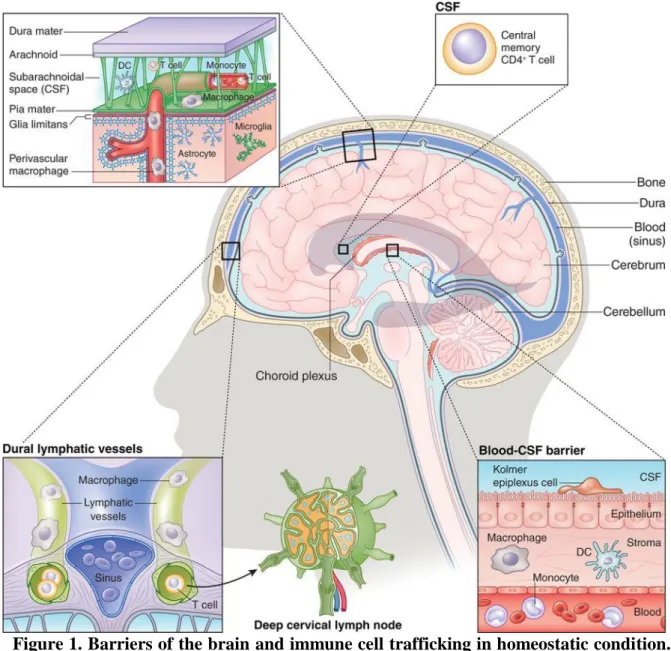
Figure 1. Barriers of the brain and immune cell trafficking in homeostatic condition.
The brain is isolated and protected by the skull and three layers of meninges. The choroid plexus produces CSF which flows both in the parenchyma and in the subarachnoid area and comprises arteries and perivascular space. Thus, the CSF contains small amount of CD4+ T cells, which patrol the brain. In healthy brain tissue, leukocytes (granulocytes, monocytes, T and B cells) stay within the blood vessels. In the brain parenchyma microglial cells are the only immune cells maintaining the healty environment. In non-parenchymal areas perivascular, meningeal and choroid plexus machrophages are patrolling and protect the brain tissue (Prinz & Priller, 2017).
10
1.2. The neurovascular unit
The key area in the brain vasculature that regulates cerebral blood flow and immune cell trafficking is called the neurovascular unit (NVU). The NVU is formed via complex functional interactions between endothelial cells, pericytes, perivascular macrophages (PVMs), microglia, astrocytes and neurons (Figure 2.) (Wong et al., 2013). All those cell types are in an integrated relationship and they maintain homeostasis and proper function of the CNS (Thurgur & Pinteaux, 2018). The blood-brain barrier can be considered as an integral part of the NVU. The low permeability and integrity of endothelial cells is the most important feature of CNS protection from blood-borne materials (Tietz & Engelhardt, 2015).
Astrocytes compose the glia limitans perivascularis, which separates the parenchyma from the blood vessels and continues as glia limitans superficialis surrounding the entire surface of the brain and spinal cord (Abbott, Rönnbäck, & Hansson, 2006). Astrocytes cover the parenchymal basement membrane, produce different growth factors for BBB maturation and maintenance and, via aquaporin channels, they can also regulate local water transport (Ransohoff & Engelhardt, 2012). Besides BBB maintenance they provide both mechanical and metabolic support for neurons (Abbott et al., 2006). Pericytes are embedded in the vascular basement membrane of microvessels. Due to their exclusive position, they have close contact with endothelial cells, astrocyte endfeet, perivascular macrophages and neurons (Rustenhoven, Jansson, Smyth, & Dragunow, 2017a). In the brain, pericytes have distinct morphologies and depending on their position in the vasculature have different functions as well. In the arteriolar end of vessels, precapillary pericytes are likely to modify vascular diameter, and true-capillary pericytes contribute more to BBB maintenance (Rustenhoven et al. 2017; Engelhardt 2008).
The main immunocompetent cell types in the NVU are microglia and perivascular macrophages. Since in this thesis the inflammatory- and immune mechanisms discussed in the context of neurotropic virus infection primarily concern microglial cells, the chapters below will focus on the origin, maintenance and effector functions of brain myeloid cell types, primarily microglia.
11
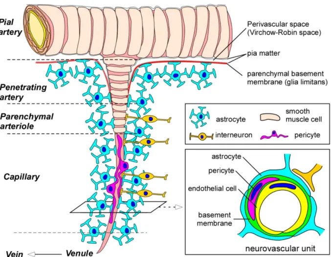
Figure 2. Structure of the blood-brain barrier (BBB) and neurovascular unit (NVU).
Pial arteries branch out into smaller arteries, called penetrating arteries. They go down into the parenchyma, branching further into arterioles and capillaries. Pial and penetrating arteries are covered by vascular smooth mucle cells and are separated from the brain parenchyma by the glia limitans. Endothelial cells form the blood-brain barrier, which are in close contact with other cell types (pericytes, astrocytes, perivascular macrophages and interneurons) composing the neurovascular unit (Yamazaki and Kanekiyo 2017).
12
1.3. Origin of myeloid cells in the CNS
Myeloid cells in the CNS represent a heterogeneous class of innate immune cells that contribute to the maintenance of tissue homeostasis differentially during development and adulthood. Myeloid cell types in the adult CNS include microglia, perivascular macrophages as well as meningeal and choroid plexus macrophages (Li and Barres 2018; Prinz, Erny, and Hagemeyer 2017). The origin and differentiation of CNS macrophages is still under intense discussion. However, novel transgenic mouse models and fate mapping studies have shed light on the origin of parenchymal microglia and other myeloid cell types. These studies have shown that microglia are derived exclusively from prenatal hematopoietic progenitor cells that reside in the yolk sac (Ginhoux et al., 2010). Microglia and most myeloid cell types in the CNS have self-renewing populations through the whole life (Li & Barres, 2018).
Microglia are the main immune cell type of the brain. Unlike ectodermal macroglial cells such as astrocytes and oligodendrocytes, microglia originate from the yolk sac, an extraembryonic mesodermal tissue, which is the first place for early hematopoiesis during development. It has been found that microglia arise from an uncommitted CD31+c-kit+
erythromyeloid precursor (EMP) cell located in the yolk sac, which further develops via the macrophage ancestor population A1 (CD45+CX3CR1loF4/80lo) into the A2 (CD45+CX3CR1hiF4/80hi) progenitor population. Microglial progenitors migrate from the yolk sac during development to the nervous tissue in two waves (Prinz & Priller, 2014b). In humans, migration happens during the first two trimesters, in rodents in between embryonic days 10 and 19. The second population of microglial cells invades the parenchyma during early postnatal days (Hanisch & Kettenmann, 2007). During fetal development, microglial progenitors enter the CNS via blood vessels, the ventricles and the meninges (Prinz &
Mildner, 2011). After their entry into the brain tissue, they migrate first tangentially and radially in a Pu.1 and Irf8 dependent manner, then undergo proliferation and apoptosis (Pósfai et al. 2018; Nikodemova et al. 2015). For healthy development and maintenance microglia need specific receptor and ligand toolkits such as CSF-1R CSF-1 axis, IL-34, Pu.1, and TGFß1 which is uniquely expressed by only microglial cells in the CNS (Erblich, Zhu, Etgen, Dobrenis, & Pollard, 2011).
Perivascular macrophages (PVM) are a distinct population of resident brain macrophages located in the perivascular (Virchow-Robin space) compartment (Figure 2.) surrounding arteries and veins. They migrate from the yolk sac into the brain during early development, like microglia. Both macrophage populations are self-renewing cells that are not replaced by
13
bone marrow-derived progenitors thorughout life under normal conditions (Faraco, Park, Anrather, & Iadecola, 2017). They can influence vascular permeability via restricting the movement of solutes, pathogens and immune cell entry from the periphery (Goldmann et al., 2016). Besides PVMs, there are two other non- parenchymal macrophage populations in different CNS compartments, namely the meningeal macrophages (MGM) and choroid plexus macrophages. MGMs patrol between the pia mater and surface of the brain and originate from the same yolk sac progenitors like PVM and microglia. Choroid plexus macrophages however, can be found in ventricles and have both embryonic and adult hematopoietic origins (Kierdorf and Prinz 2017; Goldmann et al,. 2016).
1.4. The role of microglia during development and in normal brain function
1.4.1. Developmental role of microglia
Due to their partially common origin with other tissue macrophage populations, it is difficult to define the precise contribution of microglia to CNS development. However, increasing evidence indicates that the absence or dysfunction of microglia results in severely impaired neuronal network development (Stevens & Schafer, 2018). Microglia are known to contribute to normal brain development via diverse functions, including phagocytosis of dead cells, guiding sprouting blood vessels in the parenchyma, maintanance and elimination of synapses or supporting neuronal network organization (Figure 3.). In fact, during development, the first microglial progenitors appear in the CNS when functional neuronal networks are formed and contact neurons and their processes. The importance of microglial presence was proved by a very rare case, when a child was born with CSF-1R mutation, which resulted in the complete absence of microglia in his brain. The absence of microglia led to largely deformed ventricles, undeveloped corpus callosum, serious functional deficits in general brain functions and eventually early death (Zhang, 2019). Gain-of-function and loss-of-functions studies also indicate that microglia are required for the normal development of the brain vasculature, ventricles and neuronal networks. Similarly to the human case, in CSF-1R deficient mouse model, the absence of microglia, exhibit severe defects in brain maturation with marked structural abnormalities, including olfactory bulb atrophy, expansion of lateral ventricles and dramatic thinning of the neocortex (Prinz &
Priller, 2017). During development, microglia contribute to the maintenance of neuronal networks via activity-dependent synapse elimination, which is called synaptic pruning.
14
During this process, unwanted synapses are tagged with the complement protein C1q and phagocytosed by microglia (Schafer et al. 2012; Pósfai et al. 2018). In C1q knock out mice, increased number of axonal boutons of layer V pyramidal cells has been shown, which resulted in epileptic neuronal network activity (Rubino et al., 2018). Besides the complement system, the Cx3cl1-Cx3cr1 axis is also an essential contributor in synaptic pruning, axonal growth, and normal network formation. Cx3cr1, which is a chemokine, also known as fractalkine. The deficiency of this Cx3cr1 receptor on microglial cells results in decreased survival of neurons in layer V of the neocortex, and also results in impaired network maturation (Prinz & Priller, 2017). Apart from phagocytosing synaptic elements and apoptotic neurons, microglia also support and promote neuronal survival and migration via secreting neurotrophic factors, such as BDNF or IGF-1 (Stevens & Schafer, 2018).
1.4.2. The role of microglia in adulthood
Microglia are distributed over the whole CNS parenchyma. Fate-mapping studies have revealed that microglial cells are unique in the sense that they are self-renewing throughout life. In the adult brain, microglia occupy distinct non-overlapping territories and constantly scan their environment (Davalos et al. 2005; Tremblay et al. 2011). The fine processes of microglia continuously contact neurons, axons and dendritic spines to monitor their functional status. Microglia are known to sense changes in neuronal activity via altered extracellular ion gradients, CX3CL1-CX3CR1 or CD200-CD200R interactions, purinergic signaling and other mechanisms that shape synaptic connectivity and neuronal networks under physiological and pathological conditions (Kettenmann, Kirchhoff, & Verkhratsky, 2013). Microglial process motility can change dramatically in response to extracellular stimuli, including neuronal activity and exposure to neurotransmitters (Salter & Stevens, 2017). Recent evidence suggests that the motility of microglia is controlled in part, by Thik1 potassium channel on the microglial membrane (Madry et al., 2018) and various purinergic receptors (Dissing-Olesen et al. 2014; Eyo et al. 2015). Several studies have noted that ATP released by dying cells or actively pumped out of intact cells via connexin or pannexin hemichannels, as an inflammatory amplifier induces rapid microglial responses (Färber &
Kettenmann, 2006). A key feature of microglia in the postnatal brain is the rapid identification of dying cells, followed by migration and clearance of apoptotic debris (Wolf, Boddeke, and Kettenmann 2017; Peri and Nüsslein-Volhard 2008). Although it is not entirely clear how microglia detect apoptotic cells under physiological conditions, studies
15
have identified that purinergic receptors, such as metabotropic P2Y12 as a trigger of microglial chemotaxis and phagocytosis in response to neuronal injury (Koizumi, Ohsawa, Inoue, & Kohsaka, 2013). Besides eliminating dying cells, microglia regulate the brain microenvironment via elimination of excess synaptic elements as well, directed by chemotactic signals (e.g. ATP) or phagocytic signals like C1q, and induce apoptosis without provoking inflammation (Stevens & Schafer, 2018).
Several studies are focussing on a large variety of microglial functions during either early postnatal phase or adulthood. Analyzing the exact contribution of microglia in controlling synaptic plasticity, regulating synaptic properties, especially during learning and circuit maturation have been the target of great interest. In order to study these roles, the recent development of new tools and approaches have become available. These tools include both pharmacological and genetically modified mice to deplete microglia. Pharmacological approaches include the administration of clodronate-containing liposomes (Buiting and Rooijen 1994) and the inhibition of the Csf1 signaling pathway, which is crucial for microglial survival (Elmore et al. 2014; Squarzoni et al. 2014). Genetic depletion is achieved either by removing factors indispensable for microglial maturation and survival such as CSF1R, IL-34 and PU.1 or through the expression of ’suicidal genes’, such as diphtheria toxin receptor (DTR) or viral thymidine kinase (HSVTK) under the control of specific microglial promoters, like CD11b or Cx3cr1 (Paolicelli and Ferretti 2017). However, each of these models has been essential for deepening our knowledge of microglial function, each system has its own set of drawbacks.
Elimination of microglia in adulthood via inhibition with small molecules or blocking microglia produced brain-derived neurotrophic factor (BDNF), which is a key signaling molecule important in synaptic plasticity, has to lead to impaired learning and memory and synaptic plasticity (Parkhurst et al. 2013). Those results indicate that microglia serve important physiological functions in learning and memory by promoting learning-related synapse formation through BDNF signaling (Parkhurst et al. 2013). Depletion of microglia, by using Cx3cr1-CreERT2 specific mice, which express diphtheria toxin receptor (DTR) on microglia and macrophages followed by timed injection of diphtheria toxin to eliminate receptor expressing cells, also revealed that microglia are involved in the elimination and formation of dendritic spines in the cortex both during development. Interestingly depletion of microglia during adulthood in the same model, induced only reduced synapse formation but not elimination (Paolicelli and Ferretti 2017). Such a function of microglia contributes to learning dependent motor activity. Although, it seems that Cx3cr1-CreER model has its
16
own drawbacks since specific microglia depletion via diphtheria toxin administration resulted in only 80% elimination of microglial cells. From the remaining surviving population they were quickly recuperated by hyper-proliferation (Bruttger et al., 2015).
Administration of liposome-encapsulated clodronate drug also resulted in microglia depletion in ex vivo organotypic hippocampal slices. Similarly to the previous diphtheria toxin-induced model, microglia depletion resulted in increased frequency of postsynaptic currents, which is consistent with a higher density of synapses (Frieler et al., 2015).
Replenishment of microglia in the slices restored normal synaptic currents. These results indicate that the role of microglia in synapse formation persists throughout life (Parkhurst et al., 2013a). Even though ex vivo administration of clodronate works, in vivo injection into the parenchymal tissue might induce unwanted inflammatory effects (Han et al., 2019).
The seemingly efficient microglia depletion model was the development of mice expressing the herpes-simplex virus-encoded suicide-gene thymidine kinase (HSVTK) under the CD11b-promoter (Heppner et al., 2005). Administration of i.c.v. ganciclovir resulted in up to 95% depletion of microglia (Lund, Pieber, & Harris, 2017), however, after extended delivery, the drug administration became toxic, thereby limiting this approach to a period of 4 weeks. Thus, in subsequent studies, it was demonstrated that ganciclovir delivery resulted in a complete exchange of the microglial pool by peripheral myeloid cells (Varvel et al.
2012; Prokop et al., 2015). Furthermore, BBB damage was also reported upon long-term administration of ganciclovir, resulting in the infiltration of peripheral immune cell subsets (Lund et al., 2017).
Accumulating evidence from fate-mapping and genomic studies indicates that microglia requires CSF1R both during development and adulthood for survival since CSF1R-/- mice completely lack microglia (Ginhoux et al. 2010; Elmore et al. 2014). Microglia can use both ligands of CSF1R for their survival (CSF1 and IL-34) since mice mutant for either cytokine display reduction but not complete loss of microglia (Waisman et al. 2015; Waisman et al.
2015). Pharmacological inhibition of CSF1R yields complete ablation (>99%) of microglia within 21 days. This approach is practical because it requires no mouse breeding and microglial depletion can be maintained as long as the drug is administered. Surprisingly, microglia depletion via CSF1R inhibitor PLX3397 in adult mice up to 3 weeks did not result in major changes in synaptic properties and was not associated with cognitive deficits, however, longer administration can alter the synaptic density, cause accelerated learning and increased GFAP levels (Elmore et al. 2014; Prinz, Erny, and Hagemeyer 2017). This indicates that while the mechanisms controlling microglial regulation of synaptic plasticity
17
are not completely understood, it is clear that direct manipulation of microglia alters the ability of neurons to wire and function normally.
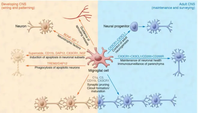
Figure 3. Microglia contribution to normal CNS functions. As resident immune cells in the CNS, microglia display many functions to maintain tissue homeostasis. Microglial cells modulate wiring and patterning during development by regulating neuron populations via phagocytosing excess synapses, releasing neurotrophic factors and guiding sprouting vessels. During postnatal development and adulhood microglia contribute to activity- dependent circuit formation, maturation of neuronal networks, regulation of adult neurogenesis and maintaining neuronal health (Kierdorf & Prinz, 2017).
18
1.5. The role of microglia in brain inflammation and injury
Besides contributing to the development and maintenance of the central nervous system, microglia function as the primary immune cells of the CNS. They provide the first line of defense against invading pathogens, and via the constant interaction with neurons, they often are the first to detect critical changes in neuronal activity and health status (Prinz & Mildner, 2011). Upon detection of danger associated molecular patterns (DAMPs, ATP, heat shock proteins, DNA, HMGB1), released by stressed or injured neurons, microglia initiate a series of responses triggered by a plethora of surface receptors, such as Toll-like receptors (TLRs), purinergic receptors, scavenger receptors, and cytokine and chemokine receptors (Salter &
Stevens, 2017). Their activation results in phagocytosis of pathogens and dying cells and the release of soluble factors involved in neuronal damage, tissue repair, and remodeling as well as recruitment of other immune cells from the blood (Stevens & Schafer, 2018). In recent years, multiple studies have indicated that microglia function as a double-edged sword, playing a protective or a detrimental role depending on the given conditions during different brain pathophysiologies (Shemer, Erny, Jung, & Prinz, 2015). Depending on the nature of the stimulus, microglia can take a number of activation states, which correspond to altered microglia morphology, gene expression, and function. For example, it has been reported that upon acute injury such as traumatic brain injury (TBI), early microglial responses may contribute to the restoration of brain homeostasis (Donat, Scott, Gentleman, & Sastre, 2017).
Selective elimination of microglia was found to exacerbate brain injury after experimental stroke (Szalay et al., 2016). On the other hand, prolonged microglial activation or the release of pro-inflammatory cytokines (IL-1, IL-6, TNFa) and chemokines (CCL2, CX3CL1, MIP- 1), may result in augmented tissue damage and this may potentially contribute to neurodegeneration.
Uncontrolled synapse loss might indicate microglial dysfunction, which eventually can lead to reduced neuroprotection and neuronal repair, and increased neurodegeneration associated with chronic neuroinflammation (Salter & Stevens, 2017). For example, microglial dysfunction contributes to neurodegeneration in experimental models Alzheimer’s disease (AD) and Parkinson’s disease (PD) (Ransohoff, 2016). Selective elimination of microglia reduces cognitive deficits in experimental AD models (Najafi et al., 2018), which is similar to that seen after blockade of complement-mediated synapse elimination (Salter & Stevens, 2017). Furthermore, large-scale genome-wide association studies (GWAS) have revealed that several CNS disorders that are considered as a primary microgliopathy whereby
19
microglial dysfunction is considered to be a primary disease mechanism, providing a direct link between microglia and neurodegeneration (Prinz et al. 2011; Arcuri et al. 2017). A good example is Nasu-Hakola disease, an autosomal recessive disorder characterized by progressive dementia and bone cystic lesions (Kaneko, Sano, Nakayama, & Amano, 2010), showing that microglia may act as a primary contributor to the disease. This rare genetic disease is caused by mutations in the DNAX activation protein 12 (DAP12) or triggering receptor expressed on myeloid cells 2 (TREM-2) in microglial cells (Paloneva et al., 2002), which results in impaired phagocytic activity and excessive pro-inflammatory microglial activation. Other studies have uncovered, that similar mutations on DAP12 and TREM-2 could be strongly linked to the progression of AD and frontotemporal dementia (Salter and Stevens 2017; Kleinberger et al. 2017). Several other CNS disorders have been considered to be caused by genetic mutations resulting in microglial dysfunction, including Rett syndrome, Fragile X syndrome and Phelan-Mc Dermic syndrome (Arcuri et al., 2017). Thus, depending on their phenotype and role in given physiological processes, microglia may also play diverse roles in differrent brain diseases. Therefore, understanding the mechanisms through which microglia recognize and respond to injury or infection and interact with other cell types in the brain may lead to the identification of novel therapeutic targets. Among many brain conditions, infections induce strong microglial reaction and experimental models of infection are valuable to study microglial responses and function.
1.6. Neurotropic viruses
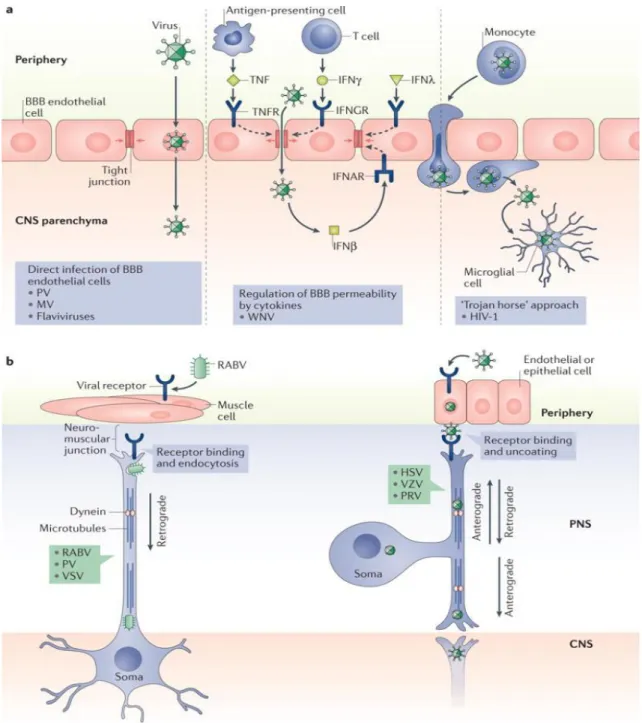
Neurotropic viruses, which are capable of infecting nerve cells, are among both DNA and RNA viruses of different families. Their infection inside the CNS leads to serious clinical syndromes of meningitis, encephalitis or meningoencephalitis. Despite the presence of the blood-brain barrier and blood-CSF barrier, as it was discussed in detail in previous chapters, viruses have evolved to find their way into the CNS (Miller, Schnell, & Rall, 2016). Three major routes of viral entry into the brain have been identified, as outlined in Figure 4.: direct infection of the cells that comprise the blood-brain barrier (BBB) and blood-cerebrospinal fluid barrier; infection of cells that are able to cross these barriers; and transneuronal migration across synapses from the peripheral nervous system (PNS) into the CNS (Koyuncu, Hogue, & Enquist, 2013).
20
Alternatively, neurotropic RNA viruses including poliovirus (PV), measles virus (MV), or some members of the Flavivirus family, like West-Nile virus infect endothelial or epithelial cells of the BBB and can act through the engagement of Toll-like receptor 3 (TLR3) to induce pro-inflammatory cytokines by circulating antigen presenting cells. As a result, BBB integrity becomes compromised by loosening its tight junctions, allowing viral particle migration into the brain tissue. Viruses may also passively access resident CNS cells by infecting lymphocytes or monocytes that can be transported across the blood-brain barrier.
This strategy is often referred to as the ‘Trojan horse’ approach because viral particles are released once the blood-borne leukocytes gain access to the parenchyma. A classic example of this mode of invasion is provided by the human immunodeficiency virus type 1 (HIV-1):
CD16+ monocytes, permissive for HIV-1, traffic across the BBB and release virions that can then infect CNS microglia (McGavern & Kang, 2011). The third mode of CNS entry is transneuronal migration, a strategy adopted by rabies virus (RABV) herpesviruses (HSVs), and pseudorabies virus (PRV). Intracellular trafficking in PNS neurons, which is necessary to shuttle cellular components to and from the synapse, can be commandeered to facilitate viral travel within and among synaptically connected neurons. The best-characterized examples of this type of spread are provided by herpesvirus family members such as Herpes simplex type 1 (HSV-1) and the closely related PRV (Koyuncu et al., 2013). After infection of epithelial cells in the oral mucosa, HSV-1 spreads to sensory and autonomic ganglia, establishing lifelong latency (Pomeranz, Reynolds, & Hengartner, 2005). Inside infected epithelial cells, these viruses become non-lytic within PNS neurons. During latency, only a few viral transcripts are synthesized, which do not encode functional proteins but prevent neuronal apoptosis via inhibition of caspase activity and disruption of both innate and adaptive immune signaling (Getts, Chastain, Terry, & Miller, 2013). However, in response to decreasing to immune monitoring, stress or other infections the virus can reactivate from its latent phase, which leads to an active infection of PNS neurons. Newly replicated viral particles can traffic along via microtubule tracks along the axons of sensory neurons, which have a pseudo-unipolar morphology. This way one axon is in contact with epithelial cells and the other synapses are in contact with CNS neurons (Kramer & Enquist, 2013). Such viral strains can reactivate long after the initial viral exposure, contributing to several neurodegenerative disorders.
21
Figure 4. Different routes of viral entry into the CNS. a, Direct infection of BBB endothelial cells, followed by release of viral particles into the parenchyma (left panel).
Some viruses can diffuse across permeable regions of the BBB, by influencing the release of pro- and anti-inflammatory cytokines or various interferons (IFNß, IFNγ, IFNλ), which can loosen barrier integrity (middle panel). With the ’Trojan horse’ approach (right panel), viruses can infect lymphocytes, monocytes or macrophages which traffic across the BBB or blood-CSF barrier, releasing virus particles once in the brain parenchyma. b, Transsynaptic spread of viral particles involves the transport of viral genomes and associated proteins via microtubules and molecular motor proteins. Rabies virus (RABV) moves from muscle, across the neuromuscular juction via dinein-mediated retrograde transport into the CNS (left panel). Herpes simplex virus (HSV), Pseudorabies virus (PRV) and Varicella zooster virus (VZV) traffic across the endothelial-neuron junction. Inside neurons retrograde transport brings virions to the neuronal soma, and anterograde transport delivers them to the PNS- CNS synaptic junctions (right panel) (Miller et al., 2016).
22
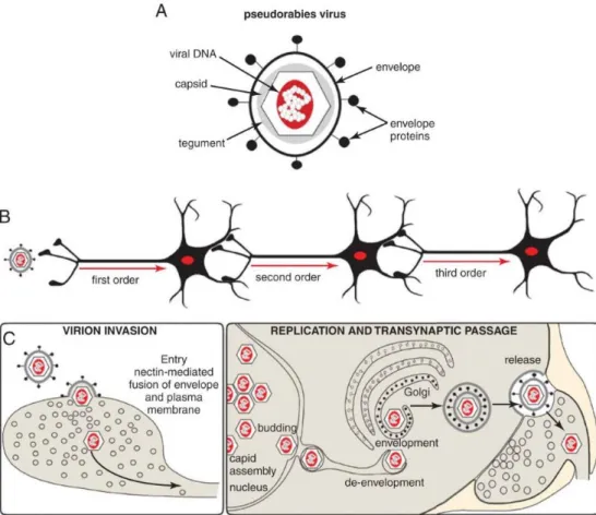
1.6.1. Pseudorabies virus
Pseudorabies virus (PRV) belongs to the same family, Alphaherpesviridae, as the human pathogen Herpes Simplex Virus (HSV) and Varicella Zoster Virus. The natural host of the virus is the pig, in which viral infection leads to Aujenszky’s disease (Schmidt, Hagemoser,
& Kluge, 1992) with similar symptoms to that caused by rabies infection. PRV has a wide range of hosts, infecting all mammals except for higher primates (McFarland, Hill, &
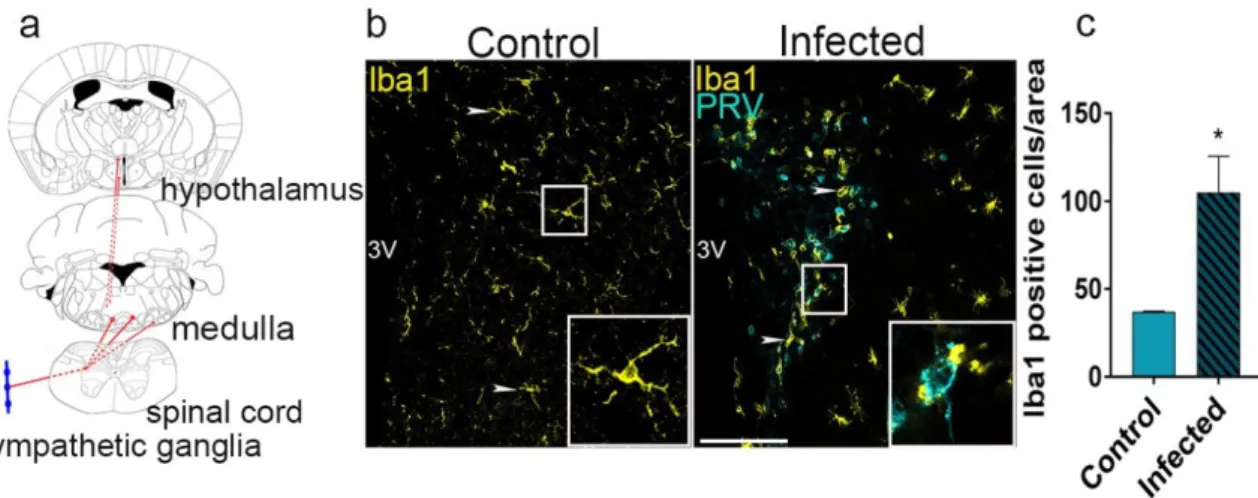
Tabatabai, 1987), probably due to circulating autoantibodies. Among other virus strains (vesicular somatitis virus, rabies virus, mouse hepatitis virus) pseudorabies virus have the unique ability to spread between exclusively synaptically connected chains of neurons. Such neuronal spread requires direct virus entry into the neuron (first-order neuron) and replication. The encapsulated viral genome is then transported at sites of synaptic contact to a second order of neuron where replication takes place again. This self-amplificating property allows the first order neuron as intensly labeled as the second or third order neurons (Pomeranz et al., 2005). Tracing studies using this feature of PRV have been successfully employed in a number of different animal models: pigs (the natural host), lambs and sheep, dogs, cats, chicken embryos, ferrets, and other rodents such as rats, mole rats, mice, gerbils, and hamsters (J. P. Card 1998; J. Card et al. 2018). Transsynaptic spread, self-amplification and broad host range made PRV an ideal tool in an extensive number of neuroanatomical studies seeking to define the architecture of multisynaptic pathways (Kramer & Enquist, 2013). Transsynaptic spread of PRV was proved by detailed electron microscopic studies which support the view that PRV spreads in the CNS primarily by direct cell-cell contact, rather than diffusion of virions through the extracellular space or via non-neuronal cells.
Analysis of infected nervous tissue by electron microscopy revealed viral capsids and structural proteins localised at the synapses of infected neurons (Pomeranz et al., 2005). This feature of the virus was well described via the analysis of different neuronal pathways between the ventral musculature of the stomach to the brainstem and higher order structures of the CNS (Pomeranz et al., 2005). PRV spread in the nervous system also requires synaptically connected intact neuronal circuit. This was tested by direct injection of PRV into brain ventricles, which result only in the infection of nearby neurons, astrocytes and ependymal cells which line the walls of ventricles, but the virus did not spread futher (Rinaman, Card, & Enquist, 1993). Even tough, PRV is able to infect non-neuronal cells in the brain, such as epitehelial cells and astrocytes, non-synaptic spread is severly limited.
Astrocytes are susceptible to PRV infection but not permissive for viral replication, they do
23
not contribute to trans-neuronal spread of the virus (Card, J. P., 2001). Rather, the infection of astroglia is tought to represent an effort of local intrinsic and innate immune defense to contain the infection (Card, J. et al., 2018). According to previous studies, microglial cells are the only cell type which can not be affected by PRV, althoug the exact reason is still unclear (Rinaman et al., 1993). When injected directly into the brain parenchyma, PRV virions diffuse very little, producing only focal infection site (Pomeranz et al., 2005). In order to effectively infect neurons, replicate and produce infectious progeny, and spread through synapses the virus needs several essential elements, including envelope proteins, which help viral capsids to interact with the extracellular matrix and gain access to permissive neurons (Figure 5.a,). PRV uses a common adhesion molecule, nectin to invade neurons through a receptor-mediated fusion of the viral envelope and plasma membrane of the target cell (Brittle, Reynolds, & Enquist, 2004). As a result, tegument protein associated viral capsids, containing the genome are released inside the host neuron, where they are transported along microtubules to the soma. The genome of virus enters the nucleus along with tegument proteins which initiate the expression of immediate early genes and initiates a transcription cascade which generates all necessary proteins for new virions (Brittle et al., 2004). During the lytic cycle viral genes (immediate-early, early and late) are expressed in a certain timeline. After achieving the right amount of copies, virus DNA is packed into capsids and exit the nucleus. Before leaving the host cell, naked capsids require envelopment, then they get transported into vesicles (Figure 5. c,) (Koyuncu et al., 2013).
The attenuated vaccine strain of PRV (PRV-Bartha) which have reduced virulence has been successfully used as a self-amplifying neural tracer after peripheral injection for its reduced neurotoxicity and prominently retrograde spread. Mutant PRV-Bartha strains are favored for tracing studies because they penetrate further into neuronal circuits due to increased host survival time. Since Bartha strains lack glicoprotein E, once introduced into the nervous system, PRV-Bartha spreads only in retrograde direction inside a circuit, while wild type strains, such as PRV-Becker and PRV-Kaplan, spread in both anterograde and retrograde directions (Pomeranz et al., 2005). Since Bartha strain is able to infect PNS neurons projecting to CNS neurons and invade specific brain regions by retrograde transport, the strain has been widely used for defining CNS ciruits that modulate the autonominc and somatic peripheral outflows (Pomeranz et al., 2005). The detailed retrograde spreading of PRV-Bartha has been effectively characterized in rats and hamsters, by injecting the virus intraocularly (Card, J. P., 1998). As different genetically modified PRV-Bartha strains have become popular tracing tools, more research has propelled the search for new techniques to
24
further enhance this powerful tool. Major autononmic pathways were defined during stress situations via dual tracing, employing ß-galactosidase (product of lacZ) expressing PRV- Bartha strain kombined with PRV-Kaplan strain (Jansen, Van Nguyen, Karpitskiy, Mettenleiter, & Loewy, 1995). Since then this dual tracing approach was used to map multiple autonomic connections in the CNS. Besides ß-galactosidase enzime, different fluorophores were inserted into PRV derivatives as well, such as EGFP or DSRed. In a model of cultured rat dorsal root ganglia the same fluorescent strains were used to uncover collateralized pathways (Miranda-Saksena, Boadle, Armati, & Cunningham, 2002).
Genetically modified viral tracers, such as PRV, HSV or rabies only recently started to become powerful tools to study viral infections and immune responses in the CNS.
Figure 5. Structure and spreading mechanism of Pseudorabies virus. a, The structure of alphaherpes virus virions compose of several capsid and tegument proteins that are contained within the virus envelope which is acquired from the host cell. The envelope contains a second set of virally encoded proteins that are crucial for target recognition, attachment and fusion that lead to the release of the capsid into permissive neurons. b, Mutant strains of PRV spread selectively in the retrograde direction through exclusively synaptically linked neural circuits. c, Multiplication of virions is a multistep process which lead to assembly of mature virions in the cell soma. Virions are trafficked through the soma and denrites of infected neurons by vesicular motor proteins and released in the vicinity of synapses (Koyuncu et al., 2013).
25
1.7. Infection-induced inflammation and anti-viral immunity in the brain
Invasion of pathogens, such as viruses, bacteria or parasites into the brain results in chronic, often lethal diseases, which are associated with severe blood-brain injury, impaired neuronal communication, innate myeloid cell activation, and leukocyte infiltration. Unlike other cell types of the CNS, neurons are generally non-renewable, thus the cytolytic and inflammatory strategies that are effective in the periphery could be damaging if deployed in the brain (Getts et al., 2013). Upon infection-induced inflammation, the immune response of the brain aims to maintain neuronal network integrity while eliminate invading pathogens and the terminally injured cells.
As discussed above, the brain is shielded from external threats at both macro- and microscopic levels: it is enveloped in bone, to prevent physical injury, and separated from peripheral tissues and blood via highly specialized barriers. To overcome this, viruses have developed sophisticated strategies to enter the CNS either via transcellular and paracellular pathways across the BBB or anterograde/ retrograde trafficking along neurites from peripheral neurons (Miller et al., 2016). If occurring via parenchymal routes, pathogen invasion into the brain is sensed immediately by specialized innate immune cells, which include microglia, perivascular macrophages, and meningeal or choroid plexus macrophages. As first responders to tissue injury and pathogen invasion, brain myeloid cells are equipped to launch an immune response (Prinz et al., 2011). In case the reaction of CNS cells to infection or tissue injury extend a certain limit, vascular activation and production of chemokines may initiate the recruitment of immune cells from the periphery. Thus, regulation of the central immune response is crucial in protecting the brain from further tissue damage due to immunopathology, and lingering inflammation that can sometimes augment BBB injury, neuronal loss and hinder the tissue repair process (Russo & McGavern, 2015).
Although immune cell migration into the CNS is tightly regulated due to the blood-brain barrier (BBB), several routes exist for peripheral leukocytes to enter the CSF, the choroid plexus (CP), the meninges, perivascular spaces and eventually the parenchymal tissue (Prinz et al., 2017). Following infection by different neurotropic viruses, both innate (monocytes, neutrophil granulocytes) and adaptive immune cells (CD8+ T cells, CD4+ T cells and B cells) are recruited, to participate in pathogen elimination and clearance. Upon pathogen entry into the CNS, most immune responses begin with pattern recognition receptors (PRRs) sensing pathogen-associated molecular patterns (PAMPs) via Toll-like receptors, RIG-like receptors
26
(RLRs), or Nod-like receptors (NLRs) expressed by neurons, astrocytes, and microglia (Klein & Hunter, 2017). At the BBB, in response to TLR and PRR activation endothelial cells and astrocytes become strongly activated, and try to control viral entry (Rinaman et al., 1993). Endothelial cells respond to these cues via modulating barrier integrity through altered expression of CAMs and Rho GTPases, while activated astrocytes produce proinflammatory cytokines such as Il-1, IL-6, TNFa, and IFNγ. These proinflammatory cytokines aid in viral clearance through recruitment of mononuclear cells but may also be detrimental long-term to neuronal function and regeneration. Among the infiltrating immune cells, T cells are the first to enter the CNS during viral infection, with the production of T cell-derived cytokines, such as IFN-γ, which is critically involved in viral clearance and the amplification of immune cell infiltration through upregulation of chemokines. Recent studies have highlighted the importance of T cell recruitment and their reactivation in multiple infection models. In an HSV-1 induced encephalitis model inhibition of T cells via the downregulation of CXCL9 T cell attractant chemokine lead to increased mortality during HSV-1 infection. Direct injection of CXCL9 into the CNS infection site enhanced HSV-1 specific CD8+ T cell accumulation, leading to marked improvements in the survival of infected mice (Koyanagi et al., 2017). In a clinical study of blood donors testing positive for West-Nile virus (WNV), T regs were found to expand after infection, and asymptomatic patients had higher levels of T reg cells compared to symptomatic patients, suggesting that T regulatory cells (T regs) control of antiviral responses may influence the severity of clinical disease. Similar results could be detected in a mouse WNV infection model, in which animals depleted of T regs showed more severe symptoms and had significantly higher mortality rate than control mice (Lanteri et al., 2009). Importantly, the specific chemokine milieu in the infected brain appears to influence T cell activity and antiviral response as well.
Cytokines expressed by T cells, also have a major role in the antiviral response, such as IFN- γ which control infectious virus replication in the periphery but clears the virus from the CNS, without affecting acute viral replication (Baxter VK. et al., 2016). Animals that lack IFN-γ or the ability to signal through its receptor IFNγR, exhibit decreased B cell recruitment (Klein & Hunter, 2017). B cell functions, including antiviral humoral immune responses, are critical for control of viral dissemination in the periphery and neuroinvasion during neurotropic viral infections (reviewed in (Kyle Austin & Dowd, 2014). Early infiltrating B cells express multiple cytokines (such, as CXCL9, CXCL10, CCL19) which are upregulated within the CNS during viral infections. Although classically considered part of the adaptive immune response, B cells are activated soon after infection by several viruses prior to the
27
generation of specific immunoglobulin G (IgG) (Rojas, Narváez, Greenberg, Angel, &
Franco, 2008). B cell responses to viruses are often initiated antigen recognition via their surface immunoglobulin receptors, such as IgG and IgM. Their activation and proliferation are also affected by T cell chemokine expression, although a recent study of neurotropic coronavirus infection suggest that activated B cells can respond to viral infection independently from CD8+ T cells (Phares, Marques, Stohlman, Hinton, & Bergmann, 2011).
Their crucial role in viral infection was also proved by an infection model induced by the neurotropic strain JHMV of mouse hepatitis virus, in which the absence of B cells resulted in uncontrolled, persistent CNS infection despite viral reduction by T cells (Phares et al., 2011). As described above, PRR detection of viral nucleic acids induces expression of chemoattractants that promote the parenchymal entry of mononuclear cells, including blood- borne monocytes, which differentiate into tissue macrophages at sites of infection or injury.
Upon neurotropic viral infections, it is still an uncleared question whether these cells aid in viral clearance and recovery or mediate continuing damaging inflammation in the CNS (Klein et al. 2019.). Macrophages can produce anti-inflammatory mediators, scavenge the infected area, phagocytose cell debris and regulate extracellular matrix and glial scar surrounding the damaged area (London, Cohen, & Schwartz, 2013). However, these cells have also been shown to have potent effector functions, including antigen presentation, T cell stimulation, and production of multiple proinflammatory mediators and reactive oxygen species (Terry et al., 2012). Monocyte recruitment can be affected by depletion of CCL2 and CCL7 proinflammatory cytokines resulting in increased viral burden and mortality (Bardina et al., 2015). Upon subsequent restoration of CCL7 by exogenous administration increased monocyte and neutrophil recruitment and improved survival.
Microglia are thought to protect CNS from viral infection via multiple mechanisms, including the production of antiviral cytokines, phagocytosis of virus-infected and dying neurons, and the induction of neuronal repair and homeostasis (Prinz et al. 2011; Ransohoff and Engelhardt 2012). In addition to restricting viral replication, microglia are suggested to orchestrate peripheral immune response against invading pathogens in the CNS. In the healthy brain, microglia do not express MHC, however, when activated by pathological conditions, they upregulate these molecules (Tsai et al., 2016). However, assessing specific immune contributions of microglia during infections has been challenging for multiple reasons, which would be discussed below.
28
1.8. The role of microglia in neurotrop viral infections of the CNS
As the main resident immune cell type of the brain parenchyma, microglia are known to shape CNS immune responses to viral infections that occur in the brain. However, for multiple reasons, the functional contribution of microglia to neurotropic viral infections has been described inadequately. Previous studies have stated, that microglia coordinate CNS immune responses via pro-inflammatory cytokine or chemokine secretion, and possibly via presenting antigen to T cells that are recruited into the brain (Zhang, Liu, & Wei, 2017). In response to a pathogen invasion microglia increase the expression of toll-like receptors (TLRs) which are critical in generating innate immune reactions. Others have indicated that in the case of HSV infection microglial TLR2 and TLR9 signaling have a pivotal role in the production of proinflammatory cytokines and chemokines (J. Card et al. 2018; Rinaman, Card, and Enquist 1993). As an anti-viral response, IFN-I production by microglia and phagocytosis of infected cells have been previously observed (Dénes, Boldogkoi, Hornyák, Palkovits, & Kovács, 2006). Rapid induction of microglia was also noticed in response to the immediate release of normally intracellular proteins and/or high-energy purine nucleotides (such as ATP, ADP, and UTP) from injured neurons (Davalos et al., 2005a). As it was previously described by others (Wolf, Boddeke, and Kettenmann 2017; Davalos et al.
2005) massive release of purines occurs after metabolic stress or trauma that might trigger microglial activation through P2 type receptor-mediated mechanisms (Dénes et al., 2006).
However, it remains unknown if these mechanisms are involved in the activation and recruitment of microglia during virus infections. In spite of this information, studying microglial responses during viral infections in vivo has been quite difficult. As previous studies have described (J. P. Card 2001; Boldogköi et al. 2004; Dénes et al. 2006), it is clear that PRV and HSV infection of the brain produces a complex cascade of innate responses that are not limited to microglia (Rinaman et al., 1993). Virulent and attenuated strains of HSV and PRV have been shown to induce neuropathological changes and ultimately lead to reactive astrogliosis, activation and recruitment of brain macrophages and different CD45- positive blood-born immune cell populations to sites of infection (Dénes et al., 2006). Using the more virulent Becker strain of PRV, trafficking, and appearance of cells positive for CD45 leukocyte common antigen as well as CD8+ cytotoxic- and CD4+ lymphocytes at the sites of infections were reported. However, activated microglia also express CD45, therefore the distinction between infiltrated CD45-positive leukocytes and resident microglia remained challenging (Rassnick, Enquist, Sved, & Card, 1998).
29
Note, that in this complex inflammatory cascade it has been very difficult to isolate immediate microglial immune responses. Discrimination of microglia from infiltrated monocytes and macrophages is difficult due to the phenotypic transformation of activated microglia rendering them morphologically similar to infiltrating mononuclear phagocytic cells. According to previous researches (Rassnick et al. 1998; Enquist and Leib 2016) it is becoming increasingly clear that multiple glial cells participate in the immune response to neurotropic viral infections by isolating afflicted neurons and thereby facilitating transsynaptic spread of the virus (Rinaman, Card, and Enquist 1993; Boldogköi et al. 2004).
Previous research proved that highly GFAP-positive astrocytes have the ability to isolate infected neurons, by wrapping afflicted cells and their synaptic contacts with processes that separate them from uninfected cells (Rinaman et al., 1993). Also, TLR3 was shown in astrocytes to sense HSV-2 infection immediately after entry into the CNS, possibly preventing HSV from spreading beyond the neurons mediating entry into the CNS (Reinert et al., 2012).However, it is likely that astrocytes do not have the ability to phagocytose compromised neurons, unlike microglia and macrophages. By using genetically modified strains of the „Bartha-Dup” strains of PRV (Boldogköi et al., 2004). Dénes et al., 2006 have described, that microglia are able to respond rapidly to early infection and isolate compromised neurons even while the infected neuronal membrane is still intact. Even though the exact molecular mechanisms of microglial responses to early infection need to be further investigated. By using the same highly specific, retrograde, transsynaptically spreading
„Bartha-Dup” PRV strain and highly selective microglia elimination tool we were able to uncover some of these precise microglial mechanisms in response to virus infections.
30
2. Objectives
Microglia are the main immunocompetent cell type of the CNS that play a role in multiple physiological and pathological processes. The importance of microglial actions in acute brain injury, such as brain trauma or stroke and in chronic neurodegenerative diseases, like Alzheimer’s is widely recognised. However, the role of microglia in anti-viral immunity of the brain is less studied. Notably, the mechanisms through which microglia recognize signs of infection at the cellular level and how infected cells are discriminated are still unclear.
Therefore, the main goal of my work was to investigate inflammatory processes in the central nervous system during neurotropic virus infection, focusing on the following three main topics:
1) To study central inflammatory processes and the functional role of microglia in a mouse model of neurotropic virus infection.
2) To identify the role of microglial P2Y12 receptor in neurotropic virus infection.
3) To study inflammation and microglial responses in human post-mortem brain tissues after herpes simplex encephalitis.
31
3. Materials and methods
3.1. Processing of human samples
Post-mortem human brain tissues
Formalin fixed, paraffin embedded (FFPE) post-mortem tissue sections from patients (n=5) with known HSV-encephalitis (age between 42-66 years) were used. Tissue samples from two additional patients with no known neurological disease were used as controls. Control samples were perfusion-fixed with Zamboni fixative (4% PFA, 15 % PIC) and postfixed overnight in the same solution. All performed procedures were approved by the Regional and Institutional Committe of Science and Research Ethics of the Scientific Council of Health (ETT-TUKEB 62031/2015/EKU, 34/2016 and 31443/20116EKU (518/pi/11)).
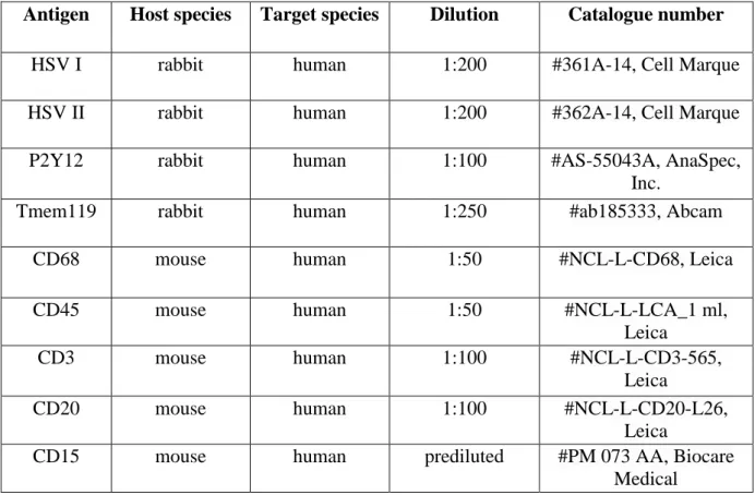
Immunohistochemistry and immunofluorescence on post-mortem human brain tissues To investigate microglia recruitment in response to neurotropic virus infection in the human brain, 4-6µm thick brain sections were cut and samples were mounted on gelatine-coated slides. After deparaffination, HIER (Novocastra Epitope Retrieval Solution pH9, Leica Biosystems) and peroxidase blocking (in 1% H2O2 solution) was carried out. Sections were blocked with 2.5% Normal Horse serum (#S-2012, Vector Laboratories) and were incubated with microglia and immune cell specific primary antibodies, see in Table 1. For signal amplification ImmPRESS anti-rabbit HRP Kit (#MP-7401, Vector Laboratories) and for visualisation either ImmPACT NovaRED HRP substrate (for detection of HSV I) or DAB- Ni HRP substrate (for detection of other markers, #SK-4805, Vector Laboratories) was used.
Representative pictures were taken on 20x magnification using a Nikon Ni-E C2+
microscope. To assess the proportion of P2Y12-positive microglia, P2Y12 and Iba1 double immunofluorescence has been performed on 50 µm thick free-floating brain sections. Images were captured using a Nikon Eclipse Ti-E inverted microscope (Nikon Instruments Europe B.V., Amsterdam, The Netherlands), with a CFI Plan Apochromat VC 60X water immersion objective (NA: 1.2) and an A1R laser confocal system.