1 Springer
1
This document is the Accepted Manuscript version of a Published Work that appeared in final form 2
in Journal of Materials Science, copyright © Springer after peer review and technical editing by the 3
publisher.
4
To access the final edited and published work see https://link.springer.com/article/10.1007/s10973- 5
017-6908-x
6
2
Synthesis and characterization of diazine-ring containing hydrazones and their Zn(II) 1
complexes 2
József Magyari1,2, Berta Barta Holló1, Marko V. Rodić1, Imre Miklós Szilágyi2, Katalin Mészáros
3
Szécsényi1
4
1 Department of Chemistry, Biochemistry and Environmental Protection, Faculty of Sciences, University of Novi Sad, 21000
5
Novi Sad, Trg Dositeja Obradovića 3, Serbia
6
2 Department of Inorganic and Analytical Chemistry, Budapest University of Technology and Economics, H-1111 Budapest,
7
Műegyetem rkp. 3, Hungary
8 9
ORCID numbers: József Magyari 0000-0002-9849-7689 ; Berta Barta Holló 0000-0002-5786-442X ;
10
Marko V. Rodić 0000-0002-4471-8001 ; Imre Miklós Szilágyi 0000-0002-5938-8543 ;
11
Katalin Mészáros Szécsényi 0000-0002-7494-7323
12
13
Corresponding author: Magyari József, dh.jozef.madjari@student.pmf.uns.ac.rs , tel.: +381691002959
14
15
Keywords: hydrazinophthalazine, hydralazine, pyridazine, dipyridyl ketone, Schiff base, zinc
16
complexes
17
18
Abstract 19
Two new zinc(II) coordination compounds have been synthesized by the reaction of diazine-ring containing
20
Schiff bases di(2-pyridyl) ketone phthalazine-1-hydrazone (HzDPK) and di(2-pyridyl) ketone 3-
21
chloropyridazine-6-hydrazone (HpDPK) with zinc(II) salts in acetonitrile in the presence of triethylamine. The
22
crystal and molecular structures of the complexes and that of the ligand HpDPK were determined by single-
23
crystal X-ray structure analysis. In both complexes, zinc atoms are situated in distorted octahedral environments,
24
formed by two meridionally coordinated NNN tridentate, mono-deprotonated ligands.
25
Since the applicability of the coordination compounds depends on their thermal properties, the thermal
26
decomposition of the ligands and their complexes was followed by simultaneous TG–DSC measurements. The
27
desolvation process of the complexes is rather slow as a consequence of a restricted diffusion through the lattice
28
and finishes ~ 200 ˚C. The desolvated compounds are stable up to 340 ˚C. In order to follow the solvent
29
evaporation and to have a better insight into the decomposition mechanism of the compounds coupled TG–MS
30
measurements were carried out.
31 32
Introduction 33
Carcinogenesis underlies complex mechanisms and to address single target approaches is inadequate to prevent
34
prevalence and deaths from the disease. The resistance of the human tumor to multiple chemotherapeutic drugs
35
was recognized as one of the most important reasons for the failure of cancer therapy so it became a focus of
36
cancer research. The phenomenon called multidrug resistance (MDR) subsequently appeared as a major
37
impediment to the curative treatment of a variety of malignancies [1,2]. MDR caused by specific membrane
38
transporters, such as ATP-binding cassette (ABC) or copper transporters, as well as other causes of drug
39
resistance, hamper successful cancer chemotherapy [3]. Schiff bases can be involved in the prevention of MDR,
40
besides, they show a broad range of biological activity, including analgesic, anti-inflammatory, antimicrobial,
41
anti-tubercular, anticancer/antitumor, anticonvulsant, anti-diabetic and anti-hypertensive properties [4]. Some of
42
them exhibit higher activity than the precursor drug [5]. Furthermore, their lower toxicity compared to
43
hydrazines is also important [6]. Compounds with diazine [7] can be used also as precursors in the synthesis of
44
new Schiff bases. One of them, 1-hydrazinophthalazine hydrochloride (Hz∙HCl) was one of the first used
45
vasodilators and still has been used in some urgent cases [8]. The hydrazino group plays a key role in its
46
reactivity in vivo [9] and in vitro environment, too. Hydrazinophthalazine itself is a chelating, practically
47
3 bidentate ligand and with metal ions forms five-membered metallocycles [10]. The other diazine compound with
1
less bulky structure, 3-chloro-6-hydrazinopyridazine (Hp), has comparable coordinational properties. Both
2
contain hydrazino group which in the reaction with carbonyl compounds give Schiff bases.
3
One of the possibilities to enhance the pharmacological potency of biologically active compounds is their
4
complexation with metals [11]. Some diazine-hydrazone coordination compounds which exhibited remarkable
5
antiproliferative effect have already been synthesized by our group [12] so, the design, synthesis and
6
characterization of similar Schiff base type ligands and their metal complexes make this topic promising for the
7
further research.
8
In this work, we present the synthesis of two Schiff base type ligands, di(2-pyridyl) ketone phthalazine-1-
9
hydrazone (HzDPK) and di(2-pyridyl) ketone 3-chloropyridazine-6-hydrazone (HpDPK) and their new zinc(II)
10
complexes. The structures of the HpDPK ligand and the complexes were determined by single crystal X-ray
11
diffraction method and confirmed by FT-IR, molar conductivity and thermal measurements, too. The
12
desolvation of the complexes and the decomposition mechanism of the compounds were evaluated using data
13
obtained by coupled TG–MS measurements.
14
Experimental 15
Materials
16
1-hydrazinophthalazine hydrochloride (Hz∙HCl), 3-chloro-6-hydrazinopyridazine (Hp), di(2-pyridyl) ketone
17
(DPK) and acetonitrile were from Sigma-Aldrich and used as received.
18
Preparation of the ligands
19
Di(2-pyridyl) ketone phthalazine-1-hydrazone (HzDPK)
20
In 50 cm3 round-bottom flask Hz∙HCl (7 mmol, 1.38 g) was dissolved by heating in 30 cm3 EtOH : H2O = 1 : 1
21
mixture. Di(2-pyridyl) ketone, DPK, (7 mmol, 1.29 g) was dissolved in 5 cm3 EtOH and combined with the
22
initial solution. The reaction mixture was refluxed for 1.5 h, then solid LiOAc∙2H2O (7.35 mmol, 750 mg) was
23
added to it and continued the reflux for further 30 minutes. The hot mixture was transferred into a beaker and
24
cooled down to room temperature. The formed precipitate was separated by filtration through a fritted glass
25
funnel, washed with 3 cm3 EtOH and twice by water (5 cm3) and dried on air. Yield: 2.0 g, 88 %.
26
Di(2-pyridyl) ketone 3-chloropyridazine-6-hydrazone (HpDPK)
27
In 25 cm3 round-bottom flask Hp (5 mmol, 723 mg) was dissolved by gently heating in 6 cm3 MeCN. Di(2-
28
pyridyl) ketone, DPK, (5 mmol, 921 mg) was dissolved in 4 cm3 MeCN and combined with the initial solution.
29
The reaction mixture was refluxing 2.5 h. The hot mixture was transferred into a beaker and cooled down to
30
room temperature. The formed precipitate was filtered off, washed with 3 cm3 MeCN and air dried. Yield: 1.2 g,
31
77.23 %.
32
The X-ray quality crystals have been obtained by slow evaporation of acetone : methanol = 1 : 1 solution of
33
HpDPK.
34
Preparation of the complexes
35
Bis(di(2-pyridylketone)phthalazine-1-hydrazone)zinc(II), [Zn(HzDPK–H)2]∙CHCl3
36
In 100 cm3 round-bottom flask HzDPK (1 mmol, 326 mg) was dissolved in 30 cm3 MeCN by heating.
37
Triethylamine (1 mmol, 0.14 cm3), then Zn(OAc)2∙2H2O (0.5 mmol, 110 mg) dissolved in 5 cm3 MeCN was
38
combined with the ligand solution. The reaction mixture was refluxed 2 h, then, it was cooled down to room
39
temperature. The resulted orange coloured precipitate was separated by filtration. The precipitate was dissolved
40
by stirring in 15 cm3 MeCN + 25 cm3 chloroform mixture. The solution was filtered off through a small pore
41
size fritted glass funnel. The liquid phase was transferred into a 100 cm3 Erlenmeyer-flask which was after
42
sealed by perforated parafilm. After one week, dark-orange single crystals were formed and separated by
43
filtration. Yield: 156 mg, 37.34 %.
44
4 Bis(di(2-pyridylketone)3-chloropyridazine-6-hydrazone)zinc(II), [Zn(HpDPK–H)2]∙CHCl3
1
In 50 cm3 round-bottom flask HpDPK (1 mmol, 311 mg) was dissolved in 10 cm3 MeCN by heating,
2
Triethylamine (1 mmol, 0.14 cm3), then Zn(NO3)2∙6H2O (0.5 mmol, 149 mg) dissolved in 3 cm3 MeCN was
3
combined with the ligand solution. The reaction mixture was refluxed 1 h then it was cooled down to room
4
temperature. The resulted orange coloured precipitate was separated by filtration. The precipitate was dissolved
5
by stirring in 10 cm3 MeCN + 20 cm3 chloroform mixture, then filtered off through a small pored-sized fritted
6
glass funnel. The solution was transferred into a 100 cm3 Erlenmeyer-flask which was after sealed by perforated
7
parafilm. After one week, dark-orange single crystals were formed and separated by filtration. Yield: 148 mg,
8
36.81 %.
9
Measurement methods 10
IR data were collected on a Thermo Nicolet Nexus 670 FT-IR spectrometer at room temperature in the range of
11
4000–400 cm-1 with resolution of 4 cm-1 using KBr pellets.
12
The molar conductivity of freshly prepared 1·10–3 mol dm–3 solutions of the complexes in N,N-
13
dimethylformamide (DMF) was determined at room temperature using a digital conductivity meter (Jenway
14
4510).
15
Thermal data were collected using TA Instruments SDT Q600 thermal analyser coupled to Hiden Analytical
16
HPR-20/QIC mass spectrometer. The decomposition was followed from room temperature to 550 oC at 10 oC
17
min-1 heating rate in nitrogen carrier gas (flow rate = 50 cm3 min–1). Sample holder / reference: alumina crucible
18
/ empty alumina crucible. Sample mass ~ 4 mg. Selected ions between m/z = 1–120 were monitored in Multiple
19
Ion Detection Mode (MID).
20
Single crystal X-ray diffraction experiments were carried out at 295 K with Mo Kα radiation using a Gemini S
21
diffractometer (Oxford Diffraction). For HpDPK, empirical absorption correction using spherical harmonics was
22
performed with the CRYSALIS PRO [13]. For [Zn(HpDPK–H)2]∙CHCl3 and [Zn(HzDPK–H)2]∙CHCl3, analytical
23
numeric absorption correction using a multifaceted crystal model, followed by empirical absorption correction
24
using spherical harmonics, has been applied. Structures were solved with the SHELXT [14] and refined with the
25
SHELXL [15]. Carbon bonded hydrogen atom parameters were refined using a riding model, while nitrogen
26
bonded hydrogen atom in HpDPK was freely refined with isotropic displacement parameter. The SHELXLE [16]
27
was used as a graphical user interface for refinement procedures. Structures were validated by using Cambridge
28
Structural Database (CSD) [17] and Mercury CSD [18]. The crystallographic data for [Zn(HzDPK–H)2]∙CHCl3,
29
Hp and [Zn(HpDPK–H)2]∙CHCl3 and have been deposited with the Cambridge Crystallographic Data Centre as
30
Supplementary Publication No. CCDC 1568439 , CCDC 1568440 and CCDC 1568441, respectively.
31
Molecular graphics were produced by ORTEP for Windows [19].
32
A disorder of CHCl3 molecule is observed in the structure of [Zn(HpDPK–H)2]∙CHCl3. To achieve reasonable
33
geometry of disordered molecules, ADP and distance restraints were applied. The specimen of [Zn(HzDPK–
34
H)2]∙CHCl3 was a non-merohedral twin, with 180° rotation around c* axis as a twin law. The Bragg reflection
35
intensities were measured in a full-sphere of reciprocal space in the range 2θ < 52.6°, with a total of 29574
36
reflections collected, 22691 of which are overlapped and 6883 isolated. Structure solution was obtained by
37
processing 18556 reflections belonging to twin component 1 in HKLF4 format using SHELXT (among these,
38
15123 were overlapped reflections, the intensities of which were determined by deconvolution). For the final
39
refinement cycles, 29574 reflections were merged to 11935 reflections in HKLF5 format.
40
Crystallographic and refinement details of HpDPK, [Zn(HpDPK–H)2]∙CHCl3 and [Zn(HzDPK–H)2]∙CHCl3are
41
shown in Table 1.
42
43
44
45
5
1
Table 1 Crystallographic and refinement details of HpDPK, [Zn(HpDPK–H)2]∙CHCl3 and
2
[Zn(HzDPK–H)2]∙CHCl3
3 4
HpDPK [Zn(HpDPK–H)2]∙CHCl3 [Zn(HzDPK–H)2]∙CHCl3
Chemical formula C15H11ClN6 C30H20Cl2N12Zn·CHCl3 C38H26N12Zn·CHCl3
Mr 310.75 804.22 835.44
Crystal system Orthorhombic Monoclinic Triclinic
Space group Pbca P21/c P1
a / Å 14.4624(5) 13.7149(2) 10.4401(5)
b / Å 11.4369(3) 25.1743(7) 11.0460(6)
c / Å 17.2563(5) 20.0693(5) 17.2978(10)
α / ° 90 90 81.733(5)
β / ° 90 95.2972(18) 85.159(4)
γ / ° 90 90 70.045(5)
V / Å3 2854.27(15) 6899.6(3) 1854.22(18)
Z 8 8 2
µ / mm−1 0.273 1.14 0.93
Crystal shape Prism Prism Irregular
Colour Off-white Orange Orange
Crystal size / mm 0.68 × 0.22 × 0.14 0.69 × 0.21 × 0.13 0.57 × 0.33 × 0.13
Tmin, Tmax 0.92, 1 0.716, 0.863 0.697, 0.894
Measured reflections 16572 45514 29574
Independent reflections 3521 15996 11935
Observed reflections 2686 10994 9909
Rint 0.025 0.031 0.034
θ range / ° 2.7–29.0 2.5–29.0 2.4–29.1
(sin θ/λ)max / Å−1 0.683 0.682 0.685
R [F2 > 2σ(F2)] 0.038 0.053 0.038
wR (F2) 0.092 0.137 0.098
S 1.03 1.02 1.06
Parameters 203 883 534
Restraints 0 0 56
(Δ/σ)max 0.001 0.001 0.001
Δρmax, Δρmin / e Å−3 0.21, −0.21 1.03, −0.79 0.31, −0.35
5
Results and discussion 6
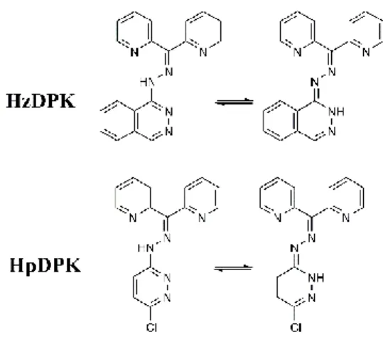
The ligands have been synthesized as free bases. By concerning the diazine ring, each ligand can be present in
7
two prototropic tautomeric forms (Figure 1). As the single crystal X-ray diffraction measurement confirmed that
8
Hz∙HCl exists in the diazine ring in –NH tautomeric form [20] it was expected to keep it in its Schiff base
9
HzDPK, too.
10
6
1
Figure 1 Prototropic tautomerism of the ligands
2
To assure the targeted coordinating properties of the ligands in the reaction with zinc(II) salts triethylamine as
3
base was applied which led to the formation of neutral bis-ligand metal complexes with deprotonated ligands.
4
MeCN/CHCl3 solvent mixture found to be the best for obtaining X-ray quality single crystals. Complexes were
5
crystallized in the form of chloroform solvates. As all the free nitrogen atoms in the complexes have tertiary
6
character, the hydrogen bond formation is not possible.
7
Crystal and molecular structures
8
The HpDPK molecule (Figure 2) significantly deviates from planarity due to twisting of the pyridine rings in
9
order to avoid steric clashes. The magnitude of the twisting is best perceived through torsion angles and τ(N3–
10
C6–C1–N1) = 10.2(2), and τ(N3–C6–C7–N2) = −138.41(13)°. The reason for unequal magnitudes of twisting is
11
the involvement of N1 atom in hydrogen bonding interaction with N4–H4A fragment of the hydrazone group.
12
Geometrical parameters of this interaction are: N4···N1 = 1.946(18) Å, N4–H4A = 0.888(18) Å, H4A···N1 =
13
2.6263(18) Å, N4–H4A···N1 = 132.1(16)°. Additionally, the valence angles around C6 significantly deviate
14
from ideal values, so that angle N3–C6–C1 equals 127.21(12)° and N3–C6–C7 angle is 111.68(11)°. These
15
peculiar geometrical parameters are also observed for some structurally related di-2-pyridyl-ketone hydrazones
16
[21–23]. The 1H NMR spectrum is in accordance with the HpDPK molecular structure and the observed
17
hydrogen bonding, too (Supplementary Material, Figure S1).
18 19
20
Figure 2 Molecular structure of HpDPK with atom numbering scheme
21
Molecular structures of [Zn(HpDPK−H)2]∙CHCl3 and [Zn(HzDPK−H)2]∙CHCl3 are depicted in Figure 3
22
furthermore, the selected structural parameters are given in Table 2. The asymmetric unit of
23
[Zn(HpDPK−H)2]∙CHCl3 complex comprises two independent complex molecules and two CHCl3 molecules,
24
while the asymmetric unit of the complex [Zn(HzDPK−H)2]∙CHCl3 consists of a complex molecule and a
25
disordered CHCl3 molecule. Zinc atoms in both complexes are situated in distorted octahedral environments,
26
formed by two meridionally coordinated NNN tridentate ligands. The amount of distortion may be appreciated
27
7 by measuring dihedral angles between two chelate planes (defined as plane through three donor atoms belonging
1
to one ligand), which for [Zn(HzDPK−H)2]∙CHCl3 equals 83.80(10)°, and for [Zn(HpDPK−H)2]∙CHCl3 equals
2
85.72(11)° and 86.41(11)°, for two independent molecules, respectively. Also, distorted octahedral geometry is
3
further evidenced by deviation of all the angles within coordination sphere from ideal geometries (trans valence
4
angles are given in Table 2).
5
Ligands are coordinated through pyridine (N1), azomethine (N3) and diazine (N5) nitrogen atoms, thus forming
6
two fused five-membered metallacycles. The metallacycles show a high degree of planarity, with the exception
7
of Zn1–N1A–C1A–C6A–N3A ring in the complex [Zn(HpDPK−H)2]∙CHCl3, which has envelope conformation
8
with N3A as the pivot atom. Metal-ligand bond lengths are within expected values, with a notable trend that
9
nitrogen atom N5 contribute to the shortest bond, and nitrogen atom N1 contribute to the longest bond within
10
the coordination sphere.
11
The lengths of chemically equivalent bonds between the metal atom and ligator atoms belonging to two
12
coordinated ligands, except of the bonds involving pyridine nitrogen N1, are in agreement within ca. 0.02 Å. In
13
[Zn(HpDPK−H)2]·CHCl3, the difference between Zn1–N1A and Zn1–N1B bond lengths is ca. 0.03 Å (2.223(3)
14
and 2.168(2) Å), while the difference between Zn2–N1D and Zn2–N1C bond lengths is ca. 0.05 Å (2.185(3) and
15
2.153(3) Å). Thus, Zn1–N1A and Zn2–N1D bonds are significantly longer compared to the rest of the bonds of
16
the coordination polyhedron. On the other hand, in [Zn(HzDPK−H)2]∙CHCl3 the Zn1–N1A and Zn1–N1B bond
17
lengths are by far the longest bonds in the coordination polyhedron (2.258(3) and 2.281(3) Å, respectively).
18
The CSD contains the structural data for three previously reported octahedral Zn(II) complexes with structurally
19
related 1-hydrazinophthalazine and 3-chloro-6-hydrazinopyridazine based Schiff bases, refcodes: DITQOO [24]
20
FARCEI [25] and CAJJAB [26]. In DITQOO and FARCEI structures, coordination mode of the tridentate
21
ligands is analogous to that of HpDPK, while in CAJJAB both nitrogen atoms of the pyridazine ring are
22
involved in coordination, thus forming a bridge between two metal atoms.
23
Intra ligand bond lengths have typical values for sp2 hybridized atoms and are in accordance with the literature
24
data [24–26]. The N3–C6 and N6–C15 bonds have lengths that correspond to localized double bonds. By
25
inspection of structures of related compounds in the CSD, it is evident that in 1-hydrazinophthalazine based
26
Schiff bases the hydrogen atom is located within pyridazine ring on nitrogen atom N5, while in 3-
27
chloropyridazine-6-hydrazone based Schiff bases the hydrogen atom is located within hydrazone group at
28
nitrogen atom N4 (which is in accordance with the structure of HpDPK). The ligands HpDPK and HzDPK are
29
coordinated in monoanionic forms, and they are deprotonated at different positions. However, the negative
30
charge in both cases is delocalized within C12–N5 and C12–N4 bonds, which eventually leads to the equivalent
31
structure of their N4–C12–N5–N6 fragments.
32
From the comparison of intra-ligand bond lengths in [Zn(HpDPK−H)2]∙CHCl3 and in HpDPK, it can be seen
33
that the most significant consequences of the monoanionic coordination form are shortening of N4–C12 and
34
elongation of N5–C12 and N5A–N6 bonds, while for the rest of the ligand molecule only subtle changes are
35
observable.
36
37
Figure 3 Molecular structures of [Zn(HpDPK−H)2]∙CHCl3 (1) and [Zn(HzDPK−H)2]∙CHCl3 (2) with selected38
atom numbering scheme. In case of [Zn(HpDPK−H)2]∙CHCl3 only one independent molecule is shown. Atoms
39
8 belonging to the other independent molecule are numbered in analogues way, with suffixes C and D for two
1
coordinated ligand molecules. Solvent molecules are omitted for clarity.
2
Table 2 Selected bond lengths and bond angles of [Zn(HpDPK−H)2]·CHCl3 (1), [Zn(HzDPK−H)2]·CHCl3 (2)
3
and HpDPK.
4
Bond Bond length / Å Bonds Bond angle / °
1 2 HpDPK 1 2
Zn1–N1A 2.223(3) 2.281(3) N1A–Zn1–N5A 147.52(10) 146.15(10)
Zn1–N1B 2.168(2) 2.258(3) N1B–Zn1–N5B 148.19(9) 147.22(10)
Zn2–N1C 2.153(3) – N1C–Zn2–N5C 146.62(11) –
Zn2–N1D 2.185(3) – N1D–Zn2–N5D 147.74(10) –
Zn1–N3A 2.145(2) 2.138(3) N3A–Zn1–N3B 164.78(10) 156.28(10)
Zn1–N3B 2.137(2) 2.136(2) N3C–Zn2–N3D 166.72(10) –
Zn2–N3C 2.159(3) –
Zn2–N3D 2.145(3) –
Zn1–N5A 2.110(3) 2.083(2) Zn1–N5B 2.105(2) 2.102(3)
Zn2–N5C 2.096(3) –
Zn2–N5D 2.114(3) –
N3A–C6A 1.290(4) 1.297(4) 1.2989(17) N3B–C6B 1.301(4) 1.297(4) –
N3C–C6C 1.301(4) – –
N3D–C6D 1.298(4) – –
N3A–N4A 1.355(4) 1.355(4) 1.3442(16) N3B–N4B 1.355(3) 1.344(3) –
N3C–N4C 1.352(4) – –
N3D–N4D 1.353(4) – –
N4A–C12A 1.356(4) 1.359(4) 1.3737(18) N4B–C12B 1.358(4) 1.357(4) –
N4C–C12C 1.364(5) – –
N4D–C12D 1.362(4) – –
N5A–C12A 1.347(4) 1.336(4) 1.3266(18) N5B–C12B 1.339(4) 1.342(4) –
N5C–C12C 1.342(4) – –
N5D–C12D 1.344(4) – –
N5A–N6A 1.361(4) 1.371(4) 1.3465(17) N5B–N6B 1.358(3) 1.369(4) –
N5C–N6C 1.353(4) – –
N5D–N6D 1.361(3) – –
N6A–C15A 1.294(5) 1.289(5) 1.3072(19) N6B–C15B 1.298(4) 1.303(5) –
N6C–C15C 1.305(4) – –
N6D–C15D 1.298(4) – –
Cl1A–C15A 1.737(4) – 1.7323(14)
Cl1B–C15B 1.739(3) – –
Cl1C–C15C 1.736(4) – –
Cl1D–C15D 1.736(3) – –
9
1
The molar conductivity values of the complexes in DMF referred to their non-electrolyte type which
2
is in agreement with the structures: [Zn(HpDPK−H)2]∙CHCl3 λM = 11,35 Scm2 mol–1;
3
[Zn(HzDPK−H)2]∙CHCl3 λM = 6,15 Scm2 mol–1.
4
FT-IR characterization
5
Due to complex formation, the νC=N and νCAr–N bands in the spectra of the complexes are shifted to lower
6
frequencies compared to those in the ligands (Table 3). The complexes have been obtained in the form of
7
chloroform solvate but due to the high volatility of CHCl3 even at room temperature, it cannot be
8
unambiguously detected in their IR spectra.
9
Table 3 Characteristic IR bands of the ligands and the complexes [Zn(HpDPK−H)2]·CHCl3 (1),
10
[Zn(HzDPK−H)2]·CHCl3 (2)
11
Vibration Wavenumber / cm–1
HpDPK 1 HzDPK 2
ν C=N 1581–1426 1588–1400 1403 1395–1376
ν CAr-N 1321 1311 1321–1282 1317–1275
δ ring, δ C=N 1130–1023 1118–1023 1163–1049 1154–1045
12
Thermal analysis
13
Simultaneous TG – DSC measurements
14
As the thermal properties of new compounds often limit the practical applicability [27–33], the ligands and the
15
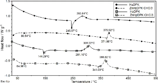
corresponding zinc(II) complexes were thermally characterized. In Figure 4 the DTG curves of the ligands and
16
the corresponding complexes are presented. DTG patterns show that the ligands have been obtained in a solvate-
17
free form. The HzDPK has a relatively high thermal stability and starts to decompose at 239 oC DTG onset. The
18
decomposition takes place in two main overlapping steps to 416 oC and afterwards slows down. HpDPK
19
decomposes in a seemingly one-step process in the temperature range of 230 – 322 oC. The successive
20
decomposition of the HzDPK can be explained by the separated fragmentation of the bulky, condensed-type
21
phthalazine ring.
22
23
Figure 4 DTG curves of the ligands and the complexes. For the sake of clarity, the curves are shifted
24
compared to zero.
25
10 The sharp endothermic peaks on the DSC curves refer to the melting of the ligands (Figure 5) The melting peak
1
(tpeak =245.6 oC) of HzDPK is immediately followed by its decomposition (tpeak = 283.8 oC). HpDPK melts at
2
much lower temperature (tpeak = 144.3 oC) and remains stable up to DSC onset 281 oC.
3
Both the complexes have been obtained as chloroform solvates and lose solvate even at room temperature
4
(Figure 4). This phenomenon is more characteristic for [Zn(HzDPK–H)2]∙CHCl3 where the desolvation process
5
occurs in a single step at lower temperatures (tpeak = 66.6 oC). The evaporation temperature of CHCl3 in
6
[Zn(HpDPK–H)2]∙CHCl3 is significantly higher. It occurs in two overlapping steps (tpeak = 140.2 oC and 206.7
7
oC) as a consequence of its restricted diffusion through the crystal lattice. The measured and the calculated
8
solvent mass loss in [Zn(HpDPK–H)2]∙CHCl3 match within the experimental error (found 15.1 %; calc. 14.84
9
%). In a freshly prepared [Zn(HzDPK–H)2]∙CHCl3 the agreement between the calculated and the measured mass
10
loss is not so good (found 15.6 %; calc. 14.29 %).
11
12
Figure 5 DSC curves of the ligands and the complexes. For the sake of clarity, the curves are shifted
13
compared to zero
14
15
TG – MS measurements
16
Coordination compounds often crystallize with solvent. However, during storage or transport the solvent might
17
be lost or replaced by water [34]. In these cases, the data obtained by TG – MS measurements give crucial data
18
for the purity check by elemental analysis. TG – MS measurements were carried out to check the solvate
19
evaporation of the complexes and to determine the decomposition processes of all the compounds. The
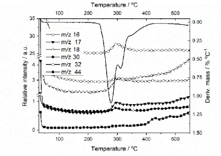
20
characteristic m/z fragments of HzDPK decomposition process are shown in Figure 6. The m/z = 16, 17 and 18
21
fragments most probably refer to the evolution of NH2+, NH3+ and NH4+ in changing proportions. Fragment m/z
22
= 30, in the accordance with structure of the compound, can be assigned to methylamine (CH3NH2), m/z = 32 to
23
hydrazine (N2H4) and m/z = 44 to ethylamine (C2H5NH2).
24
11
1
Figure 6 Selected fragments in the MS spectrum evolved during the thermal decomposition of HzDPK
2
HpDPK thermal decomposition fragments are presented in Figure 7 with the same m/z assignments.
3
4
Figure 7 Selected fragments in the MS spectrum evolved during the thermal decomposition of HpDPK
5
The presence of the CHCl3 solvent has been confirmed in both complexes. In contrast to the ligands, during the
6
decompositions of the complexes, pyridine as a fragment can also be detected in the MS spectra.
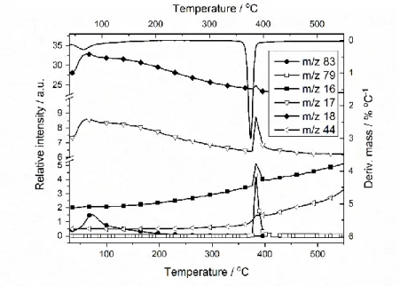
7
The characteristic fragments of [Zn(HzDPK–H)2]∙CHCl3 are shown in Figure 8. Fragments m/z = 83 and m/z =
8
18 below ~ 200 oC belong to chloroform solvate and water, respectively. The change in the relative intensities of
9
the m/z = 16, 17 and 18 signals at higher temperatures (~390 oC) refers to formation of NH2+, NH3, NH4+,
10
respectively, m/z = 44 to ethylamine (C2H5NH2), while m/z = 79 to pyridine (C5H5N).
11
12
1
Figure 8 Selected fragments in the MS spectrum evolved during
2
the thermal decomposition of [Zn(HzDPK-H)2]∙CHCl3
3
4
In [Zn(HpDPK–H)2]∙CHCl3, (Figure 9) the chloroform MS peak (m/z = 83) follow the DTG pattern. As the
5
previous complex, it also contains adsorbed water (m/z = 17, 18). Beside, at the temperatures above the onset
6
temperature of the desolvated product’s decomposition signals for NH2+–NH4+ (m/z = 16, 17, 18), ethylamine
7
(C2H5NH2; m/z = 44), and pyridine (C5H5N; m/z = 79) appear.
8 9
10
Figure 9 Selected fragments in the MS spectrum evolved during
11
the thermal decomposition of [Zn(HpDPK-H)2]∙CHCl3
12
13
14
15
16
13
Summary 1
Di(2-pyridyl)-ketone phthalazine-1-hydrazone (HzDPK), di(2-pyridyl)-ketone-3-chloropyridazine-6-hydrazone
2
(HpDPK) and their new bis-ligand zinc(II) complexes, [Zn(HzDPK-H)2]∙CHCl3 and [Zn(HpDPK-H)2]∙CHCl3
3
were synthesized and characterized by single crystal X-ray diffraction, infrared spectroscopy (FT-IR), thermal
4
analysis and coupled TG–MS measurements.
5
According to single crystal X-ray analysis, HpDPK contains intramolecular hydrogen bond which was proved
6
by NMR measurement, too (See Supporting Information). Zinc atoms in both complexes are situated in a
7
distorted octahedral environment, formed by two meridionally coordinated NNN tridentate, mono-deprotonated
8
ligands. Ligands are coordinated through pyridine, azomethine and diazine nitrogen atoms, thus forming two
9
fused five-membered metallacycles. FT-IR spectra of the complexes show the coordination of the ligands as the
10
characteristic bands are shifted to lower frequencies. By TGA and TG–MS measurements the solvent content of
11
the complexes was evaluated. It was found that chloroform partially evaporates during the storage and in part is
12
replaced by water molecules. The desolvated coordination compounds have a significantly higher thermal
13
stability than the corresponding ligands. All compounds practically decompose in one step giving small
14
fragments which mainly belong to ammonia. Fragments with a higher m/z ratio belong to pyridine or
15
alkylamines.
16
Acknowledgements 17
This research was supported by Ministry of Education, Science and Technological Development of the
18
Republic of Serbia (Grant no. 172014). József Magyari gratefully acknowledges Hungarian Academy of
19
Sciences (MTA) Domus Hungarica Grant for the research support.
20 21 22 23
References 24
25
1. Housman G, Byler S, Heerboth S, et al. Drug Resistance in Cancer: An Overview. Cancers. 2014;6:1769-
26
1792.
27
2. Zilahi G, Artigas A, Martin-Loeches I. What's new in multidrug-resistant pathogens in the ICU? Ann
28
Intensive Care. 2016;6:96-106
29
3. Locher KP. Mechanistic diversity in ATP-binding cassette (ABC) transporters. Nat Struct Mol Biol.
30
2016;23:487–493.
31
4. Kumar J, Rai A, Raj V. A Comprehensive Review on the Pharmacological Activity of Schiff Base
32
Containing Derivatives. Org Med Chem IJ. 2017;1:555564.
33
5. Popiołek Ł. Hydrazide-hydrazones as potential antimicrobial agents: Overview of the literature since 2010.
34
Med Chem Res. 2017;26:287–301.
35
6. Verma G, Marella A, Shaquiquzzaman M, Akhtar M, Ali MR, Alam MM. A review exploring biological
36
activities of hydrazones. J Pharm Bioallied Sci. 2014;6:69–80.
37
7. Azab ME, Rizk SA, Mahmoud NF. Facile Synthesis, Characterization, and Antimicrobial Evaluation of
38
Novel Heterocycles, Schiff Bases, and N-Nucleosides Bearing Phthalazine Moiety. Chem Pharm Bull.
39
2016;64:439–50.
40
8. Cohn JN, McInnes GT, Shepherd AM. Direct-acting vasodilators. J Clin Hypertens (Greenwich).
41
2011;13:690–2.
42
9. Nelson M-AM, Baba SP, Anderson EJ. Biogenic Aldehydes as Therapeutic Targets for Cardiovascular
43
Disease. Curr Opin Pharmacol. 2017;33:56–63.
44
10. Al-Falahi H, May PM, Roe AM, Slater RA, Trott WJ, Williams DR. Metal binding by pharmaceuticals.
45
Part 4. A comparative investigation of the interaction of metal ions with hydralazine, prizidilol and related
46
compounds. Agents Actions. 1984;14:113–20.
47
14 11. Abu-Dief AM, Mohamed IMA. A review on versatile applications of transition metal complexes
1
incorporating Schiff bases. Beni-Suef University Journal of Basic and Applied Sciences. 2015;4:119–33.
2
12. Barta Holló B, Magyari J, Armaković S, Bogdanović GA, Rodić MV, Armaković SJ, et al. Coordination
3
compounds of a hydrazone derivative with Co(III), Ni(II), Cu(II) and Zn(II): Synthesis, characterization,
4
reactivity assessment and biological evaluation. New J Chem. 2016;40:5885–95.
5
13. Rigaku Oxford Diffraction. CrysAlisPro Software system, version 1.171.38.46. Rigaku Corporation,
6
Oxford, UK; 2015. 2015. Rigaku Corporation.
7
14. Sheldrick GM. SHELXT - Integrated space-group and crystal-structure determination. Acta Crystallogr
8
Sect A Found Adv. 2015;71:3–8.
9
15. Sheldrick GM. Crystal structure refinement with SHELXL. Acta Crystallogr Sect C Struct Chem.
10
2015;71:3–8.
11
16. Hübschle CB, Sheldrick GM, Dittrich B. ShelXle: A Qt graphical user interface for SHELXL. J Appl
12
Crystallogr. 2011;44:1281–4.
13
17. Groom CR, Bruno IJ, Lightfoot MP, Ward SC. The Cambridge Structural Database. Acta Crystallogr Sect
14
B Struct Sci Cryst Eng Mater. 2016;72:171–9.
15
18. Macrae CF, Bruno IJ, Chisholm JA, Edgington PR, McCabe P, Pidcock E, et al. Mercury CSD 2.0 - new
16
features for the visualization and investigation of crystal structures. J Appl Crystallogr. 2008;41:466–70.
17
19. Farrugia LJ. WinGX and ORTEP for Windows: An update. J Appl Crystallogr. 2012;45:849–54.
18
20. Okabe N, Fukuda H, Nakamura T. Structure of hydralazine hydrochloride. Acta Crystallogr C Cryst Struct
19
Commun. 1993;49:1844–5.
20
21. Bakir M, Conry RR, Green O. Polymorphic di-2-pyridyl ketone 4-nitrophenylhydrazone (dpknph): The
21
structure of [beta]-dpknph. Acta Crystallogr Sect C Cryst Struct Commun. 2005;61:607–609.
22
22. Bakir M, Green O. Di-2-pyridyl ketone p-aminobenzoyl-hydrazone hydrate. Acta Crystallogr Sect C Cryst
23
Struct Commun. 2002;58:263–265.
24
23. Swearingen JK, Kaminsky W, West DX. Structural and spectral studies of di-2-pyridyl ketone 3-piperidyl-
25
and 3-hexamethyleneiminylthiosemicarbazone and their cobalt(II), nickel(II) and copper(II) complexes.
26
Transit Met Chem. 2002;27:724–31.
27
24. Deng W-T, Liu J-C, Cao J. Syntheses, crystal structures and properties of four new coordination polymers
28
involving a schiff base ligand bearing an easily abstracted proton in the hydrazone backbone. Inorg Chem
29
Commun. 2013;35:315–7.
30
25. Grünwald KR, Volpe M, Cias P, Gescheidt G, Mösch-Zanetti NC. Pyridazine-versus pyridine-based
31
tridentate ligands in first-row transition metal complexes. Inorg Chem. 2011;50:7478–88.
32
26. Goldberg AE, Kiskin MA, Popov LD, Levchenkov SI, Shcherbakov IN, Tupolova YP, Kogan VA. Crystal
33
structure of a trinuclear complex of zinc(II) with 2,6-Di-tert-butyl-p-quinone 1’-phthalazinylhydrazone. J
34
Struct Chem. 2014;55:475–80.
35
27. Di Foggia M, Bonora S, Tinti A, Tugnoli V. DSC and Raman study of DMPC liposomes in presence of
36
Ibuprofen at different pH. J Therm Anal Calorim. 2017;127:1407–17.
37
28. Li C-H, Jiang Y, Jiang J-H, Li X, Xiao S-X, Tao L-M, et al. Standard molar enthalpy of formation of
38
[(C12H8N2)2Bi(O2NO)3] and its biological activity on Schizosaccharomyces pombe. J Therm Anal
39
Calorim. 2017;128:1743–51.
40
29. Saad FA, Elghalban MG, Al-Fahemi JH, Yarkandy N, El-Metwaly NM, Abou-Melha KS, et al. Simulative
41
aurintricarboxylic acid molecular docking with antitumor activity for its VO(II), Cr(III), Mn(II) and Fe(III)
42
complexes, HF/DFT modeling and elaborated EPR studies. J Therm Anal Calorim. 2017;128:1565–78.
43
30. Srivastva U, Malhotra RK, Kaushik SC. Review of heat transport properties of solar heat transfer fluids. J
44
Therm Anal Calorim. 2017;130:605–21.
45
31. Krisyuk VV, Sysoev SV, Turgambaeva AE, Nazarova AA, Koretskaya TP, Igumenov IK, Morozova NB.
46
Thermal behavior of methoxy-substituted Pd and Cu β-diketonates and their heterobimetallic complex. J
47
Therm Anal Calorim. 2017;130:1105–10.
48
32. Abd El-Halim HF, Omar MM, Anwar MN. Preparation, characterization, antimicrobial and anticancer
49
activities of Schiff base mixed ligand complexes. J Therm Anal Calorim. 2017;130:1069–83.
50
33. Begović NN, Vasić MM, Blagojević VA, Filipović NR, Marinković AD, Malešević A, Minić DM.
51
Synthesis and thermal stability of cis-dichloro[(E)-ethyl-2-(2-((8-hydroxyquinolin-2-
52
il)methylene)hidrazinyl)acetate-κ2 N]-palladium(II) complex. J Therm Anal Calorim. 2017;130:701–11.
53
34. Jaćimović ŽK, Giester G, Kosović M, Bogdanović GA, Novaković SB, Leovac VM, Latinović N, Barta
54
Holló B, Mészáros Szécsényi. Solvent exchange reactions in coordination compounds. J Therm Anal
55
Calorim (2017) 127:1501–1509
56
15
![Table 1 Crystallographic and refinement details of HpDPK, [Zn(HpDPK–H) 2 ]∙CHCl 3 and 2 [Zn(HzDPK–H) 2 ]∙CHCl 33 4 HpDPK [Zn(HpDPK–H) 2 ]∙CHCl 3 [Zn(HzDPK–H) 2 ]∙CHCl 3 Chemical formula C 15 H 11 ClN 6 C 30 H 20 Cl 2 N 12 Zn·CHCl 3 C 38 H 26 N 12](https://thumb-eu.123doks.com/thumbv2/9dokorg/1409520.118772/5.892.103.749.199.958/table-crystallographic-refinement-details-hpdpk-hpdpk-chemical-formula.webp)
![Table 2 Selected bond lengths and bond angles of [Zn(HpDPK−H) 2 ]·CHCl 3 (1), [Zn(HzDPK−H) 2 ]·CHCl 3 (2) 3](https://thumb-eu.123doks.com/thumbv2/9dokorg/1409520.118772/8.892.99.800.210.1135/table-selected-lengths-angles-hpdpk-chcl-hzdpk-chcl.webp)
![Table 3 Characteristic IR bands of the ligands and the complexes [Zn(HpDPK−H) 2 ]·CHCl 3 (1), 10 [Zn(HzDPK−H) 2 ]·CHCl 3 (2) 11 Vibration Wavenumber / cm –1 HpDPK 1 HzDPK 2 ν C=N 1581–1426 1588–1400 1403 1395–1376 ν C Ar -N 132](https://thumb-eu.123doks.com/thumbv2/9dokorg/1409520.118772/9.892.163.733.760.1066/table-characteristic-ligands-complexes-hpdpk-hzdpk-vibration-wavenumber.webp)