Peripheral and central analgesic components of a novel opioid in the management of inflammatory and
neuropathic pain
Ph.D. thesis
Mihály Balogh Pharm.D.
Semmelweis University
Doctoral School of Pharmaceutical Sciences
Supervisor: Mahmoud Al-Khrasani, Pharm.D., Ph.D.
Official reviewers:
Éva Keller, M.D., Ph.D.
Ildikó Világi, Ph.D.
Head of the Final Examination Committee:
Éva Szökő, Pharm.D., D.Sc.
Members of the Final Examination Committee:
Gábor Pethő, M.D., Ph.D.
Ákos Zsembery, M.D., Ph.D.
Budapest 2018
1
Table of contents
List of abbreviations ... 4
1. Introduction ... 6
1.1. Opioids and analgesia ... 6
1.2. Pain transmission ... 8
1.2.1. Opioid receptors and their distribution ... 9
1.3. Different pain disorders ... 12
1.3.1. Acute and inflammatory pain disorders... 12
1.3.2. Neuropathic pain disorders ... 13
2. Objectives ... 15
3. Materials and methods ... 18
3.1. Experimental animals ... 18
3.2. Materials ... 18
3.3. Acute inflammatory pain models ... 19
3.3.1. Experimental paradigm for mouse writhing test ... 19
3.3.2. Experimental paradigm for rat formalin test ... 19
3.4. Subchronic inflammatory pain model: Complete Freund’s Adjuvant induced inflammation in rats ... 20
3.5. Neuropathic pain model: experimental paradigm for streptozocin induced chronic diabetic neuropathy in rats ... 21
3.5.1. Monitoring of diabetic and control animals ... 21
3.5.2. Investigation of the analgesic action of different opioids in rats with diabetic neuropathy... 23
3.6. Experimental paradigms for the assessment of side effects of test compounds .. 23
3.6.1. Determination of the effect of test compounds on gastrointestinal transit ... 23
3.6.2. Respiratory function tests ... 24
3.6.3. Determination of sedative effects of test compounds ... 24
3.6.4. Induction of tolerance in mice ... 25
3.7. In vitro receptor binding assays ... 26
3.7.1. Radioligand competition binding assay ... 26
3.7.2. Immunohistochemistry ... 27
3.7.3. G-protein activity assay ... 27
3.8. Analysis of data ... 28
4. Results ... 29
4.1. Acute inflammatory pain models ... 29
2
4.1.1. Mouse writhing test ... 29
4.1.2. Rat formalin test ... 32
4.2. Subchronic inflammatory pain model: CFA induced inflammation in rats ... 38
4.2.1. Antinociceptive effects of 14-O-MeM6SU and M6SU after systemic administration in CFA model in Randall-Selitto test ... 38
4.2.2. Antagonist effects of s.c. and i.pl. NAL-M on the antinociceptive actions of s.c. 14-O-MeM6SU or M6SU ... 41
4.3. Neuropathic pain model: diabetic polineuropathy... 44
4.3.1. The development of diabetic symptoms and neuropathic pain (allodynia) in STZ treated rats ... 44
4.3.2. The impairment of the antinociceptive effect of systemic 14-O-MeM6SU and morphine in advanced diabetes in rats ... 46
4.3.3. The antiallodynic effects of systemic 14-O-MeM6SU and morphine in diabetic rats ... 47
4.3.4. The antagonist effect of co-administered NAL-M on the systemic analgesic effect of 14-O-MeM6SU or morphine in diabetic rats ... 49
4.4. Side effect profiles of test products ... 50
4.4.1. Inhibitory effect of systemic 14-O-MeM6SU, M6SU and morphine on gastrointestinal transit in mice ... 50
4.4.2. Respiratory effects of 14-O-MeM6SU and M6SU compared to morphine in awake unrestrained rats ... 50
4.4.3. Sedative effects of test compounds ... 50
4.4.4. Analgesic tolerance of 14-O-MeM6SU compared to morphine in mouse tail- flick test ... 53
4.5. In vitro receptor binding assays ... 54
4.5.1. MOR immunoreactivity and binding sites in the spinal cord and DRG of diabetic and non-diabetic rats ... 54
4.5.2. The G-protein coupling activity in presence of 14-O-MeM6SU, or morphine in spinal homogenates prepared from diabetic or control rats... 56
4.5.3. The G-protein coupling activity in presence of 14-O-MeM6SU, or morphine in brain homogenates prepared from diabetic or control rats ... 56
5. Discussion ... 61
5.1. Inflammatory pain alleviation with high efficacy opioid of limited CNS penetration ... 61
5.2. Neuropathic pain alleviation with high efficacy opioid of limited CNS penetration ... 64
5.3. The side effect profile of the novel compound, 14-O-MeM6SU ... 67
6. Conclusions ... 71
7. Summary ... 73
3
8. Összefoglalás ... 74
9. Bibliography ... 75
10. List of own publications ... 88
10.1. Own publications involved in the present thesis ... 88
10.2. Own publications not involved in the present thesis ... 88
11. Acknowledgements ... 90
12. Köszönetnyílvánítás ... 91
4
List of abbreviations
14-O-MeM6SU: 14-O-methylmorphine-6-O-sulfate ACC: anterior cingular cortex
ANOVA: analysis of variance
Bmax: total density (concentration) of receptors in a sample of tissue cAMP: cyclic adenosine monophosphate
CFA: complete Freund's Adjuvant CNS: central nervous system
DAMGO: [D-Ala2,N-Me-Phe4,Gly-ol5] enkephalin DNP: diabetic neuropathic pain
DOR: -opioid receptor
DPA: Dynamic Plantar Aesthesiometer DRG: dorsal root ganglion
EC50: The concentration of an agonist that produces 50% of the maximal biological effect
ED30: antinociceptive dose necessary to produce a 30% response ED50: antinociceptive dose necessary to produce a 50% response Emax: maximal biological effect
f: frequency
GABA: γ-aminobutyric acid GDP: guanosine-5'-diphosphate GPCRs: G protein-coupled receptors GTP: Guanosine-5'-triphosphate i.c.v.: intracerebroventricular i.p.: intraperitoneal
i.pl.: intraplantar i.v.: intravenous
IASP: International Association for the Study of Pain ID50: doses caused 50% inhibition
Kd: equilibrium dissociation constant KOR: κ-opioid receptor
5 M6G: morphine-6-glucuronide
M6SU: Morphine-6-O-sulfate MOR: µ-opioid receptor MPE: maximal possible effect MV: minute ventilation NAL-M: naloxone methiodide NP: neuropathic pain
NSAIDs: non-steroidal anti-inflammatory drugs PAG: periaqueductal gray
PBS: phosphate buffered saline PEF: peak expiratory flow PIF: peak inspiratory time PPT: paw pressure threshold RT: relaxation time
RVM: rostral ventromedial medulla s.c.: subcutaneous
SEM: standard error of mean STZ: streptozocin (streptozotocin) Te: time of expiration
Ti: time of inspiration TV: tidal volume
VLO: ventrolateral orbital cortex WBP: whole-body plethysmography
6
1. Introduction
1.1. Opioids and analgesia
Pain is described by Albert Schweitzer as a more terrible lord of mankind than even death itself. His thoughts are still apposite. The clinical practice has huge successes in pain management but still lacks the proper solution in many cases; mostly chronic painful conditions are still a huge challenge. Only in the USA approximately 100 million people are suffering from some type of chronic painful condition [1].
Opioid analgesics are among the oldest pain medications applied by human beings, yet they are still the mainstay in the management of moderate to severe pain [2].
The use of opioids is going back for a long time, ancient Roman and Greek physicians described them as a powerful tool to relief pain. The start point in opioid history is the cultivation of poppy by Sumerians who described it as Hul Gil, the „joy plant” in lower Mesopotamia around 3400 B.C. Then knowledge of poppy cultivation and its euphoric effect spread to area governed by Assyrians, who passed it to Egypt and later to China.
The first opioid agent morphine was isolated by Friedrich Wilhelm Sertürner in the 19th century. At this time morphine became available to treat diverse types of occasional pain (e.g. pain originated from injuries, toothache etc.). In the same century Alexander Wood invented the hypodermic syringe for medical use, which resulted in the growing use of opioids leading to an increase in the incidence of morphine addiction especially among soldiers in the USA and Europe. As an attempt to overcome the unwanted dependence causing effect of morphine, heroin (diacetylmorphine) was developed. The new compound was widely used as an anti-cough medication. Despite the goal for which heroin was introduced, its use as favorite habit was grown. This led to a strong increase of opioid addiction in society and encouraged authorities to issue the opioid control act [3–5]. Up to this date the clinical practice still lacks an opioid compound with proper efficacy but without the unwanted central nervous system (CNS) effects like addiction or respiratory depression.
Opioid analgesics are well known to exert their antinociceptive action by the activation of opioid receptors, particularly µ-opioid receptor (MOR) at spinal and suprasinal regions [6]. MOR activation by opioids results in analgesic effect as well as adverse effects. The majority of clinically used opioid analgesics have central adverse effects such as
7
respiratory depression, development of opioid tolerance and dependence, as well as addiction liabilities. More important, when they are misused or abused, they can cause addiction, overdose and death. These effects hamper their clinical use [2, 6, 7, 8]. It is important to note that the abuse causing effect of opioids is less when they are prescribed for chronic pain management, especially when there is no history of previous drug abuse [9]. Still, the over-prescription in the USA led to a national opioid epidemic crisis, the use of opioids is estimated to cost over $700 million annually [10].
Beside the central opioid analgesic effect, several data support that antinociception could also be achieved by activation of functional opioid receptors in the periphery as well [11, 12]. In the late 1980s researchers started to pay attention to the peripheral analgesic effect of opioids, growing number of studies were conducted in order to investigate the possibility of peripheral antinociception [13]. The most investigated method of peripheral opioid analgesia is intra-articular injection of the MOR agonist morphine [13–15]. It has been shown that significant pain relief can be achieved after knee surgery or in chronic rheumatoid- and osteoarthritis. The effect may last even up to 7 days (similarly to local steroid or anesthetic injection). The most limiting factor of intra-articular injections is the enhanced risk of local infections [16, 17]. Locally injected morphine was effective in patients with chronic inflammatory tooth pain. Submucous injection of morphine also effectively alleviated the pain after dental surgery. These effects were absent in patients without preexisting inflammation (similarly as in animal studies) [17, 18]. Topically applied morphine attenuated pain in patients with unilateral corneal abrasions on the lesioned, but not the intact site. Importantly, morphine did not show any detrimental effect on wound healing [17, 19]. Locally injected morphine was also effective in postoperative visceral pain conditions (in the urinary bladder or after laparoscopic tubal ligation) [17].
On the other hand, several studies failed to prove peripheral analgesic effect of applied opioids. In the majority of these studies morphine was injected into non-inflamed tissues.
It further supports the observation, that the peripheral analgesic effect of opioids is highly elevated under inflammatory conditions [17]. MOR, δ-opiod receptor (DOR) and also κ- opioid receptor (KOR) agonists show significantly stronger antinociceptive action in injured than in non-injured tissues of animals and also of humans [13].
Many attempts have been done in order to target the peripheral source of pain (i.e. target the peripheral opioid receptors in order to inhibit the pain transmission peripherally) to
8
avoid the CNS adverse effects. However, for long time the chemical modification carried out on morphine to limit its central nervous system penetration resulted in morphine analogs of weak affinity [20]. In spite the quaternary analogs that showed limited CNS penetration and lower affinity for opioid receptors, zwitterionic molecules like morphine- 6-O-sulfate (M6SU) displayed greater antinociceptive effect than morphine [21, 22].
M6SU applied as an eye drop showed strong local antinociceptive effect in different animal models of corneal injury without any harmful effect on wound healing [23, 24].
Taken together, targeting peripheral opioid receptors offers a possible new way to treat different pain conditions, especially in the case of inflammatory pain.
1.2. Pain transmission
Nociceptors are located in the peripheral ending of primary afferents. They can be found at the end of pseudounipolar sensory neurons with cell bodies in the dorsal root-, trigeminal-, or nodose ganglia [25]. To sake of simplicity, based on axon diameter, degree of myelination and axonal conduction velocities as well as body sizes, sensory fibers are classified as A, Aδ and C. A fibers are stimulated by non-noxious stimuli. Most of them have low mechanical thresholds and are described as light-touch receptors. On the other hand, Aδ and C fibers carry the noxious sensory information into the spinal dorsal horn. These fibers are responsible for transmission of pain resulted from mechanical, thermal or chemical noxious stimuli in different parts in our body. Most of the Aδ fibers are associated with mechano- or thermoreceptors. C-fibers are polymodal fibers, because they are responding to multiple modalities: chemical, mechanical (touch, pressure, stretch) and thermal stimuli. Aδ and C primary sensory afferent fibers convey the pain from the site of injury into spinal cord, where they synapse with the secondary sensory neurons (spinothalamic tract), that further convey the pain to the thalamus, where third order neurons pass the information further to the somatosensory cortex. Two categories of pain transmission exist: fast and slow. Aδ fibers transmit the information relatively quickly (6 to 30 m/sec), C fibers are conducting at a lower speed (0.5 to 2 m/sec). The myelinated, large A fibers conduct the information (touch, pressure, vibration) at high speed (30 to 70 m/sec) [25].
The emotional aspect of the pain is conveyed by the spinoreticular tract that terminates in the reticular information in the brainstem, where information is further processed to
9
thalamus and hypothalamus. Of note, sensory C fibers are responsible for conveying visceral pain and neuropathic pain from the periphery to CNS [25]. Inflammatory mediators (e.g. bradykinin, prostaglandin, serotonin, H+, cytokines) that are released from damaged tissues surrounding the primary sensory afferents of free endings directly stimulate the nociceptors or lower their pain threshold. The later phenomenon is called primary sensitization. This will also increase the excitability of spinal neurons which can later amplify all sensory inputs including normally non-noxious stimuli conveyed by the low threshold A fibers. This central sensitization strongly contributes to the pain symptoms like allodynia (non-noxious stimuli experienced as painful stimuli) and hyperalgesia (lowered pain threshold) [14, 26].
Beside the ascending pain pathway, the descending (or inhibitory) pathway also have important role in pain sensation. Areas in the brain like the periaqueductal gray (PAG) and rostral ventromedial medulla (RVM) hosting high receptor pools and containing high endogenous opioid peptide content are major points in the control of descending pain pathways. Therefore, activation of this pathway through endogenous opioid-release results in analgesia that can explain the lack of pain sensation for example during a shock condition [25].
1.2.1. Opioid receptors and their distribution
Opioid receptors belong to G-protein coupled receptors (GPCRs), they are Gi-coupled inhibitory receptors. Currently, three opioid receptor types exist: μ-opioid receptor (MOR), κ-opioid receptor (KOR) and δ-opioid receptor (DOR) named after morphine, ketocyclazocine and vas deferens, respectively [27, 28]. The International Union of Basic and Clinical Pharmacology Committee (IUPHAR) for the Receptor Nomenclature and Drug Classification issued the abbreviations MOR, KOR and DOR. Their mRNAs as well as the gene’s structures were cloned and characterized. In addition, they were further subdivided into several subtypes as follows: MOR to μ1 (pain management) and μ2 (respiratory center) and μ3 (immune cells); KOR to κ1a, κ1b κ2, κ3 and DOR to δ1 and δ2 [29–34].
The activation of opioid receptors by opioid agonists results in decrease of intracellular cAMP levels through inhibition of adenylate cyclase, close of voltage-dependent Ca2+
channels (N type and to a lesser extent P / Q) in presynaptic nerve endings and opening
10
of K+ channels at post-synaptic neurons [35]. Consequently, beside the inhibition of the release of transmitters (neurohormones and neuropeptides), such as glutamate and substance-P from the presynaptic neurons, they do inhibit the propagation of action potential by hyperpolarizing the secondary neuron cells [36].
Opioid receptors are widely distributed in the CNS and also on the periphery.
Opioid receptors in the CNS: In the brain all types of opioid receptors can be found.
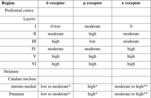
The most abundant opioid receptor with the widest distribution in the brain is MOR. Table 1 depicts the mRNA distribution of different opioid receptors in the human brain, which directly correlates with the receptor distribution [37]. MOR and DOR can be found in the whole area of cortical lamina whereas KOR can be found mostly in the deeper regions (lamina IV-VI.), which might contribute to the sedative effect of KOR agonists. Opioid receptors are also distributed in caudate putamen. The caudate putamen forwards information toward the ventral tegmental area and substantia nigra. This indicates the role in sensory-motor interactions, motivation and rewarding effects.
The opioid receptors can be found in other regions important in pain transmission, like the anterior cingular cortex (ACC), primary and secondary somatosensory cortex, ventrolateral orbital cortex (VLO). The ACC is in direct connection with the periaqueductal gray (PAG), which has an essential role in the descending inhibitory pathway. ACC is a key point in somatic- and also visceral pain processing. Endogenous opioids are among the neurotransmitters acting in the ACC. The role of opioids in pain transmission/inhibition was shown also in the motor cortex, rostral agranular cortex. In conclusion, opioids influence the pain transmission in the brain at every important area of pain processing [37–39].
Opioid neurons are also located at the spinal level. These inhibitory interneurons are influenced by the descending inhibitory pathway causing pre- and also post-synaptic inhibition. Opioid neurons in the midbrain inhibit GABAergic inhibitory neurons activating the descending pathway (inhibition of inhibition). Additionally, opioid receptors are also expressed in the dorsal horn and in the gray matter around brain ventricle IV and V [25, 40]. In the dorsal root ganglia (DRG) MORs, DORs and also KORs can be found. MORs are located on medium and large diameter cells, DORs mostly on large diameter neurons and KORs on small and medium neurons. These data suggest that these opioidergic neurons are important in the processing of different pain types and
11
they are influencing both the descending- and ascending pain pathways in the spinal cord [41].
Table 1. Opioid receptor mRNA distribution in human brain
Region δ receptor µ receptor κ receptor
Prefrontal cortex Layers:
I 0-low moderate 0
II moderate high moderate
III high low moderate
IV moderate moderate high
V high high high
VI high high high
Striatum
Caudate nucleus
interno-medial low to moderate* high* moderate to high**
Putamen low to moderate* high* moderate to high**
0 = undetectable; * = diffuse; ** = asymmetric cell clusters
Low, moderate, and high indicate the relative density of cells expressing receptor mRNA.
Table 1. was adapted from [37].
Opioid receptors at the periphery: Beside the central opioid receptors, several data support the existence of functional opioid receptors in the periphery as well [12, 42].
These receptors are localized on peripheral terminals of sensory nerves. Pharmacological evidences – on animal models and also in humans – indicate that activation of these receptors on peripheral sensory axons also results in the mitigation of pain [20, 43–45].
Based on experimental results, all three types of opioid receptors (MOR, DOR and KOR) can be found at the periphery in functionally active state [12]. These receptors can be found on small-, medium- and large- diameter sensory neurons of animals or humans.
The endogenous ligands of these receptors, opioid peptides (endorphin, enkephalin, dynorphin) were also found in immune cells infiltrating the inflamed tissues during
12
inflammation. Environmental stress is a strong factor in the mechanism of release of these peptides [13, 17, 42].
In conclusion, the three pillars of the analgesic-antinociceptive effect of opioids are:
1) Inhibition of the nociceptive stimuli transmission from the periphery to the spinal cord 2) Activation of the descending inhibitory pathway
3) Influencing the activity of the limbic system [6, 46, 47].
1.3. Different pain disorders
1.3.1. Acute and inflammatory pain disorders
Nociceptive pain originates from tissue damage surrounding peripheral sensory neurons (e.g. postoperative pain; posttraumatic pain, as broken bone pain). Opioids are the most effective tools to treat acute pain syndromes but their side effects, especially the centrally ones, limit their clinical use. NSAIDs are usually the first choice in acute pain treatment.
In severe cases they might be used in combination with opioids to achieve a synergistic antinociceptive effect, thereby reducing the opioid need [10]. Indeed, NSAIDs are good choice in the treatment of mild to moderate pain. However, limiting factors in the use of NSAIDs are their gastrointestinal damaging and cardiac adverse effects [48]. Tramadol (weak MOR agonist and serotonin- and norepinephrine reuptake inhibitor), the NMDA antagonist ketamine and in some cases the anticonvulsant gabapentinoids (gabapentin, pregabalin) are also used in the management of acute pain conditions [10].
The inevitable consequence of tissue damage is the release of inflammatory mediators such as bradykinin, serotonin, prostaglandins and cytokines as well as tissue acidification.
This will lead to the direct stimulation of nociceptors and the above mentioned primary sensitization. Beside inflammatory mediators endogenous antinociceptive agents are also released at the site of inflammation like opioid peptides, somatostatin, endocannabinoids and anti-inflammatory cytokines [14].
Opioid receptors have been reported to be upregulated in inflamed tissues as well as at the spinal level. Potentiated analgesic action of opioids was also observed [49–51]. This favorable change stimulates the researchers to develop opioid agents targeting these peripheral receptors, hence the peripheral antinociception of opioids in humans has been investigated in several randomized controlled clinical trials [52, 53]. Pooled analyses of data from 19 studies suitable for meta-analysis showed only a moderate analgesic effect
13
after intra-articular morphine compared with placebo in patients with arthroscopic knee surgery. Morphine-6-glucuronide (M6G) has been reported to have peripheral anti- hyperalgesic effects following its systemic administration in human volunteers [45]. Due to its high hydrophilicity it has a considerable delay between peek plasma concentrations and peek central opioid effects, so peripheral antinociceptive effect can be detected within this time window [45, 54]. Although in the case of M6G central side effects, like respiratory depression was observed, design of opioids with high hydrophilicity and high efficacy may provide analgesic agents of high clinical value.
1.3.2. Neuropathic pain disorders
The definition of neuropathic pain (NP) by the International Association for the Study of Pain (IASP) is “pain arising as a direct consequence of a lesion or disease affecting the somatosensory system” (IASP, 2012). Based on this definition NP is a consequence of damage to neurons in peripheral- or central nervous system or both. NP has significant undesirable impact on the economic welfare of the society [55, 56]. Therefore, to find drugs satisfactory treating NP is a major clinical goal. The management of severe acute to moderate and cancer pain by opioids is satisfactory, however opioid effectiveness in the treatment of chronic NP is controversial [2, 40, 57–61]. So far the management of NP by medication is largely based on the symptoms of disease. These medications are categorized into three groups: first, second and third line drugs. The first line medications include: tricyclic antidepressants, dual reuptake inhibitors of serotonin and norepinephrine, gabapentinoids and 5% lidocaine transdermal patch. The second line drugs are classical opioids and drugs having opioid and non-opioid actions like tramadol.
Third line medications include antiepileptics, topical capsaicin, memantine and mexiletine [62]. Of note, medications considered as second line might prescribed as first line e.g. during the titration of another drug or acute NP attack.
In addition, reports have been issued on the equivocal response of NP for opioid treatments, although this responsiveness to opioid analgesics may vary across the different types of NP syndromes [2, 59, 63]. Data obtained in diabetic animals developed NP showed a significant reduction in opioid antinociceptive efficacy following systemic administration. The loss of opioid antinociceptive efficacy has been also reported following central (spinal or supraspinal) administration in diabetic animals with NP [64–
14
66]. It was proposed that the impaired opioid analgesia occurred as a consequence of decrease in opioid receptor reserve. In addition, the reduction in opioid receptor density in spinal and supraspinal tissues was also demonstrated in diabetic animals [67, 68].
In general, less than 50% of patients respond to treatment with all of the above mentioned drugs and 40% are inadequately treated. Therefore, treatment of NP so far is considered as an unmet medical need and indicates necessity for developing new therapies for these types of painful conditions [69].
15
2. Objectives
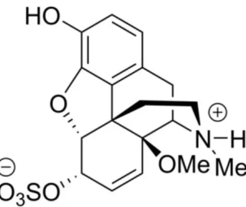
Our working group recently designed and synthesized the new MOR agonist, 14-O- methylmorphine-6-O-sulfate (14-O-MeM6SU; Fig. 1.), which displayed high affinity for MOR and strong antinociceptive effect in acute thermal nociceptive tests. Additionally, in functional in vitro [35S]GTPS binding assay, 14-O-MeM6SU showed a full agonist character whereas morphine or M6SU showed a partial agonist nature [70].
To assess the antinociceptive effect of test compounds in inflammatory conditions, three different animal models were applied: acetic acid induced writhing test in mice, formalin test in rats (acute models) and Complete Freund’s Adjuvant (CFA) induced subchronic inflammatory model.
The acetic acid-evoked writhing assay is one of the most well-established and widely used experimental models of visceral pain to assess the pain relieving actions of either NSAIDs or opioids [71, 72]. In this model chemical visceral nociception is induced by the injection of diluted acetic acid into the peritoneal cavity of the rodents.
Formalin test is frequently applied in rats to assess the antinociceptive properties of investigated compounds. The test consists of two phases: phase I (0-10 min) and phase II (11-60 min). Phase I is a direct consequence of the irritating effect of the formalin solution injected intraplantarly (i.pl.) into the hindpaw. In the second phase inflammatory mediators (bradykinin, histamine etc.) are released, hence this test is suitable to assess the effect of test compounds on acute nociceptive and inflammatory pain as well [73].
In case of Complete Freund’s Adjuvant (CFA- a mixture of mineral oils, heat- killed mycobacteria and emulsifying agent) -evoked hyperalgesia the inflammation is induced by the application of CFA into the hind paw of animals (e.g. rats or mice). This pain model has long been used as a strategy to demonstrate the (peripheral) antinociceptive actions of opioid agonists [44, 74]. CFA induces subchronic inflammation by enhancing both cell-mediated and humoral immune responses, hence modeling the clinical conditions (e.g. rheumatoid arthritis) rather properly [75].
16
Figure 1. The chemical structure of the novel compound 14-O-methylmorphine- 6-O-sulfate (14-O-MeM6SU)
Neuropathic pain is one of the hardest painful conditions to treat. Among different NP conditions diabetic polyneuropathy deserves particular attention, since it is one of the most common NP diseases. Approximately 60-70% of diabetic patients suffer from diabetic polyneuropathy (mild to severe forms) [76]. To mimic these clinical conditions streptozocin (STZ) treated diabetic rats were investigated. STZ destroys the insulin producing cells, hence causing type 1 diabetes with high blood glucose level.
First the symptoms of diabetes were confirmed: blood glucose levels, animal weights and the amount of water- and food consumption was investigated at numerous time points. Since diabetes can impair gastrointestinal motility [77], the gastric emptying of diabetic and control rats was also assessed. The elevated glucose concentration in the blood leads to neuron damage and therefore neuropathic pain, indicated by mechanical allodynia [64, 78].
In neuropathic conditions the effectiveness of different opioids is reduced [68].
Therefore, we also assessed and compared the degree of antinociception impairment of the novel compound (14-O-MeM6SU) and morphine.
The number of MOR was evaluated in DRG and dorsal horn tissues of diabetic and non-diabetic rats. G-protein coupled receptor activity was also measured in vitro to determine the efficacy of test compounds in diabetic and control rat spinal cord and brain homogenates.
We also aimed to assess the side effect profile of the novel compound in comparison with other known agents. The tail-flick test is one of the most commonly used methods to assess the acute nociceptive potency and efficacy of different opioids against
17
thermal stimuli [43, 79]. Beside the advantages of this test, like being relatively quick and easy to perform, it might be an adequate method to predict analgesic efficacy both in humans and in other more complex pain models, as well [80]. With the tail-flick test – performed after 3 days administration of test compounds – we aimed to assess the analgesic-tolerance profiles. The effects on gastrointestinal transit and respiratory functions were also investigated, since opioid induced constipation and the respiratory depressive effects of opioids are among the main limiting factors of their use [81].
Sedative effects of test compounds were also evaluated by the assessment of their anesthesia potentiating effects.
In summary, the main aims of the thesis are:
1. To assess the antinociceptive efficacy and potency of the novel compound 14-O- MeM6SU in comparison with known reference compounds (morphine or morphine-6-O- sulfate) under:
acute and subchronic inflammatory pain conditions: mouse writhing test, rat formalin test, rat CFA model
neuropathic pain conditions: rat model of diabetic polyneuropathy
2. To determine the degree of antinociception impairment in advanced diabetic neuropathy in rats
3. To investigate the peripheral component in the antinociceptive action of 14-O- MeM6SU and reference compounds in above mentioned pain conditions
4. To further analyze the actions of test opioid agonists at the spinal and supraspinal level under NP conditions applying biochemical assays
5. To investigate the side effect profile of 14-O-MeM6SU in comparison with reference compounds by analyzing the:
effects on gastrointestinal transit in mice
effects on respiratory functions in rats
sedative effects in rats
tolerance profile in mice
18
3. Materials and methods
3.1. Experimental animals
Animals (male Wistar rats or NMRI mice) were housed in the local animal house of the Department of Pharmacology and Pharmacotherapy, Semmelweis University (Budapest, Hungary) or Charité University Berlin, Campus Virchow Klinikum and Campus Charite Mitte, Berlin, Germany in the case of immunohistological assay. Housing and experiments were performed according to the European Communities Council Directives (2010/63/EU), (86/609/ECC), German science-based guidelines for laboratory animal care of the National Research Council (2003), the Hungarian Act for the Protection of Animals in Research (XXVIII.tv. 32.§) and local animal care committee (PEI/001/276- 4/2013). Animals were kept in standard cage (5 or 6 animals/cage) in a room of 20 ± 2°C temperature, 12-hour/12-hour light/dark cycle (light on at 6 A.M.). Diabetic and their control animals were kept in mash bottomed cage. Water and standard food were available ad libitum.
The animal weights, phenotypes and sources are presented under appropriate method section. Each animal was used for one experiment and only once. During work researchers did the best effort to minimize the number of animals and their suffering.
3.2. Materials
The novel compound (14-O-MeM6SU) and the reference compound M6SU used in the present work were synthesized by Sándor Hosztafi in Department of Pharmaceutical Chemistry, Semmelweis University (Budapest, Hungary) as previously described [70].
Morphine hydrochloride was obtained from Alkaloida-ICN (Tiszavasvári, Hungary). The MOR agonist enkephalin analog Tyr-D-Ala-Gly-(NMe)Phe-Gly-ol (DAMGO) was obtained from Bachem Holding AG (Bubendorf, Switzerland). The radiolabeled GTP analog, [35S]GTPγS (specific activity: 1000 Ci/mmol) was purchased from Hartmann Analytic (through Izotóp Intézet Kft., Budapest, Hungary). The UltimaGoldTM MV aqueous scintillation cocktail was purchased from PerkinElmer (through Per-Form Hungária Kft., Budapest, Hungary). Naloxone methiodide (NAL-M) and all other chemicals were of analytical grade and purchased from Sigma–Aldrich, Budapest, Hungary. Drugs were dissolved in 0.9% solution of NaCl with the exception of STZ,
19
which was dissolved in ice-cold distilled water right before injection (less than 10 min before injection). NAL-M was dissolved in saline, also right before the experiment.
Drugs or vehicle were delivered as follows: s.c. administration (under skin over the neck) 2.5 or 5 ml/kg for rats and 10 ml/kg for mice; intravenous (i.v.) injections 2.5 ml/kg for rats; intraperitoneal (i.p.) injections 2.5 ml/kg for rats; intraplantar (i.pl.) administration 100 µl/rat; intracerebroventricular (i.c.v.) injection 5 µl/animal for mice. For each dose a separate group of animals was used. All compounds were stored and handled as described in the product information sheet.
Experiments were performed in a blinded way to the drugs and doses applied. In in vivo tests morphine or morphine-6-O-sulfate (M6SU) was used as reference compound.
3.3. Acute inflammatory pain models
3.3.1. Experimental paradigm for mouse writhing test
Male NMRI mice (20-30 g) were used. The acetic acid-induced writhing test was performed as previously described [71]. Mice were injected i.p. with 0.2 ml of 0.6% acetic acid aqueous solution to induce the writhing reaction which is characterized by contractions of the abdominal musculature followed by extension of the hind limbs.
Groups of mice were injected s.c. or i.c.v. with different doses of 14-O-MeM6SU or M6SU followed 15 min later by an intraperitoneal (i.p.) injection of 0.6% acetic acid solution. Each mouse was then placed in individual transparent Plexiglass chambers, and 5 min after acetic acid injection the number of writhes was counted during a 10 min observation period. That is, assessments started 20 min after s.c. opioid agonist administration. For determination of writhes in control groups, animals were s.c. or i.c.v.
injected with 0.9% saline solution before i.p injection of 0.6% acetic acid using a similar protocol as for the test drugs. In experiments when the antagonist action was assessed, s.c. NAL-M was co-administered with the respective agonist.
3.3.2. Experimental paradigm for rat formalin test
Male Wistar rats (200–300 g) were used. The test was performed as described previously [82]. Briefly, before the experiments the animals were daily wrapped in cloth („handling”) except their right hindlimb, which was left free, for three constitutive days in order to habituate them to the experimental conditions. On the fourth day animals were
20
wrapped in cloth and 2.5% formalin solution was injected into the plantar surface of the right hind paw in a volume of 50 µl/rat. Immediately after the injection, animals were placed into Plexiglass observation chambers fixed above a mirror of 45 degree angle position allowing free viewing of the paws. Then, the antinociceptive action of the investigated agents was assessed by counting the number of nociceptive behaviors (shaking, flinching, licking and elevating the painful paw) for 60 min of 5 min time periods. The observation peridod was subdivided into two phases: Phase I: 0-10 min (caused by the irritant effect of formalin) and Phase II: 11-60 min (caused by the release of inflammatory mediators) to determine the pain events.
The test compounds were injected s.c. (2.5 ml/kg) 15 min prior to formalin injection. In order to assess the peripheral opioid system’s contribution to the whole antinociceptive action of systemically applied opioids NAL-M was co-administered. In an other set of experiments, the test compounds were injected intraplantarly into the ipsilateral or contralateral paw (100 µl/animal) 5 min prior to the i.pl. formalin solution injection.
3.4. Subchronic inflammatory pain model: Complete Freund’s Adjuvant induced inflammation in rats
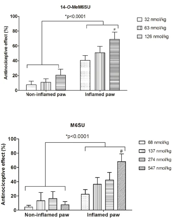
Male Wistar rats (200–300 g) were used. Rats under brief isoflurane (Willy Rüsch GmbH, Böblingen, Germany) anesthesia received i.pl. injection of 0.15 ml Complete Freund’s Adjuvant (CFA) (Calbiochem, San Diego, CA), a water-in-oil emulsion of killed mycobacteria, into the right hind paw. On the fourth and seventh day after i.pl. CFA- injection, baseline (pre-test compound) paw pressure thresholds (PPT) of inflamed and non-inflamed paws were assessed by paw pressure algesiometry (modified Randall- Selitto test; Ugo Basile, Comerio, Italy) as earlier described in [83]. Then, PPTs were reevaluated at 30, 60 and 120 min after s.c. drug administration, using an arbitrary cut off weight of twice the baseline. The cut off time was considered 100% and values are expressed as percentages.
In these experiments the antinociceptive effects of s.c. 14-O-MeM6SU and M6SU were examined. After baseline measurements separate groups of animals for each s.c. dose were used. The antinociception assessed with respect to the change in PPT of both paws after s.c. administration of drug was compared with the baseline value obtained before drug treatment in ipsilateral or contralateral paws. The doses of each drug that produced
21
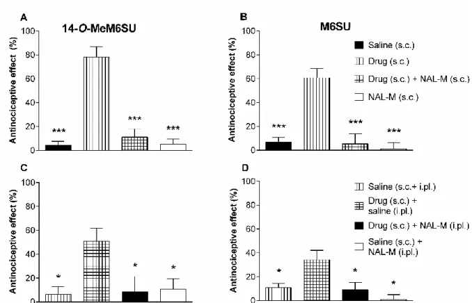
a 60-80% antinociceptive effect in inflamed paws, without a significant effect on the contralateral non-inflamed paws, were selected for experiments designed to analyze the antagonism by NAL-M. In these experiments NAL-M was co-administered with the test compounds when the peak effect was at 30 min, or 30 min after the agonist administration, when the peak effect was achieved at 60 min. In other series of experiments, NAL-M was injected locally 5 min prior to measurement (at 25 min or 55 min, when the peak effect was achieved at 30 and 60 min, respectively).
3.5. Neuropathic pain model: experimental paradigm for streptozocin induced chronic diabetic neuropathy in rats
Male Wistar rats of 200-300 g were used for STZ-induced diabetes model. Diabetes was provoked by i.p. administration of 60 mg/kg streptozocin (STZ) in a 2.5 ml/kg volume.
Vehicle treated group was used as absolute control.
The blood glucose level, the weight change, the consumption of water- and food was checked at numerous time points.
To justify the alteration in mechanical pain thresholds caused by the difference in the weights of diabetic (STZ treated) and non-diabetic (vehicle treated) animals (i.e. higher weight results in higher threshold values, data not shown), weight match control animals were used. Weight match animals were handled and kept under the same conditions described for the diabetic (and non-diabetic control) animals. The only exception was that, weight match animals were kept only for one or two weeks prior to experiments.
3.5.1. Monitoring of diabetic and control animals
Measuring the blood glucose levels: The rat blood glucose levels were determined by Accu-Chek Active blood glucose meter (Roche Diagnostics GmbH, Germany). The animals were slightly anesthetized with 3% isoflurane in oxygen via nose cone and one drop of blood was taken from the tail veins. The maximum concentration of blood glucose that is measurable is 33.3 mmol/l therefore this value was used as a maximum. The animals were considered diabetic if the value was above 14 mmol/l [78]. The blood glucose was measured before and 72 hours after the STZ treatment and on the 1st, 2nd, 3rd, 9th and 12th week after the STZ injection. Vehicle treated group was used as absolute control.
22
Measuring of animal weight and food and water consumption: In the first series of experiments control (vehicle treated) and diabetic (STZ treated) animals were kept individually. The water and food consumption of the animals was measured separately for each animal before and after the STZ treatment each day for 4 weeks and at least two times a week until the 7th week. The weight of the animals was checked during the 7 week period in the same way.
Gastric emptying assay: Phenol red method was performed as described earlier [84], with some minor modifications. Briefly, after 24 h of fasting, diabetic and non-diabetic rats received 1.5 ml of 1.5% methylcellulose solution containing 0.5 mg/ml phenol red (a non-absorbable marker compound) by intragastric gavage. After 20 min the rats were sacrificed, the pylorus and cardia were clamped and the stomach was removed. The content of stomach was mixed with 40 ml of 0.1 N NaOH, then 0.6 ml of this mixture was added to 1.2 ml of 7.4% trichloroacetic acid solution to precipitate proteins.
Following centrifugation (15 min, 3000 g) 1.2 ml of the supernatant was added to 0.6 ml of 1 N NaOH, and the absorbance was read spectrophotometrically in triplicates at 560 nm. Gastric emptying (%) was calculated as follows: [1 – (absorbance of sample / maximal absorbance)] x 100. Maximal absorbance was measured by processing the test meal alone, as described above.
In a separate experiment, weight-matched animals were treated either with saline, or with clonidine (0.1 mg/kg) subcutaneously 30 min before the methylcellulose administration.
Clonidine is an alpha2 adrenoceptor agonist, which is a well-known inhibitor of gastric emptying [85].
Assessment of neuropathic pain with Dynamic Plantar Aesthesiometer (DPA): In order to determine the allodynia caused by advanced diabetes we used the Dynamic Plantar Aesthesiometer (DPA) (Ugo Basile, Italy) as described previously [86] with slight modifications based on our pilot experiments. The animals were placed in the plastic cages of the DPA once daily for 3 subsequent days (“handling”). In addition before the experiments the animals were kept in the cages also for 5 min before starting the measurement in order to habituate them. The equipment raises a straight metal filament with a 0.5 mm diameter until it touches the paw. Then it puts pressure on the paw with an increasing force from 1 to 50 grams (cut off).
23
The withdrawal threshold was measured and expressed in grams, before and after the STZ treatment on every 3rd week. In the first series of experiments the animals were measured every week after STZ treatment in order to determine the peak of allodynia. Each of the hind paws were measured 3 times alternately and the average data of the 3 measurements were used for each animal. Both vehicle treated and weight match (i.e. animals with weights matching the diabetic ones) groups were used as control. After determination the time point of the peak effect further analyses were made at this time point. An animal was considered neuropathic, when the threshold value was decreased at least by 20%
compared to weight match animals as prescribed previously [87]. Diabetic rats that did not develop allodynia by the 9th week were excluded from further experiments.
3.5.2. Investigation of the analgesic action of different opioids in rats with diabetic neuropathy
Data were obtained 9 and 12 weeks following STZ treatment i.e. 6 and 9 weeks after the appearance of allodynia, a major sign of painful diabetic neuropathy. The baseline of withdrawal thresholds was measured before s.c. agonist treatment. The antiallodynic action of test compounds was assessed 30, 60 and 120 min after treatment. On the 9th week after STZ treatment dose-response curves were constructed. Data (100%: mean baseline values of diabetic or non-diabetic rats) were converted to log dose units and were analyzed with linear regression. The effective doses producing 30% effect (ED30; 30%
elevation in the threshold after treatment in comparison with the baseline) were determined. Since DPA elevates pressure on the paws only until 50 grams to avoid tissue damage, ED50 values could not be accurately determined. To analyze the changes in antinociceptive potency of test compounds the calculated ED30 values were compared (ED30diabetic/ED30non-diabetic). Weight match animals were used as absolute control in DPA measurement. In another set of experiments the opioid antagonist, NAL-M was co- administered with the investigated agonist.
3.6. Experimental paradigms for the assessment of side effects of test compounds 3.6.1. Determination of the effect of test compounds on gastrointestinal transit The effect of 14-O-MeM6SU and M6SU compared to that of morphine on gastrointestinal transit was measured in vivo, by using the charcoal meal method [88]. Briefly, male
24
NMRI mice (20-25 g) were fasted 6 h prior to the experiments, with free access to water.
At the time of the experiment, a charcoal suspension (10% charcoal in 5% gum arabic) was given in a volume of 0.25 ml/mouse by an oral gavage, followed by s.c.
administration of test compounds (0.1 ml/10 g). Then, 30 min after drug or saline administration mice were decapitated, their small intestines were removed, and the distance travelled by the charcoal suspension was expressed as a percentage of total small intestine length. The doses caused 50% inhibition on gastrointestinal transit (ID50) were calculated from the linear regression of dose-response curves.
3.6.2. Respiratory function tests
Respiratory function measurements were performed by unrestrained whole-body plethysmography (WBP) in conscious, spontaneously breathing male Wistar rats (200- 300 g) 30 and 60 min after s.c. injection of saline, 14-O-MeM6SU, M6SU or morphine.
Rats were placed in the chamber of a whole-body plethysmograph (PLY 3213, Buxco Europe Ltd., Winchester, UK). The flow transducers (TRD5700, Buxco Europe Ltd., Winchester, UK) were connected to the preamplifier module, which digitized the signals via an analog-to-digital converter (MAX2270 Buxco Europe Ltd., Winchester, UK).
Ventilation parameters (frequency: f, tidal volume: TV, minute ventilation: MV, time of inspiration: Ti, peak inspiratory time: PIF, time of expiration: Te, peak expiratory flow:
PEF and relaxation time: RT) were measured every 10 seconds during the 15-minute- long acquisition times and averaged by the BioSystem XA Software for Windows (Buxco Research Systems).
3.6.3. Determination of sedative effects of test compounds
To assess the anesthesia potentiating effect of test compounds righting reflex method was applied in male Wistar rats (200-300 g). The sleeping time (righting reflex, when the animals turned back on all four legs) was determined. To strengthen our results the novel compound’s potentiating effect was investigated with two different anesthetics of different routes of administration (i.v. and inhalational) and different pharmacokinetic properties.
Thiobutabarbital-induced sleeping time: Animals received i.v. saline or thiobutabarbital (153 µmol/kg), then were placed on the left side on a pillow of 30 C.
25
The sleeping time in minutes was documented. Anesthesia-potentiating effects of test drugs were studied by their s.c. administration. Thiobutabarbital was i.v. injected at the time of peak antinociceptive action of test compounds (60 min after s.c. 14-O-MeM6SU and 30 min after s.c. M6SU).
Isoflurane-induced sleeping time: The sleeping time was induced by inhaled 3%
isoflurane in oxygen for 2 min with a 2 liters/minute flow rate via nose cone using a vaporizer (Eickemeyer Isoflo Vaporiser; Eickemeyer Veterinary Equipment Inc). The sleeping time was determined as described above. The animals were treated s.c. with 14- O-MeM6SU or morphine 60 or 30 min before inhaled anesthetic, respectively. Control groups were treated with saline.
3.6.4. Induction of tolerance in mice
Male NMRI mice (18–28 g; Toxicoop, Hungary) were used. Radiant heat tail flick test was used to assess antinociceptive effect of test compounds. The assay was carried out as described by Tulunay and Takemori [79] using an ITC Life Sciences equipment. Briefly, the light intensity was adjusted to set the control tail-flick latency between 1.3 and 2.8 sec. Mice failed to respond within this range were excluded from the experiments. Cut off time was set to 6 s to avoid tissue damage. A baseline latency was measured before and 30, 60, 120, 180 min after s.c. drug or saline administration. A saline treated group was used for each experiment as control. Maximal possible effect (MPE) % was calculated for each mouse as follows: [Latency after treatment–Basal Latency]/[Cut off–
Basal Latency] × 100%. For each dose a separate group of animals (n = 5–12) was used.
Mice were rendered tolerant to morphine hydrochloride (200 µmol/kg) or 14-O- MeM6SU (12 µmol/kg) by twice daily (7 AM and 7 PM) s.c. injections for 3 days. Saline injections (10 ml/kg twice daily) were used in the control animals. The degree of tolerance was determined as the ratio of the ED50 value of the agonist in morphine or 14- O-MeM6SU injected mice to that in saline injected mice. The experiment was done on the fourth day as described previously [89]. Tolerance inducing treatment dose of 14-O- MeM6SU was calculated by using the following equation: [morphine dose] × [14-O- MeM6SU naive ED50]/ [morphine naive ED50].
Dose response curves for each drug were determined on the fourth day, following three days of chronic s.c. saline or drug treatments. Dose response curves for morphine and
26
14-O-MeM6SU were constructed in morphine or 14-O-MeM6SU treated mice. Control ED50 values for morphine and 14-O-MeM6SU were calculated from dose response curves constructed in saline treated mice. At least three groups of 5–12 mice were used to establish dose-response curves and to estimate ED50 values.
MPE% was calculated in MS Excel; for the further analysis Graph-Pad Prism 5.0 software was used. ED50 values and confidence intervals were calculated by non-linear fit (normalized response – variable slope). Differences in ED50 values were considered significant when confidence intervals did not overlap.
3.7. In vitro receptor binding assays
Male Wistar vehicle treated or STZ treated (diabetic) rats (250-400 g) were used.
Animals were decapitated and their brains and whole spinal cords were quickly removed and prepared for receptor binding assays as previously reported [90, 91].
3.7.1. Radioligand competition binding assay
The experiments were carried out as described previously [92, 93]. Specific binding of [3H]DAMGO was performed by incubating 50-100 μg of membrane protein of lumbar dorsal horn with 0.1- 4 nM [3H]DAMGO in the presence or absence of 10 μM unlabelled naloxone to determine non-specific binding. Membranes were incubated for 1 hour at 22
°C in assay buffer. The reactions were terminated by rapid filtration under vacuum through Whatman GF/B glass fibre filters, followed by four washes with cold buffer (50 mM Tris–HCl, pH 7.4). Bound radioactivity was determined by liquid scintillation spectrophotometry (Perkin Elmer, Rodgau, Germany) at 60% counter efficiency after overnight extraction of the filters in 3 ml of scintillation fluid. Unbound radioligand was separated from the receptor bound radioligand by rapid filtration under vacuum through glass fiber filter. The radioactivity of the specifically bound radioligands was determined by liquid scintillation spectrophotometry.
All experiments were performed in duplicate and carried out at least five times. Bmax and Kd values in saturation binding assays were determined by nonlinear regression analysis of concentration-effect curves using GraphPad Prism (GraphPad Software Inc., CA, USA).
27 3.7.2. Immunohistochemistry
Rats were deeply anaesthetized with isoflurane and transcardially perfused with 100 ml of phosphate buffered saline (PBS) pH 7.4, then followed by 500 ml of 4% (w/v) paraformaldehyde in phosphate buffer pH 7.4. After perfusion, DRG and spinal cord were removed from STZ treated and control animals, postfixed in the same fixatives for 90 min, and then cryo-protectedovernight at 4°C in PBS containing 10% sucrose. DRGs (10
m thick) were mounted onto gelatin coated slides. DRG mounted or spinal cord floating tissue sections were incubated with the following primary antibody rabbit polyclonal MOR antibody (1:1,000, Gramsch Laboratories, Schwabhausen, Germany). The tissue sections were washed with PBS prior to incubation with Alexa Fluor 594 donkey anti- rabbit secondary antibody (Invitrogen, Germany). Finally, the tissues were washed in PBS, mounted on vectashield (Vector Laboratories, Burlingame, CA) and viewed under a Zeiss LSM 510 laser scanning microscope (Carl Zeiss, Göttingen, Germany). To demonstrate specificity of staining, the following controls were included as mentioned in detail elsewhere [94, 95]: 1) Preabsorption of the primary antibody against MOR was verified by preabsorption with 5 µg/ml of synthetic peptide antigen for MOR (Gramsch Laboratories, Germany), for 24 hours at 4oC; 2) Omission of either the primary antisera or the secondary antibodies.
3.7.3. G-protein activity assay
In [35S]GTPγS binding experiments the GDP→GTP exchange of the Gαi/o protein is measured by a radioactive, non-hydrolysable GTP analog, [35S]GTPγS. First the brain and spinal cord were homogenized, centrifuged in ice-cold 50 mM Tris-HCl (pH 7.4) buffer and incubated at 37 oC for 30 min in a shaking water-bath (for details see [96]).
After incubation the centrifugation was repeated as described before and the final pellet was suspended in ice-cold TEM (Tris-HCl, EGTA, MgCl2) buffer and stored at –80 oC for further use. The nucleotide exchange is measured in the presence of a given ligand in increasing concentrations to measure ligand potency and the maximal efficacy. The experiments were carried out as described previously [97]. Agonist potency and maximum G-protein stimulation of reference and test compounds (see results section) were investigated in [35S]GTPγS binding assays. In the diabetes induced neuropathic pain model control and STZ treated rat whole brain, spinal cord and lumbar dorsal horn
28
membrane homogenates were examined for MOR activity. [35S]GTPγS was incubated together with the appropriate membrane homogenate together with increasing concentrations of unlabeled reference and test compounds, with excess GDP in a special buffer containing Tris, MgCl2, EGTA, NaCl, and dithiothreitol.
Total binding was measured in the absence of the test compounds, while non-specific binding was determined in the presence of 10 µM unlabeled GTPγS. The bound and unbound [35S]GTPγS were separated by rapid filtration under vacuum (Brandel M24R Cell Harvester), and washed three times with 5 ml ice-cold 50 mM Tris-HCl through Whatmann GF/B glass fibers (GE Healthcare Life Sciences through Izinta Kft., Budapest, Hungary). [35S]GTPγS binding experiments were performed in triplicates and repeated at least three times. Bound radiochemical separation and radioactivity detection were the same as discussed in radioligand competition binding assays.
3.8. Analysis of data
All the analysis was performed with a professional statistical software: GraphPad Prism version 5.00 or 6.00 for Windows (GraphPad Software, San Diego, California, USA, www.graphpad.com).
For the comparison of more than 2 groups one- or two-way ANOVA was applied with post hoc test based on the experimental setup (see Results section). Vehicle treated groups were used as control in order to decide if the applied treatment significantly influenced the parameters.
All data are represented as mean ± SEM. The results were considered to be statistically significant when p < 0.05.
29
4. Results
4.1. Acute inflammatory pain models 4.1.1. Mouse writhing test
4.1.1.1. Antinociceptive effects of 14-O-MeM6SU compared to M6SU
Injection of 0.6% acetic acid solution into the peritoneal cavity of mice administered s.c.
or i.c.v. saline resulted in an average of 43.9 ± 1.5 (n = 43) writhes during the 10 min observation period. As shown in Fig. 2., s.c. or i.c.v. administration (20 min before testing) of both agonists produced a dose-dependent antinociceptive action. The calculated ED50 values with 95% confidence intervals at 20 min are shown in Table 2.
14-O-MeM6SU produced more potent inhibitory effect than M6SU on acetic acid- induced writhing in mice both after s.c. and i.c.v. routes (Table 2).
After s.c. administration, 14-O-MeM6SU was 23- fold more potent than M6SU, while after i.c.v. administration 14-O-MeM6SU was proved to be only 5-fold more active than M6SU in inhibition of writhing. The calculated large s.c./i.c.v. potency ratios for M6SU or 14-O-MeM6SU indicate limited CNS penetration (Table 2).
Table 2. Antinociceptive potencies of 14-O-MeM6SU and M6SU against acetic acid induced nociception in mouse writhing test after 20 min of s.c. or i.c.v. administration.
Compound ED50 (nmol/kg, s.c.) ED50 (pmol/mouse, i.c.v.) s.c./i.c.v.a
M6SU 1993
(1282–3101)
9 (15-54)
221444
14-O-MeM6SU 87
(47–163)
1.7 (0.3-9.5)
51177 At least 4 animals per dose group and 3-4 doses were used for each ED50
determinations.
as.c./i.c.v. was calculated as the ratio of ED50 nmol/kg, s.c./ED50 pmol/mouse, i.c.v
30
Figure 2. Antinociceptive dose-response curves of s.c. (panel A) or i.c.v. (panel B) injected 14-O-MeM6SU and M6SU in mice writhing test.
Points represent the mean ± S.E.M. (n= 4-5)
31
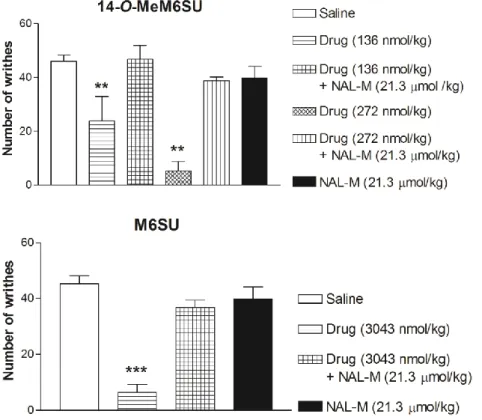
4.1.1.2. NAL-M antagonism on systemic 14-O-MeM6SU or M6SU antinociception To evaluate the opioid specificity and the site of action of 14-O-MeM6SU and M6SU in acetic acid-induced writhing in mice, the effects of the agonists were assessed after systemic co-administration with the quaternary opioid antagonist NAL-M (21.3 µmol/kg, s.c). Our results show that s.c. equipotent analgesic dose of 14-O-MeM6SU (136 nmol/kg), and M6SU (3043 nmol/kg) significantly decreased the number of writhes at 20 min after administration. Co-administration of NAL-M antagonized the antinociceptive effect of the test opioids, as presented in Fig. 3. NAL-M treatment alone failed to affect the number of writhes (Fig. 3). Higher dose of 14-O-MeM6SU (272 nmol/kg) showed also NAL-M reversible antinociception (Fig. 3).
Figure 3. The antagonist action of co-administered NAL-M (21.3 µmol/kg) on the antinociceptive effect of 14-O-MeM6SU or M6SU after 20min s.c. administration in the
writhing response induced by acetic acid (i.p.) in mice.
Data are expressed as mean ± SEM
*: significant difference versus other groups (**: p<0.01; ***: p<0.001) (one-way ANOVA, Newman-Keuls post hoc test).
32 4.1.2. Rat formalin test
4.1.2.1. Antinociceptive effect of 14-O-MeM6SU compared to morphine
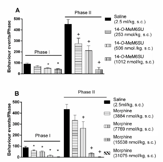
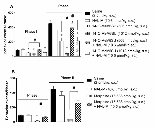
Subcutaneous 14-O-MeM6SU (253, 506 and 1012 nmol/kg) or morphine (3884, 7769, 15538 and 31075 nmol/kg) attenuated the formalin-induced pain in a dose-dependent manner (Fig. 4., panel A and B).
When both phases are considered, 14-O-MeM6SU in doses 506 and 1012 nmol/kg produced antinociception (Fig. 4., panel A), whereas 253 nmol/kg was effective only in the second phase. On the other hand, s.c. morphine caused antinociception in all tested doses in phase I and apart from the lowest dose in phase II (Fig. 4., panel B). Based on equianalgesic doses after systemic administration 14-O-MeM6SU was approx. 15 and 31 more potent than morphine in phase I and II, respectively.
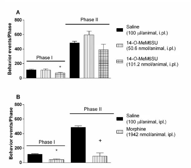
The locally (i.pl.) administered (into the ipsilateral paw) 14-O-MeM6SU at doses of (25.3, 50.6 and 101.2 nmol/rat) or morphine (971 and 1942 nmol/rat) were also tested.
The lowest dose that significantly reduced the pain in phase I was 25.3 nmol/animal for 14-O-MeM6SU and 1942 nmol/animal for morphine (Fig. 5.). When both phases considered, 14-O-MeM6SU alleviated the pain reactions in a dose of 50.6 nmol/rat and morphine only at the dose of 1942 nmol/rat (Fig. 5.). Based on equianalgesic doses after local administration 14-O-MeM6SU was approx. 77 and 38 more potent than morphine in phase I and II, respectively.
33
Figure 4. The antinociceptive effect of 14-O-MeM6SU and morphine after s.c.
administration in rat formalin test, in phase I (0-10 min) and phase II (11-60 min). Each column represents the cumulative data of the given phase (number of nociceptive
reactions). Drugs were administered in a 2.5ml/kg volume.
Each value represents the mean ± SEM.
*: significant difference vs. vehicle treated group in Phase I (p<0.05) +: significant difference vs. vehicle treated group in Phase II (p<0.05)
(one-way ANOVA followed by Fisher’s LSD post hoc test, n= 4-11)
34
Figure 5. The antinociceptive effect of 14-O-MeM6SU and morphine after local (i.pl.) administration in rat formalin test applying 50µl 2.5% formalin into the right hind paw,
in phase I (0-10 min) and phase II (11-60 min). Each column represents the cumulative data of the given phase (number of nociceptive reactions). Drugs were administered in
a 100 µl/animal volume.
Each value represents the mean ± SEM. (n=4-11)
*: significant difference vs. vehicle treated group in Phase I (p<0.05) +: significant difference vs. vehicle treated group in Phase II (p<0.05)
(one-way ANOVA followed by Fisher’s LSD post hoc test, n= 4-11)
35
4.1.2.2. NAL-M antagonism on systemic 14-O-MeM6SU or morphine antinociception
Co-administered opioid antagonist NAL-M (10.6 µmol/kg, s.c.) abolished the antinociceptive effect of s.c. 506 nmol/kg 14-O-MeM6SU in both phases (Fig. 6., panel A). On the other hand, NAL-M failed to antagonize the effect of 1012 nmol/kg 14-O- MeM6SU in phase I, yet partially affected the antinociceptive effect in phase II (Fig. 4, panel A).
In case of morphine, NAL-M antagonized its antinociceptive effect of (15 538 nmol/kg, s.c.) morphine in phase I (Fig. 6., panel B). In phase II NAL-M only partially antagonized the antinociceptive action of the same dose of morphine.
4.1.2.3. The antinociceptive effects of 14-O-MeM6SU or morphine after administration into the contralateral paw
Intraplantar (i.pl.) administration of 50.6 nmol/rat 14-O-MeM6SU into contralateral paw failed to affect formalin-induced pain in ipsilateral paw in either phases (Fig. 7., panel A), though it was effective when administered into ipsilateral (formalin treated) paw (Fig.
5.). At a higher dose (101.2 nmol/rat) 14-O-MeM6SU showed antinociception only in phase I. However, 1942 nmol/animal morphine injected into the contralateral paw (i.pl.) produced antinociceptive effect on both phases (Fig. 7., panel B). This effect is in accordance with that obtained following s.c. 7769 nmol/kg (Fig. 4., panel B).
The antagonist effect of NAL-M (section 4.1.2.2.) and these data indicate the presence of peripheral antinociceptive component of both compounds, but only 14-O-MeM6SU showed action that was of completely peripheral origin at the dose of 506 nmol/kg.
36
Figure 6. The antagonist effect of s.c. co-administered NAL-M (10.6 µmol/kg) on the antinociceptive effect of s.c. 14-O-MeM6SU (panel A) and morphine (panel B) in rat formalin test. Each column represents the cumulative data of the given phase (number
of nociceptive reactions). Drugs were administered in a 2.5 ml/kg volume.
Each value represents the mean ± SEM.
*: significant difference vs. vehicle treated group in Phase I (p<0.05) +: significant difference vs. vehicle treated group in Phase II (p<0.05)
#: significant difference between the signed groups (p<0.05) (one-way ANOVA followed by Fisher’s LSD post hoc test, n= 5-11)
37
Figure 7. The antinociceptive effect of 14-O-MeM6SU (panel A) and morphine (panel B) after administration into the contralateral paw. Drugs were administered in a
100 µl/animal volume. Each column represents the cumulative data of the given phase (number of nociceptive reactions). Each value represents the mean ± SEM.
*: significant difference vs. vehicle treated group in Phase I (p<0.05) +: significant difference vs. vehicle treated group in Phase II (p<0.05)
(one-way ANOVA followed by Fisher’s LSD hoc test in the case of 14-O-MeM6SU and unpaired t-test with two-tailed p value in the case of morphine; n= 4-5)