GUTS AND GALL: BILE ACIDS IN REGULATION OF INTESTINAL EPITHELIAL FUNCTION IN HEALTH AND DISEASE
Peter Hegyi, Jozsef Maléth, Julian R. Walters, Alan F. Hofmann, and Stephen J. Keely
Momentum Translational Gastroenterology Research Group, Hungarian Academy of Sciences–University of Szeged, Szeged, Hungary; Institute for Translational Medicine, Medical School, University of Pécs, Pécs, Hungary; Momentum Epithelial Cell Signalling and Secretion Research Group and First Department of Medicine, University of Szeged, Szeged, Hungary; Division of Digestive Diseases, Department of Gastroenterology,
Hammersmith Hospital, Imperial College London, London, United Kingdom; Division of Gastroenterology, Department of Medicine, University of California, San Diego, La Jolla, California; and Department of Molecular Medicine, Royal College of Surgeons in Ireland, Education and Research Centre, Beaumont Hospital, Dublin, Ireland
L
Hegyi P, Maléth J, Walters JR, Hofmann AF, Keely SJ.Guts and Gall: Bile Acids in Regulation of Intestinal Epithelial Function in Health and Disease. Physiol Rev 98:1983–2023, 2018. Published August 1, 2018; doi:10.1152/physrev.00054.
2017.—Epithelial cells line the entire surface of the gastrointestinal tract and its accessory organs where they primarily function in transporting digestive enzymes, nutrients, electrolytes, and fluid to and from the luminal contents. At the same time, epithelial cells are responsible for forming a physical and biochemical barrier that prevents the entry into the body of harmful agents, such as bacteria and their toxins. Dysregulation of epithelial transport and barrier function is associated with the pathogenesis of a number of conditions throughout the intestine, such as inflammatory bowel disease, chronic diarrhea, pancreatitis, reflux esophagitis, and cancer. Driven by discovery of specific receptors on intestinal epithelial cells, new insights into mechanisms that control their synthesis and enterohepatic circulation, and a growing appreciation of their roles as bioactive bacterial metabolites, bile acids are currently receiving a great deal of interest as critical regulators of epithelial function in health and disease. This review aims to summarize recent advances in this field and to highlight how bile acids are now emerging as exciting new targets for disease intervention.
I. INTRODUCTION 1983
II. BILE ACID PHYSIOLOGY 1984
III. EPITHELIAL CELL PHYSIOLOGY 1986 IV. BILE ACIDS AND EPITHELIAL SIGNALING 1989 V. BILE ACIDS AND EPITHELIAL FUNCTION 1992 VI. PATHOLOGICAL EFFECTS OF BILE ACIDS 1999 VII. THERAPEUTIC TARGETING OF ... 2005
VIII. CONCLUSIONS 2008
I. INTRODUCTION
Bile acids, classically known for their roles in facilitating the digestion and absorption of dietary lipids, are now also appreciated as a family of enteroendocrine hormones that have important roles in regulating many aspects of mam- malian physiology, both within and outside the intestinal tract. Driven largely by the discovery of two new bile acid receptors at the turn of the millennium, the past two de- cades have seen a renaissance in research activity that has firmly placed bile acids as being central to maintenance of our overall health. Disruptions to the processes that control the synthesis, recycling, and excretion of bile acids are as-
sociated with the onset of many diseases that can affect the intestine, its accessory organs, and beyond. Significant progress has been made in understanding various molecular mechanisms involved, and there is a rapidly growing inter- est in developing our capacity to target such pathways for disease treatment. Such an idea can hardly be considered as being new, given that as long as 2500 yr ago, bile was considered as comprising two of the four “humors” (black bile and yellow bile) upon which Hippocrates established his long-held system of human medicine (510), while ani- mal gall has been used for centuries in Traditional Eastern Medicine to treat many different ailments (483). However, along with the discovery of new receptors for bile acids, recent years have also seen a growing appreciation of how important these molecules are to human physiology in health and disease. This appreciation has sparked renewed interest in the potential use of bile acids as therapeutic agents.
Central to developing our understanding of how bile acids can be therapeutically exploited is an understanding of how they interact with epithelial cells throughout the intestinal tract. These cells line the entire gut and its accessory organs
and, as the interface between the body and the luminal contents, they are primary effectors of bile acid-induced responses, whether they be intestinal, hepatic, or metabolic.
While other reviews have covered recent developments with respect to bile acids in regulation of hepatic and metabolic function (97, 111, 151, 252, 370, 463), the current manu- script aims to provide an overview of how luminal bile acids interact with gastrointestinal epithelial cells, the implica- tions of such interactions for our physiology, and how we can ultimately target these fascinating molecules for the treatment of disease.
II. BILE ACID PHYSIOLOGY
A. Bile Acid Synthesis and Metabolism
Since this review is primarily concerned with the physiolog- ical effects of bile acids on extrahepatic epithelia, we aim to include only a broad overview of the processes involved in their biosynthesis and metabolism. For more detail, readers are directed to excellent reviews previously published on this topic (328, 390). Essentially, epithelial cells along the intestinal tract can come into contact with either primary and/or secondary bile acids, which are either in their con- jugated or unconjugated forms. Primary bile acids are syn- thesized from cholesterol by hepatocytes in the liver through complex enzymatic pathways, with the rate-limit- ing step being the enzyme CYP7A1. In humans, the primary bile acids are cholic acid (CA) and chenodeoxycholic acid (CDCA). However, it should be noted that other primary bile acids are synthesized in different species, such as muricholic acid in mice and hyocholic acid in pigs (168), and that these bile acids can have dramatically different bioactivities from those of CDCA and CA. Thus it is impor- tant to keep this in mind when translating experimental data from animal models into humans.
After their biosynthesis, primary bile acids are conjugated to either taurine or glycine. In most vertebrates, conjugation is with taurine, but in humans, glycine conjugation predom- inates. Glutathione conjugates have also been identified in bile, but only in trace amounts (303). Conjugation involves formation of a bile acid coenzyme A ligase (which activates the carboxyl group) and an amino transferase, in which the CoA thioester links the carboxyl group of the bile acid to the amino moiety of taurine or glycine to form a stable amide bond. Conjugation lowers the pKaof bile acids and therefore has important consequences for their physico- chemical properties and their biochemical and physiologi- cal actions, making them fully ionized at the pH of luminal contents of the proximal small intestine (170, 385). Thus, unless a transporter is present, conjugated bile acids are impermeable to epithelial cell membranes, permitting high luminal concentrations to be achieved in the biliary tract and proximal small intestine, thereby enabling micellar sol- ubilization of dietary lipids to occur (409).
Within the intestinal lumen, particularly in the colon, pri- mary bile acids are extensively metabolized by the resident microbiota into secondary bile acids, first by hydrolysis of their amide bond and subsequently by modifications to the hydroxy groups on their steroid nucleus (383). Deconjuga- tion is the gateway reaction to further bile acid metabolism and is carried out by bile salt hydrolases (BSH), enzymes that are widely expressed by gram positive, and some gram negative, bacteria within the gut lumen. Several isoforms of BSH exist which differ in their substrate specificities but which tend to have a higher affinity for glycine-conjugated bile acids (58, 200, 265). Resistance to bile acid toxicity is an important characteristic enabling bacterial survival within the gut lumen, and different species have different sensitivities to conjugated and deconjugated bile acids.
Therefore, the pattern of expression of BSHs plays a vital role in shaping the makeup of the luminal microbiota, while at the same regulating the capacity of bile acids to penetrate epithelial membranes, activate their receptors, and induce biological responses.
Upon deconjugation, the hydroxyl groups at positions 3, 7, and 12 of the steroid nucleus can then be subjected to me- tabolism by a number of bacterial enzymes, including de- hydroxylases, dehydrogenases, and epimerases. For exam- ple, CA is dehydroxylated at position 7, to yield DCA, a dihydroxy bile acid, whereas 7-dehydroxylation of CDCA yields LCA, a monohydroxy bile acid. DCA and LCA are normally the two most common of the colonic bile acids in humans (152). The 7-hydroxy group of CDCA can also by epimerized (7␣-hydroxy to 7-hydroxy) to yield UDCA, which compared with other dihydroxy bile acids is rela- tively hydrophilic and has very distinct biological proper- ties. However, UDCA is normally present at relatively low levels since epimerization at C-7 is mediated by only a few bacterial species and it is also rapidly metabolized by bac- terial hydroxysteroid dehydrogenases to LCA, a reversible reaction with 7-keto-LCA being an important intermediate.
Bile acids can also undergo epimerization at the C-3 hy- droxyl group during transit through the intestine, with such 3-hydroxy bile acids being termed “iso” bile acids;
isoCDCA, isoDCA, and isoLCA are present in the colon but upon reabsorption and recirculation to the liver, they un- dergo re-epimerization to their 3␣-hydroxy counterparts (169). The bacterial enzymes mediating alterations to bile acid hydroxyl groups are less widely expressed than BSHs, but their actions are also critically important in determining their membrane permeability, cytotoxicity, and receptor se- lectivity. Readers wishing to learn more detail of bile acid metabolism and nomenclature are directed to previously published reviews (172, 383).
B. Enterohepatic Cycling of Bile Acids
The enterohepatic circulation (EHC) of bile acids is an ex- tremely complex and elegant process that includes their
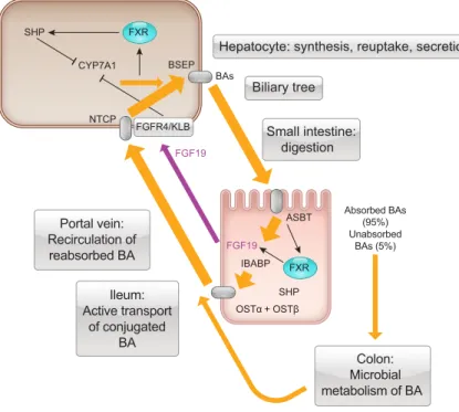
synthesis in the liver, storage in the gallbladder, transit through the intestinal tract, bacterial metabolism, reab- sorption from the small intestine and colon, transport via the portal circulation to the liver, and reuptake into hepatocytes(FIGURE 1). In the liver, recirculated bile ac- ids can be further metabolized by hepatic enzymes and reconjugated to glycine and taurine after which they are then secreted into the bile and recirculated to the gall- bladder for storage. Efficient enterohepatic cycling en- sures that a relatively constant-sized pool of bile acids is available to facilitate lipid digestion and absorption, rather than de novo synthesis being required each time food is ingested. The amount of bile acids in the EHC can be calculated by isotope dilution (261) and in humans is in the range of 2,000 –3,000 mg, which is approximately six times the daily synthesis rate (300 –500 mg/day). Nor- mally, the size of the circulating bile acid pool is kept relatively constant with fecal loss being balanced by he- patic synthesis.
After bile acids enter the small intestine, they perform their classical functions in aiding the digestion and absorption of fats. Absorption of bile acids themselves from the intestinal lumen occurs by several mechanisms. Glycine-conjugated dihydroxy bile acids (glyco-DCA and glyco-CDCA) are rel- atively hydrophobic and can undergo passive absorption from the duodenum when luminal contents are transiently acidic. These glycine-dihydroxy bile acids can also be ab- sorbed from the jejunum via organic anion transporting polypeptide (OATP) transporters (144). Postprandial levels of CDCA conjugates increase ahead of cholyl conjugates, indicating the CDCA conjugates are more efficiently ab- sorbed in the proximal small intestine (398).
Although reabsorption in the proximal small intestine con- tributes to the EHC of bile acids, the most important reab- sorptive process occurs in the terminal ileum, where epithe- lial cells express the apical sodium-dependent bile acid transporter, ASBT (also known as the ileal bile acid trans- porter, IBAT). In contrast to other transporters involved in the EHC (e.g., OST␣/OST, NTCP, BSEP) which can trans- port non-bile acid molecules, ASBT transports only bile acids and has a greater affinity for those that are conjugated over those that are not. Thus it is ASBT which confers the specificity of the EHC for bile acids (89, 91).
Following uptake by ASBT, bile acids traverse the cyto- plasm of the epithelial cells bound to ileal bile acid binding protein (IBABP) and exit the basolateral domains of the enterocyte via the heterodimeric protein, OST␣/OST, a facilitated diffusion transporter that is present throughout the small intestine (89, 90). Once in the interstitium, it is unclear how conjugated bile acids enter villus capillaries to gain access to the portal circulation. However, as capillary endothelial cells are fenestrated, similar to hepatic endothe- lial cells, entry of bile acids into the capillary plexus likely occurs by a passive process. Once in the portal venous blood, bile acids are transported bound to albumin, with dihydroxy conjugates binding more tightly (⬎90%) than conjugates of cholic acid (60 – 80%) (337).
Upon deconjugation in the terminal ileum and colon, bile acids become more hydrophobic and are primarily reab- sorbed by passive diffusion across the epithelium. Absorp- tion of DCA is 20 –50% of that formed (462), while that of lithocholic acid is less, presumably because its hydropho- bicity promotes binding to unabsorbed dietary constituents
Hepatocyte: synthesis, reuptake, secretion
Ileum:
Active transport of conjugated
BA
Colon:
Microbial metabolism of BA
BSEP BAs
NTCP SHP
FGFR4/KLB FGF19 CYP7A1
FXR
FXR ASBT
OSTα + OSTβ IBABP FGF19
SHP
Small intestine:
digestion Biliary tree
Absorbed BAs (95%) Unabsorbed
BAs (5%)
Portal vein:
Recirculation of reabsorbed BA
FIGURE 1. Enterohepatic circulation of bile acids. Bile acids (BAs) are synthesized in the liver with the enzyme, CYP7A1, being the initial step. They are transported from the liver by the bile salt export pump (BSEP) and travel via the biliary tree to the gallbladder for storage. Upon ingestion of a meal, bile acids are ejected into the small intestine, where they facilitate lipid di- gestion and absorption. In the ileum, active reuptake of conju- gated bile acids occurs via the apical bile salt transporter (ASBT), expressed in the brush border of terminal ileal epithe- lial cells. In the cytoplasm, bile acids bind to ileal bile acid binding protein (IBABP) and are transported from the ileal enterocyte via the basolateral heterodimeric protein OST␣/.
Bile acids enter the portal venous circulation and return to the liver, where reuptake takes place via the Na⫹-taurocholate polypeptide (NTCP). In both enterocytes and hepatocytes, BAs bind to FXR in the nucleus, stimulating transcription of proteins including SHP and FGF19. Approximately 5% of ileal BAs are not absorbed and enter the colon where they are metabolized into secondary bile acids by the microbiota, with some being reabsorbed and recycled.
(79). When these unconjugated bile acids return to the liver, they are reconjugated with glycine or taurine and secreted into canalicular bile. In some species, deoxycholic acid un- dergoes 7-hydroxylation to form cholic acid which is then conjugated with taurine or glycine. Additionally, in humans but not in rodents, a major fraction of lithocholic acid is esterified with sulfate after reconjugation. Such sulfolitho- cholyl amidates are secreted into the bile but cannot be absorbed from the small intestine, with the result that LCA is rapidly eliminated from the body. Thus LCA constitutes
⬍5% of biliary bile acids, with higher proportions being associated with hepatotoxicity, at least in animals.
Hepatic uptake of bile acids remains constant during meals, and under normal circumstances of feeding and fasting, it is likely to remain far below itsVmax. Uptake of bile acids is greatest in the periportal hepatocytes and is more efficient for conjugated than unconjugated molecules (337). Conju- gated bile acid uptake is mediated mostly by the sodium/
taurocholate cotransporting polypeptide (NTCP, gene name SLC10A1), which has considerable homology to ASBT, and like ASBT, uses the transepithelial sodium gra- dient to drive uptake. After they have been taken up into the liver, bile acids are then secreted into the bile via an ATP- driven transporter, known as the bile salt export pump (BSEP). This protein is present in the canalicular membrane of hepatocytes and transports bile acids against a steep con- centration gradient. The importance of NCTP and BSEP in the EHC is underscored by congenital disorders in which they are dysfunctional, with absence of NCTP causing markedly elevated plasma levels of bile acids (468), and inborn errors of BSEP being characterized by bile acid re- tention, hepatocyte death, and inflammation (187).
Each stage of the EHC, from hepatic synthesis, to intestinal reabsorption, and reuptake into hepatocytes, is intricately regulated by complex pathways of cellular and molecular communication along the gut-liver axis. Importantly, bile acids themselves play a major role in regulating this process, primarily through their actions on farnesoid X receptor (FXR) expressed in hepatocytes and epithelial cells lining the intestinal lumen. FXR reversibly binds bile acids, with CDCA being the most active natural agonist (273, 345).
When activated by bile acids, FXR forms a dimer with the retinoic acid receptor (RXR) and binds to specific FXR- responsive elements in target genes, several of which are involved in bile acid metabolism and transport. In the liver, a major FXR target gene that regulates CYP7A1 expression is short heterodimer partner-1 (SHP). SHP itself does not bind DNA but instead inhibits the activity of liver receptor homolog 1 (LRH-1), thereby reducing CYP7A1 expression and bile acid synthesis (140).
A number of other FXR-dependent pathways are also in- volved in regulating bile acid synthesis and transport in the liver and have been reviewed elsewhere (69, 88, 229) In
humans, ileal ASBT expression is downregulated by activa- tion of FXR, whereas IBABP and the basolateral transport- ers, OST␣and OST, are upregulated, effects that decrease the concentration of bile acids within the ileal enterocyte (125, 284, 519). However, the most prominent and best- characterized of the FXR responsive genes in the ileum is fibroblast growth factor 19 (FGF19), and its homolog FGF15 in rodents. These are proteins of ~24 kDa that are secreted from the basolateral aspect of ileal enterocytes into the portal venous blood. FGF15/19 then travels to the liver where it binds to FGF receptor 4 (FGFR4) and its co-recep- tor, Klotho-beta, on hepatocytes to inhibit CYP7A1 via a MAPK-dependent signaling mechanism (424). This pro- vides an elegant system of negative-feedback regulation of bile acid synthesis, whereby bile acid reabsorption in the ileum signals through the gut-liver axis to inhibit de novo synthesis in the liver. FGF19 levels peak in blood several hours after a meal and are inversely related to those of the bile acid precursor C4 (267), the plasma levels of which increase in direct relationship to the rate of bile acid synthe- sis. Under pathological conditions, such as those causing bile acid diarrhea, defective production of FGF15/19 leads to a loss of this negative-feedback loop, with increased syn- thesis resulting in epithelial cells along the upper and lower intestinal tract being exposed to considerably higher levels of bile acids (482).
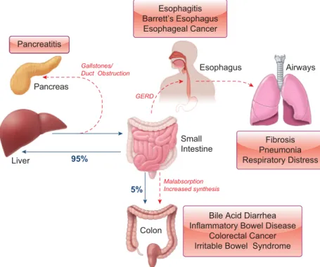
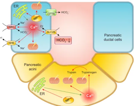
Other pathological conditions can also lead to disruptions in the EHC with the consequence that epithelial cells, both within and outside of the intestinal tract, become exposed to bile acids. For example, conditions associated with mal- absorption of bile acids in the terminal ileum (e.g., Crohn’s disease) cause their increased delivery into the colon. On the other hand, in gastroesophageal reflux disease (GERD), the EHC leaks upwards with the result that cells of the esoph- agus and airways can become exposed to bile acids. Simi- larly, common channel obstruction can cause bile acids to enter the pancreas where they come into contact with the epithelial cells lining the ducts and acini. Not surprisingly, such conditions can dramatically alter the physiology of these cells leading to the onset of disease.
III. EPITHELIAL CELL PHYSIOLOGY
Before we consider how they are regulated by bile acids, in this section we review the basic functions and physiological characteristics of intestinal epithelial cells. These cells, lin- ing the surfaces of the entire intestinal tract and its acces- sory organs, are exposed to constantly changing luminal conditions and must have the capacity to adapt appropri- ately. For example, in the intestine, the epithelium must be able to evoke rapid responses upon ingestion of a meal. This is achieved through the release of hormonal messengers, secretion of digestive enzymes, and expression of transport proteins that enable the uptake of fluid and nutrients from the lumen. At the same time, the epithelia must also act as
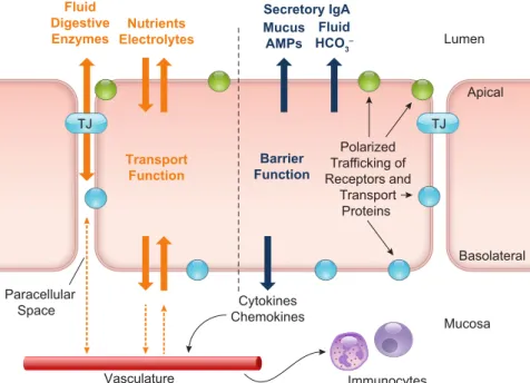
barriers that protect against the entry of harmful sub- stances, whether they originate endogenously (e.g., gastric acid, digestive enzymes, bile acids) or are ingested (e.g., bacteria, viruses, fungi, and their toxins). Given the widely diverse functions of different intestinal regions and the dif- ferent stimuli to which they are exposed, it is not surprising that epithelial cells express regional and organ-specific characteristics. However, throughout the intestine, epithe- lial cells also have common characteristics that are essential to their fundamental barrier and transport functions(FIG- URE 2).
Epithelial cells in the intestinal tract originate from stem cells located near the base of crypts. Stem cells give rise to progenitor cells which, in turn, develop into one of the four main types of epithelial cell; enterocytes, goblet cells, and enteroendocrine cells migrate upwards from the stem cell niche, while Paneth cells migrate downwards towards the crypt base. Enterocytes are the most common of the intes- tinal epithelial cells and are responsible for the surface hy- drolysis and uptake of nutrients, absorption and secretion of electrolytes and fluids, and the conservation of bile acids.
Goblet cells secrete mucus which promotes intestinal bar- rier function, protecting against pathogen invasion and
physical damage due to peristalsis (288). Paneth cells se- crete antibacterial peptides, most notably defensins and ly- sozyme, which are important in intestinal defense against bacterial infection. Paneth cells are also important in main- tenance of the stem cell niche (75). Enteroendocrine cells serve to “taste” the luminal contents and release hormones and neurotransmitters that enable coordination of both lo- cal and systemic responses to the presence of nutrients in the intestine (270). A subset of enteroendocrine cells, the L cells, can also sense bile acids in the gut lumen through their expression of TGR5 and FXR, with signals from these re- ceptors regulating the expression of the incretin hormone, glucagon-like peptide-1 (GLP-1). Such actions link changes in luminal bile acids to changes in our metabolism and energy expenditure and are the basis for a great deal of ongoing research into the potential for targeting bile acids in treatment of metabolic diseases, such as diabetes and obesity. This exciting and complex area of research is be- yond the scope of the current review, and we direct the reader to several recent publications for further information (47, 146, 344, 431, 450, 464).
The ability of epithelial cells to form barriers and to vecto- rially transport nutrients, fluid, and ions is dependent on
Mucosa Mucus
AMPs Fluid HCO3– Fluid
Digestive Enzymes
Cytokines Chemokines Paracellular
Space
Polarized Trafficking of Receptors and
Transport Proteins Transport
Function
Lumen
TJ TJ
Barrier Function Nutrients
Electrolytes
Vasculature Immunocytes
Apical
Basolateral Secretory IgA
FIGURE 2. General characteristics and functions of intestinal epithelial cells. The primary functions of epithelial cells throughout the intestinal tract are to transport nutrients, electrolytes, digestive enzymes, and fluid to and from the lumen, while at the same time acting as a protective barrier to prevent the entry of harmful pathogens and toxins. Transport of substances between the luminal contents and the body is enabled by two specific epithelial properties. First, the polarized expression of transport proteins on the apical and basolateral membranes allows for the vectorial movement of solutes in the luminal or mucosal direction. Second, the capacity of epithelial cells to form ion-selective tight junctions (TJs) with one another enables them to create selectively permeable monolayers across which electrical and osmotic gradients can be established. Barrier function is comprised of the physical barrier posed by the epithelial cells themselves and their associated TJs along with a number of factors secreted across the apical membrane, including mucus, antimicrobial peptides (AMPs), acid-neutralizing HCO3⫺, and fluid. Secretion of cytokines and chemokines across the basolateral membrane into the mucosa regulates the recruitment of innate immune cells that augment the barrier properties of the epithelium.
their ability to form tight junctions and to undergo func- tional polarization. Functional polarity refers to the ability of epithelial cells to differentially express proteins on their apical and basolateral surfaces. Such differentially ex- pressed proteins include the transport proteins that facili- tate the vectorial movement of substances across the epithe- lial layer and the receptors that are involved in their regu- lation. Tight junctions are an essential feature in the development and maintenance of functional polarity, divid- ing the membrane into distinct apical and basolateral do- mains and providing a focal point from which membrane proteins are sorted to each domain (278, 491). At the ex- tracellular side, tight junctions make contact with each other through homotypic binding of their structural trans- membrane components, occludin, claudins, and junctional adhesion molecule. Tight junctions form paracelluar pores through which solutes and water can flow, the permeability of which is determined primarily by the particular claudins expressed within the junctions (237, 456). Tight junctions are not static but open and close in response to many en- dogenous and exogenous stimuli, thereby controlling the paracellular passage of fluid and solutes to and from the lumen.
A. Epithelial Fluid and Electrolyte Transport One of the most important functions of epithelial cells is to regulate water movement across the various surfaces of the intestinal tract and its accessory organs. From beginning to end, water secretion is necessary for a number of vital pro- cesses in the intestine, including providing a liquid medium for digestion and diffusion of enzymes and nutrients, for lubricating and protecting the mucosal surface, and for di- luting potentially harmful substances, for example, diges- tive enzymes in the pancreatic ductules. On the other hand, the ability of epithelial cells to absorb water is also critical since this is required not only to prevent dehydration but also in processes that require concentration of the luminal contents, for example, in the formation of bile. The volumes of fluid that are moved across intestinal epithelial surfaces on a daily basis are great. Each day ~9 liters of fluid enters the proximal small intestine, comprised of ~2 liters of in- gested fluid and 7 liters of digestive secretions arising from the stomach, duodenum, pancreas, and liver. It is remark- able that of this fluid load,⬍200 ml are normally lost in the feces, illustrating the extraordinary efficiency with which the intestine handles fluid.
Fluid movement across epithelial cells occurs passively in response to the osmotic gradients established by active sol- ute transport. In turn, solute transport is governed by the activity of various transport proteins that are arranged into pathways specific to the location and function of the par- ticular epithelial cell. For example, the organization of transport proteins in pancreatic and biliary ductal cells, which secrete a HCO3⫺-rich fluid, is quite distinct from that of the surface cells of the colon, which avidly absorb Na⫹.
1. Secretory pathways
In the intestine, fluid secretion occurs predominantly from the crypts and is driven by electrogenic Cl⫺secretion. The molecular process of Cl⫺secretion involves the concerted activity of several basolateral transport proteins, including Na⫹-K⫹-ATPase pumps, the Na⫹/K⫹/2Cl⫺ cotransporter NKCC1, and the K⫹channels KCNQ1 and KCNN4 (34).
Together the activity of these transporters serves to elevate intracellular levels of Cl⫺above its electrochemical equilib- rium, creating a gradient for its exit when channels in the apical membrane open. The cystic fibrosis transmembrane conductance regulator (CFTR), a Cl⫺channel that opens in response to phosphorylation by cAMP-dependent protein kinase A, is the primary exit pathway for Cl⫺in intestinal epithelia. A Ca2⫹-dependent Cl⫺ channel, TMEM16A, is also expressed (319), although its contribution to transepi- thelial water transport is not well defined.
While Cl⫺secretion is the main driving force for fluid se- cretion in the distal intestine, HCO3⫺ secretion predomi- nates in more proximal regions, such as the duodenum.
Here, HCO3⫺ secretion serves to neutralize acid entering the small intestine from the stomach. HCO3⫺secretion oc- curs either through an electrogenic pathway which involves its exit across the apical membrane through CFTR or an electroneutral pathway involving the Cl⫺/HCO3⫺exchang- ers DRA and PAT-1 (405).
2. Absorptive pathways
In the ileum and colon, fluid absorption normally predom- inates and is driven by cation absorption, most notably that of Na⫹, through three main processes. In the small intes- tine, Na⫹-nutrient cotransporters, including SGLT-1 and Pept1, are primarily responsible for fluid absorption after eating a meal (504). Electroneutral Na⫹absorption, a pro- cess mediated by coordinated activity of the sodium-hydro- gen exchangers, NHE3 and NHE2, and the chloride bicar- bonate exchangers, DRA and PAT1 (211), occurs through- out the small and large intestine and is responsible for fluid absorption in interdigestive periods. Electrogenic Na⫹ ab- sorption occurs in the colon through Na⫹channels known as ENaC (176). The energy for each of these processes is derived from the activity of basolateral Na⫹-K⫹-ATPase pumps which maintains intracellular Na⫹ at low levels, thereby facilitating its influx through apically expressed SGLT1, NHE3, and ENaC.
B. Epithelial Barrier Function
While they are carrying out their roles in fluid, nutrient, and electrolyte transport, intestinal epithelia must also form a barrier to prevent mucosal damage from harmful luminal contents, such as acid, proteases, and invading pathogens.
There are two main components to this physical barrier: the
epithelial cells themselves and the tight junctions which hold them together. Normally, there is a tightly regulated balance between epithelial proliferation and programmed cell death by apoptosis, ensuring the integrity of the barrier.
However, should this balance become dysregulated, for ex- ample, as a consequence of increased apoptosis in the set- ting of inflammation, then barrier function can become compromised allowing access of the luminal contents to the mucosa. The permeability of tight junctions can also be affected in disease conditions, where alterations in the phos- phorylation or expression of tight junction proteins can lead to opening of the paracellular pores.
In addition to the physical barrier posed by epithelial cells themselves, there are several other innate factors that are crucial in augmenting barrier function. These include water secretion, whether it is driven by HCO3⫺ or Cl⫺, mucus secretion from goblet cells, defensin secretion from Paneth cells, and secretory IgA secretion. Together these compo- nents create a protective layer overlying the epithelium which protects against erosion and prevents entry of patho- gens and their toxins. Furthermore, epithelial cells also pro- duce an array of cytokines and chemokines, conferring them with the ability to recruit immune cells, such as mono- cytes and neutrophils, to enhance barrier function in times of infection or inflammation.
C. Regulation of Epithelial Transport and Barrier Function
To function effectively, intestinal epithelia must be able to adapt to the constantly changing environment of the lumen.
The activity and expression of proteins involved in mainte- nance of barrier and transport function are closely regu- lated by integrated signals arising from the luminal con- tents, the enteric nervous system, the mucosal immune system, blood-borne hormones, the resident microbiota, and the epithelium itself (34, 87, 121, 145, 219, 412, 502). In the short term, rapid changes in epithelial func- tion occur by posttranslational modifications or altered cell surface trafficking of proteins that constitute tight junctions and transport pathways. In the longer term, tran- scriptional mechanisms regulate the cellular expression of these proteins. Often, changes in junctional and transport proteins occur simultaneously, enabling the coordination of epithelial permeability to fluid, nutrient, and electrolyte transport.
IV. BILE ACIDS AND EPITHELIAL SIGNALING
Whether in the small or large intestine, the gallbladder, or the liver, epithelial cells of the lower intestine and biliary tract are constantly exposed to bile acids. However, the levels and types of bile acids to which they are exposed vary
considerably from tissue to tissue. Furthermore, pathologi- cal conditions, notably cholestasis or GERD, can result in epithelial cells of the pancreas, upper gastrointestinal tract, or the airways coming into contact with bile acids. With this in mind, it is perhaps not surprising that bile acids have emerged as important regulators of epithelial physiology and pathophysiology and, indeed, studies from more than 100 yr ago were already reporting how bile acids contribute to the pathogenesis of diseases, such as gastric ulcers and pancreatitis (122, 301, 335). Since then bile acids have been found to have the capacity to modulate many aspects of epithelial function and to play important roles in disease pathogenesis. With the discovery of nuclear and cell surface receptors for bile acids, the past decade has perhaps seen the greatest advances in our understanding of how bile acids exert their effects on epithelial cells, and with these ad- vances, we have begun to truly appreciate the critical roles that they play in intestinal homeostasis.
A. Epithelial Sensing of Bile Acids
Although bile acids have been known for many decades to alter levels of intracellular second messengers and activate signaling cascades, our knowledge of how they initially in- teract with epithelial cells to trigger such responses has only slowly evolved. However, at the turn of the millennium, the discovery of the first “dedicated” bile acid receptors stimu- lated an exciting new era of research in the field. Our un- derstanding of how these molecules exert their physiologi- cal and pathological effects has since been rapidly growing.
1. TGR5: the cell surface bile acid receptor
TGR5, a member of the G protein-coupled receptor (GPCR) superfamily, was discovered as a plasma mem- brane receptor for bile acids in 2002 (213, 282). The coding sequence of the TGR5 gene contains 993 base pairs, encod- ing 330 amino acids with the 7 putative transmembrane domains characteristic of GPCRs (213). In humans, the TGR5 gene is located on chromosome 2q35, and mapping of TGR5 mRNA expression shows that it is widely distrib- uted, being present in the immune system, adipocytes, mus- cle, and endocrine organs (213). TGR5 is expressed on enteric nerves, innate immune cells, and epithelial cells throughout the intestinal and biliary tracts (47, 173, 220, 221, 234, 488). TGR5 is characterized as a GsPCR, and its activation stimulates increases in levels of intracellular cAMP and activation of protein kinase A, leading to phos- phorylation of target proteins. Activated effector proteins can then alter cellular function either in the short term, or more chronically, through regulating the activity of tran- scription factors, such as cAMP response element binding protein (CREBP) or C/EBP (213, 322, 363). TGR5 was first recognized for its roles in energy homeostasis, through its actions on energy expenditure in brown adipose tissue and its enhancement of insulin sensitivity, thereby improv-
ing glucose metabolism (212, 490). Based on such actions, TGR5 is currently receiving a great deal of research interest as a new target to treat liver, cardiovascular, and metabolic diseases (108, 238, 364, 370, 397). However, given its widespread expression throughout the body, consequences of TGR5 activation are now known to be much broader (50). In particular, TGR5 receptors on enteric neurons are now known to be important in regulation of intestinal mo- tility (8), while receptors expressed on innate immune cells appear to dampen inflammatory responses (181, 291). The most powerful known endogenous agonists of TGR5 are bile acids with the rank order of potency of lithocholic acid ⱖdeoxycholic acid⬎chenodeoxycholic acid⬎cholic acid (213). Taurine-conjugated bile acids are more potent than unconjugated bile acids, which, in turn are more potent than glycine-conjugated bile acids. Semi-synthetic agonists of TGR5 have also been developed, the most potent of which, to date, is 6␣-ethyl-23(S)-methyl cholic acid (S- EMCA; INT-777), synthesized by Pellicciari et al. (354).
Another modified bile acid derivative, INT-767, a C24bile alcohol sulfate, activates both TGR5 and FXR (384).
2. Nuclear bile acid receptors
Although first discovered as an “orphan” receptor that is weakly responsive to farnesoids (124), FXR was subse- quently identified as a primary nuclear receptor for bile acids in 1999 (273, 484). FXR is one of the 48 members of the nuclear receptor superfamily and contains a DNA bind- ing domain (DBD), a ligand binding domain (LBD), and additional activation domains. In its ligand-free state, FXR exists as a heterodimer with RXR and is bound to specific FXR response elements (FXREs) on target genes (206).
Upon ligand binding, FXR undergoes a conformational change which induces the release of co-repressor proteins and the recruitment of co-activators, such as SRC-1, PGC1␣, and PRMT-1 (224). Although two genes encoding FXR, FXR␣ and FXR, exist, FXRis not functional in humans, while FXR␣ exists as four isoforms (FXR␣1– 4) that can differentially drive the activation of FXREs on different genes (175, 521). FXR is expressed in tissues and organs throughout the body but is at particularly high levels in tissues involved in regulating bile acid homeostasis, such as the liver, intestine, and kidneys (41, 124, 175, 273, 360).
FXR is also expressed on epithelial cells of the small intes- tine, stomach, colon, esophagus, biliary tree, and pancreas (92, 162, 201, 234, 257). It is also found on enteroendo- crine L cells (449). FXR was first recognized as being a critical contributor to intestinal/hepatic crosstalk in regula- tion of bile acid biosynthesis and transport (266, 273) but, similar to TGR5, was soon found to be also important in controlling metabolic homeostasis (55, 210, 419). Cur- rently, FXR is receiving a great deal of interest for its po- tential in treating liver diseases, such as non-alcoholic ste- atohepatitis (NASH) and primary biliary cirrhosis (PBC), and metabolic disorders, particularly diabetes and obesity.
These important applications of FXR agonists have been reviewed extensively elsewhere (3, 257, 326, 370). The structure-activity relationship for bile acids in activating FXR differs considerably from that of TGR5 with a rank order of potency of: CDCA⬎DCA ⬎LCA⬎ CA (484).
Several specific steroidal and nonsteroidal FXR agonists have been developed, including GW4064, obeticholic acid (OCA), fexaramine, and GSK2324 (408). These agonists have proved to be important tools for developing our un- derstanding of the roles of FXR in health and disease. To date, the most clinically advanced of these agonists is OCA, which is the first specific FXR agonist to receive Food and Drug Administration (FDA) approval for use in humans (166).
In addition to FXR, there are other nuclear receptors that can be activated by bile acids. For example, the pregnane X receptor (PXR) is highly expressed in the liver and intestine and is best known for its roles in detoxification of drugs and xenobiotics (57). However, PXR is a very promiscuous re- ceptor and can also be activated by numerous endogenous substances, including bile acids. It was first identified as a receptor for LCA in 2001, with its activation (in mice) in- ducing the expression of proteins required for its detoxifi- cation (by additional hydroxylation or sulfation), trans- port, and excretion of this toxic bile acid from the body (432). While PXR is activated by LCA, its oxidized metab- olite, 3-keto-cholanoic acid, and also by UDCA, it is only weakly responsive to CDCA, DCA, CA, and conjugated bile acids, making its sensitivity to bile acids quite distinct from that of FXR (401). Recent years have seen consider- able advances in our understanding of the physiology and pathophysiology of PXR, and it is now known that in ad- dition to its detoxifying roles it also exerts potent cytopro- tective and anti-inflammatory effects on intestinal epithelial cells (65, 296, 445, 524).
The vitamin D receptor (VDR) is another nuclear receptor that is widely expressed throughout the body in tissues such as bone, kidney, intestine, and innate immune cells. VDR controls numerous physiological processes, including bone and calcium metabolism, inflammatory responses, and cell growth, survival, and differentiation (71, 258, 391). While the classical endogenous ligand of VDR is 1,25-dihy- droxyvitamin D3, studies have shown that it can also be activated by LCA and that, similar to PXR, it regulates genes involved in metabolism and transport of that bile acid (272). Furthermore, VDR activation stimulates ileal epithe- lial secretion of FGF15, leading to subsequent repression of hepatic CYP7A1. This suggests that the VDR plays a com- plementary role to the FXR in regulating the synthesis and EHC of bile acids (153). Meanwhile, other studies have shown that, similar to PXR and FXR, activation of VDR exerts cytoprotective and anti-inflammatory actions in the intestine (51, 258).
Finally, the glucocorticoid receptor (GR) is another nuclear receptor that has the capacity to mediate responses to bile acids, particularly UDCA. In several experimental systems, UDCA has been shown to induce nuclear translocation of the GR and activation of GR-dependent genes that modu- late immune responses (441, 443, 496). Interestingly, the effects of UDCA appear to be mediated through its binding to a region of the GR LBD that is distinct to that bound by the classical GR agonist dexamethasone, in turn leading to the regulation of a distinct subset of GR-dependent genes (304).
3. Receptor crosstalk
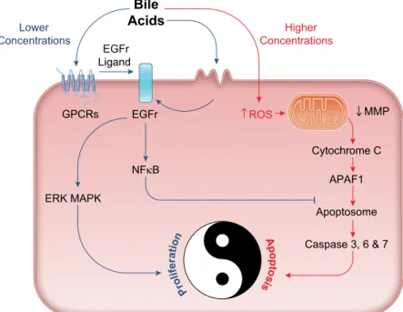
In addition to directly binding to and activating their cog- nate receptors, bile acids can also induce cellular responses by recruiting “non-bile acid” receptors. For example, recent studies have shown that TGR5 can transactivate the epider- mal growth factor receptor (EGFr), thereby enabling the recruitment of tyrosine kinase-dependent signaling path- ways to regulate cell growth (190, 380, 511). Studies by Cheng and Raufman (66) have demonstrated that taurine and glycine conjugates of LCA and DCA can also induce EGFr transactivation, but in this case, through their actions as partial agonists of the muscarinic M3 receptor. Further studies suggest that such bile acid-induced GPCR/EGFr crosstalk is mediated by metalloprotease-induced shedding of EGFr ligands from the cell membrane (14, 67, 297, 317, 497) and inhibition of EGFr degradation (56). However, although it is clear that EGFr activation plays an important role in mediating epithelial responses to GPCRs activated by bile acids, such responses are also likely to be tempered by their activation of FXR, which has been shown to inhibit EGFr-dependent signaling in intestinal epithelial cells (105, 357). Studies from mammary epithelial cells indicate that FXR activation may also inhibit EGFr-dependent signaling by downregulating expression of other members of the ErbB receptor family with which it heterodimerizes (135).
Interestingly, GR and VDR activation have also both been shown to be coupled to inhibition of EGFr signaling and expression, suggesting that bile acids may also have the capacity to influence growth factor-dependent signaling through these pathways (290, 382). Understanding how such complex mechanisms of crosstalk between bile acid and growth factor receptors ultimately impact intestinal epithelial physiology should be an important area of re- search focus in the coming years.
4. Membrane perturbations
Finally, in addition to activating specific cell surface GPCRs and intracellular nuclear receptors, bile acids can also initi- ate signaling in epithelial cells by causing perturbations in the plasma membrane. Studies have shown that treatment of colonic epithelial cells with hydrophobic bile acids cause
a redistribution of membrane cholesterol and phospholip- ids, leading to alterations in caveolin expression and mem- brane fluidity within distinct microdomains (189). Interest- ingly, such effects also bring about transactivation of the EGFr but, in contrast to TGR5- and M3R-mediated re- sponses, they appear to do so independently of EGFr ligand shedding (5, 189). While further investigation is required, it is tempting to speculate that such distinct mechanisms of EGFr transactivation would lead to different patterns of receptor phosphorylation, differential recruitment of downstream ef- fector pathways, and ultimately, different cellular responses to different bile acids.
B. Intracellular Signaling by Bile Acids
Bile acids can alter epithelial function both in the short and long term. Rapid responses to bile acids involve changes in the levels of intracellular second messengers, such as Ca2⫹, cAMP, and reactive oxygen species subsequent to activa- tion of cell surface receptors or induction of membrane perturbations. These second messengers can then alter cell physiology either through direct interactions with effector proteins or indirectly through the activation of signaling cascades that alter the phosphorylation and activity of downstream effectors. Kinase cascades commonly reported to be involved in mediating the actions of bile acids include protein kinase C (PKC), protein kinase A (PKA), ERK, JNK and p38 mitogen-activated protein kinases (MAPKs), to name but a few (19, 139, 250, 305, 394, 438). The lipid kinase phosphatidylinositol 3-kinase (PI3K) has also been shown to be an important mediator of bile acid actions in many different systems (19, 425). Another type of post- translational modification that can rapidly alter cell func- tion in response to bile acids is ubiquitination, leading to proteosomal degradation of effector proteins (13, 306).
Degradation of proteins can also occur by the process of autophagy, and recent studies suggest that bile acids are also important regulators of this process in hepatocytes, enterocytes, and pancreatic epithelial cells (45, 275, 351, 508). Rapid alterations in the trafficking of proteins to and from the cell surface can also occur in response to bile acids through regulating their internalization by endocytosis (202, 400). More long-term changes in epithelial phenotype can be brought about through altering the expression of proteins that contribute to barrier and transport function.
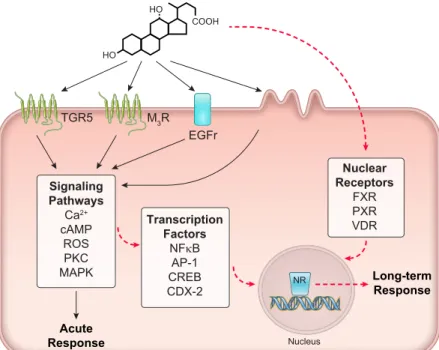
Such changes can occur in response to the activation of transcription factors (e.g., NFB, AP-1, CREB) down- stream of intracellular second messengers, or by activation of nuclear receptors (e.g., FXR, PXR, VDR). Bile acids can also alter the expression of effector proteins through regu- lating miRNA production (64, 120, 507), or by epigenetic mechanisms, involving DNA methylation or histone deacetylation (228, 262). A summary of bile acid-induced epithelial signaling mechanisms is shown inFIGURE 3.
C. Bile Acids and the Microbiota
One of the primary determinants of the bioactivity and bioavailability of bile acids in the intestine is their metabo- lism by resident microbes The colon is home to a population of trillions of bacteria, comprised of hundreds of different species, many of which have the capacity to metabolize bile acids. Normally, through deconjugation and dehydroxyla- tion, the colonic microbiome converts conjugated bile ac- ids, which have failed to be absorbed via the ASBT in the small intestine, into more hydrophobic molecules. Since there are no reports of ASBT being expressed on the apical membrane of colonic epithelial cells, increased hydropho- bicity and decreased ionization enables bile acid reabsorp- tion by passive diffusion across the cell membrane.
The most rate-limiting step in bacterial metabolism of bile acids is deconjugation of glycine or taurine by the action of bile salt hydrolases (BSH). This reaction dramatically changes the physicochemical properties of the bile acids, making them more lipophilic and partially protonated and thereby enabling further metabolism by dehydroxylases and epimerases (200, 256). The past two decades have seen enormous advances in our understanding of how the micro- biome influences human health, not only in the intestine (348), but also through its roles in regulating energy expen- diture, metabolism, and cardiovascular function (46, 154, 503). By way of the gut-brain axis, the microbiome is now becoming recognized as an important regulator of higher central functions, including emotional state and appetite (6, 82, 117). It even regulates our circadian rhythms (199, 447).
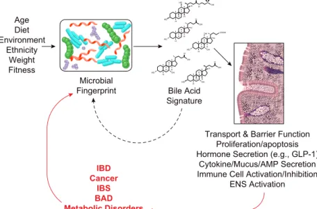
The microbiome is highly dynamic and its composition de- pends on many factors, including diet, environment, age,
fitness level, and psychological state, to name but a few (73, 264, 269, 333). It is also clear that dramatic changes in the microbiome can occur in many disease states, and being at the dividing line between the human and microbial worlds, the epithelium has a critical role to play in mediating the effects of such changes. Understanding how bacteria and epithelial cells communicate with one another under nor- mal and disease states is a critically important area of re- search that is advancing rapidly. Bile acids clearly have an important role to play in this communication network, since changes in the microbiome lead to changes in bile acid metabolism and, consequently, changes in the hydropho- bicity/hydrophilicity ratio of the colonic bile acid pool (107, 205). Thus the precise levels of different bile acids in the colon, i.e., the “colonic bile acid signature” is determined by the makeup of the microbiota. In turn, each individual’s bile acid signature determines what contributions TGR5, FXR, VDR, PXR, M3R, GR, EGFr, and membrane disrup- tions make in setting the overall tone of epithelial function at any given time. A summary of how microbiota/bile acid interactions impact on intestinal physiology in health and disease is shown inFIGURE 4.
V. BILE ACIDS AND EPITHELIAL FUNCTION
As discussed above, the two primary functions of intestinal epithelial cells are to transport fluid electrolytes and nutri- ents to and from the luminal contents and to act as a barrier to prevent the entry of harmful substances from the intesti- nal lumen to the mucosa. In their capacity as signaling mol- ecules, bile acids play important roles in regulating both of these aspects of epithelial function.
HO HO
COOH
Nuclear Receptors
FXR PXR VDR
TGR5 M3R
Signaling Pathways
Ca2+
cAMP ROS PKC MAPK
Nucleus
Long-term Response
Acute Response
Transcription Factors
NFNB AP-1 CREB CDX-2
EGFr
NR
FIGURE 3. Intracellular signaling in response to bile acids.
Bile acids induce acute responses (solid black arrows) in epithelial cells through the activation of multiple receptor types on the cell surface or through induction of membrane perturbations. This leads to the generation of numerous intracellular second messengers and the activation of signal- ing cascades. Such signaling pathways can also lead to more long-term changes in cellular function through the activation of transcription factors and regulation of gene transcription (dashed red arrows). Changes in gene transcription and protein expression can also be brought about by the activa- tion of several nuclear receptors, including farnesoid X re- ceptor (FXR), pregnane X receptor (PXR), and vitamin D receptor (VDR).
A. Bile Acids in Regulation of Epithelial Fluid and Electrolyte Transport
There has long been an association between bile acids and intestinal fluid homeostasis. The first reports of the bile acid binding resin cholestyramine being useful in treating diar- rhea were published in the late 1960s (171, 387), and soon afterwards a landmark study by Mekhjian et al. (295) di- rectly demonstrated that instillation of bile acids at high concentrations into the colons of healthy volunteers in- duced fluid secretion. Subsequent studies in animal models and cultured epithelial cell lines have revealed that the ef- fects of bile acids on luminal fluid accumulation are due to both inhibition of Na⫹absorption and stimulation of Cl⫺ secretion (40, 141, 294, 395). Although there can be quite a degree of variation in their concentration dependence in different models, it is clear that in most species, including humans, only pathophysiologically high levels of bile acids induce colonic fluid secretion. There is also a marked struc- tural specificity for bile acids in exerting their actions, with only the dihydroxy bile acids, CDCA and DCA, having prosecretory effects (141, 217, 444). UDCA, the 7-OH epimer of CDCA, is a notable exception in that it has been shown to be devoid of prosecretory activity and, in fact, has antisecretory actions (130, 223). Conjugation to glycine or taurine is also an important factor in determining bile acid actions on intestinal fluid transport. Since colonic epithelial cells do not express apical transporters for bile acids, they must first be deconjugated, to become more lipophilic and partially protonated before they can passively cross the cell membrane. However, if levels of luminal conjugated bile acids increase sufficiently, loss of tight junction integrity occurs, allowing bile acids to gain access to the basolateral side where they can then be transported into the cell to exert their effects (102, 217). Thus the colonic microbiome, through its deconjugating, dehydroxylating, and epimeriz- ing activities, is a crucial regulator of intestinal transport
responses to bile acids. While the physiological basis for such cathartic actions of bile acids in the colon is still not certain, it is thought they have likely evolved as an innate defense mechanism to protect the mucosa at times when colonic delivery of bile acids is abnormally high. Under such conditions, bile acid-induced secretion dilutes colonic con- tent and causes rapid elimination from the body, thus pre- venting damage to the epithelial barrier.
The molecular mechanisms by which intestinal epithelial transport function is altered by bile acids are still not fully elucidated. However, in vitro studies in cell culture models clearly show that they can exert their effects, at least in part, through direct actions at the epithelium itself (101, 102, 217). Such direct effects of bile acids are mediated primarily by elevations in intracellular Ca2⫹, leading to activation of basolateral KCNN4 channels, thereby creating the electri- cal driving force for Cl⫺ secretion into the lumen. Such Ca2⫹-dependent prosecretory effects of bile acids have also been reported in colonic tissues from animal models (286, 311). Studies on isolated colonic epithelial cells have shown that bile acid-induced increases in intracellular Ca2⫹ also inhibit the activity of Na⫹/H⫹and Cl⫺/HCO3⫺exchangers in the apical membrane of surface cells. In vivo, such an effect would result in reduced salt and water absorption from the lumen (11, 339). Recent studies in rats also suggest that changes in epithelial expression of aquaporins occur upon exposure to bile acids, although the contribution that these channels make to intestinal fluid secretion is still un- known (512).
While it is clear that elevations in intracellular Ca2⫹ are important in mediating bile acid actions, the upstream mechanisms involved have yet to be elucidated. The strict structure-activity relationship for bile acids in regulating epithelial transport function suggests that a receptor may be involved, but if this is so, its identity has yet to be revealed.
HO HO
OH
O OO
H H
H H
H S S N N H
HO
OH O
H H
H H
H HO
O
H H
H H
H OH
OH O
HO OH
H H
H OH
H H
COOH
HO OH
OH H
H H
H
Microbial
Fingerprint Bile Acid
Signature
Transport & Barrier Function Proliferation/apoptosis Hormone Secretion (e.g., GLP-1)
Cytokine/Mucus/AMP Secretion Immune Cell Activation/Inhibition
ENS Activation IBD
Cancer IBS BAD Metabolic Disorders Age
Diet Environment
Ethnicity Weight Fitness
FIGURE 4. Bile acid/microbial interactions in regula- tion of intestinal physiology and pathophysiology. Environ- mental and genetic factors contribute to determining the nature of the microbial fingerprint that exists within the intestinal lumen which, in turn, determines the makeup of our intestinal bile acid signatures. Luminal bile acids reg- ulate many different aspects of mucosal physiology, from epithelial transport and barrier function to immune cell and neuronal activation in the lamina propria. Bile acids also play an important feedback role in shaping the mi- crobial fingerprint. Dysregulation of microbial/bile acid interactions can negatively impact mucosal function, con- tributing to the onset of intestinal and metabolic disor- ders (red arrows). In turn, disease progression can also impact the intestinal flora, making it difficult to determine whether changes in the microbial fingerprints and bile acid signatures are a cause or consequence of disease.
Our own studies suggest that the cell surface bile acid re- ceptor TGR5 is not involved since, even though the receptor is expressed on colonic epithelial cells, its activation with the TGR5 specific ligand INT-777 inhibits, rather than stimulates, Cl⫺secretion in rat colonic tissue (488). Mean- while, the rapidity with which bile acids induce epithelial secretion (i.e., within seconds) rules out a role for nuclear receptors. An alternative possibility to the involvement of a specific receptor could be that hydrophobic bile acids cause membrane perturbations, either at the plasma membrane or within intracellular organelles, such as mitochondria and the ER, leading to the emptying of intracellular Ca2⫹stores and increased influx from the extracellular milieu (255, 453).
In addition to their direct effects on epithelial cells, bile acids can also alter intestinal fluid and electrolyte transport through indirect mechanisms. In an important series of studies carried out by Lundgren and co-workers at the Uni- versity of Gothenburg, the involvement of intrinsic neural reflexes in bile acid-induced fluid secretion in the small in- testine was defined (208, 209). Subsequent studies revealed that this reflex arc is initiated by enterochromaffin cells which, in the presence of high luminal bile acid levels, re- lease 5-HT to activate intrinsic afferent neurons (358). The efferent arm of the reflex arc appears to be mediated by both cholinergic and noncholinergic, nonadrenergic nerves that release their neurotransmitters into the neuroepithelial junction.
In addition to recruitment of the ENS, bile acids can also stimulate intestinal secretion through the activation of im- mune cells present in the lamina propria. For example, in guinea pig colon, CDCA-induced secretory responses are mediated, at least in part, by activation of mast cells and the release of histamine, which then acts at epithelial H1recep- tors to induce secretion (130). Other mediators released from activated mast cells, such as adenosine and prosta- glandins, are also likely to be involved, although this re- mains to be investigated. It should also be kept in mind that mast cells and nerves exist in close apposition to each other within the intestinal mucosa and engage in bidirectional communication (94). Such neuroimmune interactions are also likely to be important in regulating the full expression of bile acid-induced intestinal transport responses.
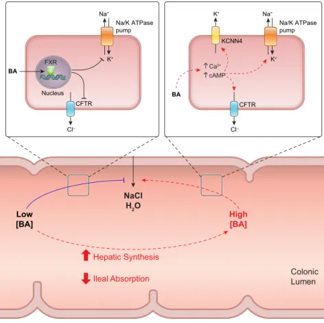
While most studies to date have focused on how bile acids acutely induce fluid and electrolyte secretion when they are present at high levels in the colon, in more recent studies we have begun to address possible roles they may play under more physiological circumstances. It was found that chronic exposure of isolated epithelial cells or rat colonic tissue to relatively low DCA concentrations (10 –200M), which by themselves do not induce secretion, inhibited sub- sequent responses to both Ca2⫹ and cAMP-dependent secretagogues. This effect was slow in onset and was not
associated with alterations in secretagogue-induced in- creases in intracellular second messengers (215). In subse- quent studies it was found that such anti-secretory actions are likely mediated by the nuclear bile acid receptor FXR, since agonists of this receptor mimicked the antisecretory actions of DCA and CDCA in cultured epithelial cells and ex vivo sections of mouse colon. Actions downstream of FXR activation appear to involve inhibition of the activity at least two key components of the Cl⫺secretory pathway, apical CFTR channels and basolateral Na⫹-K⫹-ATPase pumps (313). It was proposed that such antisecretory ac- tions of bile acids may serve a physiological role by damp- ening basal fluid secretion and thereby promoting the nor- mal absorptive function of the colon. Such dual actions of bile acids in acutely promoting and chronically inhibiting fluid secretion at high and low levels, respectively, suggests that they may have a role as colonic “osmosensors” that serve to dynamically regulate luminal fluid levels. A sum- mary of our current understanding of how bile acids regu- late colonic secretion under normal and pathological cir- cumstances is depicted inFIGURE 5.
B. Bile Acids in Regulation of Epithelial Barrier Function
The intestinal epithelial barrier is comprised of the physical barrier posed by the epithelial cells themselves, augmented by a number of secreted factors, including antimicrobial peptides, mucus, cytokines, and immunoglobulins. This barrier is by no means static but is in a constant state of flux as the cells which comprise it are continuously undergoing proliferation, migration, and differentiation. At the same time, the barrier retains the plasticity to rapidly respond to changes in the luminal environment, such as the passage of food or the presence of pathogens. The epithelium is ex- posed to a myriad of endogenous and exogenous stimuli, many of which have the capacity to alter one or several aspects of barrier function. In the following sections we discuss the role that bile acids play in this vital aspect of epithelial function.
1. Cell death and survival
Epithelial cells, particularly those of the intestinal tract, are being constantly renewed. This regeneration process re- quires programmed elimination of damaged cells, usually via the apoptotic cell death pathway, balanced with a con- stant source of newly dividing cells. This finely-tuned bal- ance between epithelial regeneration and death can become disturbed in various pathophysiological conditions, leading either to loss of barrier and transport function on the one hand, or to the development of cancer on the other. The roles of bile acids in regulating cell death and survival have been widely investigated in different epithelial cell types along the gastrointestinal tract, including those from the small and large intestine, the liver and biliary tree, and the