Diagnosis and treatment of periprosthetic joint infections
Doctoral theses
Ákos Zahár M.D.
Semmelweis University
School of PhD Studies, Clinical Medicine
Tutors: Prof. Miklós Szendrői, M.D., Ph.D., D.Sc.
Gábor Skaliczki, M.D., Ph.D.
Opponents: Imre Szerb, M.D., Ph.D.
Tamás De Jonge, M.D., Ph.D.
Examination committee:
Prof. László Hangody, M.D., Ph.D., D.Sc. (Chair) Árpád Joób-Fancsaly, M.D., Ph.D.
István Szabó, M.D., Ph.D.
Budapest
2016
1. Introduction
One of the most successful treatment options in modern orthopaedic surgery is the total joint replacement. From the very beginning it was the biggest challenge to fight the bacterial infection around the foreign material. Infection of total joint arthroplasty, caused by bacteria or other pathogens, like fungi, is referred to periprosthetic joint infection (PJI). Incidence rates of PJI in specific joints, like hip, knee, shoulder or elbow, are different. Primary implantations are less likely to get infected (infection rate 0.3-3.9%) than revision arthroplasties, where infection rate can be 10-fold higher. Therefore, the significance of PJI is quite high in modern orthopaedic surgery.
In recent years the orthopaedic community is more focused on the topic of PJI;
several research groups are involved in the diagnosis, treatment and prophylaxis of the disease. Numerous review articles, meta-analyses were published in the past five years on this topic. The first International Consensus Meeting on Prosthetic Joint Infection (ICM-PJI) was organised in 2013 in Philadelphia, PA, USA. 400 delegates from 52 countries, in 15 work groups discussed and elaborated actual challenges in PJI. The results of the consensus meeting have been published in 15 peer-reviewed journal articles and a book, which was translated into many languages. One of the work groups was dedicated to the management of fungal or atypical PJI; the author of this thesis was one of the collaborators.
Diagnostic methods of PJI
Professional orthopaedic societies from the USA (AAOS, IDSA, MSIS) published clear recommendations how to diagnose PJI when certain criteria are fulfilled, and these diagnostic algorithms are now used worldwide. The diagnostic criteria were modified by the ICM-PJI, and they are accepted like a
gold-standard. Improvement of technology makes it possible to have new diagnostic tools, which can lead to a more accurate and reliable diagnosis in PJI.
Therefore, it is important to revise the diagnostic algorithms from time to time.
Modern synovial diagnostics
Classical diagnostic methods are nowadays available everywhere, proper diagnostics of PJI are widely used. In recent years research was conducted towards new diagnostic possibilities, because conventional tools are not perfect.
None of these methods has a 100% sensitivity (Se) and specificity (Sp), this is the reason why the diagnosis of PJI cannot be proven by only one diagnostic tool.
Alpha Defensin
In the diagnosis of PJI there was an emerging demand to develop a new tool, which is quick and reliable, so the Philadelphia research group investigated several serum biomarkers as potential diagnostic options. Alpha Defensin was the biomarker with the highest Se and Sp, so further research was carried out in order to use this tool in the diagnosis of PJI. Defensins are natural peptides with bactericidal and fungicidal effect, which are produced by neutrophil leukocytes in the presence of microorganisms.
Treatment options in PJI
The diagnosis of PJI is not always easy, but with the application of the proposed diagnostic algorithms can be realised with few steps. It is important to secure the diagnosis as early as possible and to identify the pathogen along with its susceptibility. The classification of PJI, as recommended by Tsukayama provides with the proper information, which therapeutic options are reasonable.
After diagnosing and categorising PJI, surgical treatment can be carried out accordingly.
One-stage septic exchange
This treatment option of late chronic PJI is not widely used, it is performed by several specialised treatment centres only. This surgical procedure consists of explantation of the infected prosthetic device and all foreign material, excision of the infection membrane (debridement), and after a series of local measures implantation of a new prosthesis, in the most cases in combination with local antibiotics. In contrary to this option, in the treatment of PJI the gold standard of septic exchange is considered to be the two-stage septic exchange, where an antibiotic spacer is used as an interim implant.
Criteria of one-stage septic exchange are:
• known microorganism,
• known susceptibility,
• intact soft tissues, no skin defects,
• radical debridement,
• good bone stock for the new implant in terms of proper fixation,
• local antibiotic therapy with a bactericidal concentration,
• postoperative intravenous antibiotic treatment.
2. Hypothesis and purpose
2.1. Accuracy of Alpha Defensin assay
Our hypothesis consists of the reported good performance of Alpha Defensin
• with a high sensitivity and specificity, these features enable the test to diagnose PJI.
Our aim was to compare these results with the MSIS criteria which are considered to be the gold standard in diagnosis of PJI. Therefore, a prospective clinical study was conducted with synovial fluids obtained from enrolled patients in order to answer the following questions:
• What is the sensitivity, specificity, positive and negative predictive value for the Alpha Defensin assay?
• Which clinical conditions or concomitant disease may result in false negative or false positive Alpha Defensin assay result?
2.2. One-stage septic exchange of infected total knee arthroplasty
Our hypothesis consists of the reported results of one-stage septic exchange of infected total knee arthroplasty (TKA),
• being a proper surgical treatment option in the therapy of PJI after total knee arthroplasty,
• and compared with the results of two-stage exchange which is considered to be the gold standard, similarly good results can be achieved.
Therefore, we conducted a retrospective clinical and radiologic follow-up of minimum 9 years, enrolling consecutive patients with a one-stage septic exchange of infected TKA, operated with the same type of implant (rotating hinge design), in order to answer the following questions:
• In what percentage an infection control was achieved and how was the recurrence rate?
• How was the implant survival when aseptic revision of any reason or septic revision for recurrence of PJI were considered as end-points?
• How was the clinical outcome of the septic procedures when patients were investigated pre- and postoperatively with the Hospital for Special Surgery (HSS) score?
• How was the occurrence of radiologic loosening of the implanted revision total knee prostheses?
3. Patients and Methods
3.1. Accuracy of Alpha Defensin assay
After ethical approval patients were included in the study who underwent knee or hip revision arthroplasty in the period between April and October 2015, all operated at Helios ENDO Klinik, Hamburg, Germany. Indication for revision arthroplasty was a chronic deep pain of the target joint for more than 90 days.
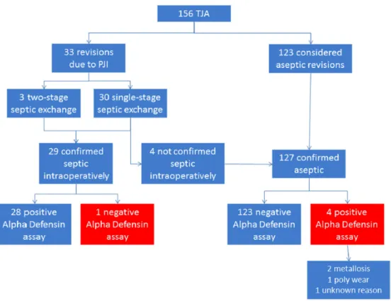
Patients’ history and clinical data, laboratory findings (CRP and WBC), results of preoperative aspiration were collected. Patients diagnosed with metallosis, systemic inflammatory disease and ongoing antibiotic therapy were not excluded. Patients with an insufficient amount of intraoperative aspirate (<2 ml) from synovial fluid were excluded. In all patients aspiration of the target joint was carried out following a strictly standardised protocol. From these aspirates Alpha Defensin assays were performed in a specialised laboratory. After consideration of all inclusion and exclusion criteria, we conducted the prospective study of 156 consecutive subjects (90 females and 66 males), where the analysis of 156 painful joints (65 knees, 91 hips) was carried out.
All patients were diagnosed according to a standardised preoperative protocol of the ENDO Klinik with blood tests and joint aspirations. Serum C-reactive protein (CRP) was measured, synovial analysis with cell count (CC) and granulocyte-percentage (PMN%) was performed and leukocyte esterase (LE) activity was tested with a colorimetric dip-stick. Aspirations were carried out twice: preoperatively and intraoperatively. Two weeks prior to preoperative aspiration any ongoing antibiotic therapy was interrupted (antibiotic holiday).
Bacterial specimens were incubated and cultured for 14 days in the Institute of Microbiology of Schleswig-Holstein University (UKSH) in Kiel, Germany.
Based on the results of preoperative aspirations and blood tests we divided the patients into two groups: aseptic patients (n=123) and patients with PJI (n=33).
Seven patients in the latter group had a draining sinus (21%).
In the group of aseptic patients, partial or complete revision arthroplasties were performed. In the PJI group one-stage septic exchange (n=30) was the therapy of choice when preoperative cultures revealed known microorganisms along with susceptibility, according to the strict protocol of the hospital. In culture negative aspirates of PJI patients a two-stage septic exchange (n=3) was carried out and antibiotic-loaded spacer was inserted.
Patients were intraoperatively aspirated by experienced surgeons on a standardised way. After sterile prepping and draping of the study patient the skin was incised, then with a new surgical knife, in order to avoid contamination, subcutaneous tissues were dissected. In all hips, a posterolateral approach was performed; in knees a medial mid-vastus approach was chosen. After preparation of muscles and soft tissues the joint capsule was exposed and the joint was aspirated without incision of the capsule. Joint fluid was obtained with a standard single-use 20 ml syringe and an 18-gauge sterile aspiration needle.
The admixture of blood was avoided in order to not to influence the test results, a minimum of 2 ml of synovial fluid was aspirated. The samples were then sent
to an independent laboratory (Labor Dr. Fenner und Kollegen, Hamburg, Germany) where quantitative Alpha Defensin immunoassays were performed within 8 hours. Test kits were donated by CD Diagnostics Inc. (Claymont, DE, USA).
Alpha Defensin immunoassay can be run with a minimum of 1 ml of native synovial fluid within 8 hours after aspiration. Quantitative determination of Alpha Defensin was carried out with the method of ELISA. The result is a numeric data which correlates with the amount of Alpha Defensin in the specimen (signal to cut-off ratio, S/CO). A result under 0.9 means a negative, over 1.0 a positive test; if the result is between 0.90 and 0.99 the interpretation of the test is not possible. A positive test suggests the presence of a PJI with a reported high (over 90%) probability.
In both patient groups, at least three tissue samples were obtained during revision surgery from pre-defined sites of the bone-implant interface and sent to UKSH microbiology lab for bacterial cultures. Histology tests were performed from two tissue samples obtained from each joint in an independent laboratory (MVZ für Histologie, Zytologie und Molekulare Diagnostik, Trier, Germany).
Metallosis was diagnosed macroscopically by the surgeon (grey or black stained soft tissues) and microscopically by the pathologist. 13 patients had a metallosis (11 subjects from the aseptic and 2 patients from the PJI group). 14 days after the revision surgery results of the intraoperative tests were compared with the preoperative investigations, and proper diagnosis of the patients in terms of PJI was confirmed or revised. In the PJI group diagnosis was confirmed by histology and microbiology in 29 cases, in 4 patients PJI could be excluded. At final evaluation, these 4 patients were added to the group of aseptic patients. 29 cases of PJI were diagnosed based on the diagnostic criteria of MSIS; in 27 patients one of the major criteria, in 2 patients 3 of 5 minor criteria were
positive. The results of Alpha Defensin assay were blinded, the microbiologist and the pathologist were not aware of these data. A dataset with all pre- and postoperative study results was established and statistically analysed.
The results of the Alpha Defensin assay were reported as a semi-quantitative signal-to-cut-off ratio of 1.0 as a threshold for diagnosis of PJI. To statistically assess the performance of the current test, the specificity (Sp), sensitivity (Se), positive predictive value (PPV), and negative predictive value (NPV) were evaluated.
3.2. One-stage septic exchange of infected total knee arthroplasty
After approval of the local ethical committee, a retrospective analysis of data collected from the ENDO Klinik, Hamburg, Germany, was performed in all patients who underwent a one-stage exchange arthroplasty for infected TKA between January 1st and December 31st, 2002.
Seventy patients with one-stage septic revision of the knee were included in our review and 11 patients with other than one-stage procedures were excluded. We excluded from the study all patients who underwent a two-stage surgical approach (n = 8) and those not re-implanted with a TKA undergoing rather an arthrodesis (n = 3). The indication for one-stage revision was the diagnosis of PJI with a known causative organism. Two-stage revision was performed if the preoperative aspiration was culture-negative. An arthrodesis nail was implanted in cases of severe damage or rupture of the extensor mechanism. The diagnosis of PJI was based on an elevated preoperative C-reactive protein (CRP) level in combination with positive culture results of a preoperative aspiration after a minimum of 14 days of bacterial culture. All patients were culture-positive and intraoperative sampling confirmed preoperative culture results. There were no exclusions on the basis of patient condition, American Society of Anesthesiologists (ASA) classification, body mass index, soft tissue status,
including the presence of a draining sinus, other patient-related factors potentially influencing the outcome, or a causative microorganism, including resistant organisms and fungi.
Of the 70 identified patients (31 women and 39 men) for our study, 11 (16%) were lost to follow-up. Although several attempts to contact them were made no records in the German federal medical database were found and it could not be determined if these patients had died. Related general practitioners (family doctors) were unable to give further information; in addition, no clinical or radiologic data for the 11 patients were available. Fifty-nine patients comprised the final study cohort; all included had complete clinical and radiologic data. A total of 13 patients died during the follow-up. In 46 patients, clinical evaluation, including radiographic follow-up, was available within the last year before the study. Average follow-up was 10 years (last follow-up 2012-13; range, 9-11 years). The mean age of the study patients at the time of surgery was 70 years (range, 60–81 years).
All patients were characterised as either “infection controlled” or “failure”.
Criteria for successful infection control were defined as no clinical signs of infection, no further surgery with the diagnosis of PJI, and no further positive cultures after the one-stage septic exchange. Failures included patients with local or systemic symptoms of infection, those needing further surgery as a result of persistent PJI according to the MSIS criteria, and patients who died after generalised sepsis. We considered reinfection to be an infection of the same joint with the same or another organism. Additionally, we considered aseptic failures of the prosthetic device to be any procedure in which a reoperation was performed for reasons unrelated to PJI or sepsis. The HSS score was measured pre- and postoperatively at the most recent follow-up visit. Patient radiographs were reviewed at the most recent postoperative follow-up and compared with those taken in the immediate index post-operative period to look for signs of mechanical failure or aseptic loosening of the prosthetic
components. All data registered before the most recent follow-up were available through the clinical database. The clinical and radiological examination was done and evaluated by the author.
A paired t-test was used for calculating the differences in the pre- and postoperative clinical results with a confidence interval (CI) of 95%. Probability
<0.05 was considered statistically significant. As a result of more than 15% of unrelated deaths during the follow-up, competing risks survival analysis was performed. The uncertainty of the fractional survival was considered as a standard error or 95% CIs. Standard errors were calculated by the method of Greenwood by using GraphPadã for Windowsã (GraphPad Software, La Jolla, CA, USA).
4. Results
4.1. Accuracy of Alpha Defensin assay
The Alpha Defensin assay was positive in 32 joints and negative in 124 (Fig. 1).
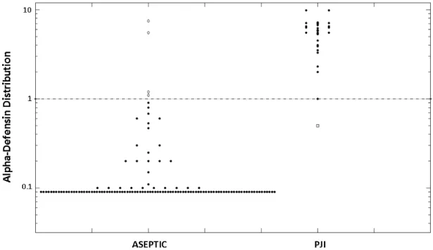
When matching these data with the diagnosis based on the MSIS criteria, it resulted in the Alpha Defensin assay being false-positive in four cases and false- negative in only one case (Fig. 2). Statistical analysis revealed that the sensitivity of the Alpha Defensin immunoassay was 97% (95% CI, 92%– 99%), the specificity was 97% (95% CI, 92%–99%), the positive predictive value was 88% (95% CI, 81%–92%), and the negative predictive value was 99% (95% CI, 96%– 99%).
Figure 1: Workflow describing the features of the patients included in the Alpha Defensin study. False-negative and false-positive cases are marked red. [reproduced after statistician Silvia Bassini (Bologna, Italy)]
Among the four patients with a false-positive alpha- defensin assay, two had a coexisting metallosis and one had severe polyethylene wear with osteolysis. In one patient no particular clinical feature was noticed. The two cases with
metallosis had a negative CRP, whereas the patient with polyethylene wear had a CRP of 15 mg/L. Cell count and LE tests were not available for any of these three patients. The false-negative case presented with a draining sinus which is a major criterion for PJI; the intraoperative cultures were negative but the CRP was 15 mg/L and the granulocyte percentage was 80%.
Figure 2: Synovial fluid Alpha-Defensin values (logarithmic scale) for aseptic and PJI patients are shown separately. The line indicates the alpha-defensin diagnostic threshold of 1.0 (signal-to-cutoff ratio [S/CO]). The five white dots represent the misdiagnosed patients, being false-negative (in the PJI group) or false-positive (in the aseptic group).
4.2. One-stage septic exchange of infected total knee arthroplasty
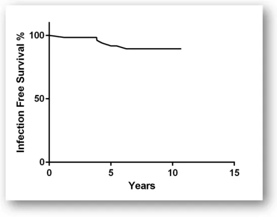
At the end of follow-up, five patients were rated as re-infected. Two of these patients died because of systemic sepsis; the affecting organism is not known. In one of the patients, the same Staphylococcus epidermidis organism was found at the revision surgery. The two other patients developed a fungal PJI with Candida parapsilosis. In the three patients with a re-infection, a second one- stage exchange was performed, whereby one of these 2 years later sustained a third PJI, then after another one-stage exchange, there was no recurrent infection until the latest follow-up. The competing risk survival analysis revealed 93%
(mean of 4.1, 95% CI,89%-96%) at 10 years for patient survival free of infection (p<0.007) (Fig. 3).
Figure 3: Survival analysis of one-stage septic exchange of infected total knee arthroplasty, septic revision as an end-point. 93% is free of infection at 10 years of follow-up. Statistical method: competing risk survival analysis (mean 4.1 and 95% CI, 89%-96%, p<0.007).
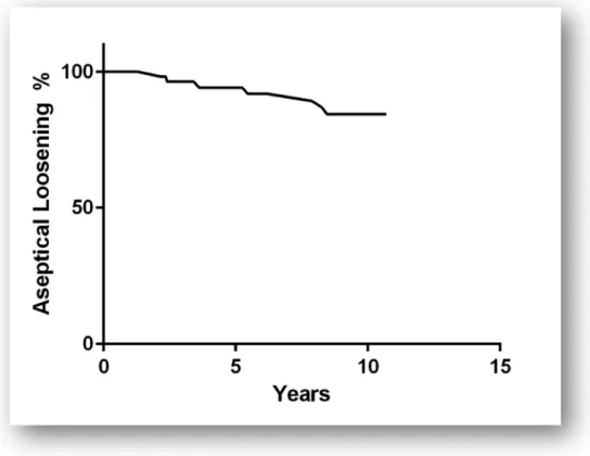
After the end of follow-up, there were seven patients with a need for revision surgery because of aseptic loosening. In three patients there was a sole loosening of the tibia and in four patients of the tibial and femoral component. Competing risk survival analysis revealed 91% survival at 10 years ( mean of 5.2 and a 95%
CI, 86%-95%) for patient survival free of reoperation for aseptic loosening reasons (p<0.002) (Fig. 4). The 10-year survivorship free of any reoperation, including both septic and aseptic causes, was 75% (95% CI, 60%-87%).
The HSS score improved significantly from a mean preoperative value of 35 (±
24.2 SD; range, 13–99) to an average of 69.6 (± 22.5 SD; range, 22–100) at latest follow-up. Postoperative excellent results (100 to 85 points) for HSS score were found in 30%, good results (84–70 points) in 33%, fair (69 to 60 points) in 11%, and poor (<60 points) in 26%. The flexion of the knee improved
significantly from a mean value of 50° (± 25.6 SD; range, 8–144) to 76° (± 33 SD; range, 8°–144°). The results for pain (maximum of 30 points) were a mean preoperative 5.7 (± 9.9; range, 0–30) and improved significantly postoperatively to 18.5 points (± 11.3; range, 0–30). A significant improvement was also found regarding the function pre- and postoperatively with a mean 10 points preoperatively (± 6; range, 4–22) and 16.7 (± 6.3; range, 4–22) postoperatively.
Figure 4: Survival analysis of one-stage septic exchange of infected total knee arthroplasty, aseptic revision due to implant loosening as an end-point. 91% is clinically and radiologically not loose at 10 years of follow-up.
Statistical method: competing risk survival analysis (mean 5.2 and 95% CI, 86%-95%, p<0.002).
In addition to the six patients who underwent revision for aseptic loosening, six other patients showed radiolucent lines in the area of the metaphysis of the tibia and femur. Because the radiolucencies were less than 2 mm in thickness and non-progressive, no further assessment was recommended.
5. Conclusions
Based on our studies the following conclusions can be derived:
1. In our investigations, Alpha Defensin assay had an excellent accuracy of 97% when diagnosing prosthetic joint infection.
2. Our results suggest that Alpha Defensin assay should be considered as one of the diagnostic criteria for prosthetic joint infection because in terms of sensitivity and specificity this tool is superior to other methods.
3. In the case of metallosis results of Alpha Defensin assay should be evaluated carefully, since false-positive results may occur.
4. Our results are outstanding in terms of the size of the study cohort (n=156) when compared with other publications related to Alpha Defensin assay. This is the first publication which is not related to the developing study group.
5. The results of one-stage septic exchange of infected total knee arthroplasty with an infection control rate of 93% at 10 years follow-up are excellent.
6. The surgical approach with radical excision of the collaterals and the posterior capsule in combination with the rotating hinge design provides good clinical and radiologic results.
7. Our results are comparable with the long-term results of two-stage septic exchange, and we were able to show that the re-infection rate in our unselected patient cohort was not higher.
8. In terms of cohort size (n=70), follow-up time (10 years) and infection control rate (93%) are our results of one-stage septic exchange of infected total knee arthroplasty unique.
9. A 91% aseptic loosening rate of the rotating hinge design total knee arthroplasty could be published first in the setup of one-stage septic exchange, with a follow-up of 10 years.
6. List of own publications
Publications related to the doctoral theses
1. Bonanzinga T, Zahar A, Dütsch M, Lausmann C, Kendoff D, Gehrke T.
(2017) How Reliable Is the Alpha-defensin Immunoassay Test for Diagnosing Periprosthetic Joint Infection? A Prospective Study. Clin Orthop Relat Res, 475(2):418-415. IF=3.127
2. Zahar A, Kendoff DO, Klatte TO, Gehrke T. (2016) Can good infection control be obtained in one-stage exchange of the infected TKA to a rotating hinge design? 10-year results. Clin Orthop Relat Res, 474(1):81- 87. IF=3.127
3. Zahar A, Hannah P. (2016) Antibiotikazumischung zum Knochenzement beim septischen Prothesenwechsel [Addition of antibiotics to bone cement for septic prosthesis exchange], Oper Orthop Traumatol, 28(2):138-144. IF=1.033
4. Zahar A, Gehrke TA. (2016) One-stage revision for infected total hip arthroplasty. Orthop Clin North Am, 47(1):11-18.
5. Zahar A, Webb J, Gehrke T, Kendoff D. (2015) One-stage exchange for prosthetic joint infection of the hip. Hip Int, 25(4):301-307.
6. Gebauer M, Frommelt L, Achan P, Board TN, Conway J, Griffin W, Heidari N, Kerr G, McLaren A, Nelson SB, Nijhof M, Zahar A. (2014) Management of Fungal or Atypical Periprosthetic Joint Infections. J Orthop Res, 32 Suppl 1:147-151.
7. Gehrke T, Zahar A, Kendoff D. (2013) One-stage exchange: it all began here, Bone Joint J, 95-B(11 Suppl A):77-83. IF=3.520
8. Szasz M, Hajdu M, Pesti N, Domahidy M, Kristof K, Zahar A, Nagy K, Szabo D. (2013) In vitro efficiency of vancomycin containing experimental drug delivery systems. Acta Microbiol Immunol Hung, 60(4):461-468. IF=0.650
9. Skaliczki G, Zahár Á, Gáti N, Prinz Gy, Szendrői M. (2011) Két lépésben történő szeptikus térdrevíziók eredményei a Semmelweis Egyetem Ortopédiai Klinika beteganyagában. Magyar Traum Ortop, 54(4):253-263.
10. Lakatos J, Zahár Á. (2004) Két lépésben végzett revízió eredményei a szeptikus csípőízületi endoprotézisek megoldásában. Magyar Traum Ortop, 47(1): 18-24.
11. Zahár Á, Kiss J, Vajda A (2003) Totál felszínpótló térdprotézis szeptikus komplikációjának megoldása antibiotikus távtartó ismételt beültetésével és célzott antibakteriális kezeléssel. Esetismertetés. Magyar Traum Ortop, 46 (1): 60-65.
Other publications
12. Zahar A, Cross MB, Lakatos T, Lakatos J, Major B, Kendoff D, Szendrői M. (2015) Conversion arthroplasty of the hip: mid-term results are good.
Hip Int, 25(6): 520-524. IF=0.763
13. C Isik, N Apaydin, HI Acar, A Zahar, M Bozkurt. (2014) The gluteal sling: an anatomical study. Surg Radiol Anat, 36(6):595-599.
14. Zahar A, Papik K, Lakatos J, Cross MB. (2014) Total hip arthroplasty with acetabular reconstruction using a bulk autograft for patients with developmental dysplasia of the hip results in high loosening rates at mid- term follow-up. Int Orthop, 38:947-951. IF=2.019
15. Guerra-Farfán E, Carrera L, Muneton D, Aguilar M, Zahar A, Giannouidis P. (2013) Vancouver type B2 fractures: best choice of treatment. Eur Orthop Traumatol, 4:81-88.
16. Haasper C, Kendoff DO, Gebauer M, Zahar A, Kreibich T, Suero EM, Gehrke T. (2013) Traumatic stem fractures in short stem THA. A rare case series, HSS Journal, 9(1):86-89.
17. Citak M, Klatte O, Zahar A, Day K, Kendoff D, Gehrke T, Gebauer M.
(2013) Intrapelvic dislocation of a femoral trial head during primary total hip arthroplasty requiring laparotomy for retrieval. The Open Orthopaedics Journal, 7: 124-126.
18. Zahar A, Rastogi A, Kendoff D. (2013) Dislocation after total hip arthroplasty. Curr Rev Musculoskelet Med, 6(4):350-356.
19. Laki M, Ludányi K, Hajdú M, Zahár Á, Szendrői M, Klebovich I, Antal
I. (2011) Determination of Gentamicin released from an orthopedic carrier system by a novel HPLC method. Journal of Chromatographic Science, 49(3):177-181. IF=0.863
20. Zahár Á, Kocsis Gy, Lengyel B, Puskás G, Laki M, Hajdú M, Antal I.
(2009) Hosszú hatóanyag leadású antibiotikus csontgraftok alkalmazása állatkísérletes osteomyelitis modellben. Magyar Traum Ortop, 52(2): 171- 178.
21. Skaliczki G, Zahár Á, Bejek Z, Lakatos J, Szendrői M. (2009) Tumor endoprotézis használata revíziós térdprotetikában, nagy csonthiány vagy szalagelégtelenség esetén. Magyar Traum Ortop, 52(3):239-244.
22. Holnapy G, Zahár Á, Tóth A, Lakatos J. (2007) Ötven éves kor alatt beültetett csípőprotézisek legalább 10 éves utánkövetése. Magyar Traum Ortop, 50(1): 15-23.
23. Skaliczki G, Zahár Á, Hüttl K, Lakatos J. (2007) Csípőízületi revíziós implantátum kilazulásának ritka vascularis szövődménye. Esetismertetés.
Magyar Traum Ortop, 50(3): 264-268.
24. Laki M, Hajdú M, Zahár Á, Sáska Zs, Klebovich I, Szendrői M, Antal I.
(2007) Designing antibiotic-containing carrier systems used in bone surgery. Acta Pharm Hung, 77: 108-115.
25. Zahár Á, Lakatos T, Lakatos J, Böröcz I, Végh G, Major B, Szendrői M.
(2005) Merev csípők arthroplasticájának eredményei. Magyar Traum Ortop, 48(4): 301-307.
26. Kiss J, Zahár Á, Nyíri P, Prinz Gy. (2005) Lactococcus okozta femur osteomyelitis (esetismertetés), Orv Hetil, 146 (13): 613-618.
27. Zahár Á, Kiss S, Sonnleitner Zs, Szőke Gy, Vízkelety T. (2004) Proximális femurvég reszekciója cerebral paresises fiatal felnőttek esetében. Magyar Traum Ortop, 47(3): 112-116.
28. Zahár Á, Skriba E, Papik K, Lakatos J. (2003) Vápatető-képzés totál csípőízületi endoprotézis beültetése során. Magyar Traum Ortop, 46 (1):
17-25.
29. Lakatos J, Bucsi L, Vajda A, Kovács Gy, Zahár Á. (1998) Cementnélküli csípőízületi endoprotézisek klinikai és radiológiai utánkövetésének eredményei. Magyar Traum Ortop, 41(3): 223-229.