Article
Biofilm Eradication by Symmetrical Selenoesters for Food-Borne Pathogens
Márta Nové1, Annamária Kincses1, Beatrix Szalontai1, Bálint Rácz1, Jessica M. A. Blair2 , Ana González-Prádena3, Miguel Benito-Lama3, Enrique Domínguez-Álvarez3,* and Gabriella Spengler1,*
1 Department of Medical Microbiology and Immunobiology, Faculty of Medicine, University of Szeged, Dóm tér 10, 6720 Szeged, Hungary; nove.marta@med.u-szeged.hu (M.N.);
kincses.annamaria@med.u-szeged.hu (A.K.); beatrixszalontai@gmail.com (B.S.);
balintracz95@gmail.com (B.R.)
2 Institute of Microbiology and Infection, College of Medical and Dental Sciences, University of Birmingham, Birmingham B15 2TT, UK; J.M.A.Blair@bham.ac.uk
3 Instituto de Química Orgánica General (IQOG-CSIC), Consejo Superior de Investigaciones Científicas, Juan de la Cierva 3, 28006 Madrid, Spain; anagonzalez.prad@gmail.com (A.G.-P.); m.benito@iqog.csic.es (M.B.-L.)
* Correspondence: e.dominguez-alvarez@iqog.csic.es (E.D.-Á.); spengler.gabriella@med.u-szeged.hu (G.S.);
Tel.:+34-91-258-7661 (E.D.-Á.);+36-62-445-115 (G.S.)
Received: 5 March 2020; Accepted: 13 April 2020; Published: 15 April 2020 Abstract:Infections caused bySalmonellaspecies andStaphylococcus aureusrepresent major health and food industry problems. Bacteria have developed many strategies to resist the antibacterial activity of antibiotics, leading to multidrug resistance (MDR). The over-expression of drug efflux pumps and the formation of biofilms based on quorum sensing (QS) can contribute the emergence of MDR.
For this reason, the development of novel effective compounds to overcome resistance is urgently needed. This study focused on the antibacterial activity of nine symmetrical selenoesters (Se-esters) containing additional functional groups including oxygen esters, ketones, and nitriles against Gram-positive and Gram-negative bacteria. Firstly, the minimum inhibitory concentrations of the compounds were determined. Secondly, the interaction of compounds with reference antibiotics was examined. The efflux pump (EP) inhibitory properties of the compounds were assessed using real-time fluorimetry. Finally, the anti-biofilm and quorum sensing inhibiting effects of selenocompounds were determined. The methylketone and methyloxycarbonyl selenoesters were the more effective antibacterials compared to cyano selenoesters. The methyloxycarbonyl selenoesters (Se-E2andSe-E3) showed significant biofilm and efflux pump inhibition, and a methyloxycarbonyl selenoester (Se-E1) exerted strong QS inhibiting effect. Based on results selenoesters could be promising compounds to overcome bacterial MDR.
Keywords: Salmonellaspecies; Staphylococcus aureus; multidrug resistance; antibacterial activity;
symmetrical selenoesters
1. Introduction
The emergence of multidrug resistant pathogens is a major problem, leading to a progressive reduction in the efficiency of many antibacterial agents. This phenomenon is a serious challenge in public healthcare and medicine [1].
The most frequent multidrug resistance (MDR) mechanisms enable the resistant bacteria to achieve one or several of the following effects: (a) limited uptake of drug; (b) target modification; (c) drug inactivation; and (d) active efflux mediated by efflux pumps. Some efflux pumps are expressed
Microorganisms2020,8, 566; doi:10.3390/microorganisms8040566 www.mdpi.com/journal/microorganisms
Microorganisms2020,8, 566 2 of 15
constitutively, whereas others are induced or over-expressed under environmental stimuli [2]. There are six families of the efflux pump systems: ATP-binding cassette (ABC) family, multidrug and toxic compound extrusion (MATE) family, small multidrug resistance (SMR) family, major facilitator family (MFS), resistance nodulation division (RND) family, and proteobacterial antimicrobial compound efflux (PACE) family [3,4]. Gram-positive bacteria mainly express the members of the MATE and MFS families, whereas Gram-negative bacteria also have transporters of the RND family [2]. The AcrAB-TolC efflux system is comprised of AcrB which belongs to the RND efflux transporters, the outer membrane protein TolC, and the periplasmic adaptor protein AcrA [5].
The formation of biofilms can also contribute to bacterial resistance. Biofilms have a dynamic structure involving a multicellular bacterial community and an extracellular polymeric matrix produced by the bacterial population. Biofilm-associated infections can lead to antibiotic resistant and persistent infections as this environment enhances the ability of the embedded bacteria to resist the action of the antibiotics [6].
One of the major food-borne illnesses is the salmonellosis caused by non-typhoidalSalmonella enterica[7]. In addition, the staphylococcal food poisoning (SFP) is a frequent food-born disease caused by staphylococcal enterotoxin (SE) producer enterotoxigenicStaphylococcus aureusstrains [8].S. aureus andSalmonella entericaserovar Typhimurium are food-borne pathogens capable of forming biofilms on various surfaces. Alkaline and acidic detergents, as well as iodophores, can be effective against biofilm.
However, these substances damage surfaces, and the inappropriate use of biocides and disinfectants could lead to a quick and undesired emergence of resistant microbes [9]. Many bacteria use a cell–cell communication system, namely quorum sensing (QS), to coordinate the population density-dependent gene expression pattern [10]. This communication system plays a major role in biofilm development, as bacteria can produce new virulence factors and thanks to them this bacterial community responds poorly to antibiotic treatment [11].
Selenium(Se)-containing compounds could provide alternative and effective scaffolds to overcome MDR [12]. Se is an essential trace element in living organisms and is crucial for the nutrient supply and energy generation of bacteria. However, overdoses of Se can be highly toxic [13,14]. There is significant evidence about the pro-oxidant effect of Se, particularly in the form of sodium selenite (Na2SeO3), while selenomethionine and selenocysteine are less toxic [14]. It has been described previously that Se-containing agents have an antibacterial effect [15,16]. Selenoesters and selenoanhydrides have exhibited anti-biofilm activity againstS. aureusandS. Typhimurium as described previously [17].
Furthermore, selenocompounds have been used as selenium nanoparticles (SeNPs) againstS. aureus, Escherichia coli, andPseudomonas aeruginosastrains [18,19].
In the present study, and based in these antecedents, symmetrical 2-oxopropyl selenoesters, methyloxycarbonylmethyl selenoesters, and methylcyano selenoesters have been investigated against Gram-negative and Gram-positive bacterial strains to determine their antibacterial, efflux pump inhibiting, and anti-biofilm properties.
2. Materials and Methods
2.1. Compounds
Nine symmetrical selenodiesters or selenotriesters were synthesized and evaluated. Three were 2-oxopropyl selenoesters (briefly, ketone selenoesters, or methylketone selenoesters; compounds Se-K1, Se-K2 and Se-K3). The next three selenocompounds were methyloxycarbonylmethyl selenoesters (methylcarbonyl selenoesters or methyloxycarbonyl selenoesters; compoundsSe-E1, Se-E2,andSe-E3) [20]. The final three compounds were methylcyano selenoesters (cyano selenoesters;
compoundsSe-C1,Se-C2,andSe-C3). For each group of three compounds, the first is the symmetrical para-disubstituted derivative, the second is the symmetrical meta-substituted derivative, and the third is the symmetrical 1,3,5-trisubstituted derivative (Scheme1). Their synthesis is described in the patent application EP17382693, and they were adequately characterized using nuclear magnetic
resonance spectroscopy (NMR), mass spectrometry (MS), and infrared spectroscopy (IR) techniques and their purity was assessed by elemental analysis [21]. Before their use in biological assays the selenocompounds were dissolved in dimethyl sulfoxide (DMSO), to obtain 10 mM concentration stock solutions.Microorganisms 2020, 8, x FOR PEER REVIEW 3 of 15
Scheme 1. Chemical structure of the symmetrical selenoesters evaluated.
2.2. Reagents and Media
DMSO (Sigma-Aldrich, St Louis, MO, USA), phosphate-buffered saline (PBS; pH 7.4), promethazine (PMZ; EGIS), verapamil, carbonyl cyanide m-chlorophenyl hydrazone (CCCP), ethidium bromide (EB), ciprofloxacin-hydrochloride (CIP) tetracycline-hydrochloride (TET), crystal violet (CV), Luria-Bertani (LB) broth, and LB agar were purchased from Sigma-Aldrich Chemie GmbH (Steinheim, Germany). The modified LB agar (LB*) was prepared from bacteriological agar 20 g/L (Difco, Detroit, USA), tryptone 10 g/L, NaCl 10 g/L, yeast extract 5 g/L, K2HPO4 1 g/L, MgSO4 × 7H2O 0.3 g/L, and FeNaEDTA 36 mg/L. pH of the agar was adjusted to 7.2. Mueller–Hinton (MH) broth, tryptic soy broth (TSB), and tryptic soy agar was purchased from Scharlau Chemie S.A.
(Barcelona, Spain).
2.3. Bacterial Strains
Compounds were evaluated against the following bacterial strains:
Gram-negative wild-type Salmonella enterica serovar Typhimurium SL1344 (SE01) expressing the AcrAB-TolC pump system and its acrB gene inactivated mutant S. Typhimurium SL1344 strain (SE02), acrA gene inactivated mutant S. Typhimurium SL1344 (SE03), and tolC gene inactivated mutant S. Typhimurium SL1344 strain (SE39) were used in the study [22–25].
Gram-positive Staphylococcus aureus American Type Culture Collection (ATCC) 25923 was used as the methicillin-susceptible reference bacterial strain, and the methicillin and ofloxacin-resistant S.
aureus 272123 clinical isolate (MRSA), which was kindly provided by Prof. Dr. Leonard Amaral (Institute of Hygiene and Tropical Medicine, Lisbon, Portugal), was used in the assays.
For QS tests we used Chromobacterium violaceum 026 (CV026) as a sensor strain and Enterobacter cloacae 31298 as a N-acyl-homoserine lactone (AHL) producer clinical bacterial isolate. If C. violaceum reaches a high cell density, it produces violacein, which is a purple pigment [26,27].
2.4. Cell Line
MRC-5 human embryonal lung fibroblast cell line (ATCC CCL-171) was purchased from LGC Promochem, Teddington, UK. The cells were cultured in Eagle’s Minimal Essential Medium (EMEM, containing 4.5 g/L glucose) supplemented with a non-essential amino acid mixture, a selection of
Scheme 1.Chemical structure of the symmetrical selenoesters evaluated.
2.2. Reagents and Media
DMSO (Sigma-Aldrich, St Louis, MO, USA), phosphate-buffered saline (PBS; pH 7.4), promethazine (PMZ; EGIS), verapamil, carbonyl cyanidem-chlorophenyl hydrazone (CCCP), ethidium bromide (EB), ciprofloxacin-hydrochloride (CIP) tetracycline-hydrochloride (TET), crystal violet (CV), Luria-Bertani (LB) broth, and LB agar were purchased from Sigma-Aldrich Chemie GmbH (Steinheim, Germany).
The modified LB agar (LB*) was prepared from bacteriological agar 20 g/L (Difco, Detroit, USA), tryptone 10 g/L, NaCl 10 g/L, yeast extract 5 g/L, K2HPO41 g/L, MgSO4×7H2O 0.3 g/L, and FeNaEDTA 36 mg/L. pH of the agar was adjusted to 7.2. Mueller–Hinton (MH) broth, tryptic soy broth (TSB), and tryptic soy agar was purchased from Scharlau Chemie S.A. (Barcelona, Spain).
2.3. Bacterial Strains
Compounds were evaluated against the following bacterial strains:
Gram-negative wild-typeSalmonella entericaserovar Typhimurium SL1344 (SE01) expressing the AcrAB-TolC pump system and itsacrBgene inactivated mutantS. Typhimurium SL1344 strain (SE02), acrAgene inactivated mutantS. Typhimurium SL1344 (SE03), andtolCgene inactivated mutantS.
Typhimurium SL1344 strain (SE39) were used in the study [22–25].
Gram-positiveStaphylococcus aureusAmerican Type Culture Collection (ATCC) 25923 was used as the methicillin-susceptible reference bacterial strain, and the methicillin and ofloxacin-resistant S. aureus272123 clinical isolate (MRSA), which was kindly provided by Prof. Dr. Leonard Amaral (Institute of Hygiene and Tropical Medicine, Lisbon, Portugal), was used in the assays.
For QS tests we used Chromobacterium violaceum 026 (CV026) as a sensor strain and Enterobacter cloacae31298 as a N-acyl-homoserine lactone (AHL) producer clinical bacterial isolate. If C. violaceumreaches a high cell density, it produces violacein, which is a purple pigment [26,27].
Microorganisms2020,8, 566 4 of 15
2.4. Cell Line
MRC-5 human embryonal lung fibroblast cell line (ATCC CCL-171) was purchased from LGC Promochem, Teddington, UK. The cells were cultured in Eagle’s Minimal Essential Medium (EMEM, containing 4.5 g/L glucose) supplemented with a non-essential amino acid mixture, a selection of vitamins, and 10% heat-inactivated fetal bovine serum. The cell lines were incubated at 37◦C, in a 5%
CO2, 95% air atmosphere.
2.5. Determination of Minimum Inhibitory Concentrations by Microdilution Method
The minimum inhibitory concentrations (MICs) of compounds were determined according to the Clinical and Laboratory Standard Institute guidelines (CLSI) [28]. MIC values of the compounds were determined by visual inspection. The solvent was also assayed to ensure there was no antibacterial effect and the concentration (1v/v%) applied in the assays had no antibacterial activity. DMSO was used at subinhibitory concentration (1v/v%) in the assays.
2.6. Cytotoxicity Assay
The adherent MRC-5 human embryonal lung fibroblast cells were cultured in 96-well flat-bottomed microtiter plates, using EMEM supplemented with 10% heat-inactivated fetal bovine serum. The density of the cells was adjusted to 1×10⁴cells in 100µL per well, the cells were seeded overnight at 37◦C, 5% CO2, then the medium was removed from the plates containing the cells, and the dilutions of selenocompounds previously made in a separate plate were added to the cells in 200µL.
The culture plates were incubated at 37◦C for 24 h; at the end of the incubation period, 20µL of MTT (thiazolyl blue tetrazolium bromide, Sigma) solution (from a stock solution of 5 mg/mL) was added to each well. After incubation at 37◦C for 4 h, 100µL of sodium dodecyl sulfate (SDS; Sigma) solution (10% in 0.01 M HCI) was added to each well and the plates were further incubated at 37◦C overnight. Cell growth was determined by measuring the optical density (OD) at 540/630 nm with Multiscan EX ELISA reader (Thermo Labsystems, Cheshire, WA, USA). Inhibition of the cell growth was determined according to the formula below:
IC50=100−[(ODsample−ODmedium control)/(ODcell control−ODmedium control)]×100 (1) Results are expressed in terms of IC50, defined as the inhibitory dose that reduces the growth of the cells exposed to the tested compounds by 50%.
2.7. Resistance Modulation Assay
The resistance modulation effect of compounds with ciprofloxacin (CIP) and tetracycline (TET) antibiotics were evaluated by the checkerboard method onS. aureusstrains. Briefly, CIP or TET was diluted in a 96-well microtiter plate by two-fold serial dilution in MH broth and then the compounds were added at subinhibitory concentrations (12MIC). In this assay, only the tested compounds with well-defined MIC values were tested. Finally, 10−4dilution of the overnight bacterial culture in MH was added to each well. The final volume was 200 µL in each well. The microtiter plates were incubated at 37◦C for 18 h. MIC values in the presence of the antibiotics alone and in combination with Se-compounds were determined by visual inspection.
2.8. Real-Time Ethidium Bromide Accumulation Assay
The impact of compounds on EB accumulation was determined by the automated EB method using a CLARIOstar Plus plate reader (BMG Labtech, UK). Firstly, the bacterial strain was incubated until it reached an optical density (OD) of 0.6 at 600 nm. The culture was washed with phosphate buffered saline (PBS; pH 7.4) and centrifuged at 13,000×gfor 3 min, the cell pellet was re-suspended in PBS. The compounds were added at12MIC concentration to PBS containing a non-toxic concentration
of EB (1µg/mL). Then, 50µL of the EB solution containing the compound were transferred into 96-well black microtiter plate (Greiner Bio-One Hungary Kft, Hungary), and 50µL of bacterial suspension (OD600 0.6) were added to the each well. Then, the plates were placed into the CLARIOstar plate reader, and the fluorescence was monitored at excitation and emission wavelengths of 530 nm and 600 nm every minute for one hour on a real-time basis. From the real-time data, the activity of the compounds, namely the relative fluorescence index (RFI) of the last time point (minute 60) of the EB accumulation assay, was calculated according to the following formula:
RFI=(RFtreated−RFuntreated)/RFuntreated (2)
where RFtreated is the relative fluorescence (RF) at the last time point of EB retention curve in the presence of an inhibitor, and RFuntreatedis the RF at the last time point of the EB retention curve of the untreated control having the solvent control (DMSO).
2.9. Measuring Biofilm Formation Using Crystal Violet
The anti-biofilm effect of the tested compounds againstS. aureusstrains and wild-typeS.Typhimurium SE01 was measured using crystal violet (CV; 0.1% (v/v)). This dye is used to detect the total biofilm biomass formed. Overnight cultures were diluted to OD of 0.1 at 600 nm in TSB medium. Then, the bacterial cultures were added to 96-well microtiter plates and the compounds were added at 12 MIC concentration. The final volume was 200µL in each well. The microtiter plates were incubated at 30◦C for 48 h with gentle agitation (100 rpm). After the incubation period, TSB medium was discarded, and the plates were washed with tap water to remove unattached cells. Then 200µL crystal violet was added to the wells and incubated for 15 min at room temperature. Then, CV was removed from the wells and the plates were washed again with tap water, and 200µL of 70% ethanol was added to the wells. Finally, the biofilm formation was determined by measuring the OD at 600 nm using Multiscan EX ELISA plate reader (Thermo Labsystems, Cheshire, WA, USA). The anti-biofilm effect of compounds was expressed in the percentage (%) of decrease in biofilm formation.
2.10. Quorum Sensing (QS) Assay
The QS inhibitory effect of selenocompounds was examined on the AHL producerE. cloacaestrain andC. violaceumsensor bacterial strain. These strains were inoculated in parallel. The QS inhibition was monitored by agar diffusion method on LB* agar plate as described previously [29]. Filter paper discs (7.0 mm in diameter) were placed between the parallel inoculated strains and impregnated with 10µL compounds. Starting concentration of the compounds was12 MIC. The agar plates were incubated at room temperature (20◦C) for 24–48 h and the inhibition of violacein production was measured.
2.11. Statistical Analysis
The values are given as the mean±standard deviation (SD) determined for three replicates from three independent experiments. The analysis of data was performed using SigmaPlot for Windows Version 12.0 software (Systat Software Inc, San Jose, CA, USA), applying the two-tailedt-test.
Microorganisms2020,8, 566 6 of 15
3. Results
3.1. Determination of Minimum Inhibitory Concentrations by Microdilution Method
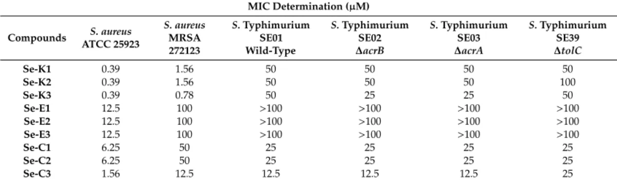
Based on the MIC values, the Se-compounds were more effective againstS. aureusstrains. The most effective compounds were the ketone selenoestersSe-K1,Se-K2, andSe-K3on the referenceS. aureus ATCC 25923, showing an MIC of 0.39µM. Interestingly, these three derivatives share a common moiety, namely a methylketone group in the alkyl moiety bound to the selenium atom. The replacement of this methylketone by a cyano or by a methyloxycarbonyl moiety reduced the activity dramatically, as the MICs were 16- and 32-fold higher againstS. aureusATCC 25923, respectively; with the exception of the trisubstituted derivativeSe-C3, as its MIC was only 4-fold higher than the MIC of the trisubstituted methylketoneSe-K3. The same tendency, but accentuated, was observed inS. aureusMRSA 272123, where the MIC values of the methylketone derivatives were in the range of 64- to 128-fold lower than the equivalent methyloxycarbonyl derivatives and in the range of 16- to 32-fold lower than the equivalent nitrile-containing selenoesters. The compounds showed a slight antibacterial effect on Salmonellastrains. The most effective compound wasSe-C3on SE01, SE02, and SE03 strains, showing an MIC of 12.5µM (Table1). Importantly, the MIC to the efflux knockout strains was unchanged suggesting that the compounds were not substrates of the AcrAB-TolC efflux pump.
Table 1. Antibacterial activity of selenocompounds. Minimum inhibitory concentrations (MICs) of compounds were determined on referenceStaphylococcus aureusATCC (American Type Culture Collection) 25923 and methicillin and ofloxacin-resistantS. aureus272123 (MRSA) strains andSalmonella Typhimurium strains.
MIC Determination (µM)
Compounds S. aureus ATCC 25923
S. aureus MRSA 272123
S.Typhimurium SE01 Wild-Type
S.Typhimurium SE02
∆acrB
S.Typhimurium SE03
∆acrA
S.Typhimurium SE39
∆tolC
Se-K1 0.39 1.56 50 50 50 50
Se-K2 0.39 1.56 50 50 50 100
Se-K3 0.39 0.78 50 25 25 50
Se-E1 12.5 100 >100 >100 >100 >100
Se-E2 12.5 100 >100 >100 >100 >100
Se-E3 12.5 100 >100 >100 >100 >100
Se-C1 6.25 50 25 25 25 25
Se-C2 6.25 50 25 25 25 25
Se-C3 1.56 12.5 12.5 12.5 12.5 25
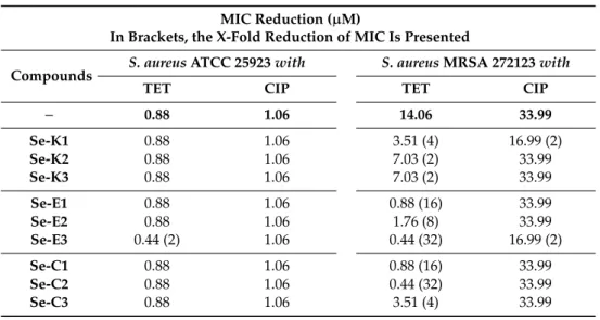
3.2. Resistance Modulation Assay
As the Se-compounds were more effective onS. aureusstrains, these strains were selected for combination studies with reference antibiotics. SelenocompoundSe-E3showed synergism with TET on the methicillin-susceptibleS. aureusATCC 25923.
Surprisingly, all selenocompounds showed synergism with TET on the methicillin-resistantS. aureus strain.Se-E3andSe-C2were the most effective compounds in combination with TET, as they reduced the MIC value of TET against this MRSA strain to a value 32-fold lower. Additionally, compoundsSe-E1and Se-C1also exerted a noteworthy reduction of the MIC value, of 16-fold in this case. On the other hand, Se-K1andSe-E3showed synergism with CIP on the MRSA strain, achieving a 2-fold reduction of the MIC value (Table2).
Table 2.Resistance modulating effect of selenocompounds in the presence of antibiotics onS. aureus strains. The resistance modulation effect of Se-compounds with ciprofloxacin (CIP) and tetracycline (TET) antibiotics on theS. aureusbacterial strains were evaluated by the checkerboard method.
MIC Reduction (µM)
In Brackets, the X-Fold Reduction of MIC Is Presented
Compounds S. aureusATCC 25923with S. aureusMRSA 272123with
TET CIP TET CIP
− 0.88 1.06 14.06 33.99
Se-K1 0.88 1.06 3.51 (4) 16.99 (2)
Se-K2 0.88 1.06 7.03 (2) 33.99
Se-K3 0.88 1.06 7.03 (2) 33.99
Se-E1 0.88 1.06 0.88 (16) 33.99
Se-E2 0.88 1.06 1.76 (8) 33.99
Se-E3 0.44 (2) 1.06 0.44 (32) 16.99 (2)
Se-C1 0.88 1.06 0.88 (16) 33.99
Se-C2 0.88 1.06 0.44 (32) 33.99
Se-C3 0.88 1.06 3.51 (4) 33.99
3.3. Ethidium Bromide Accumulation Assay
The activity of the selenocompounds on EB accumulation was determined by the automated EB method on sensitive and resistantS. aureusandS.Typhimurium SE01, -02, -03, and -39 strains. The relative fluorescence index was calculated based on the means of relative fluorescence units (RFUs;
Table3).
Table 3.Relative fluorescence indices based on real-time ethidium bromide (EB) accumulation data on S. Typhimurium andS. aureusstrains. The active compounds are presented in bold.
Relative Fluorescence Index (RFI)
Compounds
S.Typhimurium SE01 Wild-Type
S.Typhimurium SE02
∆acrB
S.Typhimurium SE03
∆acrA
S.Typhimurium SE39
∆tolC
S. aureus ATCC 25923
S. aureus MRSA 272123
Se-K1 −0.16 0.10 0.17 0.27 0.1 −0.15
Se-K2 −0.04 0.13 0.20 0.26 0.11 −0.07
Se-K3 −0.20 0.08 0.28 0.44 0.16 −0.18
Se-E1 −0.10 −0.03 0.03 0.15 0.98 0.19
Se-E2 0.09 0.70 0.56 0.59 0.67 0.33
Se-E3 0.26 0.08 0.27 0.25 4.15 0.47
Se-C1 −0.08 0.06 0.04 0.13 0.14 −0.15
Se-C2 −0.10 0.03 0.09 0.25 0.08 −0.13
Se-C3 −0.07 −0.02 0.08 0.06 0.18 −0.05
CCCP 3.50 2.46 1.81 1.32 0.52 −
Verapamil − − − − − 0.32
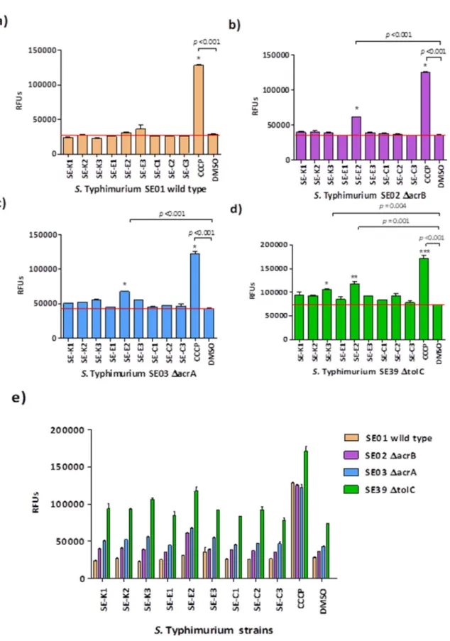
In case ofSalmonellastrains, the Se-compounds increased the intracellular EB accumulation more efficiently on thetolCgene inactivated mutantS. Typhimurium SE39 after 60 min. In contrast, RFUs obtained in the presence of Se-compounds were the lowest on the wild-typeS. Typhimurium SE01.
CCCP, the reference efflux pump inhibitor (EPI) was the positive control in case ofSalmonellaand referenceS. aureusstrain. In addition, verapamil was applied as reference EPI onS. aureusMRSA.
The solvent DMSO served as a negative control in the experiments. Se-E2significantly increased the intracellular EB accumulation onS. Typhimurium SE02, -03, and -39. In addition, a significant EB accumulation was observed forSe-K3onS. Typhimurium SE39 (Figure1).
Microorganisms2020,8, 566 8 of 15
Microorganisms 2020, 8, x FOR PEER REVIEW 8 of 15
Figure 1. Ethidium bromide (EB) accumulation in S. Typhimurium strains in the presence of Se- compounds. The graphs show the relative fluorescence units (RFUs) of (a) S. Typhimurium SE01, (b) S. Typhimurium SE02, (c) S. Typhimurium SE03, (d) S. Typhimurium SE39, and (e) all S.
Typhimurium bacterial strains in the presence of the compounds in the 60th minute of the assay. In case of S. Typhimurium SE01, -SE02 and -SE03 the level of significance was * p < 0.001. The levels of significance were * p = 0.004, ** p = 0.001, and *** p < 0.001 on S. Typhimurium SE39.
Figure 1.Ethidium bromide (EB) accumulation inS. Typhimurium strains in the presence of Se-compounds.
The graphs show the relative fluorescence units (RFUs) of (a)S.Typhimurium SE01, (b)S.Typhimurium SE02, (c)S.Typhimurium SE03, (d)S.Typhimurium SE39, and (e) allS.Typhimurium bacterial strains in the presence of the compounds in the 60th minute of the assay. In case ofS.Typhimurium SE01, -SE02 and -SE03 the level of significance was *p<0.001. The levels of significance were *p=0.004, **p=0.001, and
***p<0.001 onS.Typhimurium SE39.
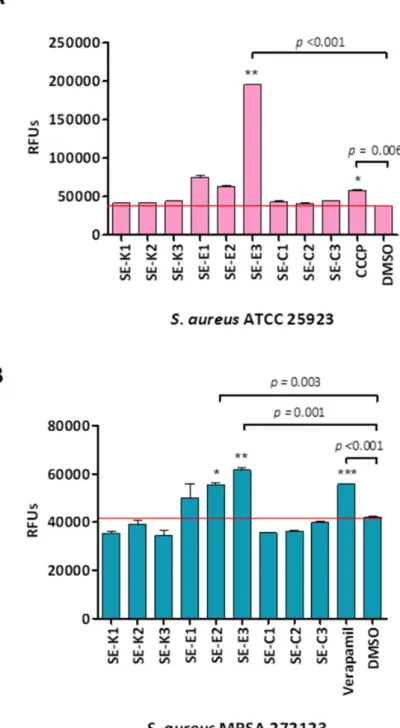
In case of the referenceS. aureusand resistant MRSA strain the highest RFUs were recorded in the presence ofSe-E3, for this reason this compound exerted the most prominent EPI activity. In addition, methylcarbonyl selenoestersSe-E1andSe-E2were proven to be effective in bothS. aureusstrains (Figure2).
Microorganisms 2020, 8, x FOR PEER REVIEW 9 of 15
In case of the reference S. aureus and resistant MRSA strain the highest RFUs were recorded in the presence of Se-E3, for this reason this compound exerted the most prominent EPI activity. In addition, methylcarbonyl selenoesters Se-E1 and Se-E2 were proven to be effective in both S. aureus strains (Figure 2).
Figure 2. EB accumulation on S. aureus strains. The graphs show the RFUs of (A) S. aureus ATCC 25923 (B) S. aureus MRSA 272123 bacterial strains in the presence of the compounds in the 60th minute of the assay. In case of S. aureus ATCC 25923 the levels of significance were * p = 0.006 and ** p < 0.001.
The levels of significance were * p = 0.003, ** p = 0.001, and *** p < 0.001 on S. aureus MRSA 272123.
Figure 2.EB accumulation onS. aureusstrains. The graphs show the RFUs of (A)S. aureusATCC 25923 (B)S. aureusMRSA 272123 bacterial strains in the presence of the compounds in the 60th minute of the assay. In case ofS. aureusATCC 25923 the levels of significance were *p=0.006 and **p<0.001.
The levels of significance were *p=0.003, **p=0.001, and ***p<0.001 onS. aureusMRSA 272123.
Microorganisms2020,8, 566 10 of 15
3.4. Measuring Biofilm Formation Using Crystal Violet
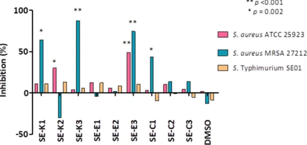
The effect of selenocompounds on biofilm formation of sensitive and resistantS. aureusstrains and wild-typeS.Typhimurium SE01 was evaluated. The biofilm inhibition (%) was calculated based on the mean of absorbance units (AUs). The absorbance expressed in AUs was the following on non-treated samples: referenceS. aureusshowed an absorbance of 2.4±0.1, the resistantS. aureus exhibited 1.3±0.1 AU, and the wild-typeS.Typhimurium presented 2.2±0.3 AU. Selenocompounds Se-K1(AU: 0.45±0.17; inhibition: 64.5%),Se-K3(AU: 0.16±0.06; inhibition: 84.7%),Se-E3(AU:
0.32±0.07; inhibition: 74.6%), andSe-C1(AU: 0.72±0.15; inhibition: 43.7%) could efficiently inhibit the biofilm formation ofS. aureusMRSA. In case of the referenceS. aureusstrain, the anti-biofilm effect was observed forSe-K2(AU: 1.67±0.10; inhibition: 30.3%) andSe-E3(AU: 1.22±0.17; inhibition:
74.6%). The compounds showed no significant anti-biofilm effect onS.Typhimurium SE01 (Figure3).
Microorganisms 2020, 8, x FOR PEER REVIEW 10 of 15
3.4. Measuring Biofilm Formation Using Crystal Violet
The effect of selenocompounds on biofilm formation of sensitive and resistant S. aureus strains and wild-type S. Typhimurium SE01 was evaluated. The biofilm inhibition (%) was calculated based on the mean of absorbance units (AUs). The absorbance expressed in AUs was the following on non- treated samples: reference S. aureus showed an absorbance of 2.4 ± 0.1, the resistant S. aureus exhibited 1.3 ± 0.1 AU, and the wild-type S. Typhimurium presented 2.2 ± 0.3 AU. Selenocompounds Se-K1 (AU: 0.45 ± 0.17; inhibition: 64.5%), Se-K3 (AU: 0.16 ± 0.06; inhibition: 84.7%), Se-E3 (AU: 0.32 ± 0.07;
inhibition: 74.6%), and Se-C1 (AU: 0.72 ± 0.15; inhibition: 43.7%) could efficiently inhibit the biofilm formation of S. aureus MRSA. In case of the reference S. aureus strain, the anti-biofilm effect was observed for Se-K2 (AU: 1.67 ± 0.10; inhibition: 30.3%) and Se-E3 (AU: 1.22 ± 0.17; inhibition: 74.6%).
The compounds showed no significant anti-biofilm effect on S. Typhimurium SE01 (Figure 3).
Figure 3. Anti-biofilm effect of Se-compounds on S. Typhimurium SE01 wild-type and on sensitive and resistant S. aureus strains. The levels of significance were ** p < 0.001 and * p = 0.002, respectively.
3.5. Quorum Sensing (QS) Assay
The sensor strain C. violaceum 026 and the AHL producer strains E. cloacae 31298 were inoculated as parallel lines. Interactions between the strains and compounds were evaluated for the reduction in the size of the zone of pigment production and the zone of growth inhibition of the affected strains, in millimeters. Promethazine (PMZ) was applied as a QS inhibitor and its zone of inhibition was 46 mm. Selenocompounds Se-K1, Se-K2, and Se-E1 had QS inhibitory effect. In addition, Se-K1 and Se- K2 showed inhibition zones of 37 mm and 40 mm, respectively, whereas the methyloxycarbonyl selenoester Se-E1 was the most effective QS inhibitor with an inhibition zone of 41 mm (Figure 4).
Figure 3.Anti-biofilm effect of Se-compounds onS. Typhimurium SE01 wild-type and on sensitive and resistantS. aureusstrains. The levels of significance were **p<0.001 and *p=0.002, respectively.
3.5. Quorum Sensing (QS) Assay
The sensor strainC. violaceum026 and the AHL producer strainsE. cloacae31298 were inoculated as parallel lines. Interactions between the strains and compounds were evaluated for the reduction in the size of the zone of pigment production and the zone of growth inhibition of the affected strains, in millimeters. Promethazine (PMZ) was applied as a QS inhibitor and its zone of inhibition was 46 mm. SelenocompoundsSe-K1,Se-K2, andSe-E1had QS inhibitory effect. In addition,Se-K1and Se-K2showed inhibition zones of 37 mm and 40 mm, respectively, whereas the methyloxycarbonyl selenoesterSe-E1was the most effective QS inhibitor with an inhibition zone of 41 mm (Figure4).
Figure 4. Quorum Sensing (QS) inhibition by selenocompounds. The QS-inhibition assay was performed using the parallel inoculation disk diffusion method. The ineffective compounds are not shown. Promethazine (PMZ) was used as a positive control.
3.6. Cytotoxicity Assay on Normal Human Fibroblasts
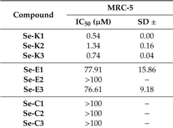
In order to determine the toxicity and safety of the selenocompounds on human cells, a cytotoxicity assay was performed using normal MRC-5 human embryonal lung fibroblast cells (Table 4).
Based on the data obtained, ketone selenoesters Se-K1, Se-K2, and Se-K3 presented high toxicity on normal cells (IC50 between 0.5 and 1.5 μM). Fortunately, the methylcarbonyl selenoesters (Se-E1, Se-E2, and Se-E3) and the cyano selenoesters (Se-C1, Se-C2, and Se-C3) showed no toxicity on normal cells as all their IC50 values were above 75 μM.
Table 4. Cytotoxic activity of selenocompounds on MRC-5 human embryonal fibroblast cells, expressed in Inhibitory Concentration 50 (IC50) and with the calculated standard deviation (SD).
Compound MRC-5
IC50 (µM) SD ±
Se-K1 0.54 0.00
Se-K2 1.34 0.16
Se-K3 0.74 0.04
Se-E1 77.91 15.86
Se-E2 >100 −
Se-E3 76.61 9.18
Se-C1 >100 −
Se-C2 >100 −
Se-C3 >100 −
4. Discussion
In case of MIC determination, the symmetrical selenoesters evaluated herein (whose selenium- bound alkyl moiety contains functional groups as a ketone, oxygen ester or nitrile) were more effective on sensitive and resistant S. aureus strains compared to the four S. Typhimurium bacterial strains. This suggests that these symmetrical selenoesters are more active against Gram-positive
Figure 4. Quorum Sensing (QS) inhibition by selenocompounds. The QS-inhibition assay was performed using the parallel inoculation disk diffusion method. The ineffective compounds are not shown. Promethazine (PMZ) was used as a positive control.
3.6. Cytotoxicity Assay on Normal Human Fibroblasts
In order to determine the toxicity and safety of the selenocompounds on human cells, a cytotoxicity assay was performed using normal MRC-5 human embryonal lung fibroblast cells (Table4).
Table 4.Cytotoxic activity of selenocompounds on MRC-5 human embryonal fibroblast cells, expressed in Inhibitory Concentration 50 (IC50) and with the calculated standard deviation (SD).
Compound MRC-5
IC50(µM) SD±
Se-K1 0.54 0.00
Se-K2 1.34 0.16
Se-K3 0.74 0.04
Se-E1 77.91 15.86
Se-E2 >100 −
Se-E3 76.61 9.18
Se-C1 >100 −
Se-C2 >100 −
Se-C3 >100 −
Based on the data obtained, ketone selenoestersSe-K1,Se-K2, andSe-K3presented high toxicity on normal cells (IC50between 0.5 and 1.5µM). Fortunately, the methylcarbonyl selenoesters (Se-E1, Se-E2, andSe-E3) and the cyano selenoesters (Se-C1,Se-C2, andSe-C3) showed no toxicity on normal cells as all their IC50values were above 75µM.
4. Discussion
In case of MIC determination, the symmetrical selenoesters evaluated herein (whose selenium-bound alkyl moiety contains functional groups as a ketone, oxygen ester or nitrile) were more effective on sensitive and resistantS. aureusstrains compared to the fourS.Typhimurium bacterial strains.
This suggests that these symmetrical selenoesters are more active against Gram-positive bacteria (as Staphylococcus aureus) than against Gram-negative bacteria (asSalmonella entericaserovar Typhimurium).
This fact is in accordance with the antibacterial activity of non-symmetrical selenoesters, which were evaluated in a previous work of the group [27]; only three non-symmetrical ketone selenoesters
Microorganisms2020,8, 566 12 of 15
(9–11in [27]) were active againstS. aureus, whereas none of them were active againstEscherichia coli.
Interestingly, all of them were active againstChlamydia trachomatis(Gram-negative), but sinceChlamydia is an intracellular bacterium this may affect its sensitivity to the compounds [27].
The methylketone selenoestersSe-K1,Se-K2, andSe-K3were the most potent antibacterials on referenceS. aureus. In contrast, the methyloxycarbonyl selenoestersSe-E1,Se-E2, andSe-E3and the cyano selenoestersSe-C1andSe-C2showed strong resistance modulating activity with tetracycline against the MRSA strain. Comparing the antibacterial activity with the previously reported data [27], two observations are of interests. First, the symmetrical selenoesters are more potent antibacterials againstS. aureusATCC 25923 than the respective asymmetrical derivatives. This is observed when we compare the 0.39µM MIC values ofSe-K1,Se-K2, andSe-K3with the 3.12µM MIC value of 9in [27] (methylketone selenoesters), and the 12.5µM MIC values ofSe-E1,Se-E2andSe-E3with 7in [27], which was not active at concentrations below 100µM (methyloxycarbonyl selenoesters).
Second, the symmetrical methyl selenoesters2–5in [27] were not active againstS. aureusATCC 25923 (MIC>100µM), whereas all the functionalized selenoesters evaluated in this work (-CH2COCH3, -CH2COOCH3, -CH2CN) showed MIC values against this strain at 12.5µM or lower. This indicates that these second-generation selenoesters have improved antibacterial activity compared with those that have been previously reported.
If we compare the antibacterial activity of the symmetrical selenocompounds with its toxicity against MRC-5 normal embryonal lung fibroblast cell line, we observe that the MIC values of the compounds againstS. aureusATCC 25923 were lower than the IC50values against this cell line.
In the resistance modulation assay, the selenocompounds were tested at 12 of their MIC in combination with tetracycline and ciprofloxacin in the twoS. aureusstrains (ATCC 25923 and MRSA 272123). As mentioned previously, all compounds were able to modulate the activity of tetracycline againstS. aureusMRSA 272123. The results were somehow comparable with the antibacterial activity.
Interestingly, the –CH2COOCH3and –CN containing symmetrical selenoesters were more potent modulators than the –CH2COCH3selenoesters (X-fold reductions of 2–4, 8–32, and 4–32, respectively).
However, as MIC values of the selenocompounds were higher against thisS. aureusstrain, onlySe-C1 andSe-C2could be used at a safe concentration (25µM, non-toxic in MRC-5 cells) with a noteworthy effect (16- and 32-fold reduction of MIC value of tetracycline).
Real-time EB accumulation was applied in order to monitor the EPI activity of the compounds.
The intracellular EB accumulation was the highest on thetolCgene inactivated mutantS. Typhimurium SE39, and the lowest EB accumulation was obtained in the wild-typeS. Typhimurium SE01 in the presence of methyloxycarbonyl selenoester Se-E2. This compound significantly increased the EB accumulation in the efflux pump gene inactivated (∆acrA,∆acrB, and∆tolC) mutantS. Typhimurium strains due to efflux independent mechanisms, e.g., membrane destabilizing effect. In addition, methyloxycarbonyl selenoesterSe-E3 showed significantly effective pump inhibition on sensitive (p<0.001) and resistant (p=0.001)S. aureusstrains. Unfortunately, these two Se-compounds have to be applied at a high concentration (50µM, which is12of their MIC) againstS.Typhimurium (Se-E2) orS. aureusMRSA 272123 (Se-E3), respectively. CompoundSe-E3could be used in this application againstS. aureusATCC 25923, as in this case its concentration would be 6.25µM, much lower.
Regarding the anti-biofilm effect, the methyloxycarbonyl selenoesterSe-E3showed significant biofilm inhibition on both of sensitive and resistantS. aureusstrains. Furthermore, the methylketone selenoesterSe-K3was the most effective anti-biofilm agent on resistantS. aureusMRSA. In addition, Se-K1was also interesting, as it showed a biofilm inhibiting effect higher than 50% against MRSA.
It was surprising that Se-K2promoted the biofilm formation of S. aureus MRSA, because it has the same chemical formula asSe-K1(both are 2-oxopropyl selenodiesters); they only differ in the substitution pattern at the phenyl ring, such thatSe-K1has aparasubstitution (1,4) andSe-K2has ametasubstitution (1,3). It is interesting to see how such a small change in the substitution pattern at the core phenyl ring leads to completely different activities. What is more, inSe-K2the inclusion of a third –COSeCH2COCH3at the position five of the core phenyl ring led toSe-K3, recovering
the biofilm inhibition in respect toSe-K2and enhancing it in respect toSe-K1. In the case of the methyloxycarbonyl selenoesters, only the trisubstituted derivativeSe-E3was capable of significantly inhibiting the biofilm formation in both strains ofS. aureus(reference and MRSA), whereas the two disubstituted ones were inactive. Methylcyano selenoesters showed a lower inhibition than the other two families of compounds, however, one of them (thepara-disubstituted (Se-C1)) was close to exerting a 50% inhibition ofS. aureusMRSA.
Finally, QS inhibiting effect of compounds was evaluated based on the inhibition of violacein production. The methylketone selenoesterSe-K1andSe-K2and the methyloxycarbonyl selenoester Se-E1were potent QS-inhibitors, with Se-E1 being the most effective QS inhibitor of these three derivatives by showing an inhibition close to the reference promethazine (positive control).
All these findings reveal that the symmetrical selenoesters have a potent antibacterial activity, mainly againstS. aureusstrains. Furthermore, the methylcyano selenoesters could be used as potential novel antibiotics. Additional studies to evaluate the ADME-Tox properties of these compounds is needed to evaluate their applicability in medicine more in-depth. Besides, the methylketone selenoesters, which are less selective, still could be used, for example, in disinfection of surfaces or in the coating of surfaces to prevent biofilm formation.
5. Conclusions
It can be concluded that all the symmetrical selenoesters evaluated have a potent antibacterial activity againstS. aureusATCC 25923. The most potent derivatives were the methylketone selenoesters (Se-K1,Se-K2, andSe-K3), followed by the cyano selenoesters (Se-C1,Se-C2, andSe-C3), and at the end by the methyloxycarbonyl selenoesters (Se-E1,Se-E2, andSe-E3). After determining the toxicity on normal fibroblasts, the more selective ones were the cyano selenoesters, followed by the methyloxycarbonyl selenoesters, and the ones by the methylketone selenoesters. Combining both the antibacterial activity and the cytotoxic activity, the most promising compound againstS. aureusATCC 25923 wasSe-C3. The tested selenocompounds also showed antibacterial activity againstS. aureus MRSA 272123 and against different strains ofS.Typhimurium, although with higher MIC values.
In addition to the antibacterial activity, the methyloxycarbonyl selenoesters and two cyano selenoesters showed strong resistance reversing activity in the presence of tetracycline against the MRSA strain. Additionally, the methyloxycarbonyl selenoesterSe-E3was the most effective compound concerning the reversal of resistance, efflux pump inhibition, and anti-biofilm activity onS. aureusstrains.
6. Patents
This work explores the antibacterial activity of compounds covered by the patent EP18382693 [21]
(filed on 28 September 2018 by Enrique Domínguez-Álvarez, Gabriella Spengler, Claus Jacob and Carmen Sanmartín) more in-depth.
Author Contributions: G.S. conceived and designed the study. A.G.-P., M.B.-L., and E.D.-Á. synthesized the selenocompounds used in the study. M.N., B.S., B.R., and A.K. performed the laboratory work. M.N., G.S., and E.D.-Á. wrote the article. J.M.A.B. revised the manuscript critically. All authors read and approved the final manuscript.
Funding: The study was supported by the projects SZTE ÁOK-KKA 2018/270-62-2 of the University of Szeged, Faculty of Medicine and GINOP-2.3.2-15-2016-00038 (Hungary). M.N. was supported by EFOP 3.6.3-VEKOP-16-2017-00009. E.D-A. was supported by ‘Iniciativas Ropelanas’ and ‘Asociación Cultural Trevinca’, two associations from Zamora (Spain), that promote cancer research.
Conflicts of Interest: The authors declare no conflict of interest. The funders had no role in the design of the study; in the collection, analyses, or interpretation of data; in the writing of the manuscript, or in the decision to publish the results.
Microorganisms2020,8, 566 14 of 15
References
1. Carlet, J.; Pulcini, C.; Piddock, L.J. Antibiotic resistance: A geopolitical issue.Clin. Microbiol. Infect.2014,20, 949–953. [CrossRef] [PubMed]
2. Reygaert, W.C. An overview of the antimicrobial resistance mechanisms of bacteria.AIMS Microbiol.2018,4, 482–501. [CrossRef] [PubMed]
3. Hassan, K.A.; Liu, Q.; Elbourne, L.D.H.; Ahmad, I.; Sharples, D.; Naidu, V.; Chan, C.L.; Li, L.; Harborne, S.P.D.;
Pokhrel, A.; et al. Pacing across the membrane: The novel PACE family of efflux pumps is widespread in Gram-negative pathogens.Res. Microbiol.2018,169, 450–454. [CrossRef] [PubMed]
4. Sharma, A.; Gupta, V.K.; Pathania, R. Efflux pump inhibitors for bacterial pathogens: From bench to bedside.
Indian J. Med. Res.2019,149, 129–145. [CrossRef] [PubMed]
5. Colclough, A.L.; Alav, I.; Whittle, E.E.; Pugh, H.L.; Darby, E.M.; Legood, S.W.; McNeil, H.E.; Blair, J.M. RND efflux pumps in Gram-negative bacteria; regulation, structure and role in antibiotic resistance.Future Microbiol.
2020,15, 143–157. [CrossRef]
6. Santos, A.; Galdino, A.C.M.; Mello, T.P.; Ramos, L.S.; Branquinha, M.H.; Bolognese, A.M.; Columbano Neto, J.; Roudbary, M. What are the advantages of living in a community? A microbial biofilm perspective!
Mem. Inst. Oswaldo Cruz2018,113, e180212. [CrossRef]
7. Li, W.; Li, Y.; Liu, Y.; Shi, X.; Jiang, M.; Lin, Y.; Qiu, Y.; Zhang, Q.; Chen, Q.; Zhou, L.; et al. Clonal Expansion of Biofilm-Forming Salmonella Typhimurium ST34 with Multidrug-Resistance Phenotype in the Southern Coastal Region of China.Front. Microbiol.2017,8, 2090. [CrossRef]
8. Hennekinne, J.A.; De Buyser, M.L.; Dragacci, S. Staphylococcus aureus and its food poisoning toxins:
Characterization and outbreak investigation.FEMS Microbiol. Rev.2012,36, 815–836. [CrossRef]
9. Knowles, J.R.; Roller, S.; Murray, D.B.; Naidu, A.S. Antimicrobial action of carvacrol at different stages of dual-species biofilm development by Staphylococcus aureus and Salmonella enterica serovar Typhimurium.
Appl. Environ. Microbiol.2005,71, 797–803. [CrossRef]
10. Abisado, R.G.; Benomar, S.; Klaus, J.R.; Dandekar, A.A.; Chandler, J.R. Bacterial Quorum Sensing and Microbial Community Interactions.mBio2018,9. [CrossRef]
11. Saxena, P.; Joshi, Y.; Rawat, K.; Bisht, R. Biofilms: Architecture, Resistance, Quorum Sensing and Control Mechanisms.Indian J. Microbiol.2019,59, 3–12. [CrossRef] [PubMed]
12. Huang, T.; Holden, J.A.; Heath, D.E.; O’Brien-Simpson, N.M.; O’Connor, A.J. Engineering highly effective antimicrobial selenium nanoparticles through control of particle size. Nanoscale2019,11, 14937–14951.
[CrossRef] [PubMed]
13. Staicu, L.C.; Oremland, R.S.; Tobe, R.; Mihara, H. Bacteria versus selenium: A view from the inside out.
InSelenium in Plants; Springer: Cham, Switzerland, 2017; pp. 79–108.
14. Verma, P. A review on synthesis and their antibacterial activity of Silver and Selenium nanoparticles against biofilm forming Staphylococcus aureus.World J. Pharm. Pharmaceut. Sci.2015,4, 652–677.
15. Mosolygo, T.; Kincses, A.; Csonka, A.; Tonki, A.S.; Witek, K.; Sanmartin, C.; Marc, M.A.; Handzlik, J.;
Kiec-Kononowicz, K.; Dominguez-Alvarez, E.; et al. Selenocompounds as Novel Antibacterial Agents and Bacterial Efflux Pump Inhibitors.Molecules2019,24, 1487. [CrossRef]
16. Witek, K.; Nasim, M.J.; Bischoff, M.; Gaupp, R.; Arsenyan, P.; Vasiljeva, J.; Marc, M.A.; Olejarz, A.; Latacz, G.;
Kiec-Kononowicz, K.; et al. Selenazolinium Salts as “Small Molecule Catalysts” with High Potency against ESKAPE Bacterial Pathogens.Molecules2017,22, 2174. [CrossRef]
17. Spengler, G.; Kincses, A.; Mosolygo, T.; Marc, M.A.; Nove, M.; Gajdacs, M.; Sanmartin, C.; McNeil, H.E.;
Blair, J.M.A.; Dominguez-Alvarez, E. Antiviral, Antimicrobial and Antibiofilm Activity of Selenoesters and Selenoanhydrides.Molecules2019,24, 4264. [CrossRef]
18. Medina Cruz, D.; Mi, G.; Webster, T.J. Synthesis and characterization of biogenic selenium nanoparticles with antimicrobial properties made by Staphylococcus aureus, methicillin-resistant Staphylococcus aureus (MRSA), Escherichia coli, and Pseudomonas aeruginosa. J. Biomed. Mater. Res. A2018,106, 1400–1412.
[CrossRef]
19. Khiralla, G.M.; Bahig, A.E.D. Antimicrobial and antibiofilm effects of selenium nanoparticles on some foodborne pathogens.LWT Food Sci. Technol.2015,63, 1001–1007. [CrossRef]
20. La Cruz-Claure, D.; María, L.; Cèspedes-Llave, A.A.; Ulloa, M.T.; Benito-Lama, M.; Domínguez-Álvarez, E.;
Bastida, A. Inhibition–Disruption of Candida glabrata Biofilms: Symmetrical Selenoesters as Potential Anti-Biofilm Agents.Microorganisms2019,7, 664. [CrossRef]
21. DomínguezÁlvarez, E.; Spengler, G.; Jacob, C.; Sanmartín Grijalba, M.C. Selenoester-Containing Compounds for Use in the Treatment of Microbial Infections or Colorectal Cancer. European Patent EP18382693, 28 September 2018.
22. Wray, C.; Sojka, W.J. Experimental Salmonella typhimurium infection in calves. Res. Vet. Sci. 1978,25, 139–143. [CrossRef]
23. Blair, J.M.; La Ragione, R.M.; Woodward, M.J.; Piddock, L.J. Periplasmic adaptor protein AcrA has a distinct role in the antibiotic resistance and virulence of Salmonella enterica serovar Typhimurium.J. Antimicrob.
Chemother.2009,64, 965–972. [CrossRef] [PubMed]
24. Eaves, D.J.; Ricci, V.; Piddock, L.J. Expression of acrB, acrF, acrD, marA, and soxS in Salmonella enterica serovar Typhimurium: Role in multiple antibiotic resistance. Antimicrob. Agents Chemother. 2004, 48, 1145–1150. [CrossRef]
25. Buckley, A.M.; Webber, M.A.; Cooles, S.; Randall, L.P.; La Ragione, R.M.; Woodward, M.J.; Piddock, L.J.
The AcrAB-TolC efflux system of Salmonella enterica serovar Typhimurium plays a role in pathogenesis.
Cell Microbiol.2006,8, 847–856. [CrossRef] [PubMed]
26. Ballantine, J.; Beer, R.; Crutchley, D.; Dodd, G.; Palmer, D. The synthesis of violacein and related compounds.
Proc. Chem. Soc. Lond.1958,8, 232–233.
27. Kothari, V.; Sharma, S.; Padia, D. Recent research advances on Chromobacterium violaceum.Asian Pac. J.
Trop Med.2017,10, 744–752. [CrossRef]
28. CLSI. Susceptibility Testing Process. InMethods for Dilution Antimicrobial Susceptibility Tests for Bacteria that Grow Aerobically, 10th ed.; Christopher, P.J., Polgar, E.P., Eds.; Clinical and Laboratory Standards Institute:
Wayne, MI, USA, 2015; Volume 32, pp. 15–49.
29. Gajdacs, M.; Spengler, G. The Role of Drug Repurposing in the Development of Novel Antimicrobial Drugs:
Non-Antibiotic Pharmacological Agents as Quorum Sensing-Inhibitors. Antibiotics (Basel)2019, 8, 270.
[CrossRef]
©2020 by the authors. Licensee MDPI, Basel, Switzerland. This article is an open access article distributed under the terms and conditions of the Creative Commons Attribution (CC BY) license (http://creativecommons.org/licenses/by/4.0/).