Supported by
Accepted Article
Title: Flexibility of the CueR metal site probed by instantaneous change of element and oxidation state from AgI to CdII
Authors: Ria Katalin Balogh, Bela Gyurcsik, Mikael Jensen, Peter Waaben Thulstrup, Ulli Köster, Niels Johan Christensen, Frederik J. Mørch, Marianne L. Jensen, Attila Jancsó, and Lars Hemmingsen
This manuscript has been accepted after peer review and appears as an Accepted Article online prior to editing, proofing, and formal publication of the final Version of Record (VoR). This work is currently citable by using the Digital Object Identifier (DOI) given below. The VoR will be published online in Early View as soon as possible and may be different to this Accepted Article as a result of editing. Readers should obtain the VoR from the journal website shown below when it is published to ensure accuracy of information. The authors are responsible for the content of this Accepted Article.
To be cited as: Chem. Eur. J. 10.1002/chem.202000132
Link to VoR: http://dx.doi.org/10.1002/chem.202000132
PAPER
Flexibility of the CueR metal site probed by instantaneous change of element and oxidation state from Ag I to Cd II
Ria K. Balogh,
[b]Bela Gyurcsik,
[b]Mikael Jensen,
[c]Peter W. Thulstrup,
[a]Ulli Köster,
[d]Niels Johan Christensen,
[e]Frederik J. Mørch,
[a]Marianne L. Jensen,
[f]Attila Jancsó,*
[b]Lars Hemmingsen*
[a][a] Prof. P.W. Thulstrup, F.J. Mørch, Prof. L. Hemmingsen Department of Chemistry, University of Copenhagen Universitetsparken 5, 2100 Copenhagen, Denmark E-mail: lhe@chem.ku.dk
[b] R.K. Balogh, Prof. B. Gyurcsik, Prof. A. Jancsó
Department of Inorganic and Analytical Chemistry, University of Szeged Dóm tér 7, 6720 Szeged, Hungary
E-mail: jancso@chem.u-szeged.hu [c] Prof. M. Jensen
Hevesy Laboratory, Center for Nuclear Technologies, Technical University of Denmark Frederiksborgvej 399, 4000 Roskilde, Denmark
[d] Dr. U. Köster Institut Laue-Langevin
71 avenue des Martyrs, 38042 Grenoble, France [e] Asst. Prof. N.J. Christensen
Department of Chemistry, Faculty of Science, University of Copenhagen Thorvaldsensvej 40, 1871 Frederiksberg C, Denmark
[f] M.L. Jensen
Niels Bohr Institute, University of Copenhagen Universitetsparken 5, 2100 Copenhagen, Denmark
Supporting information for this article is given via a link at the end of the document.
Abstract: Selectivity for monovalent metal ions is an important facet of the function of the metalloregulatory protein CueR. 111Ag perturbed angular correlation of -rays (PAC) spectroscopy probes the metal site structure and the relaxation accompanying the instantaneous change from AgI to CdII upon 111Ag radioactive decay. That is, a change from AgI which activates transcription to CdII which does not. In the frozen state (-196 °C) two nuclear quadrupole interactions (NQIs) are observed; one (NQI1) agrees well with two coordinating thiolates and an additional longer contact to the S77 backbone carbonyl, and the other (NQI2) reflects that CdII has attracted additional ligand(s). At 1 °C only NQI2 is observed, demonstrating that relaxation to this structure occurs within ~10 ns of the decay of 111Ag. Thus, transformation from AgI to CdII rapidly disrupts the functional linear bis(thiolato)AgI metal site structure. This inherent metal site flexibility may be central to CueR function, leading to remodeling into a non-functional structure upon binding of non-cognate metal ions. In a broader perspective,
111Ag PAC spectroscopy may be applied to probe the flexibility of protein metal sites.
Introduction
Maintaining the intracellular concentration of metal ions at an optimal level is important for all organisms. For example, the copper homeostasis of Escherichia coli is managed partially by the cue system including CopA, CueO and CueR proteins.[1, 2]
CopA is a P-type ATPase, transporting CuI ions into the periplasmic space. CueO is a periplasmic multicopper oxidase
that converts CuI to less harmful CuII.[3] CueR functions as a transcriptional metalloregulatory protein.[4] Under normal conditions, apo-CueR is bound to the promoter region of the cue operon. Upon cognate metal ion binding, it activates the transcription of the downstream copA and the cueO genes.[1, 2, 4]
CueR belongs to the MerR metalloregulator family sharing the common fold of the dimeric structure. The main units are: An N-terminal winged helix DNA binding domain, followed by a dimerization helix and a C-terminal metal binding domain (Figure 1A).[5, 6] The C-terminal region of MerR family proteins is more diverse in terms of sequence and structure than the other segments and this region is responsible for the recognition of various types of effectors, including metal ions.[7-9] Crystal structures of the metal ion bound form of the MerR[10, 11], ZntR[5], CueR[5, 6] and CadR[12] proteins suggest that the metal ion binding region of MerR proteins has evolved to provide an appropriate number and geometry of ligands for the regulated metal ions.[8] In addition, other residues surrounding the metal site may influence the metal ion recognition via charge neutralization, hydrophobic or steric restrictions.[8, 13]
CueR activates the transcription only in the presence of the monovalent group 11 transition metal ions CuI, AgI and AuI.[4, 5, 14]
These cognate metal ions bind to two cysteine thiolates (C112, C120) of the metal binding loop in a linear coordination geometry (Figure 1B).[5, 6] The structure of the AgI-bound activator form of CueR co-crystallized with DNA[6] shows that DNA binding does not alter the coordination characteristics of the metal ion to the protein.
Accepted Manuscript
FULL PAPER
Figure 1. A: Overall structure of the activator form of CueR (PDB id.: 4WLW).[6]
AgI ions are shown as grey spheres. B: Close-up view of the metal binding loop.
Residues from C112 to C120 of one monomer, and S77 of the other monomer are indicated explicitly. AgI is coordinated by C112 and C120 in a linear dithiolate fashion.
Analysis of the electrostatic- and hydrogen-bonding interactions in the crystal structure suggested that net negative charge of the S-Cu-S center might be partly neutralized by a helix dipole and a fairly distant lysine residue.[5] Furthermore, the S77 residue of the other monomer was found to interact with the C112 and D115 residues of the metal binding loop stabilizing its structure.[5, 15]
In this work, the structure and flexibility of the metal ion binding site of the Wild Type E. coli CueR was probed by 111Ag and 111mCd perturbed angular correlation of γ-rays (PAC) spectroscopy.[16, 17] The radioactive decay of 111Ag is described in some detail here, as a prerequisite for the interpretation of the PAC spectroscopic data in terms of metal site structure. 111Ag decays by β− emission to 111Cd, and with a ~5% probability an excited state of the 111Cd nucleus (342 keV) is populated (Figure 2). This state may decay by successive emission of two γ-rays, thereby providing the γ-γ cascade required for PAC spectroscopy.[16] The intermediate (245 keV) level in this γ-γ cascade has a half-life of 85 ns, and it is the nuclear quadrupole interactions (NQI) experienced by the Cd nucleus in this state that are recorded in the 111Ag PAC experiments. Accordingly, the observed spectra may reflect one of three possible scenarios: 1) Structural relaxation from the AgI coordination geometry to that of CdII occurs rapidly (faster than ~10 ns), and the 111Ag PAC experiments reflect the relaxed structure; 2) relaxation occurs on the PAC time scale (10-200 ns), and the recorded NQIs change as a function of time on this ns time scale, or 3) the relaxation is slower than ~200 ns, and the coordination geometry of AgI is observed.
An instantaneous change of element and oxidation state from AgI to CdII accompanies the decay of 111Ag, vide supra.
Moreover, in the nuclear decay the Cd-nucleus receives a recoil
energy corresponding to the momenta of the emitted β− and antineutrino. The continuous spectrum of 111Cd recoil energies in the β− decay of 111Ag extends up to a maximum of 557 kJ/mol, but the phase space for such high values is negligibly small. In fact, the recoil energies are typically smaller by a factor of 2 or more.
Due to the change of element, change of redox state, and the recoil energy, the system is shifted out of equilibrium, and the subsequent relaxation process reflects the rigidity/flexibility of the metal site. In the current case the change from AgI to CdII is particularly interesting, because it is a change from a metal ion which induces activation of transcription (AgI) to a metal ion which does not (CdII). Moreover, while both metal ions favour the binding of thiolate donors, the AgS2 coordination mode is highly unusual for Cadmium, and linear coordination geometry is only observed for CdMe2 and the dihalides in the gas phase.[18-20] Thus, a fundamental question is if the CueR metal site is sufficiently rigid that CdII remains transiently in the linear two-coordinate structure, or if the preference of CdII for higher coordination numbers dominates and leads to remodelling of the metal site coordination geometry. In a broader perspective, 111Ag PAC spectroscopy provides insight into the local potential energy minima that may be realized at protein metal sites by the change from AgI to CdII and its initial recoil kinetic energy. This local metal site adaptability/rigidity is likely to be an important facet – beyond structure - of metalloprotein function, and it is related to the so- called entatic state proposed to account for the properties of Type 1 Cu centres in blue copper proteins.[21-23]
Figure 2. Decay schemes of 111Ag and 111mCd including information about the nuclear spin, the energy and the half-life of the energy levels. The solid arrows indicate the γ-γ-cascade used in PAC spectroscopy. Notice that the 111Ag and
111mCd decay pass through the same intermediate state (I = 5/2), and it is the NQIs experienced by the Cd-nucleus in this state, that are recorded in 111Ag and
111mCd PAC spectroscopy.
Results and Discussion
The bacterial metalloregulator protein CueR discriminates very effectively between mono- and divalent metal ions. It is a central tenet that the selectivity is achieved by the unique two- coordinated site occupied by CuI, AgI and AuI, while divalent metal ions usually require higher coordination numbers.
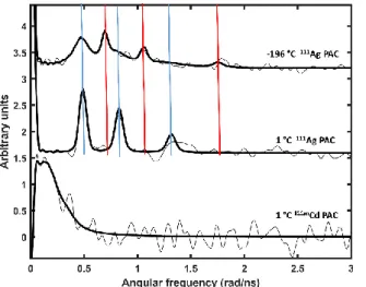
Initially we conducted experiments on AgI-CueR at −196 °C to prevent or slow down the structural relaxation occurring after the decay of 111Ag to 111Cd. Two NQIs were observed, see Figure 3. The high frequency NQI1 is analysed unambiguously, given that all three peaks characteristic of a 111Ag PAC signal rise above the noise level, and they satisfy the requirement ω1+ω2=ω3.[16] The second NQI2 is characterized by a lower frequency, and we present the best fit in Table 1. Analysing the first 100 ns of the
Accepted Manuscript
PAC data gives essentially the same two NQIs as analysis of the data from 100 ns to 200 ns after the decay. This indicates that no structural changes occur within the time span from ~10 ns to 200 ns after the decay, and we therefore rule out structural relaxation on the 10-200 ns time scale. That is, the two observed NQIs reflect structures which are stable on the PAC time scale (up to
~200 ns) at a temperature of -196 °C
Figure 3. 111Ag- and 111mCd-PAC spectra of CueR. Top: at −196 °C two different NQIs characterized by a high frequency signal (red), NQI1, and a low frequency signal (blue), NQI2, are observed; Middle: at 1 °C only the low frequency signal, NQI2, is observed; Bottom: the 111mCd-PAC spectrum of CdII bound to CueR gives a very low frequency signal. The experimental 111Ag data are multiplied by -1 to allow for comparison to the 111mCd PAC data. Experimental and fitted data are shown by thin and thick black lines, respectively. cCueR = 11 µM, cAgI = 5.5 µM, cCdII = 11 µM, cDNA= 5.5 µM, w/w%sucrose = 55%, pH = 7.3 at room temperature
The high frequency signal is relatively straight forward to interpret in terms of metal site structure. Both semi-empirical AOM[24] and DFT calculations indicate that CdII remains essentially in the AgI coordination geometry, see Figure 4, possibly establishing a long contact (2.59 Å) with the S77 carbonyl oxygen. An overlay with the AgI-CueR crystal structure is presented in Figure S5, illustrating that the protein atoms move very little, while CdII shifts by ~0.7 Å towards the Ser77 carbonyl oxygen. The significant deviation of the asymmetry parameter from zero (η = 0.51) indicates that the structure is distorted, i.e.
the linear coordination observed in the crystal structures is not maintained, but the very high frequency (ω0 = 0.555 rad/ns) agrees well with coordination by two thiolates and no coordinating ligands in the equatorial plane. Typical Cd-O bond lengths for a carbonyl oxygen found in the Cambridge Structural Database are in the range of 2.24-2.41 Å,[12] and are expected to be even shorter for the 2-3 coordinate CdII observed in this work. Thus, the long contact given by the constrained DFT geometry optimization, Figure 4, is clearly beyond normal bond length. That is, this signal reflects distorted linear bis(thiolato)CdII with an additional interaction with the carbonyl oxygen of S77.
The other signal, NQI2, is more difficult to interpret unambiguously because several possible structures map into the observed frequency range. However, the lower frequency relative to NQI1 strongly indicates a coordination number higher than 2, either as a result of coordination by a nearby amino acid residue
or an extraneous ligand such as a solvent water molecule. Finally, there is a minor fraction (< 10%) of the signal that is fitted as an exponential decay of the perturbation function. This may reflect CdII ions ejected from the metal site due to the recoil energy in the β-decay, vide supra, and this small fraction of the signal is therefore affected either by dynamics or unspecific binding. There
are two possible explanations of the observation of the two well defined NQIs (NQI1 and NQI2), indicated hereafter as Scenario A and B.
Figure 4. Geometry-optimized metal site structure with CdII at the CueR metal site (BP86-D3BJ/def2-TZVP) and calculated electric field gradient (EFG) parameters (DKH2 Hamiltonian, BHandHLYP with def2-QZVPP on CdII and def2-TZVP on remaining atoms), see the text for details. The S-CdII-S angle is indicated in transparent blue. The EFG parameters derived from the experimental data presented in Table 1 for NQI1 are: Vzz = 3.9 au and η = 0.51 in fair agreement with the calculated EFG (using Q = 0.64 barn for the intermediate nuclear level of 111Cd[25]). Coordinates for the optimized geometry are included in the Supporting Information.
Scenario A: There are two co-existing coordination geometries for AgI bound to the CueR metal site. One of the signals (NQI1) would then reflect that CdII remains in the CdS2(O) structure (indicating that there is a long contact to the carbonyl oxygen of S77). The other could involve coordination by a water molecule or another extraneous ligand, giving rise to NQI2. A coordination number of 3 at the CueR metal site is in conflict with the EXAFS and XANES data measured for the CuI-CueR complex,[26] although it is conceivable that the XANES data would exhibit the characteristic 1s to 4p transition at 8983 eV even upon bending of the S-CuI-S unit.[27] Thus, a structure similar to that presented in Figure 4, with a remote equatorial oxygen ligand might exist. The similarity of the CuI-CueR XANES spectra to those of the linear model complex presented by Chen et al.[26] is however striking, making the alternative interpretation of a bent CuS2 structure unlikely. An alternative structural interpretation may be inspired by our previous suggestion for the CueR metal site with one of the cysteines coordinating in the protonated form.[15] Within this model, it is conceivable that the low frequency signal could originate from a species where AgI is coordinated by one thiolate and one thiol. If so, a pH dependent change is expected for the relative intensities of the low and high frequency signals, reflecting the pKa of the coordinated thiol. In order to explore if this type of protonation equilibrium exists, we carried out a pH series (pH 5.7 - 8.0) of 111Ag PAC experiments. The data do not exhibit any clear pH dependence, see Figure S1, i.e. we were not able to detect a pKa for the deprotonation of a coordinated thiol, and thus we can exclude that NQI2 represents a species where CdII is coordinated by a thiolate and a protonated thiol ligand. However, the decay from the monovalent AgI to the
Accepted Manuscript
FULL PAPER
divalent CdII provides a considerable driving force for the deprotonation. Even at −196 C it is possible that the proton would rapidly dissociate from the thiol group, possibly by tunnelling. Therefore, the results presented here cannot confirm nor reject the existence a coordinated thiol for the monovalent metal ions. Finally, we tested if chloride might coordinate in AgI- CueR, and this does not appear to be the case, see Figure S2.
Similarly, we explored the effect of DNA binding and the presence of sucrose (used to limit the rotational diffusion), neither of which changed the NQI parameters significantly, and thus the observed structures were not affected, although the population of the two species may change, see Figures S3 and S4.
Scenario B: There is only one coordination geometry for AgI which relaxes into two different CdII coordination geometries via the radioactive decay. At −196 °C structural relaxation is limited for the atoms in the protein, but CdII may move, possibly with no barrier, to a local minimum on the free energy surface. Moreover, the β− emission gives rise to a recoil energy of up to 557 kJ/mol, which may drive CdII into (two) different local energy minima, depending on the direction of the velocity vector with respect to
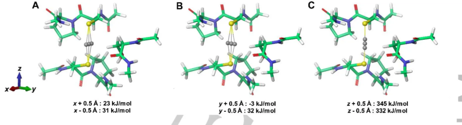
the molecular structure. In Figure 5 we present a very simple analysis of the potential energy change upon moving CdII 0.5 Å from the position of AgI either along the S-Cd-S axis (z-axis) or within the equatorial plane. Clearly, the potential energy surface is relatively shallow for movement of CdII within the equatorial plane, while the potential energy rises steeply upon movement along the S-Cd-S axis. A recoil kinetic energy of some hundreds of kJ/mol may therefore give rise to considerable movement of CdII, if the velocity vector lies within equatorial plane, while a much larger energy barrier is encountered for movement along the S- Cd-S axis, as expected. Thus, in a very simple model, about 1/3 of the decays give rise to recoil along the z-axis, but the energy is quickly dissipated and CdII relaxes to the structure presented in Figure 4. The other 2/3 of the decays give rise to recoil in the equatorial plane, and the much lower potential energy change along this “reaction coordinate” allows CdII to be shifted to a different (local) energy minimum. This very simple model predicts a ratio of 1/2 of the two structures, in fair agreement with the observed ratio of amplitudes (2.3/7.4) of the two NQIs, see Table 1. In summary, we find scenario B more likely than scenario A.
Figure 5. Change of potential energy upon translation of CdII away from the position occupied by AgI in CueR calculated using DFT (BP86/def2-T2VP). A) translation along x, B) translation along y, C) translation along Z corresponding to S-Cd-S axis.Movement of CdII by 0.5 Å along the S-Cd-S axis gives rise to changes comparable to the recoil kinetic energy which 111Cd receives in the nuclear decay, while the energy upon movement of 0.5 Å in the equatorial plane gives rise to potential energy changes which are an order of magnitude smaller.
Table 1. Parameters fitted to the 111Ag PAC and 111mCd PAC-data for CueR.
The rotational correlation time was fixed to infinite at −196 °C because it is assumed that the protein is completely immobilized. See reference [16] for a description of the parameters.
Isotope T ω0 η δ 1/τc A χr2
(°C) (rad/ns) ×100 µs-1 ×100
111Ag −196 0.555(2)
0.41(1) 0.510(8) 0.39(4) 2.5(6)
16(2) 0(fix)
0(fix) −2.3(4)
−7.4(1) 0.93
111Ag 1 0.426(1) 0.384(6) 1.5(6) 6(4) −6.3(6) 0.90
111mCd 1 0.11(2) 0.5(5) 9(14) 6(4) 9(2) 0.78
The 111Ag PAC experiment conducted at 1 °C exclusively displays one NQI, with essentially the same parameters as the low frequency NQI2 observed at −196 °C. Consequently, this species must represent the lower free energy structure at 1 °C, and the barrier to relaxation from the high frequency species to the low frequency species must be small enough to be overcome within
~10 ns at 1 °C. A simple estimate using transition state theory
gives a barrier of less than 25 kJ/mol. The structural model with a water molecule relatively close to the metal site, and relaxation to a 3- or 4-coordinate structure, could account for the data recorded at 1 °C. To explore this further, we conducted a molecular dynamics (MD) simulation on CueR (PDB-ID: 4WLW with CuI parameters), monitoring if a solvent molecule may be located in proximity of the metal ion at any time point. The shortest distance between water and CuI during 1 µS was 3.95 Å (Cu-O), see Figure 6. In this structure, there are multiple hydrogen bonds between the water molecule and hydrogen bond donors and acceptors within 4.0 Å of CuI, including the metal-coordinating C112 sulfur.
Similarly, Rao et al. suggest that ZnII bound to CueR may attract solvent water molecules to form a 4-coordinate site.[28] Thus, a solvent water molecule or other extraneous ligands may be present close to the metal ion, and potentially be recruited by CdII very quickly after the decay from 111Ag to 111mCd, allowing for the formation of a 3- or 4-coordinate species.
From a theoretical chemistry point of view, the experimental data presented here may be modelled by placing CdII in the coordination geometry of AgI, and then simulating the dynamics of the system with an initial velocity (in addition to the Maxwell-
Accepted Manuscript
Boltzmann kinetic energy) of CdII in a random direction and corresponding to a spectrum of recoil energies up to 557 kJ/mol.
It is likely that this energy is rapidly dissipated (< 1 ps),[29, 30] so the application of quantum mechanics based dynamics should be feasible, but it is beyond the scope of the current work.
Figure 6. Structure selected from a 1 µs MD simulation on CueR (PDB−ID:
4WLW) with CuI demonstrating that a solvent water molecule may approach the metal site, and, as an additional ligand, be attracted by CdII after the decay from
111Ag.
111mCd decays to the nuclear ground state of 111Cd, i.e. it is an isomeric transition with no change of element or oxidation state, and 111mCd PAC spectroscopy therefore reflects the thermodynamic equilibrium of CdII binding to CueR. The 111mCd PAC spectrum of CueR exhibits low frequencies, see Figure 3 and Table 1, reflecting symmetric coordination, possibly distorted tetrahedral CdS4, although other coordination geometries cannot be excluded. The C-terminal CCHH motif could provide the two additional cysteines, in analogy to what has been proposed for HgII bound to CueR.[34] It is noteworthy that the 111Ag and 111mCd PAC experiments at 1 °C give very different NQIs, demonstrating that the structure occupied by CdII at 1 °C derived from the 111Ag spectrum is a transient species. The rotational correlation time of the protein, see Table 1, is the same in both experiments. The theoretical value for the global rotational correlation time is 1 μs−1, using the Stokes-Einstein approximation for the rotational diffusion,[31] a molecular weight of the protein dimer of 30500 Da, and a radius of hydration of 3 Å. This is slightly smaller than the experimental value of 6(4) μs−1, implying that there may be a component of local dynamics contributing to the observed rotational diffusion, in agreement with the disordered metal ion binding loop observed by x-ray diffraction.[5] Thus dynamics affects the metal ion binding loop at the level of thermal energy.
Moreover, the structure of the metal site changes upon a change of metal ion and oxidation state from AgI to CdII, where a considerably larger free energy change as well as recoil energy may be realized.
The flexibility of the CueR metal site reported in this work is likely to be an inherent facet of the protein function, where transcriptional activation is only desired for binding of the monovalent coinage metal ions, and other metal ions may disrupt the functional CueR binding site. This is in contrast to the metal site properties reported for the small blue copper protein azurin[32],
which is often described as exhibiting an entatic state.[21-23]
Accordingly, only very little structural change was observed by
111Ag PAC at the azurin metal site upon the decay from 111Ag to
111Cd.[32] It is remarkable that this correlates with the function of the two proteins: Azurin is an electron transporter, and is required to accommodate change of oxidation state between CuI and CuII without much structural reorganization, while CueR must essentially do the opposite, i.e. discriminate against other metal ions binding to the same metal site. That is, not only the metal site structure, but also the metal site flexibility may be central to the function, and 111Ag PAC spectroscopy may be applied to probe this property.
Conclusion
With this work we have probed the response of the AgI- CueR metal site to instantaneous change of element and oxidation state from AgI to CdII, due to the radioactive β-decay of
111Ag to 111Cd. Two coordination geometries are observed at low temperature (-196 °C). One which reflects CdII trapped in a distorted two-coordinate structure with an additional long contact to the S77 carbonyl oxygen, reminiscent of the site occupied by AgI bound to CueR. In the other structure CdII has attracted one or more additional ligands, presumably because the recoil caused by the β-emission is directed in the equatorial plane of the S-Cd- S structure. At 1 °C the structure rapidly relaxes this second species with higher coordination number. Nevertheless, the equilibrium CdII binding is different as probed by 111mCd PAC spectroscopy. This demonstrates that the CueR metal site is relatively flexible, and that CdII rapidly disrupts the functional S- Ag-S structure. In a more general perspective, 111Ag PAC spectroscopy may be applied to probe the local potential energy surface near protein metal sites, because the CdII nucleus is equipped with recoil energy of a few hundred kJ/mol in the nuclear decay, i.e. kinetic energy comparable to relevant chemical bond energies.
Experimental Section
Preparation of the 111Ag solution
Metallic Pd powder (2.54 mg and 2.8 mg respectively), enriched to 98.6%
in 110Pd (Oak Ridge National Lab, batch 214301) was enclosed in a quartz ampoule and irradiated for 4 days and 5 days respectively in a thermal neutron flux of (1.1-1.3)1015 cm-2s-1 in the beam tube V4 of the high flux reactor at Institut Laue-Langevin in Grenoble, France. Thermal neutron capture on 110Pd produces short-lived 111Pd which beta-decays with 23.4 minutes half-life to 111Ag. The samples were shipped to Hevesy Lab, Risø, Denmark, where radiochemical separation of 111Ag from 110Pd was carried out as described previously.[32]
Preparation of protein samples for PAC measurements
First, the radioactive 111Ag or 111mCd solutions in 0.1 M HNO3 were mixed with a non-radioactive AgClO4 or Cd(ClO4)2 solution, respectively. The acidic metal ion solutions were neutralized with NaOH. Then, the CueR protein (in storage buffer 20 mM Tris/HClO4, pH = 7.5, 1 mM TCEP) and
35 bp PcopA dsDNA (5’-
AAAGGTTAAACCTTCCAGCAAGGGGAAGGTCAAGA-3’, in storage buffer 20 mM Tris/HClO4, pH = 8.0, 0.1 mM NaClO4) were added. CueR was expressed in E. coli and purified as described previously.[33, 34] Next,
Accepted Manuscript
FULL PAPER
the pH was adjusted to the required value at RT by adding Tris or MES buffers and solutions of NaOH or HClO4.Finally, sucrose (55 % w/w) was dissolved into the mixtures in order to reduce the rotational diffusion of the protein.
To investigate the effect of the addition of Cl− ions on the 111Ag PAC spectra, NaCl (cNaCl,stock = 4 M) was added to a sample of AgI-CueR in a final concentration of 100 mM.
PAC measurements
Measurements were carried out at the University of Copenhagen with a PAC instrument consisting of six BaF2 scintillation detectors.[35] During the measurements the temperature was maintained at 1 °C by a Peltier element or at −196 °C by liquid nitrogen.
PAC Data evaluation
Evaluation of the PAC data was carried out using the Prelude32 and Winfit programs. 200 to 400 data points, depending on their information content, were used in the fitting procedures, except the first 7 points due to systematic errors experienced in these. Fourier transformation of the data and fits were carried out using a Keiser-Bessel window with a window parameter equal to 6. The time resolution and time per channel values were 0.981 ns and 0.562 ns, respectively. A component with pure exponential decay was included in the fit of the 111Ag PAC data recorded at −196 °C with an amplitude of −0.9(1) and a decay rate of 22(9) µs−1. This component is not included in Table 1, as it constitutes less than 10 % of the total amplitude. The difference in amplitude of the 111Ag experiments at −196 °C and 1 C is a consequence of a shorter sample detector distance for the latter experiment. The 111Ag spectrum at -196 °C presented in Figure 3 is s sum of several experiments, with and without sucrose and DNA, because they all gave the same to species (NQI1 and NQI2).
Ultrafiltration
In order to test if AgI was found in the protein fraction, a PAC AgI-CueR sample (without DNA) was treated as follows. First it was diluted with MilliQ water for reducing the viscosity (caused by the sucrose). The diluted sample was divided into two centrifugal filters (Vivaspin 500, 3 kDa MWCO, GE Healthcare) and centrifuged at 4 °C and 10 krpm for 160 min.
The low Mw fractions from the two filters were pooled into one Eppendorf tube, and likewise for the high Mw fractions. The activity of the two fractions and the two inner cells of the ultrafilter units were measured with a PAC detector for 300 s. The sample – detector distance was 36.2 mm.
Molecular Dynamics Simulations
In order to examine solvent behaviour at the metal site, a 1 µs MD simulation of CueR (PDB-ID: 4WLW) with CuI parameters was carried out using Desmond[36] and the OPLS3 force field.[37] The CueR complex was placed in a cubic box of TIP3P waters[38] ensuring a 15 Å solvent buffer on all sides of the complex. Na+ counterions were automatically added to yield an overall charge neutral system. Desmond default settings were used for the NPT ensemble with thermostat and barostat set points of 300 K and 1 bar, respectively. Analysis of the MD trajectory and visualization was carried out with VMD[39]. The approach of water molecules to the CuI sites was analyzed by parsing the MD trajectory in VMD, using a dynamically updated selection of all atoms within 4.5 Å of CuI during the simulation.
DFT Calculations
DFT calculations on CdII model complexes (Figure 4 and 5) built from the PDB entry 4WLW were carried out with Orca 3.0.3[40]. During optimization the C120 and C112 side chains, S77 backbone, Cl−, H2O, metal ion, and hydrogen atoms were allowed to relax, while other atoms were frozen to
their initial crystal structure positions. Optimizations were done using the BP86 functional[41] and the def2-TZVP basis set[42, 43]. Electric field gradients (EFG) were calculated with the DKH2 relativistic approximation[44-46] using BHandHLYP[47] with def2-QZVPP on the metal ion and def2-TZVP on other atoms. Atom-pairwise dispersion corrections with the Becke-Johnson damping scheme (D3BJ) were used in all DFT- calculations[48, 49].
Acknowledgements
Financial support from the Hungarian National Research, Development and Innovation Office (GINOP-2.3.2-15-2016- 00038, and K 16/120130) is acknowledged.
Keywords: protein metal sites, protein function, flexibility, cadmium, silver, relaxation
[1] C. Rademacher, B. Masepohl, Microbiology 2012, 158, 2451-2464.
[2] K. Yamamoto, A. Ishihama, Molecular Microbiology 2005, 56, 215-227.
[3] F. W. Outten, D. L. Huffman, J. A. Hale, T. V. O'Halloran, Journal of Biological Chemistry 2001, 276, 30670-30677.
[4] J. V. Stoyanov, J. L. Hobman, N. L. Brown, Molecular Microbiology 2001, 39, 502-512.
[5] A. Changela, K. Chen, Y. Xue, J. Holschen, C. E. Outten, T. V. Halloran, A. Mondragón, Science 2003, 301, 1383.
[6] S. J. Philips, M. Canalizo-Hernandez, I. Yildirim, G. C. Schatz, A.
Mondragón, T. V. O'Halloran, Science (New York, N.Y.) 2015, 349, 877- 881.
[7] K. R. Brocklehurst, J. L. Hobman, B. Lawley, L. Blank, S. J. Marshall, N.
L. Brown, A. P. Morby, Molecular Microbiology 1999, 31, 893-902.
[8] Z. Ma, F. E. Jacobsen, D. P. Giedroc, Chemical reviews 2009, 109, 4644- 4681.
[9] N. L. Brown, J. V. Stoyanov, S. P. Kidd, J. L. Hobman, FEMS Microbiology Reviews 2003, 27, 145-163.
[10] C. C. Chang, L. Y. Lin, X. W. Zou, C. C. Huang, N. L. Chan, Nucleic acids research 2015, 43, 7612-7623.
[11] D. Wang, S. Huang, P. Liu, X. Liu, Y. He, W. Chen, Q. Hu, T. Wei, J.
Gan, J. Ma, H. Chen, Scientific Reports 2016, 6, 33391.
[12] X. Liu, Q. Hu, J. Yang, S. Huang, T. Wei, W. Chen, Y. He, D. Wang, Z.
Liu, K. Wang, J. Gan, H. Chen, Proceedings of the National Academy of Sciences 2019, 116, 20398.
[13] M. M. Ibáñez, S. K. Checa, F. C. Soncini, Journal of Bacteriology 2015, 197, 1606.
[14] J. V. Stoyanov, N. L. Brown, Journal of Biological Chemistry 2003, 278, 1407-1410.
[15] D. Szunyogh, H. Szokolai, P. W. Thulstrup, F. H. Larsen, B. Gyurcsik, N.
J. Christensen, M. Stachura, L. Hemmingsen, A. Jancsó, Angewandte Chemie International Edition 2015, 54, 15756-15761.
[16] L. Hemmingsen, K. N. Sas, E. Danielsen, Chemical Reviews 2004, 104, 4027-4062.
[17] A. Jancso, J. G. Correia, A. Gottberg, J. Schell, M. Stachura, D.
Szunyogh, S. Pallada, D. C. Lupascu, M. Kowalska, L. Hemmingsen, Journal of Physics G: Nuclear and Particle Physics 2017, 44, 064003.
[18] R. B. King, Encyclopedia of Inorganic Chemistry, Vol. 2, John Wiley &
Sons Ltd, England, 1994.
[19] M. Hargittai, Chemical Reviews 2000, 100, 2233-2302.
[20] F. Hanke, S. Hindley, A. C. Jones, A. Steiner, Chemical Communications 2016, 52, 10144-10146.
[21] W. R. Hagen, Metallomics 2019, 11, 1768-1778.
[22] B. Dicke, A. Hoffmann, J. Stanek, M. S. Rampp, B. Grimm-Lebsanft, F.
Biebl, D. Rukser, B. Maerz, D. Göries, M. Naumova, M. Biednov, G.
Neuber, A. Wetzel, S. M. Hofmann, P. Roedig, A. Meents, J. Bielecki, J.
Andreasson, K. R. Beyerlein, H. N. Chapman, C. Bressler, W. Zinth, M.
Rübhausen, S. Herres-Pawlis, Nature Chemistry 2018, 10, 355-362.
[23] J. Stanek, A. Hoffmann, S. Herres-Pawlis, Coordination Chemistry Reviews 2018, 365, 103-121.
Accepted Manuscript
[24] R. Bauer, S. J. Jensen, B. Schmidt-Nielsen, Hyperfine Interactions 1988, 39, 203-234.
[25] H. Haas, S. P. A. Sauer, L. Hemmingsen, V. Kellö, P. W. Zhao, EPL (Europhysics Letters) 2017, 117, 62001.
[26] K. Chen, S. Yuldasheva, J. E. Penner-Hahn, T. V. O'Halloran, Journal of the American Chemical Society 2003, 125, 12088-12089.
[27] R. Zhang, J.-S. McEwen, The Journal of Physical Chemistry Letters 2018, 9, 3035-3042.
[28] L. Rao, Q. Cui, X. Xu, Journal of the American Chemical Society 2010, 132, 18092-18102.
[29] J. Jolie, N. Stritt, H. G. Börner, C. Doll, M. Jentschel, S. J. Robinson, E.
G. Kessler, Zeitschrift für Physik B Condensed Matter 1996, 102, 1-7.
[30] N. Stritt, J. Jolie, M. Jentschel, H. G. Börner, C. Doll, Physical Review B 1998, 58, 2603-2613.
[31] R. Fromsejer, L. Hemmingsen, Hyperfine Interactions 2019, 240, 88.
[32] R. Bauer, E. Danielsen, L. Hemmingsen, M. J. Bjerrum, Ö. Hansson, K.
Singh, Journal of the American Chemical Society 1997, 119, 157-162.
[33] R. K. Balogh, B. Gyurcsik, É. Hunyadi-Gulyás, H. E. M. Christensen, A.
Jancsó, Protein Expression and Purification 2016, 123, 90-96.
[34] R. K. Balogh, B. Gyurcsik, É. Hunyadi-Gulyás, J. Schell, P. W. Thulstrup, L. Hemmingsen, A. Jancsó, Chemistry – A European Journal 2019, 25, 15030-15035.
[35] T. Butz, S. Saibene, T. Fraenzke, M. Weber, Nuclear Instruments and Methods in Physics Research Section A: Accelerators, Spectrometers, Detectors and Associated Equipment 1989, 284, 417-421.
[36] K. J. Bowers, E. Chow, H. Xu, R. O. Dror, M. P. Eastwood, B. A.
Gregersen, J. L. Klepeis, I. Kolossvary, M. A. Moraes, F. D. S. ., et al., Scalable algorithms for molecular dynamics simulations on commodity clusters, Association for Computing Machinery, Tampa, Florida, 2006.
[37] E. Harder, W. Damm, J. Maple, C. Wu, M. Reboul, J. Y. Xiang, L. Wang, D. Lupyan, M. K. Dahlgren, J. L. Knight, J. W. Kaus, D. S. Cerutti, G.
Krilov, W. L. Jorgensen, R. Abel, R. A. Friesner, Journal of Chemical Theory and Computation 2016, 12, 281-296.
[38] W. L. Jorgensen, J. Chandrasekhar, J. D. Madura, R. W. Impey, M. L.
Klein, The Journal of Chemical Physics 1983, 79, 926-935.
[39] W. Humphrey, A. Dalke, K. Schulten, Journal of Molecular Graphics 1996, 14, 33-38.
[40] F. Neese, WIREs Computational Molecular Science 2012, 2, 73-78.
[41] A. D. Becke, Physical Review A 1988, 38, 3098-3100.
[42] A. Schäfer, H. Horn, R. Ahlrichs, The Journal of Chemical Physics 1992, 97, 2571-2577.
[43] F. Weigend, R. Ahlrichs, Physical Chemistry Chemical Physics 2005, 7, 3297-3305.
[44] R. J. Buenker, P. Chandra, B. A. Hess, Chemical Physics 1984, 84, 1-9.
[45] B. A. Hess, Physical Review A 1985, 32, 756-763.
[46] B. A. Hess, Physical Review A 1986, 33, 3742-3748.
[47] A. D. Becke, The Journal of Chemical Physics 1993, 98, 1372-1377.
[48] S. Grimme, S. Ehrlich, L. Goerigk, Journal of Computational Chemistry 2011, 32, 1456-1465.
[49] S. Grimme, J. Antony, S. Ehrlich, H. Krieg, The Journal of Chemical Physics 2010, 132, 154104.
Accepted Manuscript
FULL PAPER
Entry for the Table of Contents
AgI activates the transcriptional regulator protein CueR, while CdII does not activate CueR. In this work we exploit the fact that 111Ag decays to 111Cd, and elucidate the subsequent relaxation at the metal site from transcriptional activator to repressor by PAC spectroscopy. Considerable metal site flexibility is observed, and we hypothesize that this is relevant to the function of CueR.
![Figure 1. A: Overall structure of the activator form of CueR (PDB id.: 4WLW). [6]](https://thumb-eu.123doks.com/thumbv2/9dokorg/964403.57096/3.892.134.367.142.503/figure-overall-structure-activator-form-cuer-pdb-wlw.webp)