Endoscopic Arytenoid Abduction Lateropexy with Endolaryngeal Thread Guide Instrument for Bilateral Vocal Fold Paralysis
Shahram Madani1, Ádam Bach2, Gholam Hossein Alim Marvasti2*, László Rovó2
1. Department of Oto-Rhino- Laryngology, Csongrád Megyi Dr. Bugyi István Hospital, Szentes, Hungary.
2. Department of Oto-Rhino- Laryngology & Head- Neck Surgery, University of Szeged, Szeged, Hungary.
Article Info Abstract
Article Note:
Received: August, 2019 Accepted: September, 2019 Publish Online: September, 2019
Corresponding Author:
Dr. Gholam Hossein Alim Marvasti
Email:
alimmarvasti@gmail.com
Keywords:
Bilateral vocal fold paralysis;
Endoscopic arytenoid abduction lateropexy;
Endolaryngeal thread guide instrument;
Minimally invasive surgery.
The treatment of upper airway stenosis is considered to be one of the most difficult fields in laryngology. In the 100-year-old history of airway stenosis surgery several important works of Hungarian authors (Rethi, Lichtenberger, Pytel) are found.
At the Department of Oto-Rhino- Laryngology and Head- Neck Surgery, University of Szeged our workgroup has been working on the treatment of upper airway stenosis for more than 30 years.
Hereby we introduce our surgical concept for bilateral vocal fold paralysis, the minimally invasive endoscopic arytenoid abduction lateropexy (EAAL), which provides an immediate adequate airway with acceptable voice quality, and good swallow function.
A new Endolaryngeal Thread Guide instrument (ETGI) is also presented here, which is essential for a safe, accurate, and fast suture loop creation around the arytenoid cartilage for this surgical procedure.
Conflicts of Interest: The first author is also the Medical Director of Mega ltd., which has developed the presented surgical device. (Mega ltd., Szeged, Hungary; megakft@megakft.com).
Please cite this article as: Madani S, Bach A, Alim Marvasti GH, Rovó L. Endoscopic Arytenoid Abduction Lateropexy with Endolaryngeal Thread Guide Instrument for Bilateral Vocal Fold Paralysis. J Otorhinolaryngol Facial Plast Surg 2019;5(3):1-5. https://doi.org/10.22037/orlfps.v5i3.29834
Introduction
Bilateral vocal fold immobility (BVFI) is a collective term used to describe vocal folds that are restricted secondary to mechanical fixation, neuropathy or muscular disorders (1).
Bilateral vocal fold paralysis (BVFP) is the most common form of the BVFI syndrome, caused by injury to both recurrent laryngeal nerves. The leading causes include surgery (mainly thyroid), intubation, trauma, neurological disorders and extralaryngeal malignancies (2-5). The moderate to severe dyspnoea generally requires surgical intervention. The treatment of bilateral vocal fold paralysis is a great surgical challenge to
find the ideal balance between breathing and voicing (6). The optimal surgical intervention should be both quick and reversible as well as provide an immediate adequate airway, acceptable voice quality, and good swallow function. In the past decades, several open and endoscopic surgical techniques were introduced for the treatment. Procedures that utilize suture loop(s) for lateralization of glottis structures alone or in combination with other resection methods can be traced back to the beginning of the 20th century (7).
Hereby we present our surgical concept for BVFP, the endoscopic arytenoid abduction lateropexy (EAAL) and the Endoscopic
--- Thread Guide Instrument (ETGI), which was
purposely designed for safe, accurate, and fast suture loop creation for this surgical procedure.
SURGICAL TECHNIQUE AND DEVICE
Endolaryngeal Thread Guide Instrument (ETGI)
The principle of the Endolaryngeal Thread Guide Instrument (ETGI) is the utilization of a built-in, movable curved blade with a hole at its tip allowing a suture thread to be guided in and out between the exterior surface of the neck and the internal laryngeal cavity (Figures 1, 2).
Figure 1. Schematic model of the Endolaryngeal Thread Guide Instrument (ETGI)
b = the built-in, movable, curved blade in a pushed-out position with a hole at its tip. s = steel pipe stem; r= rod;
h= handle; c = clamping screw; f =finger clip.
Figure 2. Endolaryngeal Thread Guide Instrument (ETGI)
a) Handling, stem-pipe and curved blade in pushed-out position (stem-pipe and blade designed for infants).
b)1. stem-pipe and blade designed for infants; 2. stem- pipe and blade designed for women, 3. stem-pipe and
blade designed for men.
The stem of the instrument is a rigid steel pipe, curved at its distal, blade-holding end, created to fit into midsized, closed laryngoscopes. The second component is a rod, largely cased within the steel pipe stem. At the uncased proximal end of the rod is a freely rotating finger clip. At the distal end of the rod is the curved blade, appropriately designed to fit the curvature of its stem casing. The connection between the blade and the rod is fixed but flexible, ensuring forceful blade movement on exit and re-entry of the curved stem end. The pull and push of the finger clip (with the thumb) causes the in-and-out blade movement from the stem end. At rest, the blade is inside the curved stem end. The third component of the instrument is the ergonomic handle, which also serves as a shaft to hold the instrument in a straight position. The steel stem of the instrument is fixed to the handle with a clamping screw after turning it to the desired direction. The structural rigidity of the ETGI ensures easy penetration through the thyroid cartilage.
Endoscopic Arytenoid Abduction Lateropexy (EAAL)
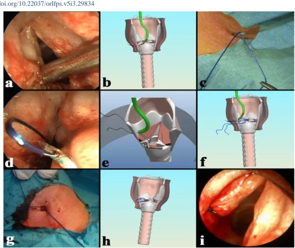
General anaesthesia combined with supraglottic jet ventilation is suggested for the intervention. For the sake of manoeuvrability, the larynx is exposed with a modified Weerda laryngoscope (8). After the disinfection of the mucosa, the ETGI is led through the laryngoscope to the glottic level. The mobile (or mobilized) arytenoid cartilage is tilted backward and upward with the end of the instrument (Figure 3.a). The built-in, curved blade is then pushed through under the vocal process out to the surface of the neck (Figure 3.b). A non-absorbable suture thread (Prolene 1.0; Ethicon, Somerville, NJ) is laced through the hole at the tip of the blade by an assistant surgeon (Figure 3.c). The doubled-over thread is pulled back with the blade, into the laryngeal cavity (Figure 3.d).
Figure 3. Intraoperative pictures and schematic drawing of EAAL on the left side a) The opening of the curved steel pipe stem is placed under the vocal process.
b) The built-in, curved blade is pushed through under the vocal process out to the surface of the neck.
c) A non-absorbable thread is inserted through the eye of the blade.
d) The doubled-over thread is pulled back with the blade, into the laryngeal cavity by pulling the thumb holder outward.
e, f) The tip of the stem pipe is placed above the arytenoid cartilage and the blade is pushed through the thyroid cartilage toward the skin to make a double loop.
g) A small skin incision is created to withdraw the ends of the thread to the surface of the sternohyoid muscle. The corresponding ends are knotted above it.
h,i) The left arytenoid cartilage is lateralised via EAAL.
After a repeated tilting of the arytenoid cartilage, the blade is pushed out with the
thread above the vocal process to the outer surface of the neck (Figure 4.e, f).
Figure 4. Endoscopic picture of a 45-year-old female patient with bilateral vocal fold paralysis a) Preoperative picture. b) Postoperative (6th week) picture after left-sided endoscopic arytenoid abduction lateropexy.
--- The assistant surgeon then cuts the double-
folded thread to remove it from the blade tip.
The blade is then pulled back into the laryngeal cavity, and the ETGI can be removed. A small skin incision (approximately 5 mm) is then created to withdraw the ends of the thread by a Jansen hook to the surface of the sternohyoid muscle (Figure 3.g). The corresponding ends are knotted above it. This simple procedure enables the endoscopic creation of two fixating loops in one step at suitable laryngeal locations, providing maximal physiological abduction of the arytenoid cartilage within 5 minutes (Figure 3.h, i; Figure 4.). In the peri - and postoperative period parenteral antibiotic and steroid is administered for 7 days.
Discussion
The structure of the ETGI gives a possibility to penetrate the thickest, posterior part of thyroid cartilage (9). This special device allows the creation of a double thread loop in a single step around the arytenoid cartilage. Hence, the arytenoid cartilage can be stabilized in a maximally abducted position in a physiological manner without resection of delicate phonatory structures and without consequential scarring (10). The procedure does not damage the interarytenoid region either, which is essential for safe, aspiration- free swallowing. In case of EAAL the laryngeal anatomical structures remain intact, thus EAAL considered a completely reversible procedure. Moreover, EAAL does not interfere with the potential neural regeneration process after neural injury either (11). This way, the fixating sutures can be removed in case of temporary recurrent laryngeal nerve injury.
In our earlier cadaver studies, performed on one hundred fresh cadaver larynges, we proved that EAAL provided the most beneficial glottis configuration compared to other suture lateralizing techniques (12, 13).
Moreover, after EAAL, the vocal folds become straighter and tenser than after other
published endoscopic glottis enlarging interventions. In addition, the EAAL technique produces a relatively small anterior angle compared to other interventions. This further facilitates better phonatory closure along with improved breathing potential (14).
The endoscopic laryngeal manipulation reduces the possible postoperative complications, and makes the external neck incision unnecessary, which has high priority in the treatment of a mostly iatrogenic disease.
Therefore, EAAL is a cost-effective procedure as well by lowering the time a patient spends in the operating theatre.
Considering the laryngeal conformation and size differences between genders, adults and infants; three different blades have been created for the ETGI (Figure 2.b).
According to our earlier study, EAAL is an optimal solution for (congenital) bilateral vocal fold paralysis even in new-borns to avoid tracheotomy and consequent, long term structural and functional damage of the larynx (15, 16). EAAL can be used in the treatment of uni/bilateral vocal fold immobility such as vocal fold paralysis, posterior laryngeal commissure stenosis or ankylosis of the cricoarytenoid joint. EAAL has been successfully used in revision surgeries of previously failed other glottis enlarging procedures and it can be a useful technique in cases of laryngotracheal reconstruction surgeries as well (17, 18).
Conclusion
Endoscopic arytenoid abduction lateropexy, which was developed by the anatomical observations of the cricoarytenoid joint is a minimally invasive, effective primary solution for BVFP. The Endolaryngeal Thread Guide Instrument, which was specially designed for this sugical intervention, can facilitate this procedure with the rapid and safe creation of fixating loops at the proper position. The simple surgical procedure, the great extent of reversibility, the easily detectable laryngeal
function recovery, and good functional results may simplify the management of BVFI patients.
Acknowledgments Not declared.
Conflicts of Interest
The first author is also the Medical Director of Mega ltd., which has developed the presented surgical device. (Mega ltd., Szeged, Hungary;
megakft@megakft.com).
Financial Support Not declared.
References
1. Rosenthal LH, Benninger MS, Deeb RH. Vocal fold immobility: a longitudinal analysisofetiology over 20 years. Laryngoscope. 2007;117(10):1864- 70.
2. Benninger MS, Gillen JB, Altman JS. Changing etiology of vocal fold immobility. Laryngoscope.
1998;108:1346–50.
3. Leon X, Venegas MP, Orus C, Quer M, Maranillo E, Sanudo JR. Inmovilidad glotica:estudo retrospectivo de 299 casos. Acta Otorrinolaringol Esp. 2001;52:486–92.
4. Maisel RH, Ogura JH. Evaluation and treatment of vocal cord paralysis. Laryngoscope.
1974;84:302–16.
5. Sessions DG, Ogura JH, Heeneman H. Surgical management of bilateral vocal cord paralysis.
Laryngoscope. 1976;86:559–66.
6. Belafsky PC. Bilateral vocal fold immobility.
Curr Opin Otolaryngol HeadNeck Surg.2011;19:415.
7. Sapundzhiev N, Lichtenberger G, Eckel HE, et al. Surgery of adult bilateral vocal fold paralysis in adduction: history and trends. Eur Arch Otorhinolaryngol.2008;265:1501–14.
8. Rovó L, Bach Á, Szakács L, Bella Z, Lázár G, (Application of modified Weerda laryngoscopes in the ear, nose, and throat practice). Orv Hetil.
2019;160:264-9. (Hungarian).
9. Madani S, Bere Z, Perényi A, Rovó L.
Morphometric Analysis of human laryngeal images for optimailsation of suture lateralization instruments. 11th Congress of the European Laryngological Society, Genoa, Italy, 8-11 June 2016.
10. Rovó L, Madani S, Sztanó B, Majoros V, Smehák G, Szakács L, Jóri J. A new thread guide instrument for endoscopic arytenoid lateropexy. Laryngoscope. 2010;120(10):2002-7.
11. Isolated Recovery of Adductor Muscle Function Following Bilateral Recurrent Laryngeal Nerve Injuries. Bach Á, Sztanó B, Matievics V, Bere Z, Fabian V, Andreas M, Förster G, Castellanos PF, Rovó L.Laryngoscope.
2019;129(10):2334-40.
12. Sztanó B, Szakács L, Madani S, Tóth F, Bere Z, Castellanos PF, Rovó L.Comparison of endoscopic techniques designed for posterior glottic stenosis—
a cadavermorphometric study. Laryngoscope.
2014;124(3):705-10.
13. Szakács L, Sztanó B, Matievics V, Bere Z, Bach A, Castellanos PF, Rovó L. A comparisonbetween transoral glottis-widening techniques for bilateral vocal fold immobility.Laryngoscope, 2015;125(11):2522-9.
14. Functional outcomes of endoscopic arytenoid abduction lateropexy for unilateral vocal cord paralysis with dyspnea. Matievics V, Bach A, Sztano B, Bere Z, Tobias Z, Castellanos PF, Mueller AH, Rovo L. Eur Arch Otorhinolaryngol.
2017;274(10):3703-10.
15. A new solution for neonatal bilateral vocal cord paralysis: Endoscopic arytenoid abduction lateropexy. Madani S, Bach Á, Matievics V, Erdélyi E, Sztanó B, Szegesdi I, Castellanos PF, Rovó L. Laryngoscope. Laryngoscope.
2017;127(7):1608-14.
16. Endoscopic arytenoid abduction lateropexy for the treatment of neonatal bilateral vocal cord paralysis - Long-term results. Sztanó B, Bach Á, Matievics V, Erdélyi E, Szegesdi I, Wootten CT, Rovó L. Int J Pediatr Otorhinolaryngol.
2019;119:147-50.
17. Rovó L, Venczel K, Torkos A, Majoros V, Sztanó B, Jóri J. Endoscopic Arytenoid Lateropexy for Isolated Posterior Glottic Stenosis.Laryngoscope. 2008;118(9):1550-5.
18. Rotational thyrotracheopexy after cricoidectomy for low-grade laryngeal chrondrosarcoma. Rovó L, Bach Á, Sztanó B, Matievics V, Szegesdi I, Castellanos PF.Laryngoscope. 2017;127(5):1109-15.