http://journal.pan.olsztyn.pl Original research article
Section: Nutritional Research
© Copyright by Institute of Animal Reproduction and Food Research of the Polish Academy of Sciences
© 2018 Author(s). This is an open access article licensed under the Creative Commons Attribution-NonCommercial-NoDerivs License (http://creativecommons.org/licenses/by-nc-nd/3.0/).
ABBREVIATIONS
AAPH – 2,2’-azobis (2-amidinopropane) dihydrochloride;
MDR – multidrug resistance;
MTT – 3-(4,5-dimethylthiazol-2-yl)-2,5-diphenyltetrazolium bromide;
RBCs – red blood cells
INTRODUCTION
Mini kiwi (Actinidia arguta, family Actinidiaceae, type:
vine), commonly called the hardy kiwi, is a variety from north- ern China. Leaves of this plant are a rich source of phenolic substances, mainly composed of phenolic acids (neochloro- genic, chlorogenic and cryptochlorogenic acid), B-type pro- cyanidin dimers, (+) catechin and fl avonols (quercetin glyco- sides, mostly triglycosides and kaempferol with 1, 2 or 3 sugar substituents) [Cyboran et al., 2014]. The presence of these substances ensures the biological activity, which includes an- tioxidant activity, hypoglycemic and antiproliferative action,
* Corresponding Author: Tel.: +48713205275; Fax: +48713205167;
E-mail: sylwia.cyboran@up.wroc.pl (S. Cyboran-Mikołajczyk, PhD)
and support of the digestive system [Montoya et al., 2014;
Zuo et al., 2012].
One of the main causes of many pathological states and ag- ing of the organism is believed to be disturbance of the oxida- tion-reduction balance that results in oxidative stress caused by a high concentration of free radicals [Kaur et al., 2014]. As a consequence, this leads to deterioration of basic life processes of cells, irreversible structural changes, mutations, and ulti- mately to cell death [Valko et al., 2007]. Therefore, currently, special attention is devoted to substances with high ability to reduce free radicals, mostly through looking for compounds of plant origin that do not show side effects on the organism.
Erythrocytes (RBC), due to their high content of unsatu- rated fatty acids and the ability of oxygen transfer [Chai et al., 2014], are constantly exposed to oxidative stress, which leads to changes in physical properties of the cell membrane responsible for integrity and mechanical properties of the cells, and conse- quently hinders their movement, in particular in the blood cap- illary veins. Oxidative damage occurs not only in membrane lipids but also in proteins, e.g. hemoglobin and spectrin, which in turn leads to irreversible structural changes and blood cell hemolysis [Becker et al., 1986; Buchwald et al., 2000]. The ef- fect of oxidative stress on the physical properties and physi-
In Vitro Studies of Anti-Hemolytic and Cytotoxic Activity
of Procyanidin-Rich Extract from the Leaves of Actinidia arguta Sylwia Cyboran-Mikołajczyk
1*, Ákos Csonka
2, Joseph Molnar
2,
Diana Szabó
3, Jan Oszmiański
4, Halina Kleszczyńska
11
Department of Physics and Biophysics, Wroclaw University of Environmental and Life Sciences, Norwida 25,50–375 Wrocław, Poland
2
Department of Medicinal Microbiology and Immunobiology, University of Szeged, Dóm tér 10, H-6720 Szeged, Hungary
3
Department of Otorhinolaryngology and Head-Neck Surgery, University of Szeged, Tisza L. krt. 111, H-6725 Szeged, Hungary
4
Department of Fruit, Vegetable and Nutraceutical Technology,
Wroclaw University of Environmental and Life Sciences, Norwida 25, 50–375 Wrocław, Poland
Key words: anti-hemolytic activity, cytotoxic activity, L5178Y cells, multidrug resistance, mini-kiwi (Actinidia arguta)
The leaves of mini kiwi (Actinidia arguta) are a rich source of phenolic compounds, in particular the B-type procyanidins that exhibit e.g. antioxi- dant, anticancer, antiviral, and anti-infl ammatory activities. The aim of this study was to determine the biological activity of the extract from leaves of kiwi in relation to cells of erythrocytes and lymphoma. This activity was determined by studying kiwi leaves extract anti-hemolytic, cytotoxic and an- tiproliferative activity, and its ability to change the physical properties of the cell membrane and inhibit multidrug resistance of mouse lymphoma cells. It was shown that the extract ingredients bound to the cells, caused changes in erythrocyte shape and slightly affected the granularity and size of lymphoma cells. They effectively protected the red blood cells from oxidative damage, but were not toxic to lymphoma cells and did not affect their multidrug resistance. The extract of kiwi leaves is an effective antioxidant but it does not exhibit cytotoxic activity. Therefore, it can be used in the pre- vention of diseases, especially those related to oxidative stress.
ological functions of RBCs depends on the nature of the oxida- tive agent [Borst et al., 2000; Hale et al., 2011]. In our previous work we found that mini-kiwi leaves extract protects biologi- cal membranes from oxidation induced by different physico- chemical factors, i.e. UVB and UVC radiation, and the AAPH compound [Cyboran et al., 2014]. This high antilipoperoxi- dant activity is mainly associated with the location of these compounds in the hydrophilic area of the membrane, where they form a protective barrier against diffusion of free radicals.
Moreover, literature data indicate that some polyphenolic com- pounds can also effectively protect not only the lipids of bio- logical membrane but also whole erythrocytes against oxidative damage [Fofi e et al., 2014; Senguttuvan et al., 2014]. Polyphe- nols can also modify the shape of erythrocytes without showing cytotoxic effects [Bonarska-Kujawa et al., 2012; Suwalsky et al., 2006]. Our earlier research has shown the mini-kiwi leaves extract to be nontoxic in relation to erythrocytes and even to strengthen them in operating under the conditions of os- motic shock [Cyboran et al., 2014]. Therefore, the main aim of the present research was to determine whether polyphenolic compounds contained in the extract from the leaves of mini- -kiwi have the ability to change erythrocyte shape and protect the cells from oxidative damage.
The second very important direction for research on sub- stances of plant origin is their use in the prevention and treat- ment of cancer. Polyphenolic compounds, and in particular procyanidins, show the ability to inhibit the growth of various tumor cell lines, among others pancreatic, lung and colorec- tal cancer cells [Akhtar el al., 2009; Chung et al., 2012; Kaur et al., 2008; Zhao et al., 2013]. In addition, these compounds may also inhibit the multidrug resistance of tumor cells and increase effi cacy of cytostatics [Zhao et al., 2013]. Multi- drug resistance (MDR) of human tumors is one of the major reasons for the failure of chemotherapy in refractory cancer patients. The reversal of MDR in cancer cells can be im- proved by the application of chemotherapy in combination with resistance modifi ers [Szabo & Molnar, 1998; Szabo et al., 2000]. Great numbers of natural plant compounds, in- cluding procyanidins and kiwi fruit extract, have been shown to block the P-glycoprotein activity [He et al., 2009; Mo- tohashi et al., 2002]. It was found that grape procyanidins reversed the MDR effect in blood-brain barrier by blocking the function of P-gp [He et al., 2009] and strongly inhibited P-gp expression by blocking MDR1 gene transcription. They also increased the intracellular accumulation of the P-gp sub- strate rhodamine-123 in A2780/T cells [Zhao et al., 2013].
In the literature there is a lack of studies on anticancer/cy- totoxic and MDR-reversal activity of phenolics contained in the leaves of mini-kiwi. This prompted us to undertake a study on the infl uence of kiwi leaves extract (contain- ing large amount of bioactive procyanidins) on the survival and multidrug resistance of lymphoma L5178 cells.
MATERIALS AND METHODS Plant material
Mini-kiwi leaves (Actinidia arguta, family Actinidia- ceae, type: vine) extract was obtained from the Department of Fruit, Vegetable and Plant Nutraceutical Technology, Wro-
claw University of Environmental and Life Sciences, Poland.
The Garden of Medicinal Plants herbarium of the Medical University in Wroclaw, Poland, was the provider of kiwi leaves used in the study. The research material was obtained from the stolon using a knife, and liquid nitrogen was used to freeze the leaves; and a dryer (Alpha 1–4 LSC, Christ, Germany) to freeze-dry them for 24 h. The procedure for obtaining a poly- phenolic extract from the leaves of mini kiwi and a detailed analysis of the composition of the extract were published ear- lier in the work by Cyboran et al. [2014].
Cells and other compounds of interest
The study was conducted on pig erythrocytes and L5178 mouse lymphoma T cells. The choice of pig erythrocytes was dictated by the fact that this cell’s percentage share of lipids is closest to that of the human erythrocyte, and the blood was easily available. The choice of L5178 mouse lymphoma T cells was dictated by the fact that these cells are commonly used to study antitumor and MDR- reversal activity of chemicals.
The L5178 mouse lymphoma T cells (ECACC cat. no.
87111908; U.S. FDA, Silver spring, MD, USA) were treated as a model of cancer cells. The cells were cultured in McCoy’s 5A medium supplemented with L-glutamine and antibiotics (penicillin/streptomycin solution) at 37°C and in an atmo- sphere with 5% CO2. The reagents for cell culture were pur- chased from Sigma Aldrich (Steinheim, Germany).
The oxidation inducer 2,2’-azobis(2-amidinopropane) dihydrochloride (AAPH) and antioxidant standard L(+) ascorbic acid (AA) were purchased from Sigma-Aldrich, Inc., Steinheim, Germany. Verapamil and 3-(4,5-dimethyl- thiazol-2-yl)-2,5-diphenyltetrazolium bromide (MTT) were purchased from Sigma-Aldrich Quimica SA, Madrid, Spain.
Procyanidin B3 was obtained from the Department of Fruit, Vegetable and Cereal Technology, Wroclaw University of En- vironmental and Life Sciences, Poland, and was extracted from pine. All other reagents were analytically pure.
Spectrophotometric assay for anti-hemolytic activity To test the effect of extract on hemolysis induced by free radicals, RBCs were pre-incubated with varying concen- trations of the extract (1–8 μg/mL) dissolved in ethanol at 37°C for 1 h. In order to compare the anti-hemolytic activ- ity of the extract with other substances the RBCs were also incubated, in the same conditions, with procyanidin B3 (1–8 μg/mL) and L(+) ascorbic acid (1–40 μg/mL) dis- solved in ethanol. Hemolysis of RBCs was carried out by mix- ing a 3% suspension of RBCs (unmodifi ed or modifi ed) in a phosphate buffer (pH=7.4) with AAPH solution (fi nal concentration 40 mmol/L). This reaction mixture was incu- bated at 37°C for 3 h. After incubation, the samples were centrifuged at 23°C (2000×g) for 15 min. The extent of he- molysis was determined spectrophotometrically by measuring the absorbance of the supernatant at 540 nm. For reference, RBCs were treated with redistilled water and the absorbance of the hemolysate was used as 100% hemolysis. The percent- age of hemolysed cells was calculated as the ratio of ab- sorbance of extract’s modifi ed and AAPH oxidized cells to the absorbance of unmodifi ed and AAPH oxidized cells (con- trol). The IC50 value of extract, responsible for 50% inhibi-
tion of hemolysis induced by AAPH, was determined and was compared with IC50 values determined for standard antioxi- dant L(+) ascorbic acid and procyanidin B3.
Microscopic studies of erythrocyte shapes
The impact of the extract on the shape of erythrocytes was determined by using the optical microscope and scanning electron microscope (SEM).
After separating from plasma, the red cells were washed four times in a 0.9% NaCl solution in order to study them with the optical microscope. Next, the erythrocyte solution at 2% hematocrit was incubated with kiwi leaves extract used at 0.01 and 0.1 mg/mL for 1 h at 37°C. After this modifi cation, a 0.2% solution of glutaraldehyde was added to the erythro- cytes to fi x them. The red cells thus prepared were observed under a biological optical microscope (Nikon Eclipse E200) equipped with a digital camera. On the basis of the obtained photographs, the percentage of the two basic forms, i.e. echi- nocytes and stomatocytes, in a population of ca. 800 cells was specifi ed. Individual forms of erythrocyte cells were ascribed morphological indices according to the Bessis scale [Ber- nhardt & Ellory, 2003; Bessis, 1997]. Negative values from –1 to –4 were ascribed for stomatocytes and positive ones from 1 to 4 for echinocytes.
For study with the electron microscope the erythrocytes were prepared in a way similar to that of optical microscope but slightly modifi ed. After incubating the erythrocytes with the extract, the cells were fi xed in a 2.5% solution of glutar- aldehyde for 48 h. Next, the phosphate buffer was used for washing the cells for 20 min, followed by a rising series of ace- tone concentrations (30, 50, 60, 70, 80, 90 and 100%) in order to dehydrate the cells. Every sample was washed for 15 min in a proper concentration of acetone, and next left in pure acetone for 30 min. After that, red cells were dried at 23°C for 12 h. The prepared cells were deposited on object stages and subjected to X-ray microanalysis by means of an X-ray analyzer (Brucker AXS Quantax) combined with the ESPRIT ver. 1.8.2. software. The Scancoat 6 (Edwards, London) sprinkler was used to coat the samples with gold. To analyze material ultrastructure, we used a scanning microscope (EVO LS15 ZEISS) with SE1 detector, under high vacuum and ac- celerating voltage of EHT=20 kV.
Spectrophotometric assays for cytotoxic and antiproliferative activities – MTT test
The antiproliferative and toxic activity of the kiwi leaves extract in relation to mouse lymphoma T-cells were deter- mined in MTT (3-(4,5-dimethylthiazol-2-yl)-2,5-diphenyl- tetrazolium bromide) assays. The cells 6×10³/well for anti- proliferative assay or 2×10⁴/well for cytotoxic assay in 50 μL of the medium were added into 96-well, fl at-bottom culture plates. Next, the extract dissolved in ethanol (10 mg/mL) and diluted in 100 μL of the culture medium of increasing concentrations from 1 to 400 μg/mL were added to the cells which were further incubated at 37°C for 72 h and 24 h for antiproliferative and cytotoxic assay, respectively. At the end of the incubation period, 15 μL of 5 mg/mL MTT solution were added to each well and the cells were incubated at 37°C for 4 h. After incubation, 100 μL of sodium dodecyl sulfate
(10% SDS in 0.01 mol.L HCl) were added and the plates were further incubated overnight. The cell growth was then deter- mined spectrophotometrically by measuring the absorbance at 550 nm (ref. 630 nm) with a Multiscan EX ELIXA reader (ThermoLabsystem, Cheshire, WA, USA). Inhibition of cell growth (Icg) was determined according to the formula:
Icg = Acell modified – Amedium modified × 100%
Acell control – Amedium
Flow cytometry assay for reversal of MDR in mouse lymphoma cells
The impact of the extract on MDR reversal was determined by treating the mouse lymphoma L5178Y cells transfected with the human MDR1 gene with different concentrations of kiwi leaves extract. The rhodamine-123 accumulation test was used. In general, the pHa MDR1/A retrovirus was used to infect the L5178 mouse T-lymphoma cells. By way of cultur- ing the infected cells with 60 ng/mL colchicine, the MDR-1-ex- pressing cell lines were obtained. Both MDR and PAR (parent) were grown in McCoy’s 5A medium with 10% heat-inactivated horse serum supplemented with L-glutamine and antibi- otics (penicillin/streptomycin solution). The fi nal density of the cells in the samples was 2×10⁶/mL, and they were re- suspended in the serum-free McCoy’s 5A medium and distrib- uted in 0.5 mL aliquots into the Eppendorf centrifuge tubes.
Next, to the cell solutions the kiwi extract (dissolved in ethanol) was added, to the fi nal concentrations of 40 and 400 μg/mL, and the cells were incubated for 10 min at room temperature.
Verapamil, treated as a standard MDR modulator, was used as a positive control. Next, the cells were incubated with 10 μL (5.2 μmol/L fi nal concentration) of rhodamine 123 (an indica- tor) for further 20 min at 37°C, washed twice and re-suspend- ed in 0.5 mL phosphate buffered saline (PBS) for analysis.
A fl ow cytometer (Beckton-Dickinson FACS scan) was used to measure the light scattering and fl uorescence of the cell popu- lation. On the basis of the obtained results the forward scatter count (FCS) and side scatter count (SSC) that depend on size, shape and optical homogeneity of the cells were determined.
Furthermore, the percentages of the control mean fl uorescence intensity were calculated for MDR and parental (PAR) cells as compared with untreated cells. The fl uorescence activity ratio (FAR) was calculated from the measured fl uorescence values using the following formula:
FAR = MDRtreated / MDRcontrol PARtreated / PARcontrol Statistical analysis
Statistical analysis of results was performed using the STA- TISTICA 12.0 (StatSoft PL) software. For results obtained from microscopic and anti-hemolytic tests, the statistical analysis was conducted using the Dunnett test (post-hoc test – ANOVA) at a signifi cance level α=0.01 or α=0.05. Statisti- cal analysis of results obtained from studies with lymphoma cells was conducted using Student’s t-test with the accepted level of signifi cance at α=0.05. All the experiments were done in at least three replicates, the results being presented as mean
± standard deviation.
RESULTS AND DISCUSSION
The extract was prepared from the leaves of Actinidia ar- gute obtained from the Garden of Medicinal Plants herbari- um of the Medical University in Wroclaw, Poland. The con- tent of phenolic substances in the extract was analyzed by using liquid chromatography UPLC/DAD and UPLC/
ESI/MS methods and is ca. 600 mg in 1 g of preparation [Cyboran et al., 2014]. The main polyphenolic components of the extract include different phenolic acids (neochlorogenic and chlorogenic acid, cryptochlorogenic), catechin, quercetin and kaempferol glycosides and, in particular, the B-type pro- cyanidin dimers of high biological activity which constitute ca. 25% of the identifi ed polyphenolic compounds.
Spectrophotometric assay for anti-hemolytic activity By using spectrophotometric methods, the anti-hemolytic activity of the extract was determined on the basis of the con- centration of hemoglobin released from cells damaged by oxi- dation. The obtained results showed that kiwi leaves extract ef- fectively protects erythrocytes against the adverse effects of free radicals, because a concentration-dependent decrease in hemo- lysis of erythrocytes was observed (Figure 1). Procyanidin B3 – one of the most active extract constituents, and L (+) ascorbic acid – the standard hydrophilic antioxidant, were included into this investigation. The IC50 value of kiwi leaves extract, procyan- idin B3, and L (+) ascorbic acid responsible for 50% inhibition of AAPH-induced hemolysis of erythrocytes were as follows:
6.89±0.64, 2.50±0.12 and 32.57±1.16 μg/mL, respectively.
The results indicate that procyanidin B3 better protects cells from damage induced by free radicals than the extract, but both the extract and procyanidin B3 are much more effective than the standard hydrophilic antioxidant which is ascorbic acid.
Similar results, i.e. higher activity of procyanidin B3 than that of the extract, were observed in our previous work, where
we examined the antilipoperoxidant activity of kiwi leaves ex- tract and procyanidin B3 [Cyboran et al., 2014]. Furthermore, B type procyanidin dimers constitute about 25% of extract ingredients [Cyboran et al., 2014]. Therefore, to establish if activity of procyanidin is modifi ed by other extract compo- nents, we compared the activity of procyanidin B3 used at a concentration of 2 μg/mL with the activity of extract used
0 10 20 30 40 50 60
0 1 2 3 4 5 6 7 8
Hemolysis of erythrocytes (%)
Concentration (µg/mL)
Procyanidin B3 Kiwi leaves extract
** **
** **
**
* *
**
**
** **
**
**
**
** **
FIGURE 1. Percentage of hemolysed cells vs. concentration of kiwi leaves extract and its constituent procyanidin B3. Statistical analysis was con- ducted using the Dunnett test. Statistically signifi cant results are denoted, respectively: *α=0.05, **α=0.01.
FIGURE 2. Shapes of unmodifi ed erythrocytes (A) and those modifi ed with kiwi leaves extract used at the concentration of 0.01 mg/mL (B) and 0.1 mg/mL (C), observed with an electron microscope.
at 8 μg/mL (the same procyanidin concentration). The re- sults included in Figure 1 clearly indicate that procyanidin B3 in the extract better protects erythrocytes against oxidative damage than when used alone. The obtained results confi rm that biological activity, in particular antioxidant/anti-hemo- lytic activity, of the extract depends not only on the number, amount, and type of its constituents but also on their mu- tual interaction. Hence extract components, by preserving whole erythrocytes and their membrane phospholipids intact, and due to their anti-hemolytic and antilipoperoxidant activ- ity, may maintain in vivo the integrity of RBC in capillaries and effectively counteract free radical damage.
Microscopic studies of erythrocyte shapes
To determine the location of extract constituents in the erythrocyte membrane, their impact on the shape of erythrocytes was examined. The study was conducted us- ing an electron and optical microscope, coupled with a digi- tal camera. Sample photographs of the blood cells (control and extract’s modifi ed) taken with the electron microscope are shown in Figures 2 (A), (B) and (C).
The photographs registered by using the optical micro- scope allow to quantify the individual forms of erythrocytes in a population of 800 cells. The classifi cation of shapes was carried out based on the scale of Bessis & Brecher [Ber- nhardt & Ellory, 2003; Bessis, 1997], where specifi c shapes are assigned the following morphological indexes: sphero- stomatocytes (-4), stomatocytes II (-3), stomatocytes I (-2), discostomatocytes (-1), discocytes (0), discoechinocytes (1), echinocytes (2), spheroechinocytes (3), and sphero- cytes (4). Figure 3 shows the percentage of specifi c shapes of erythrocytes modifi ed with the kiwi leaves extract at 0.01 and 0.1 mg/mL concentrations.
The obtained results have shown that erythrocytes modi- fi ed by the extract used at 0.01 mg/mL practically do not differ from control cells. At a higher extract concentration
(0.1 mg/mL) a decrease were observed in the number of dis- cocites and increase in the number of discoechinocytes (1) and echinocytes (2). The formation of echinocytes means, according to the Sheetz and Singer theory [Sheetz & Singer, 1974], that extract components may bind only to the outer monolayer of erythrocyte membrane. Our earlier studies showed changes in membrane fl uidity and packing order of the polar heads of lipids of the erythrocyte membrane caused by a mini-kiwi leaves extract [Cyboran et al., 2014].
Literature data indicate that various phenolic extracts and free phenolic compounds, that are able to modulate the hydro- philic and hydrophobic parts of erythrocyte membrane, can induce changes in erythrocyte shape [Bonarska-Kujawa et al., 2015; Suwalsky et al., 2007], this being in good agreement with our results. The main extract ingredients, like procyanidin B3 or chlorogenic acid, used at the same concentration as that in the extract, induce the formation of echinocytes to a much greater extent (results not included). Taking into account this result, the slight induction of echinocytes caused by the extract is probably due to the presence of other compounds which have a lower affi nity to the membrane. It is possible, because among the polyphenolic components of the extract, aside of monomers and procyanidin dimers which are readily identi- fi ed by HPLC methods, there might also occur procyanidin oligomers which have been detected in, e.g., a hydroalcoholic extract from plants of the Actinidia family [Pinelli et al., 2013].
Due to their size, such components are not expected to pen- etrate deep into the membrane but can interact with its sur- face. The presence of such compounds can also impair bind- ing of other compounds which exhibit that ability, i.e. B type procyanidins or phenolic acids, to the membrane.
MTT cytotoxicity assay
The in vitro research on antiproliferative and toxic activity of the kiwi leaves extract in relation to the mouse lymphoma cell line was done by using an MTT assay. The results of both experiments (24 and 72 h) are shown in Table 1. The optical density of cancer cells in the presence of 1–400 μg/mL kiwi extract was from 0.512 to 0.485 after 24 h and from 0.612 to 0.694 after 72 h, respectively, whereas the values determined for the control non-treated cells were from 0.515 after 24 h to
0 10 20 30 40 50 60 70 80 90
-3 -2 -1 0 1 2 3
Percentage share of the cells
Morfological index of erythrocytes Control 0.01 mg/mL 0.1 mg/mL
**
**
**
*
FIGURE 3. Mean percentage of erythrocyte shapes formed in the pres- ence of kiwi leaves extract applied at 0.01 mg/mL and 0.1 mg/mL. Statis- tical analysis was conducted using the Dunnett test. Statistically signifi - cant results are denoted, respectively: *α=0.05, **α=0.01.
TABLE 1. Optical density of cancer cells non-treated (control) and incu- bated for 24 and 72 h with different concentrations of Actinidia arguta leaf extract in the MTT assay.
Concentration
(μg/mL) Incubation time
24 h Incubation time
72 h
0 (control) 0.515±0.056 0.630±0.051
1 0.512±0.041 0.612±0.037
10 0.527±0.050 0.600±0.051
25 0.505±0.031 0.675±0.062
50 0.505±0.040 0.635±0.041
100 0.528±0.048 0.660±0.043
200 0.497±0.041 0.625±0.058
400 0.485±0.062 0.694±0.072
0.630 after 72 h. The inhibition of cells grown (Icg) calculated on the basis of these results was less than 2% and indicated that, in the applied concentrations, the compounds contained in the extract had no antiproliferative and cytotoxic effect on mouse lymphoma cells.
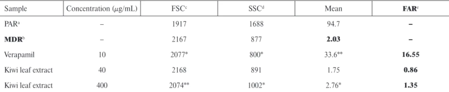
Assay for the reversal of MDR in mouse lymphoma cells The effects of the extract on reversion of multidrug resis- tance in the L5178Y mouse T-cell lymphoma drug-sensitive (parent) and multidrug resistant (MDR) cells were exam- ined. The well-known MDR modifi er verapamil was used as a control. The reversal effect was determined on the basis of the calculated fl uorescence activity ratio (FAR) between treated and untreated groups of cells. The kiwi extract was ineffective at both applied concentrations, because the FAR values calculated for the extract-modifi ed cells were less than MDR means and also several times lower than the values cal- culated for verapamil-treated cells (Table 2). It means that the extract was ineffective as a resistance modifi er of lympho- ma cells. However, the slightly increased values of side scatter count (SSC is proportional to cell granularity) and slightly decreased values of forward scatter count (FCS) obtained for the extract-modifi ed cells compared to the control (Ta- ble 2) may refer to some non-specifi c membrane effects.
This research has shown that the kiwi leaves extract, de- spite the high content of procyanidins and other phenolic compounds, is ineffective as a resistance modifi er of lym- phoma cells, because its ingredients have no inhibitory effect of P-glycoprotein. However, this result indicates that the ex- tract can interact with lymphoma cells in an unspecifi c way, because it caused a slight change in cell granularity and cell size. However, this observation requires more accurate and de- tailed research. This study proves that the well-documented anticancer/cytotoxic activity of procyanidins strongly depends on mutual interaction between them and other phenolic compounds, when present in a mixture and also on the type of cancer cells. In future studies, the fraction of procyanidins should be isolated from the kiwi leaves extract and their bio- logical activity examined in order to determine the suitability of those compounds, not only in the prevention of diseases but also in the treatment of cancer cells.
CONCLUSION
Kiwi leaves extract has high anti-hemolytic activity and ability to modulate the physical properties of the cells.
The extract effectively protects erythrocytes from oxida- tive damage caused by water-soluble free radicals. Its in- gredients bind to the cell membrane, change its physical properties, inducing alteration in the shape of erythrocytes, and slightly affect granularity and size of lymphoma cells.
Owing to the binding they may form a barrier on the mem- brane which protects against the harmful effect of free radi- cals. On the other hand, the B type procyanidins contained in a large amount in the extract, of documented anticancer activity, are nontoxic and do not exhibit the antiprolifera- tive activity with respect to lymphoma cells and do not af- fect their multidrug resistance. The extract from the leaves of the kiwi may fi nd an application in the prevention of dis- eases especially those caused by the oxidative stress. Fur- thermore, it may be a useful material for obtaining procy- anidins, which after further research may prove effective against tumor cells, as it has been in the case of procyani- dins contained in kiwi fruit.
ACKNOWLEDGEMENTS
We are grateful to Mrs. Anikó Vigyikán Váradi for the preparation of the tissue cultures and technical assis- tance, and to Imre Ocsovszki for the fl ow cytometric mea- surements.
STUDY FUNDING
This work was supported from funds of the statutory ac- tivities of the Department of Physics and Biophysics, Wroclaw University of Environmental and Life Sciences. The study was also supported by Szeged Foundation for Cancer Research, by the project TAMOP-4.2.2A- 11/1/KONV-2012- 0035.
CONFLICT OF INTEREST
The authors declare no confl icts of interest.
TABLE 2. Flow-cytometric analysis of the effect of Actinidia arguta leaf extract on rhodamine 123 accumulation of human MDR gene transfected mouse lymphoma cells.
Sample Concentration (μg/mL) FSCc SSCd Mean FARe
PARa – 1917 1688 94.7 –
MDRb – 2167 877 2.03 –
Verapamil 10 2077* 800* 33.6** 16.55
Kiwi leaf extract 40 2168 891 1.75 0.86
Kiwi leaf extract 400 2074** 1002* 2.76* 1.35
aparental cells without transfection of MDR1 gene; bparental cells transfected with MDR 1 gene; cFSC – forward scatter count (cell size ratio); dSSC – side scatter count; eFAR – fl uorescence activity ratio.
*Statistical analysis was conducted using the Student’s t-test. Statistically signifi cant results are denoted, respectively: *p < 0.05, **p < 0.01.
REFERENCES
1. Akhtar S., Meeran S.M., Katiyar N., Katiyar S.K., Grape seed proanthocyanidins inhibit the growth of human non-small cell lung cancer xenografts by targeting insulin-like growth factor binding protein-3, tumor cell proliferation and angiogenic fac- tors. Clin. Cancer Res., 2009, 15, 821–831
2. Becker P.S., Cohen C.M., Lux S.E., The effect of mild diamide oxidation on the structure and function of human erythrocyte spectrin. J. Biol. Chem., 1986, 261, 4620–4628.
3. Bernhardt I., Ellory J.C. (eds.), Membrane lipids and proteins as a basis of red cell shape and its alterations. 2003, in: Red Cell Membrane Transport in Health and Disease. Berlin: Springer- -Verlag, pp. 26–60.
4. Bessis M., Erythrocyte form and deformability for normal blood and some hereditary hemolytic anemias. Nouv. Rev. Fr. Hema- tol., 1997, 18, 75–94.
5. Bonarska-Kujawa D., Pruchnik H., Cyboran S., Żyłka R., Oszmiański J., Kleszczyńska H., Biophysical mechanism of the protective effect of blue honeysuckle (Loniceracaerulea L.
var. kamtschatica Sevast.) polyphenols extracts against lipid per- oxidation of erythrocyte and lipid membranes. J. Membr. Biol., 2012, 247, 611–625.
6. Bonarska-Kujawa D., Cyboran-Mikołajczyk S., Kleszczyńska H., Molecular mechanism of action of chlorogenic acid on erythro- cyte and lipid membrane. Mol. Membr. Biol., 2015, 32, 46–54.
7. Borst J.W., Visser N.V., Kouptsova O., Visser A.J.W.G., Oxida- tion of unsaturated phospholipids in membrane bilayer mixtures in accompanied by membrane fl uidity changes. Biochim. Bio- phys. Acta, 2000, 1487, 61–73.
8. Buchwald H., O’Dea T.J., Menchaca H.J., Michalek V.N., Rohde T.D., Effect of plasma cholesterol on red blood cell oxygen trans- port. Clin. Exp. Pharmacol. Physiol., 2000, 27, 951–955.
9. Chai W.M., Shi Y., Feng H.L., Xu L., Xiang Z.H., Gao Y.S., Chen Q.X., Structure characterization and anti-tyrosinase mechanism of polymeric proanthocyanidins fractionated from kiwifruit peri- carp. J. Agric. Food Chem., 2014, 62, 6382–6389.
10. Chung Y.C., Huang C.C., Chen C.H., Chiang H.C., Chen K.B., Chen Y.J., Liu C.L., Chuang L.T., Liu M., Hsu C.P., Grape-seed procyanidins inhibit the in vitro growth and invasion of pancre- atic carcinoma cells. Pancreas, 2012, 41, 447–454.
11. Cyboran S., Oszmiański J., Kleszczyńska H., Modifi cation of the properties of biological membrane and its protection against oxidation by Actinidia arguta leaf extract. ChemBiol. In- teract., 2014, 222, 50–59.
12. Fofi e C.K., Wansi S.L., Nguelefack-Mbuyo E.P., Atsamo A.D., Watcho P., Kamanyi A., Nole T., Nguelefack T.B., In vitro anti-hy- perglycemic and antioxidant properties of extracts from the stem bark of Ceiba pentandra. J. Complement. Integr. Med., 2014, 11, 185–193.
13. Hale J., Winlove C.P., Petrov P.G., Effect of hydroperoxides on red blood cell membrane mechanical properties. Biophys. J., 2011, 101, 1921–1929.
14. He L., Zhao C., Yan M., Zhang L.Y., Xia Y.Z., Inhibition of P-glycoprotein function by procyanidin on blood-brain barrier.
Phytother. Res., 2009, 23, 933–37.
15. Kaur R., Kaur J., Mahajan J., Kumar R., Arora S., Oxidative stress-implications, source and its prevention. Environ. Sci. Pol- lut. Res., 2014, 21, 1599–1613.
16. Kaur M., Mandair R., Agarwal R., Agarwal C., Grape seed ex- tract induces cell cycle arrest and apoptosis in human colon car- cinoma cells. Nutr. Cancer, 2008, 60 Suppl., 2–11.
17. Montoya C.A., Rutherfurd S.M., Olson T.D., Purba A.S., Drum- mond L.N., Boland M.J., Moughan P.J., Actinidin from kiwifruit (Actinidia deliciosa cv. Hayward) increases the digestion and rate of gastric emptying of meat proteins in the growing pig. Brit.
J. Nutr., 2014, 111, 957–967.
18. Motohashi N., Shirataki Y., Kawase M., Tani S., Sakagami H., Satoh K., Kurihara T., Nakashima H., Mucsi I., Varga A., Molnár J., Cancer prevention and therapy with kiwifruit in Chinese folk- lore medicine: a study of kiwi fruit extracts. J. Ethnopharmacol., 2002, 81, 357–364.
19. Pinelli P., Romani A., Fierini E., Remorini D., Agati G., Char- acterization of the polyphenol content in the kiwifruit (Actinidia deliciosa) exocarp for the calibration of a fruit-sorting optical sensor. Phytochem. Anal., 2013, 24, 460–466.
20. Senguttuvan J., Paulsamy S., Karthika K., Phytochemical analy- sis and evaluation of leaf and root parts of the medicinal herb Hypochaeris radicata L. for in vitro antioxidant activities. Asian Pac. J. Trop. Biomed., 2014, 4 Suppl., S359–67.
21. Sheetz M.P., Singer S.J., Biological membranes as bilayer cou- ples. A molecular mechanism of drug-erythrocyte interactions.
Proc. Natl. Acad. Sci., 1974, 71, 4457–4461.
22. Suwalsky M., Orellana P., Avello M., Villena F., Sotomayor C.P. Human erythrocytes are affected in vitro by extracts of Ugni molinae leaves. Food Chem. Toxicol., 2006, 44, 1393–1398.
23. Suwalsky M., Orellana P., Avello M., Villena F., Protective effect of Ugni molinae Turcz against oxidative damage of human eryth- rocytes. Food Chem. Toxicol., 2007, 45, 130–113.
24. Szabó D., Keyzer H., Kaiser H.E., Molnár J., Reversal of mul- tidrug resistance of tumor cells. Anticancer Res., 2000, 20(6B), 4261–4274.
25. Szabó D., Molnár J., The role of stereoselectivity of chemosensi- tizers in the reversal of multidrug resistance of mouse lymphoma cells. Anticancer Res., 1998, 18(4C), 3039–3044.
26. Valko M., Leibfritz D., Moncol J., Cronin M.T.D., Mazur M., Telser J., Free radicals and antioxidants in normal physiological functions and human disease. Int. J. Biochem. Cell. Biol., 2007, 39, 44–84.
27. Zhao B.X., Sun Y.B., Wang S.Q., Duan L., Huo Q.L., Ren F., Li G.F. Grape seed procyanidin reversal of P-glycoprotein as- sociated multi-drug resistance via down-regulation of NF-κB and MAPK/ERK mediated YB-1 activity in A2780/T cells. PLoS One, 2013, 8(8):e71071.
28. Zuo L.L., Wang Z.Y., Fan Z.L., Tian S.Q., Liu J.R., Evaluation of antioxidant and antiproliferative properties of three Actinidia (Actinidia kolomikta, Actinidia arguta, Actinidia chinensis) extracts in vitro. Int. J. Mol. Sci., 2012, 13, 5506–5518.
Submitted: 9 December 2016. Revised: 21 March, 9 May, and 24 May 2017. Accepted: 26 May 2017. Published on-line:
13 September 2017.