Cardiac effects of acute exhaustive exercise in a rat model☆
Attila Oláh
a,⁎ , Balázs Tamás Németh
a, Csaba Mátyás
a, Eszter Mária Horváth
b, László Hidi
a, Ede Birtalan
a, Dalma Kellermayer
a, Mihály Ruppert
a, Gerg ő Merkely
a, Gábor Szabó
c, Béla Merkely
a,1, Tamás Radovits
a,c,1aHeart and Vascular Center, Semmelweis University, Városmajor u. 68, 1122 Budapest, Hungary
bInstitute of Human Physiology and Clinical Experimental Research, Semmelweis University, Tűzoltó u. 37-47, 1094 Budapest, Hungary
cDepartment of Cardiac Surgery, University of Heidelberg, 2. OG. INF 326, 69120 Heidelberg, Germany
a b s t r a c t a r t i c l e i n f o
Article history:
Received 24 January 2014
Received in revised form 31 October 2014 Accepted 20 December 2014
Available online 23 December 2014
Keywords:
Exhaustive exercise Cardiac function Pressure-volume analysis Gene expression Oxidative stress Cardiac mechanoenergetics
Background:The role of physical exercise in the prevention and treatment of cardiovascular diseases has been well described, however, elevations in cardionecrotic biomarkers after prolonged exercise (i.e. ultramarathon running) were observed. We aimed at understanding the biochemical, molecular biological, structural and func- tional alterations in the heart after exhaustive exercise in a rat model.
Methods:Rats of the exercise group were forced to swim for 3 h with 5% body weight (workload) attached to the tail, control rats were taken into the water for 5 min. After a 2-hour recovery period we performed left ventricular (LV) pressure-volume analysis to investigate LV function and mechanoenergetics. Additionally, blood and myo- cardium samples were harvested for biochemical and histological examinations. Gene expression changes were detected by qRT-PCR.
Results:When compared to controls, elevated plasma levels of cardiac troponin T and creatine kinase were de- tected after exhaustive exercise. Histological analysis showed sporadic fragmentation of myocardial structure and leukocyte infiltration in the exercised group. We observed increased end-systolic volume, decreased ejection fraction, impaired contractility (preload recruitable stroke work) and mechanoenergetics (ventriculoarterial coupling, mechanical efficiency) of LV after exercise. Myocardial expression of major antioxidant enzymes was increased along with increased myocardial nitro-oxidative stress. Bax/Bcl-2 ratio and TUNEL staining showed en- hanced apoptotic signaling. Exhaustive exercise also resulted in the dysregulation of the matrix metalloprotein- ase system.
Conclusions:Excessive physical activity has an adverse effect on the heart. The observed functional impairment is associated with increased nitro-oxidative stress, enhanced apoptotic signaling and dysregulation of the matrix metalloproteinase system after exhaustive exercise.
© 2014 The Authors. Published by Elsevier Ireland Ltd. This is an open access article under the CC BY-NC-ND license (http://creativecommons.org/licenses/by-nc-nd/4.0/).
1. Introduction
The role of regular physical activity is widely recognized in the pre- vention, management and treatment of several cardiovascular diseases, such as coronary heart disease[1,2]. Although regular exercise training reduces cardiovascular risk, recent studies have documented elevations in biomarkers consistent with cardiac damage (i.e. cardiac troponin (cTn)) after bouts of prolonged exercise in apparently healthy indi- viduals without cardiovascular disease[3–6], which has raised con- cerns about cardiovascular health consequences of such exercise[7].
Prolonged and intensive physical activities such as ironman triathlon,
cross country biking or ultramarathon running are becoming increas- ingly popular.
Numerous studies on humans[4,5]and animals[8,9]have reported elevated serum cardiac troponins—as highly specific and sensitive laboratory markers for biochemical detection of myocardial injury— after prolonged, exhaustive exercises that can exceed clinical cut-off value of myocardial infarction. Increased levels of reactive oxygen and nitrogen species, thus nitro-oxidative stress have been demonstrated after excessive physical exercise in human studies[10,11]. It is also well described that exhaustive exercise induced reactive oxygen species (ROS) generation leads to oxidative damage in rat myocardium[12]. Ex- cessive ROS formation causes cellular dysfunction, protein and lipid per- oxidation and DNA injury. These processes can lead to irreversible cell damage and death, which have been implicated in a wide range of path- ological cardiovascular conditions[13].
Cardiac functional effects of acute bouts of exhaustive exercise have been investigated in human studies using echocardiography and MRI [14,15]. The observed transient impairment in left ventricular (LV)
☆ All of the authors take responsibility for all aspects of the reliability and freedom from bias of the data presented and their discussed interpretation.
⁎ Corresponding author at: Experimental Research Laboratory, Heart and Vascular Center, Semmelweis University, Városmajor u. 68, 1122 Budapest, Hungary.
E-mail address:o.attilio@gmail.com(A. Oláh).
1 Equal contribution.
http://dx.doi.org/10.1016/j.ijcard.2014.12.045
0167-5273/© 2014 The Authors. Published by Elsevier Ireland Ltd. This is an open access article under the CC BY-NC-ND license (http://creativecommons.org/licenses/by-nc-nd/4.0/).
Contents lists available atScienceDirect
International Journal of Cardiology
j o u r n a l h o m e p a g e : w w w . e l s e v i e r . c o m / l o c a t e / i j c a r d
function has been described as“exercise-induced cardiac fatigue”[16], however controversial data and study design differences result in uncer- tain conclusions[15,17]. Small animal studies also investigated exhaus- tive exercise induced cardiac functional changes by echocardiography [18,19]and although a few basic hemodynamic parameters were previ- ously measured, they were largely dependent on loading conditions.
Therefore, appropriate, hemodynamically validated standards are re- quired for reliable investigation of exhaustive exercise induced cardiac functional changes. Pressure-volume (P-V) analysis is a useful approach for investigating several aspects of in vivo LV function independently from loading conditions. Advances of the last decade in the technical development and validation of miniature P-V catheters have made it possible to use this approach for studies in small animals[20].
The purpose of this study was to provide thefirst detailed in vivo characterization of left ventricular hemodynamic changes after an acute bout of exhaustive exercise using P-V analysis. For validation of our rat model we determined key markers of already described cellular and molecular mechanisms leading to myocardial injury as a conse- quence of excessive exercise.
2. Materials and methods 2.1. Animals
All animals received humane care in compliance with the Principles of Laboratory Animal Care formulated by the National Society for Medical Research and the Guide for the Care and Use of Laboratory Animals prepared by the Institute of Laboratory Animal Resources and published by the National Institutes of Health (NIH Publication No. 86-23, revised 1996). All procedures and handling of the animals during the study were reviewed and approved by the Ethical Committee of Hungary for Animal Experimentation.
Forty-eight young adult (12-wk old, m = 325–375 g) male Wistar rats (Toxi-Coop, Dunakeszi, Hungary) were housed in a room with constant temperature of 22 ± 2 °C with a 12/12 h light–dark cycle and fed a standard laboratory rat diet ad libitum and free access to water.
2.2. Experimental design
A 150-l water tank was divided into 6 lanes with a surface area of 20 × 25 cm and a depth of 45 cm per lane andfilled with tap water maintained at 30–32 °C to allow individ- ual swim exercise. Attempting to minimize the general stress response, all rats were familiarized with swimming for 20 min 48 h before the experiments. Acute exercised rats (n = 26) were forced to swim for 3 h with a workload (5% of body weight) attached to the tail, as previously described[9]. A total of four rats needed to be rescued from the water and removed from the study due to an inability to complete the exercise protocol.
Control rats (n = 22) were taken into the water for 5 min. After the exercise, rats were dried and twenty-four animals (12–12 exercised and control, group A) were sacrificed 2 h after swimming to collect blood and tissue samples. The 2 h resting period was chosen according to previous literature data[9]. The remaining twenty rats (10–10 exercised and control, group B) underwent hemodynamic measurements after the 2 h period of obser- vation. In order to eliminate diurnal effects, the experiments were performed at the same time of the day.
2.3. Biochemical measurements
Twenty-four rats (group A) were anesthetized with a mixture of ketamine (100 mg/kg ip.) and xylazine (3 mg/kg ip.). The abdominal cavity was quickly opened, and blood samples were collected from the inferior vena cava in tubes pre-rinsed with EDTA. Blood samples were centrifuged and plasma cardiac troponin T (cTnT), creatine ki- nase (CK), lactate dehydrogenase (LDH), aspartate transaminase (AST) and creatinine were measured by automated clinical laboratory assays on a Cobas Integra 400 (Roche Di- agnostics, Mannheim, Germany) autoanalyzer.
2.4. Histology
Myocardial samples of twenty rats (group B) were removed for histological process- ing immediately after completing the LV P-V analysis. Tissue samples were snap frozen orfixed in buffered paraformaldehyde solution (4%) and embedded in paraffin. Transverse transmural slices of the ventricles were sectioned (5μm) and processed conventionally for histological examination. One series of sections were stained with hematoxylin and eosin (HE). Light microscopic examination was performed with a Zeiss microscope (Axio Observer.Z1, Carl Zeiss, Jena, Germany) at a magnification of 400×, and digital images were captured by an imaging software (QCapture Pro 6.0, QImaging, Canada).
In situ detection of ROS was performed by using the oxidativefluorescent dye dihydroethidium (DHE; Sigma-Aldrich, St. Louis, USA). DHE is freely permeable to cell membranes and emits a redfluorescent signal when oxidized by ROS to ethidium, which is intercalating into DNA (typically nuclear localization). Fresh frozen LV myocardial
sections (16μm) were incubated with 1μM DHE (in PBS; pH 7.4) at 37 °C for 30 min in a dark humidified chamber[21]. Fluorescence in myocardial sections was visualized using a fluorescence microscope (Axio Observer.Z1, Carl Zeiss, Jena, Germany) with a 590 nm long-passfilter after background corrections to saline treated negative control. Eight images were taken randomly each of the slides andfluorescence area and intensity were analyzed using ImageJ (NIH, Bethesda, MD, USA) image analysis software.
To demonstrate nitrative stress, tyrosine nitration was detected in LV myocardial sections by immunohistochemistry[22]. Paraffin-embedded sections of myocardium were deparaffinized. After antigen retrieval (0.1 M citrate buffer, pH3, heating in micro- wave oven for 15 min) and quenching endogenous peroxidase with 0.3% H2O2in 100%
methanol for 15 min, slides were immunostained with a rabbit anti-nitrotyrosine antibody (1:200, Millipore, Bedford, MA, USA) overnight at 4 °C. Specific labeling was detected by incubation for 30 min at room temperature with a biotin-conjugated anti-rabbit goat an- tibody (Vector Laboratories, Burlingame, CA, USA) and amplified with an avidin–biotin peroxidase complex (Vector Laboratories). Nickel–cobalt enhanced diaminobenzidine (Vector Laboratories) was used as chromogen. Nitrotyrosine positive area was calculated using ImageJ.
Apoptosis in cardiomyocytes was determined with terminal deoxynucleotidyl transferase-mediated dUTP nick-end labeling (TUNEL) technique. TUNEL staining was performed using DeadEnd™Colorimetric TUNEL System (Promega, Madison, WI, USA) ac- cording to the manufacturer's instructions. Sections were counterstained by the red col- ored Nuclear Fast Red (Sigma-Aldrich, St. Louis, MO, USA). Thirty visualfields of heart sections were randomly selected in each animal, and TUNEL-positive cells were counted.
Data are expressed as mean number of apoptotic cells perfield.
2.5. Cardiac mRNA analysis
After the blood sample collection from twenty-four (group A) rats the chest cavity was quickly opened and the heart was removed. Left ventricular tissues were immediately snap frozen in liquid nitrogen, and stored at−80 °C. LV tissue was homogenized in RLT buffer, RNA was isolated from the ventricular samples by using the RNeasy Fibrous Tissue Mini Kit (Qiagen, Hilden, Germany) according to the manufacturer's instructions and quantified by measuring optical density (OD) at 260 nm. RNA purity was ensured by obtaining a 260/280 nm OD ratio approximately 2.0. Reverse transcription reaction (1μg total RNA of each sample) was completed by using the QuantiTect Reverse Transcription Kit (Qiagen). Quantitative real-time PCR was performed with the StepOnePlusTMReal- Time PCR System (Applied Biosystems, Foster City, USA) in triplicates of each sample in a volume of 10μl in each well containing cDNA (1μl), TaqMan® Universal PCR MasterMix (5μl) and a TaqMan® Gene Expression Assay for the following targets (0.5μl): atrial na- triuretic factor (ANF; Rn00561661_m1), catalase (assay ID: Rn00560930_m1), glucose- 6-phosphate dehydrogenase (G6PD; assay ID: Rn00566576_m1), gluthathione peroxidase 1 (GPX-1, Rn00577994_g1), glutathione reductase (GSR, assay ID: Rn01482159_m1), thioredoxin-1 (assay ID: Rn00587437_m1), superoxide dismutase 2 (SOD-2, assay ID:
Rn00690587_g1), endothelial nitric oxide synthase (eNOS, assay ID: Rn02132634_s1), Bcl- 2 associated X protein (Bax, assay ID: Rn02532082_g1), Bcl-2 (assay ID: Rn99999125_m1), matrix metalloproteinase-2 (MMP-2, assay ID: Rn01538170_m1), tissue inhibitor of metalloproteinase-2 (TIMP-2, assay ID: Rn00573232_m1), matrix metalloproteinase-9 (MMP-9, assay ID: Rn00579162_m1), tissue inhibitor of metalloproteinase-1 (TIMP-1, assay ID: Rn00587558_m1) and transforming growth factorβ1 (TGF-β1, assay ID:
Rn00572010_m1), all purchased from Applied Biosystems. Gene expression data were nor- malized to glyceraldehyde-3-phosphate dehydrogenase (GAPDH; reference gene; assay ID:
Rn01775763_g1) and expression levels were calculated by the CTcomparative method (2−ΔCT). All results are expressed as values normalized to a positive calibrator (a pool of cDNA's from all samples of the control group).
2.6. Hemodynamic measurements—left ventricular pressure-volume (P-V) analysis Twenty rats (group B) were anesthetized with pentobarbital (60 mg/kg ip.), tracheotomized, and intubated to facilitate breathing. Animals were placed on controlled heating pads, and the core temperature, measured via rectal probe, was maintained at 37
°C. A polyethylene catheter was inserted into the left external jugular vein forfluid administration. A 2-Fr microtip pressure-conductance catheter (SPR-838, Millar Instru- ments, Houston, TX, USA) was inserted into the right carotid artery and advanced into the ascending aorta and after stabilization for 5 min, mean arterial blood pressure (MAP) was recorded. After that, the catheter was advanced into the left ventricle under pressure control. After stabilization for 5 min, signals were continuously recorded at a sampling rate of 1000 samples/s using a P-V conductance system (MPVS-Ultra, Millar In- struments) connected to the PowerLab 16/30 data acquisition system (AD Instruments, Colorado Springs, CO, USA), stored and displayed on a personal computer by the LabChart5 Software System (AD Instruments). Hemodynamic parameters were computed and calcu- lated as described before[20,23]. Briefly, with the use of a special P-V analysis program (PVAN, Millar Instruments), heart rate (HR), LV end-systolic pressure (LVESP), LV end-diastolic pressure (LVEDP), the maximal slope of LV systolic pressure increment (dP/dtmax) and diastolic pressure decrement (dP/dtmin), time constant of LV pressure decay [τ; according to the Weiss method and Glantz method], LV end-systolic volume (LVESV), LV end-diastolic volume (LVEDV), stroke volume (SV), cardiac output (CO), ejec- tion fraction (EF) and stroke work (SW) were computed and calculated. CO was normal- ized to body weight [cardiac index (CI)]. Total peripheral resistance (TPR) was calculated by the following equation: TPR = MAP/CO. Using P-V loops recorded during the transient compression of vena cava inferior (reducing preload) we measured load-independent
contractility indexes: the slope (Ees, end-systolic elastance) of the LV end-systolic P-V rela- tionship (ESPVR, according to the parabolic curvilinear model); preload recruitable stroke work (PRSW, the slope of the linear relation between SW and EDV) and the slope of the dP/dtmax-end-diastolic volume relationship (dP/dtmax-EDV). The slope of the LV end- diastolic P-V relationship (EDPVR) was calculated as a reliable index of LV stiffness. Arterial elastance (Ea) was calculated as LVESP/SV. To characterize LV mechanoenergetics stroke work (SW), pressure-volume area (PVA) and maximal power of LV work were computed, ventriculoarterial coupling (VAC) was determined by the quotient of Eaand Ees, while me- chanical efficiency (Eff) was calculated as the ratio of SW and PVA. After completion of the hemodynamic measurements all animals were euthanized by exsanguination.
2.7. Statistical analysis
Statistical analysis was performed on a personal computer with a commercially avail- able software (Origin 7G; OriginLab, Northampton, MA, USA). All data are expressed as means ± SEM. An unpaired two sided Student's t-test was used to compare parameters of exercised and control rats, after normal distribution of data was confirmed by Shapiro-Wilk test. Differences were considered statistically significant when pb0.05.
3. Results
3.1. Body weight and heart weight
The body weight did not differ before (368 ± 9 g exercised vs. 356 ± 8 g control, p = 0.3050) and after (350 ± 8 g exercised vs. 347 ± 7 g con- trol, p = 0.7417) the acute exhaustive exercise. Body weight loss to base- line body weight ratio (4.69 ± 0.31% exercised vs. 2.41 ± 0.39% control, pb0.0001) during the exercise protocol was significantly increased in exercised rats. Heart weight measured immediately after the hemody- namic measurements was similar in exercised and control animals (1.14 ± 0.04 g exercised vs. 1.13 ± 0.04 g control, p = 0.8777).
3.2. Biochemical parameters
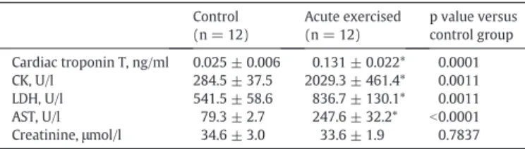
When compared to the control group, serum cTnT concentrations increased significantly after exhaustive exercise (Table 1). Serum CK and LDH enzyme activity levels as well as that of AST were also marked- ly increased after exhaustive exercise. Serum creatinine did not differ between the groups (Table 1).
3.3. Histology
In contrast to control myocardium, sporadic fragmentation of myocardialfibers, leukocyte infiltration, tissue edema and cytoplasmic eosinophilia could be observed in the LV myocardium of acute exercised rats (Fig. 1A).
The redfluorescence signal intensity of DHE stained LV myocardial sections was markedly increased after exhaustive exercise suggesting a robust generation of ROS (Fig. 1B; area-intensity score: 38.2 ± 7.8 exercised vs. 5.7 ± 0.9 control, pb0.0001).
Nitrotyrosine-staining showed increased tyrosine nitration in the myocardium of rats that underwent excessive exercise (Fig. 1C; frac- tional area: 16.7 ± 1.5% exercised vs. 8.6 ± 0.8% control, p = 0.0011).
The number of TUNEL-positive cardiomyocyte nuclei was signifi- cantly increased in the exercised group (Fig. 1D; mean TUNEL-positive cardiomyocyte nuclei/visualfield: 1.26 ± 0.17 exercised vs. 0.59 ± 0.06 control, pb0.01).
3.4. Gene expression analysis
Myocardial gene expression analysis showed a significant increase of endogenous antioxidants glucose-6-phosphate dehydrogenase (G6PD), glutathione reductase (GSR), thioredoxin-1 and superoxide dismutase-2 (SOD-2), while catalase and glutathione peroxydase-1 (GPX-1) had a strong tendency toward higher expression values with- out reaching the level of statistical significance in rats after exhaustive exercise. Myocardial expression of endothelial nitric oxide synthase (eNOS) was increased in the exercised group (Fig. 2A).
The myocardial gene expression of proapoptotic mediator Bax sig- nificantly increased, while the antiapoptotic mediator Bcl-2 expression significantly decreased which led to a markedly significant augmenta- tion of Bax/Bcl-2 ratio (Fig. 2B).
Table 1
Biochemical parameters measured from blood plasma in control and acute exercised rats.
Control (n = 12)
Acute exercised (n = 12)
p value versus control group Cardiac troponin T, ng/ml 0.025 ± 0.006 0.131 ± 0.022* 0.0001
CK, U/l 284.5 ± 37.5 2029.3 ± 461.4* 0.0011
LDH, U/l 541.5 ± 58.6 836.7 ± 130.1* 0.0011
AST, U/l 79.3 ± 2.7 247.6 ± 32.2* b0.0001
Creatinine,μmol/l 34.6 ± 3.0 33.6 ± 1.9 0.7837
Values are means ± SEM. CK, creatine kinase; LDH, lactate dehydrogenase; AST, aspartate aminotransferase. *:pb0.05 vs. controls.
Fig. 1.Histological analysis of left ventricular (LV) myocardium. Panel A: Hematoxylin- eosin stained LV tissue sections showed sporadic fragmentation of myocardialfibers, in- terstitial edema, cytoplasmic eosinophilia (see star symbol) and leukocyte infiltration (see arrows)—signs of myocardial injury—in the LV myocardium of acute exercised rats compared to intact myocardium of control animals (magnification 400×, scale bar 60μm). Panel B: Myocardial dihydroethidium staining showed more intense redfluores- cent signal (typically nuclear position) in LV myocardium after exhaustive exercise indi- cating increased superoxide formation (magnification 200×, scale bar 100μm). Panel C:
Nitrotyrosine immunostaining revealed increased tyrosine nitration (dark gray color indi- cates nitrotyrosine positive area) in the myocardium of exercised group (magnification 200×, scale bar 100μm). Panel D: Acute exercised rats showed increased number of TUNEL-positive cardiomyocyte nuclei (see arrows) compared to control, suggesting enhanced myocardial apoptotic activation (magnification 400×, scale bar 60μm).
Matrix metalloproteinase (MMP)-2 and MMP-9 expression values were both increased after intense exercise. The expression of tissue in- hibitor of metalloproteinase (TIMP)-2 did not differ between the
groups, while TIMP-1 was significantly upregulated in exercised rats leading to increased MMP-2/TIMP-2 and decreased MMP-9/TIMP-1 ratio (Fig. 2C). Myocardial gene expression of transforming growth Fig. 2.Myocardial gene expression analysis. Relative myocardial expression of genes related to oxidative stress (Panel A: glucose-6-phosphate dehydrogenase (G6PD), glutathione perox- idase 1 (GPX-1), glutathione reductase (GSR), thiordeoxin-1, catalase, superoxide dismutase-2 (SOD-2) and endothelial nitric oxide synthase (eNOS)), apoptotic signaling (Panel B: Bax, Bcl-2 and Bax/Bcl-2 ratio) and extracellular matrix turnover (Panel C: matrix metalloproteinase (MMP)-2, tissue inhibitor of metalloproteinase (TIMP)-2, MMP-2/TIMP-2 ratio, MMP-9, TIMP-1, MMP-9/TIMP-1 ratio) in control (Co) and acute exercised (AEx) rats. *: pb0.05 vs Co.
factorβ1 (TGF-β1) showed a strong tendency toward higher values after exercise, however without reaching the level of statistical signifi- cance (relative expression: 1.36 ± 0.13 exercised vs. 0.98 ± 0.11 con- trol, p = 0.0552). Atrial natriuretic factor (ANF) expression did not differ between the groups (relative expression: 1.71 ± 0.38 exercised vs. 1.59 ± 0.16 control, p = 0.7761).
3.5. Hemodynamic parameters
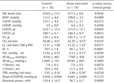
Baseline hemodynamic data. MAP, HR, LVESP, LVEDP,τ, dP/dtmaxand dP/dtminwere not different in acute exercised animals compared to the control group (Table 2).Fig. 3shows representative original steady- state P-V loops obtained from acute exercised and control rats. The de- creased width of baseline P-V loops after exhaustive swimming reflects reduced SV along with unaltered LVEDV and increased LVESV. EF, CO and CI decreased significantly suggesting deteriorated systolic perfor- mance in rats after exhaustive exercise. TPR and Eawere increased in rats after intense exercise (Table 2).
Systolic and diastolic functional indexes derived from P-V analysis at different preloads.Fig. 4A shows representative original P-V loops re- corded during transient occlusion of the inferior vena cava in exercised and control animals. The slope (Ees) of end-systolic P-V relationship (ESPVR) was less steep in exhaustive exercised than in control animals suggesting decreased contractility. The EDPVR did not differ between the groups indicating unchanged LV stiffness of myocardium after ex- cessive exercise. Slope values of PRSW were significantly decreased after intense exercise indicating impaired contractility (Fig. 4B). As Fig. 4C shows, the linear relation of dP/dtmaxand EDV was less steep in exhausted animals than in controls. The overall dP/dtmax-EDV slope values were significantly lower in exercised rats (Fig. 4C).
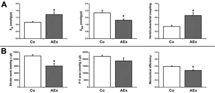
Cardiac mechanoenergetics. SW was decreased in acute exercised animals. P-V analysis revealed a significant decrease in Eesand an in- crease of Ea. Subsequently, ventriculoarterial coupling was significantly increased in exhaustively exercised rats suggesting contractility- afterload mismatch (Fig. 5A). Despite the decreased SW in exercised rats, pressure-volume area did not differ between the groups.
Mechanical efficiency and maximum power of LV work were also im- paired in exercised rats compared to the controls (Fig. 5B,Table 2).
4. Discussion
In the current study we validated our rat model of acute exhaustive exercise induced myocardial injury by confirming key mechanisms of cardiac damage. We provided thefirst detailed hemodynamic charac- terization and described several aspects of LV dysfunction after acute exhaustive exercise. Our results suggest that deteriorated cardiac per- formance (impaired contractility and mechanoenergetics) and myocar- dial injury might be associated with increased nitro-oxidative stress and enhanced proapoptotic activity in the LV myocardium.
4.1. Biomarkers of myocardial injury
In the present study an obvious myocardial injury was observed after exhaustive swimming exercise. The release of cTnT as well as non-specific cardiac enzymes CK, LDH and AST was markedly increased after exhaustive exercise (Table 1), which is in line with previous obser- vations on animal models[8,9,19]and numerous human exercise studies[5,6]. In accordance with recent literature data our HE staining of LV myocardium showed signs of sporadic cardiomyocyte damage after exhaustive exercise[19](Fig. 1A).
4.2. Cardiac dimensions and function
Although numerous human studies investigated cardiac function after a prolonged exercise both in nonelite subjects and in elite athletes by using echocardiography, there is controversial literature data about systolic and diastolic functional changes of LV[15]. Li et al. showed a sig- nificant impairment of cardiac function of experimental animals sub- jected to exhaustive physical activity both in vivo (echocardiography) and in vitro (Langendorff model)[19]. To the best of our knowledge, the present study is thefirst one that describes LV pressure and volume relations in detail and provides characterization of LV dysfunction in vivo after exhaustive exercise. P-V analysis clearly demonstrated significantly increased LVESV along with unchanged LVEDV, thus decreased SV and EF in rats that underwent our exhaustive exercise pro- tocol (Fig. 3). These cardiac dimensions are consistent with previous ex- perimental results[19]and correspond with human echocardiography data suggesting systolic impairment after exhaustive exercise[24,25].
Table 2
Hemodynamic parameters in control and exhaustive exercised rats.
Control (n = 10)
Acute exercised (n = 10)
p value versus control group
HR, beats/min 418.6 ± 13.1 417.5 ± 8.0 0.9436
MAP, mmHg 112.5 ± 4.4 109.8 ± 3.5 0.6400
LVESP, mmHg 125.7 ± 4.4 125.1 ± 3.7 0.9272
LVEDP, mmHg 3.9 ± 0.6 4.4 ± 0.3 0.4208
LVEDV,μl 214.7 ± 15.5 211.9 ± 13.7 0.8943
LVESV,μl 100.1 ± 6.7 140.4 ± 8.5* 0.0015
SV,μl 139.1 ± 9.4 102.3 ± 11.3* 0.0226
CO, ml/min 58.48 ± 4.67 42.39 ± 4.53* 0.0235
CI, (ml/min)/100 g BW 17.31 ± 1.68 12.32 ± 1.22* 0.0271
EF, % 59.5 ± 1.8 44.1 ± 3.4* b0.0001
SW, mmHg · ml 13.42 ± 0.55 9.11 ± 0.11* 0.0030
dP/dtmax, mmHg/s 8350 ± 465 7700 ± 458 0.3329
dP/dtmin, mmHg/s 11095 ± 741 10183 ± 693 0.3805
τ(Weiss), ms 7.8 ± 0.3 7.9 ± 0.3 0.6876
τ(Glanz), ms 10.9 ± 0.6 11.2 ± 0.6 0.7297
TPR, mmHg/(ml/min) 2.02 ± 0.16 2.84 ± 0.29* 0.0228 Slope of EDPVR, mmHg/μl 0.036 ± 0.004 0.041 ± 0.004 0.3133 Maximal power, mW 110.4 ± 16.5 58.7 ± 6.1* 0.0009 Values are means ± SEM. Hemodynamic parameters were measured by the Millar pres- sure-volume conductance catheter system. HR, heart rate; MAP, mean arterial pressure;
LVESP, left ventricular (LV) end-systolic pressure; LVEDP, LV end-diastolic pressure;
LVEDV, LV end-diastolic volume; LVESV, LV end-systolic volume; SV, stroke volume; CO, cardiac output; CI, cardiac index; BW, body weight; EF, ejection fraction; SW, stroke work; dP/dtmaxand dP/dtminmaximal slope of the systolic pressure increment and the di- astolic pressure decrement, respectively;τ, time constant of LV pressure decay; TPR, total peripheral resistance; EDPVR, end-diastolic pressure-volume relationship. *:pb0.05 vs.
controls.
Fig. 3.Steady-state left ventricular pressure-volume loops. Original recordings of steady- state pressure-volume (P-V) loops in representative rats from control (Co) and acute exercised (AEx) groups. The decreased width of P-V loops in exercised animals indicate reduced stroke volume as a result of unaltered end-diastolic volume and increased end- systolic volume, thus decreased ejection fraction.
4.3. Systolic function and cardiac contractility
In our rat model exhaustive exercise was associated with unchanged dP/dtmaxand LVESP, but markedly decreased EF (Table 2). Although dP/dtmax and EF have been widely used as cardiac contractility parameters, it is well recognized that they are dependent on loading con- ditions, and therefore cannot reliably be used to assess LV contractile function in models where loading is altered[26]. The method of simulta- neous LV pressure and volume analysis performed by the miniaturized pressure-conductance catheter allowed us to calculate highly sensitive load-independent indexes of LV contractility.
The slope of ESPVR (Ees) has been described as a fairly load- insensitive index of LV contractility, which was significantly decreased after intense exercise, indicating a deteriorated inotropic state of the LV myocardium (Fig. 4A). However, because this relation can be altered not only by changes in the contractile state but also by changes in chamber geometry and other diastolic factors, we also calculated other reliable contractility parameters[26].
PRSW represents the slope of the linear relation between SW and EDV. It has been described as a sensitive LV contractility index
independent of chamber size and mass. PRSW was also significantly de- creased in exhaustive exercised rats compared to controls (Fig. 4B). Al- though this parameter integrates data from the entire cardiac cycle, it is influenced most of all by the systole[26].
Analysis of dP/dtmax-EDV relationship allowed us to compare dP/dtmax
values at a given LVEDV between the experimental groups. A previous investigation has demonstrated[27]that the slope of the relation be- tween dP/dtmaxand EDV, another P-V loop-derived index, represents a less load-dependent parameter of LV contractility. This sensitive con- tractility index was also impaired after exhaustive exercise compared to controls (Fig. 4C).
4.4. Diastolic function
Indices of LV stiffness (LVEDP and slope of EDPVR) were not signifi- cantly different between exercised and control animals, as well as unchanged ventricular relaxation has been observed after intense exer- cise (as indicated byτand dP/dtmin;Table 2). The observed unchanged diastolic function is in line with recentfindings on isolated rat hearts [28]. Nevertheless there are several human studies describing a Fig. 4.Pressure-volume loop derived load independent left ventricular contractility parameters. The slope (Ees) of end-systolic pressure-volume relationship (ESPVR) (A); preload recruitable stroke work (PRSW), the slope of the relationship between stroke work (SW) and end-diastolic volume (EDV) (B); and maximal slope of the systolic pressure increment (dP/dtmax)—EDV relationship (dP/dtmax-EDV) (C) in representative rats from control (Co) and acute exercised (AEx) groups. As seen on the bar graphs, all of these contractility param- eters are decreased in the AEx group, suggesting reduced systolic performance after exhaustive exercise. *: pb0.05 vs. Co.
transient diastolic dysfunction (decreased early to late diastolic ratio assessed by echocardiography; E/A) after exhaustive exercise[15].
Further experimental reports are required for appropriate comparison of human and animal diastolic values.
4.5. Mechanoenergetics
P-V analysis was used to characterize mechanoenergetic changes after exhaustive exercise. We determined Eesand Ea, which are relative load-independent indices of LV contractility and vascular loading, respectively. Eais an integrative index that incorporates the principal ele- ments of arterial load, including peripheral vascular resistance, total arte- rial compliance and characteristic impedance[29]. Ventriculoarterial coupling (Ea/Ees) is an important determinant of net cardiac perfor- mance and energetics[30]. The parallel decrease of myocardial contrac- tility (Ees) and the increase of Ealed to a significant impairment of the ventriculoarterial coupling ratio in acute exercised animals (Fig. 5A).
Impaired ventriculoarterial coupling in the exercised group reflects an inadequate matching between LV contractility and afterload after ex- haustive exercise, which results in a suboptimal transfer of blood from the LV to the periphery with more excessive changes in pressure.
Stroke work is determined as the area within the P-V loop and rep- resents the external mechanical work performed by the left ventricle during a single heart cycle. P-V area (PVA) has been described as an index of LV total mechanical energy and is linearly related to total myo- cardial oxygen consumption[31]. Decreased SW along with unaltered PVA leads to decreased mechanical efficiency in exhaustive exercised animals compared with controls, suggesting a deterioration of metabol- ic efficiency after such exercise: decreased mechanical work along with similar myocardial oxygen consumption (Fig. 5B). According to our knowledge the present study is thefirst to demonstrate decreased me- chanical efficiency after exhaustive exercise.
4.6. Oxidative stress
Even though the exact mechanisms responsible for exercise-induced myocardial injury are still not well understood, there have been accu- mulated evidence indicating that exhaustive exercise causes imbalance between ROS and antioxidant defense, resulting in oxidative stress in the myocardium[12]. Increased formation of ROS (by the mitochondrial electron transport chain, NAPDH and xanthine oxidases[32]) activates a
broad variety of hypertrophy signaling kinases and transcription factors and induces apoptosis by DNA and mitochondrial damage and activa- tion of proapoptotic signaling kinases[33]. A robust generation of ROS and thus increased oxidative stress was observed by DHE-staining in the myocardium of exhaustive exercised rats compared to controls (Fig. 1B). A recent experimental study of exhaustive exercise showed the key role of Nrf2, the primary transcriptional regulator of antioxi- dants, including G6PD, GPX-1, GSR and catalase, which are upregulated as a compensatory reaction to ROS overproduction[12]. Corresponding- ly we found significantly increased myocardial gene expressions of en- dogenous antioxidants, such as G6PD, GSR as well as thioredoxin-1 after exhaustive exercise. In accordance with Muthusamy et al.[12], we observed an upregulation of mitochondrial superoxide dismutase- 2 (SOD-2) in the myocardium, suggesting mitochondrial superoxide generation as a result of exhaustive exercise (Fig. 2A). Increased su- peroxide concentration reduces the bioavailability of nitric oxide (NO) by chemical inactivation to form toxic peroxynitrite. A marked- ly increased protein nitration was observed by nitrotyrosine staining (Fig. 1C), suggesting increased nitrative stress, which could be the consequence of peroxynitrite formation[32]. Peroxynitrite can also
“uncouple”eNOS to become a dysfunctional superoxide-generating enzyme that contributes to vascular oxidative stress[34]. In our study the myocardial expression of eNOS increased in response to oxidative stress, which might reflect upregulation as a reaction to decreased NO bioavailability and eNOS uncoupling. Moreover, nitration of several myocardial proteins can have a potential delete- rious effect on myocardial contractility and increased peroxynitrite formation can induce apoptosis as well as matrix metalloproteinase activation[32].
4.7. Apoptosis
The ratio of proapoptotic and antiapoptotic proteins (e.g. Bax/Bcl-2) regulates myonuclei integrity and cell survival by controlling mitochon- drial membrane permeability[35]. According to our results exhaustive exercise resulted in a markedly increased Bax/Bcl-2 ratio, thus en- hanced apoptotic signaling in the myocardium. Thisfinding has further been supported by the increased number of TUNEL positive cardiomyo- cyte nuclei, suggesting increased DNA fragmentation after acute exer- cise (Fig. 1D).
Fig. 5.Cardiac mechanoenergetics. Panel A: Decreased end-systolic elastance (Ees) and increased arterial elastance (Ea) resulted in deteriorated ventriculoarterial coupling (Ea/Ees) in acute exercised (AEx) rats compared with controls (Co). Panel B: Decreased stroke work (SW) along with unaltered pressure-volume area (P-V area) led to reduced mechanical efficiency (SW/
P-V area) in the swimming group. All of these parameters suggest impaired LV mechanoenergetics after exhaustive exercise. *: pb0.05 vs. Co.
4.8. Oxidative stress induced dysregulation of MMPs
ROS can stimulate cardiacfibroblast proliferation and activate ma- trix metalloproteinases, which play a key role in the homeostasis of extracellular matrix in the myocardium. Sustained MMP activation (increased expression of MMPs or downregulation of their endogenous inhibitors, TIMPs) might influence the structural properties of the myo- cardium by providing an abnormal extracellular environment, which the cardiomyocytes interact with[13,36]. As recent investigations showed, expression and activity of MMP-2 and MMP-9 are increased in the skeletal muscle after a single bout of exercise[37]. The observed significant changes of MMP-2/TIMP-2 as well as MMP-9/TIMP-1 ratio suggest MMP dysregulation in myocardium of exercised rats. These findings raise the possibility that enhanced oxidative stress can be a stimulus for myocardial MMP activation, which might play an impor- tant role in the development of exhaustive exercise-induced cardiac dysfunction. TGF-β1 is a pleiotropic cytokine, which is involved in cardi- ac injury, repair andfibrotic remodeling[38]. A strong tendency toward upregulation of TGF-β1 expression after exhaustive exercise suggests the activation of reparative and profibrotic mechanisms after myocardi- al injury induced by prolonged exercise.
4.9. Study limitations
The interpretation of results from the present study is limited to young male rats. The possible influence of gender, age or species should be assessed in future studies. Swimming with weight attached to the tail has been suggested to be effective to induce exhaustive exercise in- duced cardiac injury and oxidative stress in animals[9]even though swimming is associated with an important stress response[39]. In order to minimize the influence of stress all rats were familiarized with swimming 48 h before the formal swimming protocol.
Our current data on increased myocardial gene expression of MMPs and TIMPs within 2 h suggest initiation of matrix degradation and profibrotic activity, however these phenomena are more relevant in a longer period after myocardial insult.
This study was designed to observe hemodynamic alterations induced by one bout of exhaustive exercise, however impact of the ob- served molecular and functional changes on the longer term remains unanswered. Further studies are warranted to elucidate the possible re- versibility of myocardial dysfunction described in the present work.
5. Conclusions
In conclusion we confirmed exhaustive exerciseinduced cardiac in- jury and nitro-oxidative stress in our rat model and for thefirst time we described in vivo cardiac dysfunction in detail by using the P-V con- ductance catheter system. Based on reliable load-independent indices we demonstrated systolic functional deterioration (reduced contractility) in exhaustive exercised animals along with unchanged diastolic function and impaired mechanoenergetics (decreased efficiency, ventriculo- arterial mismatch). Elevations in cardiac enzymes, sporadic cardiac in- juries and possible impaired myocardial function along with the activa- tion of proapoptotic and profibrotic activity raise the question whether prolonged endurance exercise could induce persistent myocardial dam- age and dysfunction.
Conflict of interest
The authors declare that they have no conflict of interest.
Acknowledgments
This work was supported by the Hungarian Research Fund (OTKA 105555 to B.M.), by the János Bolyai Research Scholarship of the
Hungarian Academy of Sciences (to T.R.) and by a grant from the National Development Agency of Hungary (TÁMOP-4.2.2/B-10/1-2010-0013).
The expert technical assistance of Henriett Biró, Gábor Alt and Gábor Fritz is gratefully acknowledged.
References
[1] C. Lee, The definition and assessment of physical activity in cardiovascular risk re- duction research, Aust. J. Public Health 17 (1993) 190–194.
[2] P.D. Thompson, D. Buchner, I.L. Pina, et al., Exercise and physical activity in the pre- vention and treatment of atherosclerotic cardiovascular disease: a statement from the Council on Clinical Cardiology (Subcommittee on Exercise, Rehabilitation, and Prevention) and the Council on Nutrition, Physical Activity, and Metabolism (Subcommittee on Physical Activity), Circulation 107 (2003) 3109–3116.
[3] N. Middleton, K. George, G. Whyte, D. Gaze, P. Collinson, R. Shave, Cardiac troponin T release is stimulated by endurance exercise in healthy humans, J. Am. Coll. Cardiol.
52 (2008) 1813–1814.
[4] R. Shave, K.P. George, G. Atkinson, et al., Exercise-induced cardiac troponin T release:
a meta-analysis, Med. Sci. Sports Exerc. 39 (2007) 2099–2106.
[5] J. Scharhag, K. George, R. Shave, A. Urhausen, W. Kindermann, Exercise-associated increases in cardiac biomarkers, Med. Sci. Sports Exerc. 40 (2008) 1408–1415.
[6] R. Shave, A. Baggish, K. George, et al., Exercise-induced cardiac troponin elevation:
evidence, mechanisms, and implications, J. Am. Coll. Cardiol. 56 (2010) 169–176.
[7] A. La Gerche, D.L. Prior, Exercise—is it possible to have too much of a good thing?
Heart Lung Circ. 16 (Suppl. 3) (2007) S102–S104.
[8] Y. Chen, R.C. Serfass, S.M. Mackey-Bojack, K.L. Kelly, J.L. Titus, F.S. Apple, Cardiac troponin T alterations in myocardium and serum of rats after stressful, prolonged intense exercise, J. Appl. Physiol. 88 (2000) 1749–1755.
[9] J. Nie, G. Close, K.P. George, T.K. Tong, Q. Shi, Temporal association of elevations in serum cardiac troponin T and myocardial oxidative stress after prolonged exercise in rats, Eur. J. Appl. Physiol. 110 (2010) 1299–1303.
[10] Z. Radák, H. Ogonovszky, J. Dubecz, et al., Super-marathon race increases serum and urinary nitrotyrosine and carbonyl levels, Eur. J. Clin. Investig. 33 (2003) 726–730.
[11]N.B. Vollaard, J.P. Shearman, C.E. Cooper, Exercise-induced oxidative stress:myths, realities and physiological relevance, Sports Med. 35 (2005) 1045–1062.
[12]V.R. Muthusamy, S. Kannan, K. Sadhaasivam, et al., Acute exercise stress activates Nrf2/ARE signaling and promotes antioxidant mechanisms in the myocardium, Free Radic. Biol. Med. 52 (2012) 366–376.
[13]H. Tsutsui, S. Kinugawa, S. Matsushima, Oxidative stress and heart failure, Am. J.
Physiol. Heart Circ. Physiol. 301 (2011) H2181–H2190.
[14] N. Mousavi, A. Czarnecki, K. Kumar, et al., Relation of biomarkers and cardiac mag- netic resonance imaging after marathon running, Am. J. Cardiol. 103 (2009) 1467–1472.
[15] D. Oxborough, K. Birch, R. Shave, K. George,“Exercise-induced cardiac fatigue”—a re- view of the echocardiographic literature, Echocardiography 27 (2010) 1130–1140.
[16] P.S. Douglas, M.L. O'Toole, W.D. Hiller, K. Hackney, N. Reichek, Cardiac fatigue after prolonged exercise, Circulation 76 (1987) 1206–1213.
[17]G.P. Whyte, Clinical significance of cardiac damage and changes in function after exercise, Med. Sci. Sports Exerc. 40 (2008) 1416–1423.
[18]C.C. Huang, T.J. Lin, C.C. Chen, W.T. Lin, Endurance training accelerates exhaustive exercise-induced mitochondrial DNA deletion and apoptosis of left ventricle myo- cardium in rats, Eur. J. Appl. Physiol. 107 (2009) 697–706.
[19] T. Li, D. Zhu, R. Zhou, W. Wu, Q. Li, J. Liu, HBOC attenuates intense exercise-induced cardiac dysfunction, Int. J. Sports Med. 33 (2012) 338–345.
[20] P. Pacher, T. Nagayama, P. Mukhopadhyay, S. Bátkai, D.A. Kass, Measurement of car- diac function using pressure-volume conductance catheter technique in mice and rats, Nat. Protoc. 3 (2008) 1422–1434.
[21]T. Csont, E. Bereczki, P. Bencsik, et al., Hypercholesterolemia increases myocardial oxidative and nitrosative stress thereby leading to cardiac dysfunction in apoB- 100 transgenic mice, Cardiovasc. Res. 76 (2007) 100–109.
[22] G. Masszi, R. Benko, N. Csibi, et al., Endothelial relaxation mechanisms and nitrative stress are partly restored by Vitamin D3 therapy in a rat model of polycystic ovary syndrome, Life Sci. 93 (2013) 133–138.
[23]T. Radovits, A. Oláh, Á. Lux, et al., Rat model of exercise-induced cardiac hypertro- phy: hemodynamic characterization using left ventricular pressure-volume analy- sis, Am. J. Physiol. Heart Circ. Physiol. 305 (2013) H124–H134.
[24] T.G. Neilan, D.M. Yoerger, P.S. Douglas, et al., Persistent and reversible cardiac dys- function among amateur marathon runners, Eur. Heart J. 27 (2006) 1079–1084.
[25]N. Middleton, R. Shave, K. George, G. Whyte, E. Hart, G. Atkinson, Left ventricular function immediately following prolonged exercise: a meta-analysis, Med. Sci.
Sports Exerc. 38 (2006) 681–687.
[26]D.A. Kass, Clinical ventricular pathophysiology: a pressure-volume view, in: D.C.
Warltier (Ed.), Ventricular Function, 1st ed.Williams & Wilkins, Baltimore, 1995, pp. 131–151.
[27]W.C. Little, The left ventricular dP/dtmax-end-diastolic volume relation in closed- chest dogs, Circ. Res. 56 (1985) 808–815.
[28] P.O. Reger, S.C. Kolwicz, J.R. Libonati, Acute exercise exacerbates ischemia-induced diastolic rigor in hypertensive myocardium, Springerplus 1 (2012) 46.
[29]K. Sunagawa, W.L. Maughan, D. Burkhoff, K. Sagawa, Left ventricular interaction with arterial load studied in isolated canine ventricle, Am. J. Physiol. 245 (1983) H773–H780.
[30] D.A. Kass, R.P. Kelly, Ventriculo-arterial coupling: concepts, assumptions, and appli- cations, Ann. Biomed. Eng. 20 (1992) 41–62.
[31] H. Suga, Cardiac energetics: from E(max) to pressure-volume area, Clin. Exp.
Pharmacol. Physiol. 30 (2003) 580–585.
[32] P. Pacher, J.S. Beckman, L. Liaudet, Nitric oxide and peroxynitrite in health and dis- ease, Physiol. Rev. 87 (2007) 315–424.
[33] A. Sabri, H.H. Hughie, P.A. Lucchesi, Regulation of hypertrophic and apoptotic signal- ing pathways by reactive oxygen species in cardiac myocytes, Antioxid. Redox Sig- nal. 5 (2003) 731–740.
[34] U. Förstermann, Nitric oxide and oxidative stress in vascular disease, Pflugers Arch.
459 (2010) 923–939.
[35] M.O. Hengartner, The biochemistry of apoptosis, Nature 407 (2000) 770–776.
[36] A.D. Kandasamy, A.K. Chow, M.A. Ali, R. Schulz, Matrix metalloproteinase-2 and myo- cardial oxidative stress injury: beyond the matrix, Cardiovasc. Res. 85 (2010) 413–423.
[37] S.O. Koskinen, W. Wang, A.M. Ahtikoski, et al., Acute exercise induced changes in rat skeletal muscle mRNAs and proteins regulating type IV collagen content, Am. J.
Physiol. Regul. Integr. Comp. Physiol. 280 (2001) R1292–R1300.
[38] M. Dobaczewski, W. Chen, N.G. Frangogiannis, Transforming growth factor (TGF)-β signaling in cardiac remodeling, J. Mol. Cell. Cardiol. 51 (2011) 600–606.
[39] R.V. Contarteze, B. Manchado Fde, C.A. Gobatto, M.A. De Mello, Stress biomarkers in rats submitted to swimming and treadmill running exercises, Comp. Biochem. Phys- iol. A Mol. Integr. Physiol. 151 (2008) 415–422.