The Rockefeller University Press $30.00 J. Gen. Physiol. Vol. 142 No. 1 61–73
www.jgp.org/cgi/doi/10.1085/jgp.201210954 61
I N T R O D U C T I O N
CFTR (ABCC7; Riordan et al., 1989) is a member of the asymmetric C subfamily of ATP-binding cassette (ABC) proteins. Most ABC proteins are active transporters that move a variety of substrates across biological mem- branes. The substrate translocation pathway, formed by two transmembrane domains (TMDs), cycles between inward- and outward-facing conformations driven by an ATP hydrolysis cycle catalyzed by the two cytosolic nu- cleotide-binding domains (NBDs). ABC NBDs contain three highly conserved sequence motifs: the Walker A (GXXXXGKS/T) and B (DE, with represent- ing hydrophobic residues), and the ABC signature motif (LSGGQR/K). In each catalytic cycle, ATP binding to the Walker motifs of both NBDs promotes formation of a tight head-to-tail NBD dimer that occludes two ATP molecules in composite interfacial catalytic sites formed by the Walker motifs of one NBD and the signature motif
Correspondence to László Csanády:
c s a n a d y . l a s z l o @ m e d . s e m m e l w e i s - u n i v . h u
Abbreviations used in this paper: ABC, ATP-binding cassette; NBD, nucleotide-binding domain; P-ATP, N6-(2-phenylethyl)-ATP; Po, channel open probability; RI, regulatory insertion; TMD, transmembrane domain.
of the other. ATP hydrolysis is required to disrupt this dimer to allow nucleotide exchange and initiation of a new cycle (Hollenstein et al., 2007). Members of the C subfamily share this overall architecture but show a marked asymmetry in the primary sequences of their two NBDs. Because of noncanonical substitutions in the Walker B and in other conserved motifs of the N-terminal NBD (NBD1) and in the signature sequence of the C-terminal NBD (NBD2), the catalytic site con- taining these motifs (site 1) is inactive (Aleksandrov et al., 2002; Basso et al., 2003), leaving ABC-C proteins with a single catalytically active site (site 2, formed by Walker motifs of NBD2 and signature sequence of NBD1). Non- canonical substitutions in the same key positions, resulting in reduced or absent hydrolysis at one composite site, are also seen in the clinically important transporter as- sociated with antigen processing (formed by TAP1 and TAP2 of the human ABC-B subfamily; Procko et al., 2009),
Conformational changes in the catalytically inactive nucleotide-binding site of CFTR
László Csanády,1,2 Csaba Mihályi,1 Andras Szollosi,1 Beáta Töröcsik,1 and Paola Vergani3
1Department of Medical Biochemistry and 2MTA-SE Ion Channel Research Group, Semmelweis University, Budapest H-1094, Hungary
3Department of Neuroscience, Physiology and Pharmacology, University College London, London WC1E 6BT, England, UK
A central step in the gating of the cystic fibrosis transmembrane conductance regulator (CFTR) chloride channel is the association of its two cytosolic nucleotide-binding domains (NBDs) into a head-to-tail dimer, with two nucleo- tides bound at the interface. Channel opening and closing, respectively, are coupled to formation and disruption of this tight NBD dimer. CFTR is an asymmetric adenosine triphosphate (ATP)-binding cassette protein in which the two interfacial-binding sites (composite sites 1 and 2) are functionally different. During gating, the canonical, catalytically active nucleotide-binding site (site 2) cycles between dimerized prehydrolytic (state O1), dimerized post-hydrolytic (state O2), and dissociated (state C) forms in a preferential C→O1→O2→C sequence. In contrast, the catalytically inactive nucleotide-binding site (site 1) is believed to remain associated, ATP-bound, for several gating cycles. Here, we have examined the possibility of conformational changes in site 1 during gating, by studying gating effects of perturbations in site 1.
Previous work showed that channel closure is slowed, both under hydrolytic and nonhydrolytic conditions, by occupancy of site 1 by N6-(2-phenylethyl)-ATP (P-ATP) as well as by the site-1 mutation H1348A (NBD2 signature sequence). Here, we found that P-ATP prolongs wild-type (WT) CFTR burst durations by selectively slowing (>2×) transition O1→O2 and decreases the nonhydrolytic closing rate (transition O1→C) of CFTR mutants K1250A (4×) and E1371S (3×). Mutation H1348A also slowed (3×) the O1→O2 transition in the WT background and decreased the nonhydrolytic closing rate of both K1250A (3×) and E1371S (3×) background mutants. Neither P-ATP nor the H1348A mutation affected the 1:1 stoichiometry between ATP occlusion and channel burst events characteristic to WT CFTR gating in ATP. The marked effect that different structural perturbations at site 1 have on both steps O1→C and O1→O2 suggests that the overall conformational changes that CFTR undergoes upon opening and coincident with hydrolysis at the active site 2 include significant structural rearrangement at site 1.
© 2013 Csanády et al. This article is distributed under the terms of an Attribution–
Noncommercial–Share Alike–No Mirror Sites license for the first six months after the publi- cation date (see http://www.rupress.org/terms). After six months it is available under a Creative Commons License (Attribution–Noncommercial–Share Alike 3.0 Unported license, as described at http://creativecommons.org/licenses/by-nc-sa/3.0/).
The Journal of General Physiology
on November 4, 2014jgp.rupress.orgDownloaded fromSupplemental Material can be found at:
gating cycles. This notion obtained further support from thermodynamic studies (Szollosi et al., 2011) that examined state-dependent changes in energetic cou- pling between pairs of residues on opposing faces of the site-1 interface. A lack of change in energetic coupling for three such pairs was interpreted to be consistent with the model suggested by Tsai et al. (2010), i.e., with an “immobile” site 1 remaining closed throughout the gating cycle.
Nevertheless, site 1 clearly does play a role in gating energetics, as perturbations at site 1 can profoundly alter channel gating kinetics. For instance, the K464A mutation, which—by removing the side chain of the conserved NBD1 Walker A lysine—perturbs the NBD1 side of site 1, dramatically alters the mechanism of gating (Csanády et al., 2010). In addition, the H1348A mutation in the NBD2 signature sequence, which per- turbs the NBD2 side of site 1, was found to slow channel closure (Szollosi et al., 2011). Finally, gating is affected by perturbations of the nucleotide structure at site 1, as P-ATP bound at site 1 also slows channel closure (Tsai et al., 2010). However, the exact mechanisms by which the H1348A mutation, or P-ATP bound at site 1, affects gating have not yet been elucidated. To gain further in- sight into the energetic consequences of perturbations at site 1, we examined in detail the effects of these two perturbations on CFTR channel gating. Our observations suggest that, despite maintained contact between resi- dues on opposite sides of site 1, this site does not remain
“frozen”; rather, significant conformational changes are likely to take place there both during channel opening and concomitant with ATP hydrolysis at site 2.
M A T E R I A L S A N D M E T H O D S Molecular biology
Human WT CFTR and CFTR segment 433–1480 in the pGEMHE plasmid (Chan et al., 2000) served as templates for mutants H1348A, K1250A, E1371S, K1250A/H1348A, E1371S/H1348A, E1371S/K464A, and 433–1480(K1250A), which were created using the QuikChange kit (Agilent Technologies). The entire coding sequence of each construct was verified by automated sequenc- ing (LGC Genomics). T7 polymerase was used for in vitro tran- scription (Mmessage kit; Ambion), and purified cRNA was stored at 80°C.
Isolation and injection of Xenopus laevis oocytes
Xenopus oocytes were extracted and treated with collagenase as described previously (Chan et al., 2000). Isolated oocytes were stored at 18°C in a solution containing (mM) 82 NaCl, 2 KCl, 1 MgCl2, 5 HEPES, pH 7.5 with NaOH, 1.8 CaCl2, and 50 µg/ml gentamycin. To obtain expression levels appropriate for single- channel or macroscopic recordings, oocytes were microinjected with 0.1–10 ng of CFTR cRNA, and further incubated at 18°C for 2–3 d.
Excised inside-out patch-clamp recordings
For inside-out patch recordings, the pipette solution contained (mM) 136 NMDG-Cl, 2 MgCl2, and 5 HEPES, pH 7.4 with NMDG.
as well as in numerous yeast and prokaryotic transporters (Szollosi et al., 2011). The latter group includes TM287/
288, a heterodimeric transporter from Thermotoga mari- tima, whose high resolution structure has been recently solved (Hohl et al., 2012).
CFTR, the protein mutated in cystic fibrosis patients (Riordan et al., 1989), is the only member of the ABC-C family whose TMDs are known to form the pore of a Cl ion channel. In addition to the canonical ABC domains, CFTR contains a unique regulatory (R) domain, phos- phorylation of which by cAMP-dependent protein kinase (PKA) is a prerequisite for channel gating (Anderson et al., 1991). In phosphorylated CFTR, the pore opens to a burst upon ATP-induced dimerization of its NBDs and closes from a burst upon dimer dissociation (Vergani et al., 2005) after ATP hydrolysis at site 2; thus, in WT CFTR, each gating cycle (or burst of openings) is cou- pled to hydrolysis of one ATP molecule (Csanády et al., 2010). Deviations from this strict 1:1 coupling between ATP hydrolysis events and bursts of openings have been suggested to happen in mutants or in the presence of drugs. Catalytic site mutations can lower this cou- pling ratio leading to bursts that involve ATP occlusion/
deocclusion but no hydrolysis (Csanády et al., 2010). In contrast, [ATP]-dependent prolongation of open burst durations for the W401F CFTR mutant (Jih et al., 2012b), or for WT CFTR gating in the presence of the potentiator compound Vx-770 (Jih and Hwang, 2013), has led to the suggestion that post-hydrolytic ADP/ATP exchange in site 2 might occur without intervening pore closure, thereby increasing the coupling ratio by allowing more than one ATP to be hydrolyzed in site 2 within a single gating cycle (Jih et al., 2012a).
Biochemical work using -32P-labeled ATP analogues revealed that ATP remains bound at CFTR’s NBD1 for pe- riods much longer (several minutes) than a channel gat- ing cycle (1 s), without being hydrolyzed (Aleksandrov et al., 2002; Basso et al., 2003). In a recent study (Tsai et al., 2010), nucleotide exchange time constants at the two sites were measured using a functional approach.
The high affinity ATP analogue N6-(2-phenylethyl)-ATP (P-ATP) was found to speed the opening and slow the closing of CFTR channels, but the onset of these two effects upon sudden replacement of ATP with P-ATP in the bath (cytosolic) solution followed distinct time courses. Whereas the effect on opening rate appeared instantaneously, suggesting that it was caused by P-ATP binding to the rapid-turnover site 2, the onset of slowed closure followed with a delay of 30–50 s, consistent with ATP/P-ATP exchange occurring more slowly at degen- erate site 1. Furthermore, because the nucleotide ex- change rate at site 1 was affected by mutations in the NBD2 signature sequence, which completes site 1 only in a formed dimer, the authors suggested only partial separation of the NBD dimer in closed channels, with the interface remaining closed around site 1 for several
on November 4, 2014jgp.rupress.orgDownloaded from
ms were excluded from the analysis. The probability density func- tions of the two fitted models are given by f(k, t) = k · exp(kt) and f(k1, k2, t) = (k1k2/(k2 k1)) · (exp(k1t) exp(k2t)), respectively;
the improvement of the fit caused by introduction of the second free parameter was evaluated using the log-likelihood ratio test and found significant for both distributions in Fig. 7 (A and B).
Analysis of macroscopic current relaxations
Macroscopic current decay time courses were fitted with single- exponential functions using nonlinear least squares (pClamp9), and the closing rate was defined as the inverse of the fitted time constant. For the nonhydrolytic mutants (K1250A, E1371S, and double mutants), the decay time courses after nucleotide removal often required a double-exponential function—of the form I(t) = I0(A1exp(t/1) + (1 A1)exp(t/2))—with two slow time con- stants for a satisfactory fit (e.g., Fig. 3, B and E), suggesting the presence of two populations of open-channel bursts. In such cases, the average steady-state burst duration (*) was estimated as * = 12/(A12 + A21), and the average closing rate was defined as 1/* (Szollosi et al., 2011).
Mutant cycle analysis
Mutant cycle analysis (Fersht, 2002) was used to give a measure of the energetic coupling (Gint) between pairs of target structural elements, as described previously (Vergani et al., 2005; Szollosi et al., 2010, 2011). In brief, perturbation-induced changes (between corners i and j of a mutant cycle) in activation free energy barriers (G‡) for closing were calculated from the changes in (hydro- lytic and nonhydrolytic) closing rates (r) as G‡ji = RTln(rj/ri), and Gint was defined as the difference between G‡ values along parallel sides of the mutant cycle (e.g., G‡43 G‡21).
All Gs are given as mean ± SEM. Because the numbers of obser- vations, n, for each corner of the cycle are similar, SEMs were calculated using the mean value for n.
Statistics
Data are given as mean ± SEM of at least five measurements. Sta- tistical significance was evaluated using Student’s t test (*, P <
0.05; **, P < 0.01).
Online supplemental material
Fig. S1 compares single-channel conductances in ATP versus P-ATP. Fig. S2 shows the effect of the K464A mutation on nonhy- drolytic closing rate measured in the E1371S background. Figs. S1 and S2 are available at http://www.jgp.org/cgi/content/full/jgp .201210954/DC1.
R E S U L T S
P-ATP stimulates CFTR channel opening with high affinity and prolongs steady-state burst durations
We first determined rough kinetic parameters for CFTR channels gating in P-ATP in our experimental system.
Apparent affinities for ATP and P-ATP were compared in macroscopic patches by applying test concentrations of the nucleotide bracketed by applications of a satu- rating (2 mM and 32 µM, respectively) concentration (Fig. 1, A and B). Fractional currents plotted against nucleotide concentration (Fig. 1 C) were well fit by the Michaelis–Menten equation and yielded Km values of 40 ± 2 µM for ATP but 2.4 ± 0.2 µM for P-ATP (Fig. 1 C, blue and red). We next compared steady-state gating of CFTR channels exposed either to a close-to-saturating
The bath solution was continuously flowing and contained (mM) 134 NMDG-Cl, 2 MgCl2, 5 HEPES, and 0.5 EGTA, pH 7.1 with NMDG. MgATP (Sigma-Aldrich) was added from a 400-mM aque- ous stock solution (pH 7.1 with NMDG) to achieve a final concen- tration of 2 mM (or 10 mM for channels bearing the K1250A mutation). A 10-mM aqueous stock solution of P-ATP Na+ salt (BIOLOG Life Science Institute) was stored at 80°C and diluted into the bath solution immediately before recording to achieve a final concentration of 10 µM (or 50 µM for K1250A mutants).
CFTR channels were fully activated by a 1–2-min cytosolic expo- sure to 300 nM of catalytic subunit of PKA (Sigma-Aldrich);
all experiments shown were done in the partially dephosphory- lated state after PKA removal, which remains stable over the time course of several minutes (Csanády et al., 2000). Switching between various bath solutions was achieved using computer- controlled electronic valves (HEKA); solution exchange time con- stant was 20–50 ms. Experiments were performed at 25°C. Unitary CFTR currents were recorded (Axopatch 200B; Molecular De- vices) at a pipette holding potential of +80 mV (Vm = 80 mV), filtered at 2 kHz, and digitized at 10 kHz (Digidata 1322A, Pclamp9;
Molecular Devices). As CFTR gating is largely voltage independent (Cai et al., 2003), macroscopic currents were recorded at mem- brane potentials between 20 and 80 mV.
Steady-state kinetic analysis of multichannel patches
Mean burst and interburst durations were extracted from steady segments of record with one to six active CFTR channels as de- scribed previously (Csanády et al., 2000). Currents were digitally filtered at 100 Hz and idealized by half-amplitude threshold cross- ing. Events lists were fitted with a simple model in which ATP- dependent slow gating is pooled into a closed-open scheme and ATP-independent brief closures are modeled as pore-blockage events (Ishihara and Welsh, 1997). Rate constants (rCO, rOC, rOB, and rBO) of the resulting closed–open–blocked (C↔O↔B) scheme were extracted by a simultaneous maximum likelihood fit to the dwell-time histograms of all conductance levels while ac- counting for the filter dead time (Csanády, 2000). Mean burst duration was then calculated as (1/rOC)(1 + rOB/rBO) (Figs. 1 F and 2 C), and mean interburst duration was calculated as 1/rCO
(Figs. 1 G and 2 D). The estimates of interburst duration, and consequently of channel open probability (Po), are sensitive to correct estimation of the number of channels (N) in the patch.
These parameters were therefore used only if N could be deter- mined with reasonable confidence; for this subset of patches, N was ≤5.
To extract the gating pattern of channels loaded with either ATP or P-ATP at both sites, segments immediately following nu- cleotide exchange were not included in our analysis. The starting point for idealization was assigned individually for each patch, based on visual inspection, with analysis restricted to segments in which the gating pattern had already stabilized. The time re- quired for stabilization after nucleotide exchange was typically between 10 and 60 s. To facilitate nucleotide loss from site 1, be- lieved to happen faster in closed channels (Tsai et al., 2010), in some cases patches were rinsed with nucleotide-free solution for 20–30 s before applying a different nucleotide (e.g., Figs. 4 and 6).
Burst analysis of single-channel patches
Burst analysis was done as described previously (Csanády et al., 2010). In brief, open bursts from records with a single active channel were isolated by ignoring closures shorter than a cutoff, chosen using the method of Jackson et al. (1983). The distribu- tions of the durations of bursts obtained in this way were fitted using maximum likelihood (Colquhoun and Sigworth, 1995) to either a single-exponential distribution (Fig. 7, A and B, blue dot- ted lines) or to the scheme in Fig. 7 C, with the slow rate k-1 fixed to 0 (Fig. 7, A and B, red solid lines); events shorter than tlow = 12
on November 4, 2014jgp.rupress.orgDownloaded from
earlier studies that have established P-ATP as a high affinity, potent CFTR stimulator (Zhou et al., 2005; Tsai et al., 2010).
[ATP] does not affect the prolongation of steady-state burst duration by the H1348A mutation
In an earlier study, we found that the H1348A mutation prolongs burst durations of CFTR channels gating in 2 mM ATP by approximately threefold (Szollosi et al., 2011).
concentration of 10 µM P-ATP or to 2 mM ATP, in patches containing a few (≤2) channels in which indi- vidual gating transitions remained clearly resolved (Fig. 1 D). Kinetic analysis (see Materials and methods) revealed significantly higher Po in P-ATP as compared with that seen in ATP (Fig. 1 E), which was largely caused by approximately twofold longer mean burst durations (Fig. 1 F) together with a small reduction in interburst durations (Fig. 1 G). These results confirm
Figure 2. Steady-state mean burst duration of H1348A CFTR is insensitive to elevation of [ATP]. (A) Steady-state current recording from a patch containing two active H1348A CFTR channels gating in 2 mM (green segment) or 10 mM (dark blue segment) ATP. (B–D) Steady-state open probabilities (B), mean burst durations (C), and mean interburst durations (D) of H1348A CFTR in the presence of 2 mM (green bars) or 10 mM (dark blue bars) ATP, extracted from records with ≤5 active channels, as described in Materials and methods.
Figure 1. Effects of P-ATP on rough channel gating parameters of WT CFTR. (A and B) Macroscopic currents of prephosphorylated CFTR channels elicited by step applications (bars) of various concentrations of ATP (A) or P-ATP (B). (C) Dose–response curves for current stimulation by ATP (blue symbols) and P-ATP (red symbols). Mean steady currents in the presence of test nucleotide concen- trations were normalized to the average of those measured in bracketing segments in the presence of 2 mM (ATP) or 32 µM (P-ATP) nucleotide. Fits to the Michaelis–Menten equation (solid lines) yielded Km values shown. (D) Steady-state current recording from a patch containing two active WT CFTR channels gating in 2 mM ATP (blue segments) or 10 µM P-ATP (red segment). (E–G) Steady-state open probabilities (E), mean burst durations (F), and mean interburst durations (G) of WT CFTR in the presence of 2 mM ATP (blue bars) or 10 µM P-ATP (red bars), extracted from records with ≤2 active channels, as described in Materials and methods.
on November 4, 2014jgp.rupress.orgDownloaded from
mutation (Fig. 3, B and E, green fit lines and time con- stants) caused markedly slower current relaxations than those observed for the two background constructs upon removal of ATP (Fig. 3, A and D, blue fit lines and time constants; cf. Zhou et al., 2005; Szollosi et al., 2011).
Thus, the nonhydrolytic closing rate (Fig. 3, C and F, bars; calculated from the fitted relaxation time con- stants as described in Materials and methods) was simi- larly affected by both site-1 perturbations, and this was true regardless of whether the K1250A (Fig. 3 C) or the E1371S (Fig. 3 F) mutant was chosen as the nonhydro- lytic model; i.e., both site-1 perturbations decreased this rate by two- to threefold.
Nonadditive effects of P-ATP and site-1 mutations on hydrolytic closure support slowing of this gating step by P-ATP bound in site 1
The slowing of both hydrolytic and nonhydrolytic clo- sure by P-ATP was suggested to result from occupation of site 1 by this nucleotide, based on the delayed onset of both effects upon application of the analogue (Tsai et al., 2010). We used thermodynamic mutant cycles (Fersht, 2002) as an independent approach to test this hypothesis. Nonadditive effects on gating of two struc- tural perturbations suggest that those two structural ele- ments are energetically coupled. Channels opened by Because for another site-1 mutant (W401F) mean burst
durations were shown to increase at high millimolar ATP concentrations (Jih et al., 2012b), we tested whether that was the case for H1348A CFTR. However, comparing the patterns of gating in 2 and 10 mM ATP (Fig. 2 A) did not reveal any significant effect of raising [ATP]
above 2 mM; kinetic analysis revealed identical Po
(Fig. 2 B), mean burst (Fig. 2 C), and interburst (Fig. 2 D) durations under these two conditions.
Both P-ATP and the H1348A mutation slow nonhydrolytic channel closure
To quantitatively compare the effects of our site-1 per- turbations on the slow rate of nonhydrolytic closure, we studied macroscopic closing rates after nucleotide re- moval for channels in which nucleotide hydrolysis at site 2 is abrogated by mutagenesis. Because different NBD2 mutations, in addition to disrupting ATP hy- drolysis, likely differentially alter the stability of the open state, we do not know which nonhydrolytic mutant is the best model for the O1 state of WT CFTR. We there- fore compared the effects of our site-1 perturbations on the closing rates of two nonhydrolytic mutants, NBD2 Walker A mutant K1250A (Fig. 3, A–C) and NBD2 Walker B mutant E1371S (Fig. 3, D–F). Both P-ATP (Fig. 3, A and D, red fit lines and time constants) and the H1348A
Figure 3. P-ATP and the H1348A mutation slow nonhydrolytic CFTR closure. (A and D) Macro- scopic currents of prephosphory- lated K1250A (A) and E1371S (D) CFTR channels elicited by exposure (bars) to either 10 mM ATP alternating with 50 µM P-ATP (A) or 2 mM ATP alternating with 10 µM P-ATP (D); the fivefold higher nucleotide concentrations for the K1250A constructs were used to compensate for the large decrease in apparent ATP affinity caused by this mutation (Vergani et al., 2003). Current decay time courses after sudden removal of the nucleotide were fitted with single exponentials (colored solid lines); colored numbers are time constants (in milliseconds), which reflect mean burst durations.
(B and E) Macroscopic currents of prephosphorylated K1250A/
H1348A (B) and E1371S/
H1348A (E) CFTR channels elic- ited by transient exposure (bars) to either 10 mM (B) or 2 mM (E) ATP. Current decay time courses after sudden ATP removal were fitted with double exponentials (colored solid lines); colored numbers are calculated average burst durations (*, in milliseconds; see Materials and methods). The fit parameters were 1 = 37 s, 2 = 165 s, A1 = 0.34, and A2 = 0.66 for B, and 1 = 24 s, 2 = 179 s, A1 = 0.07, and A2 = 0.93 for E. (C and F) Nonhydrolytic closing rates of channels opened by ATP (blue bars) or P-ATP (red bars), or of channels bearing the H1348A mutation opened by ATP (green bars), measured in the K1250A (C) or E1371S (F) background. Closing rates were calculated as the inverse of the decay time constant after nucleotide removal; for traces that were not well fit by a single exponential, the average closing rate was calculated as 1/*.
on November 4, 2014jgp.rupress.orgDownloaded from
gating largely intact (Csanády et al., 2005) but is never- theless expected to cause local distortion in areas of NBD1 that contact the adenine base of the site-1–
bound nucleotide.
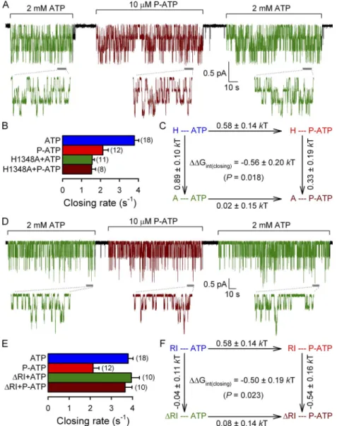
We first characterized the effect of P-ATP on hydro- lytic closing rate in these two mutant backgrounds (Fig. 4) by recording single-channel activity of H1348A (Fig. 4 A), or of RI channels (Fig. 4 D), alternately exposed to either 2 mM ATP (Fig. 4, A and D, green segments) or 10 µM P-ATP (Fig. 4, A and D, brown segments). Consistent with previous work, the H1348A and RI perturbations differentially affected hydrolytic closure: the closing rate in ATP was unchanged for RI (Fig. 4 E; compare green bar with blue bar; cf. Csanády et al., 2005), but two- to threefold slowed for H1348A (Fig. 4 B; compare green bar with blue bar; cf. Fig. 2 C).
However, switching from ATP to P-ATP (Fig. 4, A and D, brown segments) did not elicit any noticeable change in the length of channel open bursts for either con- struct (Fig. 4, A and D; compare brown with green seg- ments in expanded insets). Thus, P-ATP does not slow P-ATP contain two structural “perturbations”: a phenyl-
ethyl group (P group) linked to N6 of the adenine of the nucleotide bound in site 1, and another P group on the nucleotide bound in site 2. If the presence of the P group in site 2 affects closing rate, then this effect is expected to be additive with effects on closing rate of mu- tations in the spatially distant site 1. In contrast, if the P group in site 1 is responsible for the effects of P-ATP on closing, then P-ATP and mutations of site-1 residues (which are energetically coupled to this P group) will affect closing rate in a nonadditive manner. We chose two site-1 perturbations to test for additivity of effects on closing rate with those of P-ATP. On the NBD2 side of site 1 we chose mutation H1348A, be- cause this perturbation causes a large effect on closing rate in ATP, which we had already characterized (Fig. 2).
To perturb the NBD1 side of site 1, we chose to delete most of a nonconserved insertion in CFTR’s NBD1 (called the “regulatory insertion” [RI]; Lewis et al., 2004) by coexpression of CFTR segments 1–414 and 433–
1480. This “RI” perturbation leaves ATP-dependent
Figure 4. Mutation H1348A and deletion of segment 415–432 (RI) abolish the effect of P-ATP on hydrolytic channel closure. (A and D) Steady-state recordings of single-channel cur- rents in the presence of 2 mM ATP (green segments) or 10 µM P-ATP (brown segments) for H1348A (A) or RI CFTR (D). Insets show at an expanded time scale 10-s intervals taken from the stable steady section of each segment (gray bars). In A, Vm was 40 mV.
(B and E) Closing rates, obtained as the in- verses of the steady-state mean burst duration, for H1348A (B) and RI (E) CFTR channels gating in ATP (green bars) or P-ATP (brown bars). Steady-state closing rates of WT CFTR in ATP (blue bars) and P-ATP (red bars) were extracted from the data shown in Fig. 1 F.
(C and F) Thermodynamic mutant cycles built on (hydrolytic) closing rates for the in- teraction of the P-ATP P group with residue 1348 (C) or the RI region (F). Each corner is represented by the particular site-1 protein structure (H or A at position 1348 [C]; main- tained or deleted RI [F]) and the nucleotide driving gating (ATP or P-ATP), respectively.
G‡ values (mean ± SEM) on arrows show perturbation-induced changes in the stability of the closing transition state with respect to the open ground state and were used to calcu- late (see Materials and methods) the coupling energy for the P group–1348 (C) and the P group–RI (F) interaction (Gint(closing)).
on November 4, 2014jgp.rupress.orgDownloaded from
The H1348A and RI perturbations affected nonhydro- lytic closing rate in opposite ways; whereas mutation H1348A slowed it by approximately threefold (Fig. 5 B;
compare green bar with blue bar; cf. Fig. 3 C), the RI perturbation accelerated it by approximately threefold (Fig. 5 E; compare green bar with blue bar; cf. Csanády et al., 2005). Interestingly, neither perturbation abol- ished the effect of P-ATP on nonhydrolytic closure (Fig. 5, A and D; compare brown with green single-exponential fit lines and time constants); rather, both perturbations seemed to potentiate it. Thus, whereas P-ATP slowed nonhydrolytic closure by approximately fourfold for K1250A channels with an intact site 1 (Fig. 3 C; com- pare red with blue bar; replotted in Fig. 5, B and E), this effect increased to greater than sixfold and to approxi- mately ninefold, respectively, in the presence of the site-1 perturbations H1348A and RI (Fig. 5, B and E;
compare brown with green bars), again suggesting non- additivity. Indeed, mutant cycles built on nonhydro- lytic closing rates (Fig. 5, C and F) yielded interaction free energies between the P group and residue 1348 (Fig. 5 C), as well as between the P group and the RI re- gion (Fig. 5 F), significantly different from zero (P < 0.05 hydrolytic closure when applied in the H1348A or RI
mutant background (Fig. 4, B and E; compare brown with green bars). Although the twofold effect of P-ATP on hydrolytic closing rate of WT CFTR is small in ener- getic terms, the difference in the effects of the muta- tions on closing rates in P-ATP versus those in ATP yielded interaction free energies (Gint(closing))—both between the P group and residue 1348 (Fig. 4 C) and between the P group and the RI region (Fig. 4 F)—
which were significantly different from zero (P < 0.05).
This is consistent with the effect on hydrolytic closing rate being caused by the P group in site 1.
Nonadditive effects of P-ATP and site-1 mutations on nonhydrolytic closure support the slowing of this gating step by P-ATP bound in site 1
Additivity of effects on nonhydrolytic closure of the same site-1 perturbations with those of P-ATP was tested in the K1250A nonhydrolytic background (Fig. 5) by mea- suring the macroscopic closing rate of K1250A/H1348A channels (Fig. 5 A), and of channels obtained by co- expression of segments 1–414 and 433–1480(K1250A) (K1250A/RI; Fig. 5 D) upon nucleotide removal.
Figure 5. Mutation H1348A and deletion of segment 415–432 (RI) potentiate the effect of P-ATP on nonhydrolytic channel closure. (A and D) Macroscopic currents from K1250A/H1348A (A) and K1250A/RI (D) CFTR channels elicited by exposures to 10 mM ATP or 50 µM P-ATP (bars). Solid lines are single-exponential fits to the current relaxation time courses upon nucleotide re- moval, with time constants indicated. (B and E) Nonhydrolytic closing rates for K1250A/
H1348A (B) and K1250A/RI (E) CFTR channels, obtained as the inverses of the re- laxation time constants upon removal of ATP (green bars) or P-ATP (brown bars); clos- ing rates of K1250A CFTR upon removal of ATP (blue bars) and P-ATP (red bars) were replotted from Fig. 3 C. (C and F) Thermo- dynamic mutant cycles built on nonhydrolytic closing rates for the interaction of the P-ATP P group with residue 1348 (C) or the RI re- gion (F). Each corner is represented by the particular site-1 protein structure (H or A at position 1348 [C]; maintained or deleted RI [F]) and the nucleotide driving gating (ATP or P-ATP), respectively. G‡ values (mean ± SEM) on arrows show perturbation- induced changes in the stability of the tran- si tion state for nonhydrolytic closure with respect to the open ground state and were used to cal culate the coupling energy for the P group–1348 (C) and the P group–RI (F) in- teraction (Gint(closing)).
on November 4, 2014jgp.rupress.orgDownloaded from
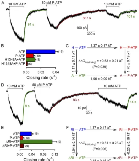
burst, thereby prolonging the mean duration of bursts at steady state. A simply testable prediction of such a model is a discrepancy between the burst durations measured at steady state and the time constant of the macroscopic current relaxation upon sudden removal of ATP. This is because the latter closing rate is mea- sured under conditions that preclude a reentry cycle, i.e., in the absence of nucleotides. Because the W401F mutation also resides in site 1, we asked whether a reen- try mechanism might explain the longer burst dura- tions in P-ATP or in the presence of the H1348A mutation. To this end, we measured macroscopic clos- ing rates after nucleotide removal for WT and RI channels opened by either 2 mM ATP or by 10 µM P-ATP (Fig. 6, A and C), and for H1348A channels opened by 2 or 10 mM ATP (Fig. 6 B, top) or by 10 µM P-ATP (Fig. 6 B, bottom). Current relaxation time courses were fitted by single exponentials (Fig. 6, A–C, colored and P < 0.01, respectively), consistent with the effect
on nonhydrolytic closing rate also being caused by the P group in site 1.
Neither P-ATP nor the H1348A mutation disrupts near 1:1 stoichiometry between ATP occlusion and channel burst events
Both P-ATP and the H1348A mutation prolong steady- state mean burst durations (Figs. 1 F and 2 C). Recent studies suggested a novel mechanism for a prolonga- tion of steady-state CFTR burst durations for NBD1 mutant W401F (Jih et al., 2012a,b), by proposing the presence of a short time window at the end of each burst during which the hydrolysis products ADP and phosphate at site 2 can be replaced by a new ATP mol- ecule without intervening channel gate closure. Such a “reentry” mechanism should allow for more than one ATP hydrolysis cycle to happen within a single
Figure 6. Relaxation time courses of macroscopic WT, H1348A, and RI CFTR currents upon sudden nucleotide removal. (A–C) Mac- roscopic currents of prephosphorylated WT (A), H1348A (B), and RI (C) CFTR channels elicited by exposure (bars) to either 2 mM ATP alternating with 10 µM P-ATP (A and C), or 2 mM ATP alternating with either 10 mM ATP (B, top) or 10 µM P-ATP (B, bottom).
Current decay time courses upon sudden removal of the nucleotide were fitted with single exponentials (colored solid lines); colored numbers are time constants (in milliseconds). (D) Closing time constants of WT CFTR currents upon removal of 2 mM ATP (blue bar) or 10 µM P-ATP (red bar), of H1348A CFTR currents upon removal of 2 (left green bar) or 10 mM ATP (dark blue bar) or 10 µM P-ATP (left brown bar), and of RI CFTR currents upon removal of 2 mM ATP (right green bar) or 10 µM P-ATP (right brown bar). As a com- parison, striped bars replot mean burst durations of the respective constructs measured at steady state in the presence of the respective nucleotide (from Figs. 1 F, 2 C, and 4, B and E).
on November 4, 2014jgp.rupress.orgDownloaded from
Both P-ATP and the H1348A mutation prolong bursts by slowing the O1→O2 transition
Thus, under the conditions studied here, the duration of each open burst includes some time spent in a prehy- drolytic open state (O1; Fig. 7 C) in which a nucleoside triphosphate is occluded at site 2, followed by a shorter time interval in a less stable post-hydrolytic open state (O2; Fig. 7 C). Because the rate (k-1) of nonhydrolytic closure (step O1→C1; Fig. 7 C) is very slow (see Fig. 3), the overall mean burst duration is mostly determined by the rates of the O1→O2 and O2→C2 transitions (k1
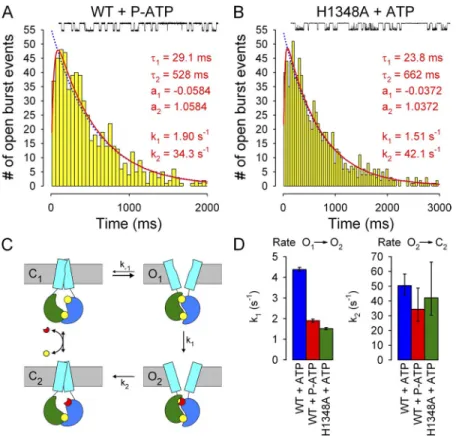
and k2; Fig. 7 C). The latter two rates can be estimated by fitting the scheme in Fig. 7 C to the distributions of open burst durations using maximum likelihood (Colquhoun and Sigworth, 1995). To determine which of these two rates is affected by P-ATP and the H1348A mutation, respectively, we recorded currents from patches containing a single active channel (Fig. 7, A and B, insets).
Open burst events were isolated by burst analysis (see Materials and methods), pooled from several patches, and fitted by maximum likelihood. The histograms of burst durations were distinctly peaked for both WT channels gating in 10 µM P-ATP (Fig. 7 A) and H1348A channels gating in 2 mM ATP (Fig. 7 B), consistent with a non-equilibrium gating cycle that involves nucleotide lines). The time constants of these exponentials reflect
the mean duration of bursts, which do not contain re entry events. When we compared these time constants (Fig. 6 D, colored bars) with the steady-state mean burst durations measured in the presence of the respective nucleotide (Fig. 6 D, striped bars; data replotted from Figs. 1 F, 2 C, and 4, B and E), we found in each case a very close agree- ment; importantly, in no case was the steady-state burst duration longer than the macroscopic estimate. This suggests that both for WT (and RI) channels gating in P-ATP, and for H1348A channels gating in 2 or 10 mM ATP or 10 µM P-ATP, each steady-state burst involves oc- clusion of a single nucleotide at site 2, just as suggested for WT channels gating in ATP (Csanády et al., 2010).
Of note, enhancement of macroscopic WT CFTR cur- rent by P-ATP, relative to ATP, was slightly smaller (<1.5-fold; see Fig. 6 A) than expected from the frac- tional increase in Po (1.55-fold; Fig. 1 E). This small discrepancy is accounted for by the slightly smaller aver- age conductance of channels opened by P-ATP, which likely reflects rapid flickery closures not fully resolved at our recording bandwidth (Fig. S1). Thus, at 80 mV, average unitary current of open channels was 0.59 ± 0.01 pA (n = 6) in ATP but significantly (P < 0.01) smaller, 0.55 ± 0.01 pA (n = 6), in P-ATP.
Figure 7. P-ATP and the H1348A mutation slow the O1→O2 transition of CFTR channels. (A and B) Histograms of open burst durations compiled from 621 open burst events of single WT CFTR channels gating in 10 µM P-ATP (A) and from 908 open burst events of single H1348A CFTR channels gating in 2 mM ATP (B). Both distri- butions were fitted by maximum likelihood to either a single exponential (blue dotted lines) or to the scheme in C, with rate k-1 fixed to zero (red lines); the latter fits proved significantly (P < 0.01 for A and P ≈ 0.05 for B) better by the log-likelihood ratio test. Fit parameters, as well as calculated time constants and fractional am- plitudes of the exponential components of the fitted distributions, are plotted in both panels.
(Insets) 30-s segments of single-channel record- ings. (C) Simplified cyclic gating scheme (from Csanády et al., 2010) used for maximum likelihood fitting of open burst distributions, depicting closed and open states with either ATP (states C1 and O1) or ADP (states C2 and O2) bound at site 2. Pore opening/closure is assumed quasi-simultaneous with formation/disruption of the NBD dimer.
Cyan rectangles, TMDs; green, NBD1; blue, NBD2; yellow, ATP; red, ADP. (D) Summary of rates k1 and k2 obtained from the fits in A and B for WT CFTR gating in 10 µM P-ATP (red bars) and H1348A CFTR gating in 2 mM ATP (green bars); as a comparison, the values measured for WT CFTR gating in 2 mM ATP (blue bars) are replotted from Csanády et al. (2010). Error bars represent 0.5-unit log-likelihood inter- vals. Note that in the presence of the site-1 perturbations, rate k2 could be estimated only to a limited precision because of the increased discrepancy between the values of k1 and k2 (compare Csanády, 2006; k2/k1 is 11, 18, and 29, respectively, for WT+ATP, WT+P-ATP, and H1348A+ATP).
on November 4, 2014jgp.rupress.orgDownloaded from
Hwang, 2013). Because one such mutation (W401F) re- sides in site 1, we have evaluated the possibility that the H1348A mutation, or P-ATP bound in site 1, might also act by such a mechanism. However, the lack of effect of increasing [ATP] on steady-state burst durations of H1348A CFTR (Fig. 2 C) and the close agreement of macroscopic closing time constants upon nucleotide re- moval with steady-state mean burst durations (Fig. 6) ruled out this possibility and confirmed that the two site-1 perturbations studied here do not disrupt the near 1:1 stoichiometry between ATP occlusion events and pore opening events, characteristic to WT CFTR gating in ATP (Csanády et al., 2010). Although compar- ison of macroscopic and steady-state closing rates (Fig. 6) might not be sensitive enough to rule out slight devia- tions from 1:1 stoichiometry, it is clear that small effects would not be sufficient to explain the two- to threefold prolongation of burst durations by the H1348A muta- tion and P-ATP. We have therefore used a simplified cy- clic scheme (Fig. 7 C) as a framework for interpreting our kinetic results and have dissected the effects of site-1 perturbations on the three rates that determine the stability of the open states: those of steps O1→O2, O1→C1, and O2→C2 (Fig. 7 C).
Interestingly, the detailed kinetic consequences of these two perturbations turned out to be very similar, amounting to a simultaneous two- to threefold decrease in the rates of steps O1→O2 and O1→C1. This pheno- type affords a simple explanation in terms of the free energy profile of the CFTR gating cycle (Fig. 8, blue energy profile). For a structural perturbation that selec- tively stabilizes the O1 state, relative to both states C1
and O2 (by GO1; Fig. 8, red energy profile), a simulta- neous increase in the heights of the transition-state bar- riers for both step O1→O2 (G‡1; Fig. 8) and step O1→C1 (G‡1) is expected (compare red and blue ver- tical double arrows in Fig. 8), resulting in slowed rates k-1 and k1, as observed. In addition, the distortion of the energy profile, as drawn, also suggests a lower energy barrier for step C1→O1, predicting an increase in open- ing rate. Interestingly, this is exactly what we observed for the H1348A mutant, the opening rate of which is approximately twofold increased relative to WT (com- pare Fig. 2 D, green bar, with Fig. 1 G, blue bar). We also observed a more modest increase in WT opening rate in the presence of 10 µM (subsaturating) P-ATP (com- pare red with blue bar in Fig. 1 G), but interpretation of this small effect is limited by the fact that stimulation of opening rate by P-ATP has been mostly attributed to P-ATP bound at site 2 (Tsai et al., 2010).
From an energetic point of view, the effects of the site-1 perturbations we have studied are modest, on the order of 1 kT. However, two facts have to be consid- ered when interpreting these effects. First, the mea- sured G values by no means represent the overall energetic change of the site-1 structure between states hydrolysis. The most striking difference between these
distributions and that found earlier for WT channels gating in ATP (Csanády et al., 2010) was in the tail, which was distinctly longer in the presence of the site-1 per- turbations, whereas the positions of the peaks were little moved. Accordingly, the maximum likelihood fit yielded two- to threefold slower k1 values (Fig. 7 D, left) in the presence of P-ATP (red bar) or the H1348A mutation (green bar) compared with the earlier estimate for WT channels gating in ATP (blue bar; replotted from Csanády et al., 2010). In contrast, neither P-ATP nor the H1348A mutation seemed to dramatically affect the faster rate k2 (Fig. 7 D, right).
D I S C U S S I O N
In this paper, we have investigated in detail the effects on channel gating of two structural perturbations at CFTR’s degenerate interfacial ATP-binding site (site 1).
The H1348A mutation removes a histidine side chain from the signature sequence of NBD2, thereby perturb- ing the NBD2 side of site 1, whereas the use of P-ATP introduces a phenylethyl group into this composite binding site. Although in our experiments, which in- volved prolonged exposure to P-ATP, both composite sites were presumably loaded with this ATP analogue, an elegant study by Tsai et al. (2010) convincingly dem- onstrated that the effect of P-ATP on burst length (com- pare Fig. 1 F) is caused by the P-ATP molecule bound at site 1, whereas P-ATP bound at site 2 is responsible for the high affinity, potent stimulation of opening rate (compare Fig. 1, B and C). In addition, experiments using the nonhydrolyzable ATP analogue AMPPNP together with P-ATP to lock WT channels in the open state sug- gested that the slowing of nonhydrolytic closure by P-ATP is also attributable to P-ATP bound at site 1 (Zhou et al., 2005). Here, we provide further independent support for this assignment by demonstrating nonaddi- tive effects on both the hydrolytic and nonhydrolytic closing rate of P-ATP and of mutations on either face of composite site 1 (Figs. 4 and 5). Furthermore, com- bining maximum likelihood fitting of steady-state sin- gle-channel records with the analysis of macroscopic current relaxations, we have determined which micro- scopic rates are affected by these site-1 perturbations re- sulting in the increased stability of channel open states.
In two recent studies, Jih et al. (2012a,b) proposed the existence of a short time window at the end of each burst, which would follow the release of hydrolysis prod- ucts from site 2 but precede gate closure, and allow a new ATP molecule to bind at site 2, returning the chan- nel into a new prehydrolytic open state without in- tervening gate closure. They further proposed that mutations or pharmacological modulation might pro- long CFTR channel bursts by increasing the frequency of such “reentry” events (Jih et al., 2012a,b; Jih and
on November 4, 2014jgp.rupress.orgDownloaded from
Furthermore, the selective energetic stabilization of state O1 relative to O2 (Fig. 8) by both perturbations implies that in a WT channel, the physico-chemical en- vironment of the H1348A side chain, or of the phenyl- ethyl group of a P-ATP molecule bound at site 1, experiences a significant change also during the O1→O2
transition, i.e., upon ATP hydrolysis at the active com- posite site 2. The implication is a significant movement in site 1 upon ATP hydrolysis at site 2: a classical alloste- ric effect. Interestingly, the K464A mutation, which per- turbs site 1 by removing the conserved Walker A lysine, was also shown to affect the energetics of both of the C1↔O1 and O1→O2 gating steps (Csanády et al., 2010), although in a different way: in this mutant, rate k1
decreased approximately fourfold, whereas the rate of nonhydrolytic closure, in a K1250A mutant background, increased by 10-fold (this is also replicated in the E1371S background; Fig. S2). Thus, although the distor- tion of the energetic profile by the K464A mutation dif- fers from that seen for the two perturbations studied here, the K464A result is again consistent with movements in site 1 occurring in both of the above gating steps.
In principle, we cannot exclude the possibility of a
“static allosteric effect” as an alternative scenario to ex- plain our results. That is, perturbations in site 1 might statically impose a slightly different overall conforma- tion of the NBDs, which leads to altered transition rates associated with reactions and conformational changes occurring at the active site 2, without movements in site 1 during those transitions. However, such an interpre- tation is at odds with the generally accepted view of protein allostery. Thus, manipulations at one site in a protein (the “allosteric site”) that affect conformational C1 and O1, or O1 and O2, but the change in environ-
ment of a single chemical moiety (the histidine side chain at position 1348 or the phenylethyl group of P-ATP in site 1) associated with those gating transitions. Sec- ond, our G values reflect changes in barrier heights, which suggest larger changes between ground states (see Fig. 8); e.g., an 1-kT change in the barrier height for nonhydrolytic closure might be associated with a
G of 2 kT between states C1 and O1. (As a com- parison, Gint between C1 and O1 is 2 kT for residues 555 and 1246 on opposing faces of the active site 2, which completely separates during the O1→C1 transi- tion [Vergani et al., 2005].)
What mechanistic insight can be gained from these observations? The recent suggestion that composite site 1 of CFTR does not separate upon channel closure (Tsai et al., 2010; Szollosi et al., 2011) has fostered a view of the degenerate site as a simple rigid scaffold, which does not participate in the functional movements of asymmetric ABC-C proteins (although this proposal has remained a matter of contention; Artur et al., 2011).
Our present data are hard to reconcile with such an assignment. The fact that removal of the histidine side chain at position 1348 selectively stabilizes state O1 rela- tive to C1 (Fig. 8) is most simply explained by postulat- ing that in the WT channel, the environment of that histidine side chain is not identical in those two states.
The similar effect of P-ATP bound at site 1 suggests that the phenylethyl group also experiences a change in en- vironment during the C1↔O1 transition. It follows then that significant movement is likely to take place at site 1 during the channel opening step, even if the compos- ite site does not actually separate in a closed channel.
Figure 8. Energetic interpretation of the gating effects of P-ATP and the H1348A mutation. Free energy profiles (top; not drawn to scale) of channels moving sequentially through the gating states depicted below (from Fig. 7 C), in the absence (blue energy profile) or presence (red energy profile) of a perturbation (red star in cartooned states) in composite site 1. A selective stabilization of the O1
ground state (by GO1, relative to both C1 and O2) is predicted to increase the heights of the free energy barriers for exiting O1 in both directions (G‡-1 and G‡1; compare red and blue vertical double arrows), thereby slowing both rate k-1 and k1 (cartoon).
on November 4, 2014jgp.rupress.orgDownloaded from
AMPPNP. Interestingly, the corresponding positions in site 1 of CFTR formed one of three interface pairs (T460- H1375) for which a lack of state-dependent changes in energetic coupling was interpreted to suggest a main- tained interaction throughout the gating cycle (Szollosi et al., 2011). However, consistent with the data we pres- ent here, a comparison between TM287/288 and crystals with tightly dimerized NBDs highlights clear differences in composite site-1 structure. The distance between the Walker A motif and the opposing signature sequence of the degenerate site is larger by 3 Å in the TM287/288 structure (Hohl et al., 2012) than that seen in tight di- mers of ABC NBDs (e.g., Smith et al., 2002; Dawson and Locher, 2007). The TM287/288 structure therefore also suggests some opening, although not complete separa- tion, of the degenerate site.
In conclusion, the data presented here strongly suggest that significant movements occur in CFTR’s composite site 1 concomitant with gating conformational changes.
The precise extent and direction of these movements await further structural and functional studies.
We thank Dorottya Mayer for oocyte isolation and injection.
This work is supported by National Institutes of Health grant R01-DK051767, MTA Lendület grant LP2012-39/2012, and an In- ternational Early Career Scientist grant from the Howard Hughes Medical Institute (to L. Csanády).
Christopher Miller served as editor.
Submitted: 21 December 2012 Accepted: 16 May 2013 R E F E R E N C E S
Aleksandrov, L., A.A. Aleksandrov, X.B. Chang, and J.R. Riordan.
2002. The first nucleotide binding domain of cystic fibrosis transmembrane conductance regulator is a site of stable nucleo- tide interaction, whereas the second is a site of rapid turnover.
J. Biol. Chem. 277:15419–15425. http://dx.doi.org/10.1074/jbc .M111713200
Anderson, M.P., D.P. Rich, R.J. Gregory, A.E. Smith, and M.J. Welsh.
1991. Generation of cAMP-activated chloride currents by expres- sion of CFTR. Science. 251:679–682. http://dx.doi.org/10.1126/
science.1704151
Artur, L., P. Chaves, and D.C. Gadsby. 2011. Extent of nucleotide- binding domain (NBD) separation when a CFTR channel closes.
Biophys. J. 100:265a–266a. http://dx.doi.org/10.1016/j.bpj.2010 .12.1662
Basso, C., P. Vergani, A.C. Nairn, and D.C. Gadsby. 2003. Prolonged nonhydrolytic interaction of nucleotide with CFTR’s NH2- terminal nucleotide binding domain and its role in channel gat- ing. J. Gen. Physiol. 122:333–348. http://dx.doi.org/10.1085/jgp .200308798
Cai, Z., T.S. Scott-Ward, and D.N. Sheppard. 2003. Voltage- dependent gating of the cystic fibrosis transmembrane conduc- tance regulator Cl channel. J. Gen. Physiol. 122:605–620. http://
dx.doi.org/10.1085/jgp.200308921
Chan, K.W., L. Csanády, D. Seto-Young, A.C. Nairn, and D.C. Gadsby.
2000. Severed molecules functionally define the boundaries of the cystic fibrosis transmembrane conductance regulator’s NH2- terminal nucleotide binding domain. J. Gen. Physiol. 116:163–180.
http://dx.doi.org/10.1085/jgp.116.2.163
changes at a distant site (the “active site”) generally involve movement at the allosteric site itself: allosteric modulators affect the conformational equilibrium of pro- teins because the affinity of their binding sites changes during the conformational transition. Examples range from 2,3-bisphosphoglycerate or proton modulation of oxygen binding in hemoglobin (Monod et al., 1965) to activation of ligand-gated channels (Changeux and Edelstein, 2005); in contrast, we are not aware of any example of a “frozen” allosteric site. We therefore feel that a conventional “dynamic allosteric effect,” which involves movements in site 1 of CFTR, is a more likely explanation of our findings.
Does site 1 separate then, or not, during a normal gat- ing cycle? The answer might lie somewhere in between.
The recent determination of the TM287/288 structure (Hohl et al., 2012) at least suggests such an intermedi- ate scenario. In this structure, the TMDs are seen in an inward-facing orientation, generally believed to corre- spond to the closed-channel state in CFTR. At the same time, the NBD dimer interface is only partially sepa- rated (Fig. 9): the active site 2 is open and devoid of nucleotide (Fig. 9, top composite site), whereas the de- generate site (Fig. 9, bottom composite site) contains a bound AMPPNP molecule, and in it, the opposing faces remain in contact. In particular, the -carbons of an asparagine in the TM288 D-loop (Asn521; Fig. 9, violet space fill) and that of a conserved threonine in the TM287 Walker A loop (T368; Fig. 9, red space fill) are separated by only 5.7 Å, and the asparagine is seen to make contact with the -phosphate of the bound
Figure 9. Partial separation of degenerate site in an asymmetrical bacterial ABC exporter. Ribbon diagram of NBD dimer from the crystal structure of TM287/288 (Protein Data Bank accession no.
3QF4), with AMPPNP (yellow) bound at the degenerate site. Con- served Walker A threonines (T368 in TM287 and T390 in TM288;
red), as well as D-loop residue N521 of TM288 and corresponding TM287 residue S499 (violet), are highlighted in space fill. The empty active site is wide open, whereas the nucleotide-bound de- generate site retains some contact between the two NBD surfaces through opposing Walker A and D-loop motifs. NBD color coding follows that used in Fig. 7 C: green, TM287; blue, TM288.
on November 4, 2014jgp.rupress.orgDownloaded from
![Figure 2. Steady-state mean burst duration of H1348A CFTR is insensitive to elevation of [ATP]](https://thumb-eu.123doks.com/thumbv2/9dokorg/1361918.110967/4.918.118.815.92.464/figure-steady-state-burst-duration-cftr-insensitive-elevation.webp)